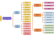

Hydrogels are increasingly explored as functional wound-dressing materials because they combine high water content, biocompatibility, structural tunability, and the ability to localize therapeutic delivery at injured tissue. As biomaterial platforms, hydrogel dressings can maintain a moist microenvironment, absorb exudate, protect the wound bed, and carry bioactive agents that modulate infection, inflammation, oxidative stress, and tissue regeneration. This review examines hydrogel wound dressings from a materials centered perspective. First, skin structure, wound-healing physiology, and major barriers to repair are outlined to define the biological requirements for effective dressings. Next, the chemical composition of natural, synthetic, and composite hydrogels, their crosslinking strategies, swelling behavior, and drug-loading and release mechanisms are discussed in relation to wound healing performance. Recent progress in infection responsive, stimuli responsive, growth factor delivering, antimicrobial peptide loaded, and self-healing hydrogel systems is then summarized. The present review highlights how composition, network architecture, and responsiveness govern biomedical function and localized drug delivery in wound care. These insights provide a materials centered framework that connects hydrogel composition, network architecture, responsiveness, and localized delivery behavior with wound healing performance, thereby supporting the rational design of next generation hydrogel biomaterials for difficult to heal wounds.
Hydrogels are increasingly explored as functional wound-dressing materials because they combine high water content, biocompatibility, structural tunability, and the ability to localize therapeutic delivery at injured tissue. As biomaterial platforms, hydrogel dressings can maintain a moist microenvironment, absorb exudate, protect the wound bed, and carry bioactive agents that modulate infection, inflammation, oxidative stress, and tissue regeneration. This review examines hydrogel wound dressings from a materials centered perspective. First, skin structure, wound-healing physiology, and major barriers to repair are outlined to define the biological requirements for effective dressings. Next, the chemical composition of natural, synthetic, and composite hydrogels, their crosslinking strategies, swelling behavior, and drug-loading and release mechanisms are discussed in relation to wound healing performance. Recent progress in infection responsive, stimuli responsive, growth factor delivering, antimicrobial peptide loaded, and self-healing hydrogel systems is then summarized. The present review highlights how composition, network architecture, and responsiveness govern biomedical function and localized drug delivery in wound care. These insights provide a materials centered framework that connects hydrogel composition, network architecture, responsiveness, and localized delivery behavior with wound healing performance, thereby supporting the rational design of next generation hydrogel biomaterials for difficult to heal wounds.
DOI: https://doi.org/10.37349/ebmx.2026.101368
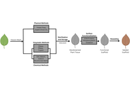
The development of biomaterials capable of supporting complex tissue growth remains a central challenge in regenerative medicine and tissue engineering, particularly in replicating the structural, mechanical, and transport functions of native extracellular matrices. While decellularized animal tissues have demonstrated significant success as scaffolds for tissue engineering, they are still constrained by cost, immunogenicity, and ethical concerns. In recent years, decellularized plant tissues have emerged as a compelling alternative scaffold platform due to their inherent vascular architectures, ethical sourcing, tunable mechanical properties, cytocompatibility, and sustainability. This review summarizes current strategies for the decellularization of plant tissues, including chemical, enzymatic, and physical approaches, and discusses how these methods preserve plant cell wall structure while removing immunogenic components. Advances in surface loading and functionalization, including protein coatings, oxidation, nanoparticle incorporation, peptide conjugation, and bioactive molecule loading, have further enhanced cell adhesion, differentiation, biodegradability, and immunomodulation. Recent applications of decellularized plant scaffolds in cardiac, skeletal muscle, bone, nerve, and wound healing contexts are reviewed, highlighting proof-of-concept successes and remaining challenges. Beyond therapeutic applications, plant-derived scaffolds have also enabled physiologically relevant in vitro models for vascular biology, mechanotransduction, cancer, metabolic tissues, and drug response studies. Collectively, these advances position decellularized plant tissues as versatile, low-cost, and ethically favorable biomaterials with growing relevance for both regenerative medicine and tissue modeling.
The development of biomaterials capable of supporting complex tissue growth remains a central challenge in regenerative medicine and tissue engineering, particularly in replicating the structural, mechanical, and transport functions of native extracellular matrices. While decellularized animal tissues have demonstrated significant success as scaffolds for tissue engineering, they are still constrained by cost, immunogenicity, and ethical concerns. In recent years, decellularized plant tissues have emerged as a compelling alternative scaffold platform due to their inherent vascular architectures, ethical sourcing, tunable mechanical properties, cytocompatibility, and sustainability. This review summarizes current strategies for the decellularization of plant tissues, including chemical, enzymatic, and physical approaches, and discusses how these methods preserve plant cell wall structure while removing immunogenic components. Advances in surface loading and functionalization, including protein coatings, oxidation, nanoparticle incorporation, peptide conjugation, and bioactive molecule loading, have further enhanced cell adhesion, differentiation, biodegradability, and immunomodulation. Recent applications of decellularized plant scaffolds in cardiac, skeletal muscle, bone, nerve, and wound healing contexts are reviewed, highlighting proof-of-concept successes and remaining challenges. Beyond therapeutic applications, plant-derived scaffolds have also enabled physiologically relevant in vitro models for vascular biology, mechanotransduction, cancer, metabolic tissues, and drug response studies. Collectively, these advances position decellularized plant tissues as versatile, low-cost, and ethically favorable biomaterials with growing relevance for both regenerative medicine and tissue modeling.
DOI: https://doi.org/10.37349/ebmx.2026.101367
This article belongs to the special issue Nature-Based Biomaterials for Biomedical Applications
Aim:
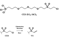
In biosensor technology, reliable attachment of protein-based probes requires careful control of the orientation of the probe molecule on the biosensor surface. In this regard, polyhistidine peptide became an attractive target for on surface immobilization. The present contribution details the total synthesis and the surface chemistry of a new antifouling organotrichlorosilane linker bearing a head function designed to immobilize the imidazole side chain of histidine for future immobilizations with polyhistidine peptide onto biosensor surface.
Methods:
A novel organotrichlorosilane linker bearing the ethylene glycol backbone and a 2-chloroethyl sulfone head function (which can be converted to the vinyl sulfone group for subsequent attachment with imidazole) were synthesized via a multiple-step synthesis and carefully characterized. Surface modifications using the synthesized novel organotrichlorosilane linker, subsequent conversion to vinyl sulfone head function, and treatment with N-protected histidine were demonstrated on black lithium niobate substrate.
Results:
Novel organotrichlorosilane linker was successfully synthesized, though it was also observed that organotrichlorosilane linker was quite moisture reactive. Surface characterizations also indicated successful modification of lithium niobate with the novel organotrichlorosilane linker as well as presence of N-protected histidine on the lithium niobate surface post-immobilization.
Conclusions:
A novel organotrichlorosilane linker bearing the 2-chloroethylsulfone group was successfully synthesized and successful immobilization with N-protected histidine was demonstrated. The surface chemistry demonstrated onto lithium niobate herein is immediately applicable for future on-surface immobilization of protein-based probe molecules bearing polyhistidine moieties.
Aim:
In biosensor technology, reliable attachment of protein-based probes requires careful control of the orientation of the probe molecule on the biosensor surface. In this regard, polyhistidine peptide became an attractive target for on surface immobilization. The present contribution details the total synthesis and the surface chemistry of a new antifouling organotrichlorosilane linker bearing a head function designed to immobilize the imidazole side chain of histidine for future immobilizations with polyhistidine peptide onto biosensor surface.
Methods:
A novel organotrichlorosilane linker bearing the ethylene glycol backbone and a 2-chloroethyl sulfone head function (which can be converted to the vinyl sulfone group for subsequent attachment with imidazole) were synthesized via a multiple-step synthesis and carefully characterized. Surface modifications using the synthesized novel organotrichlorosilane linker, subsequent conversion to vinyl sulfone head function, and treatment with N-protected histidine were demonstrated on black lithium niobate substrate.
Results:
Novel organotrichlorosilane linker was successfully synthesized, though it was also observed that organotrichlorosilane linker was quite moisture reactive. Surface characterizations also indicated successful modification of lithium niobate with the novel organotrichlorosilane linker as well as presence of N-protected histidine on the lithium niobate surface post-immobilization.
Conclusions:
A novel organotrichlorosilane linker bearing the 2-chloroethylsulfone group was successfully synthesized and successful immobilization with N-protected histidine was demonstrated. The surface chemistry demonstrated onto lithium niobate herein is immediately applicable for future on-surface immobilization of protein-based probe molecules bearing polyhistidine moieties.
DOI: https://doi.org/10.37349/ebmx.2026.101366
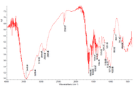
The search for inexpensive raw materials for chitin production has led to the exploration of various natural resources, including some less conventional ones, such as plants and waste from the processing of various animals. In this context, the production of chitin from chicken bones and feet has been reported, attracting attention as a cheap and widely available source in some regions. However, to the best of our knowledge, birds do not possess genes that encode chitin synthases, the enzymes responsible for chitin biosynthesis. Therefore, this study analyzes the results reported in related articles, especially their FTIR spectra, to assess when the obtained material can be identified as chitin. The analysis revealed that, in some cases, there is poor agreement between the signals in these spectra and the characteristic signals established for well-characterized chitins, while in others, the spectra exhibit signals with a high noise-to-signal ratio that limits their use for identification. Furthermore, the X-ray diffraction studies reported in some of these works provide scarce support to confirm the presence of chitin in these materials. A search of two specialized databases confirmed that, to date, no results have been reported for genes expressing chitin synthases in birds. Finally, some recommendations are offered for properly addressing the studies necessary for the unambiguous identification of these materials.
The search for inexpensive raw materials for chitin production has led to the exploration of various natural resources, including some less conventional ones, such as plants and waste from the processing of various animals. In this context, the production of chitin from chicken bones and feet has been reported, attracting attention as a cheap and widely available source in some regions. However, to the best of our knowledge, birds do not possess genes that encode chitin synthases, the enzymes responsible for chitin biosynthesis. Therefore, this study analyzes the results reported in related articles, especially their FTIR spectra, to assess when the obtained material can be identified as chitin. The analysis revealed that, in some cases, there is poor agreement between the signals in these spectra and the characteristic signals established for well-characterized chitins, while in others, the spectra exhibit signals with a high noise-to-signal ratio that limits their use for identification. Furthermore, the X-ray diffraction studies reported in some of these works provide scarce support to confirm the presence of chitin in these materials. A search of two specialized databases confirmed that, to date, no results have been reported for genes expressing chitin synthases in birds. Finally, some recommendations are offered for properly addressing the studies necessary for the unambiguous identification of these materials.
DOI: https://doi.org/10.37349/ebmx.2026.101365
Aim:
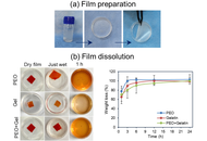
Cell sheet technology is a transformative approach in epithelial tissue engineering, offering scaffold-free constructs that preserve cell-cell and cell-matrix interactions, enabling better integration with host tissues. However, safe and efficient transfer of these fragile sheets remains a critical challenge, limiting their broader clinical adoption. This study aims to develop a facile method for the retrieval and transfer of epithelial cell sheets cultivated over thermo-responsive polymer surfaces (TRPS) using sacrificial films.
Methods:
Three epithelial cell lines, HCE-S (cornea), HaCaT (skin), and A549 (lung), were cultured on poly(N-isopropylacrylamide-co-glycidyl methacrylate) (P(NIPA-GMA)) coated TRPS and conventional tissue culture surfaces. Upon reaching confluence, the dishes were incubated below the lower critical solution temperature to induce phase transition in TRPS. Subsequently, sacrificial films made of polyethylene oxide, gelatin and their blend were used to lift and transfer the cell sheets to new culture dishes containing a minimal amount of culture medium. Additional medium was then added to dissolve the film, allowing the cell sheet to settle gently onto the dish surface.
Results:
In all three epithelial cell types, a continuous, confluent cell sheet was visible on the TRPS prior to transfer. Subsequent to temperature lowering and sacrificial film assisted transfer, the master TRPS dish exhibited a distinct void corresponding to the sheet removal, confirming successful detachment. The transferred sheets reattached successfully and maintained over a one-week observation period.
Conclusions:
The sacrificial film-based transfer method provided a gentle, efficient and scalable alternative for handling cell sheets from TRPS. This approach enhances the translational potential of cell sheet engineering and supports its integration into clinical workflows for epithelial tissue regeneration.
Aim:
Cell sheet technology is a transformative approach in epithelial tissue engineering, offering scaffold-free constructs that preserve cell-cell and cell-matrix interactions, enabling better integration with host tissues. However, safe and efficient transfer of these fragile sheets remains a critical challenge, limiting their broader clinical adoption. This study aims to develop a facile method for the retrieval and transfer of epithelial cell sheets cultivated over thermo-responsive polymer surfaces (TRPS) using sacrificial films.
Methods:
Three epithelial cell lines, HCE-S (cornea), HaCaT (skin), and A549 (lung), were cultured on poly(N-isopropylacrylamide-co-glycidyl methacrylate) (P(NIPA-GMA)) coated TRPS and conventional tissue culture surfaces. Upon reaching confluence, the dishes were incubated below the lower critical solution temperature to induce phase transition in TRPS. Subsequently, sacrificial films made of polyethylene oxide, gelatin and their blend were used to lift and transfer the cell sheets to new culture dishes containing a minimal amount of culture medium. Additional medium was then added to dissolve the film, allowing the cell sheet to settle gently onto the dish surface.
Results:
In all three epithelial cell types, a continuous, confluent cell sheet was visible on the TRPS prior to transfer. Subsequent to temperature lowering and sacrificial film assisted transfer, the master TRPS dish exhibited a distinct void corresponding to the sheet removal, confirming successful detachment. The transferred sheets reattached successfully and maintained over a one-week observation period.
Conclusions:
The sacrificial film-based transfer method provided a gentle, efficient and scalable alternative for handling cell sheets from TRPS. This approach enhances the translational potential of cell sheet engineering and supports its integration into clinical workflows for epithelial tissue regeneration.
DOI: https://doi.org/10.37349/ebmx.2026.101364
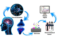
Electrochemical sensors have emerged as powerful tools for the detection and monitoring of neurotransmitters, offering high sensitivity, selectivity, and potential for real-time analysis. Neurotransmitters play a crucial role in regulating various physiological and neurological processes, and imbalances in their levels are linked to a wide range of neurological disorders, including Parkinson’s disease, depression, Alzheimer’s disease, and epilepsy. This review highlights recent advancements in electrochemical sensor technologies for neurotransmitter detection, focusing on innovations that enhance performance through the use of nanomaterials, wearable devices, and multiplexed sensing techniques. The integration of nanomaterials such as graphene, carbon nanotubes, and metal nanoparticles has significantly improved sensor sensitivity and selectivity, enabling more accurate detection even at low concentrations. Furthermore, the development of flexible, wearable, and implantable sensors is facilitating continuous, non-invasive monitoring of neurotransmitter levels in real time. Advances in multiplexed sensors are enabling the simultaneous detection of multiple neurotransmitters, providing a more comprehensive approach to disease diagnosis and management. Despite these promising developments, challenges remain, including issues of selectivity, stability, and long-term monitoring. Nevertheless, electrochemical sensors hold great potential for transforming the way neurological disorders are diagnosed and managed, offering opportunities for personalized, real-time monitoring and more effective treatment strategies.
Electrochemical sensors have emerged as powerful tools for the detection and monitoring of neurotransmitters, offering high sensitivity, selectivity, and potential for real-time analysis. Neurotransmitters play a crucial role in regulating various physiological and neurological processes, and imbalances in their levels are linked to a wide range of neurological disorders, including Parkinson’s disease, depression, Alzheimer’s disease, and epilepsy. This review highlights recent advancements in electrochemical sensor technologies for neurotransmitter detection, focusing on innovations that enhance performance through the use of nanomaterials, wearable devices, and multiplexed sensing techniques. The integration of nanomaterials such as graphene, carbon nanotubes, and metal nanoparticles has significantly improved sensor sensitivity and selectivity, enabling more accurate detection even at low concentrations. Furthermore, the development of flexible, wearable, and implantable sensors is facilitating continuous, non-invasive monitoring of neurotransmitter levels in real time. Advances in multiplexed sensors are enabling the simultaneous detection of multiple neurotransmitters, providing a more comprehensive approach to disease diagnosis and management. Despite these promising developments, challenges remain, including issues of selectivity, stability, and long-term monitoring. Nevertheless, electrochemical sensors hold great potential for transforming the way neurological disorders are diagnosed and managed, offering opportunities for personalized, real-time monitoring and more effective treatment strategies.
DOI: https://doi.org/10.37349/ebmx.2026.101363
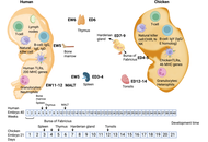
Antibiotic resistance is a global threat, driven by limited new antimicrobials and rising multidrug-resistant infections. Lipid nanoparticles (LNPs) combine tunable material properties with antimicrobial functionality, providing biocompatibility, controlled release, and biofilm penetration. LNPs provide key advantages over metallic and polymeric nanocarriers, including high biocompatibility, the ability to encapsulate both hydrophilic and hydrophobic agents, controlled release profiles, and reduced cytotoxicity and immune activation. These features enhance drug stability and bioavailability and may help circumvent bacterial defences such as biofilms and efflux pumps. Robust preclinical evaluation platform of antimicrobial biomaterials requires platforms that capture biologically relevant interactions while remaining ethically and economically feasible. The chick embryo model (CEM) has emerged as a versatile platform for infection studies, bridging conventional in vitro assays and mammalian in vivo models. Its vascularized and developing tissue environment enables assessment of nanoparticle biodistribution, local toxicity, and antimicrobial efficacy within a dynamic biological context. This review critically examines the application of the CEM for evaluating LNP-based antimicrobial systems, highlighting current methodological variability and limitations in experimental standardization. By identifying gaps in protocol harmonisation and comparative assessment, this work outlines opportunities to improve reproducibility and translational relevance. Overall, integrating rationally designed LNP systems with optimised CEMs may accelerate the development of next-generation antimicrobial biomaterials to combat antibiotic-resistant infections.
Antibiotic resistance is a global threat, driven by limited new antimicrobials and rising multidrug-resistant infections. Lipid nanoparticles (LNPs) combine tunable material properties with antimicrobial functionality, providing biocompatibility, controlled release, and biofilm penetration. LNPs provide key advantages over metallic and polymeric nanocarriers, including high biocompatibility, the ability to encapsulate both hydrophilic and hydrophobic agents, controlled release profiles, and reduced cytotoxicity and immune activation. These features enhance drug stability and bioavailability and may help circumvent bacterial defences such as biofilms and efflux pumps. Robust preclinical evaluation platform of antimicrobial biomaterials requires platforms that capture biologically relevant interactions while remaining ethically and economically feasible. The chick embryo model (CEM) has emerged as a versatile platform for infection studies, bridging conventional in vitro assays and mammalian in vivo models. Its vascularized and developing tissue environment enables assessment of nanoparticle biodistribution, local toxicity, and antimicrobial efficacy within a dynamic biological context. This review critically examines the application of the CEM for evaluating LNP-based antimicrobial systems, highlighting current methodological variability and limitations in experimental standardization. By identifying gaps in protocol harmonisation and comparative assessment, this work outlines opportunities to improve reproducibility and translational relevance. Overall, integrating rationally designed LNP systems with optimised CEMs may accelerate the development of next-generation antimicrobial biomaterials to combat antibiotic-resistant infections.
DOI: https://doi.org/10.37349/ebmx.2026.101362
Aim:
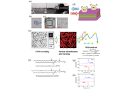
This study aims to develop and validate a transmission electron microscopy (TEM)–based approach for probing nanoscale lipid membrane dynamics by tracking the motion of gold nanoparticles dispersed on membrane surfaces.
Methods:
Lipid thin films composed of dipalmitoylphosphatidylcholine (DPPC) or dioleoylphosphatidylcholine (DOPC) were prepared over 2 μm holes in Quantifoil grids, and 5 nm gold nanocolloids were introduced as tracer particles. Sequential TEM imaging was performed during controlled heating and cooling cycles, and nanoparticle trajectories were analyzed to obtain mean squared displacement (MSD) curves. These measurements enabled quantification of thermally driven membrane dynamics. The temperature dependent behavior was further compared with differential scanning calorimetry (DSC) of dehydrated lipid samples.
Results:
DPPC exhibited a pronounced MSD peak near 52.5 °C during the first heating cycle, corresponding to its main phase transition, whereas DOPC showed gradual and continuous mobility changes consistent with its intrinsically disordered acyl chains. Differences between electron beam molecular dynamics (EBMD) and DSC transition temperatures likely arose from dehydration and thin film geometry. Across repeated thermal cycles, DPPC membranes displayed cycle dependent changes in MSD profiles, suggesting annealing induced homogenization and potential beam induced structural alterations.
Conclusions:
EBMD provides real space, time resolved visualization of nanoscale membrane fluctuations and complements ensemble techniques such as DSC, fluorescence recovery after photobleaching (FRAP), and nuclear magnetic resonance (NMR). The TEM based particle tracking approach reliably distinguishes ordered versus disordered lipid systems and offers a versatile platform for investigating soft biological membranes, including systems containing proteins or heterogeneous lipid compositions.
Aim:
This study aims to develop and validate a transmission electron microscopy (TEM)–based approach for probing nanoscale lipid membrane dynamics by tracking the motion of gold nanoparticles dispersed on membrane surfaces.
Methods:
Lipid thin films composed of dipalmitoylphosphatidylcholine (DPPC) or dioleoylphosphatidylcholine (DOPC) were prepared over 2 μm holes in Quantifoil grids, and 5 nm gold nanocolloids were introduced as tracer particles. Sequential TEM imaging was performed during controlled heating and cooling cycles, and nanoparticle trajectories were analyzed to obtain mean squared displacement (MSD) curves. These measurements enabled quantification of thermally driven membrane dynamics. The temperature dependent behavior was further compared with differential scanning calorimetry (DSC) of dehydrated lipid samples.
Results:
DPPC exhibited a pronounced MSD peak near 52.5 °C during the first heating cycle, corresponding to its main phase transition, whereas DOPC showed gradual and continuous mobility changes consistent with its intrinsically disordered acyl chains. Differences between electron beam molecular dynamics (EBMD) and DSC transition temperatures likely arose from dehydration and thin film geometry. Across repeated thermal cycles, DPPC membranes displayed cycle dependent changes in MSD profiles, suggesting annealing induced homogenization and potential beam induced structural alterations.
Conclusions:
EBMD provides real space, time resolved visualization of nanoscale membrane fluctuations and complements ensemble techniques such as DSC, fluorescence recovery after photobleaching (FRAP), and nuclear magnetic resonance (NMR). The TEM based particle tracking approach reliably distinguishes ordered versus disordered lipid systems and offers a versatile platform for investigating soft biological membranes, including systems containing proteins or heterogeneous lipid compositions.
DOI: https://doi.org/10.37349/ebmx.2026.101361
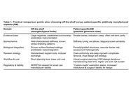
Three-dimensional metal printing has made anatomical perfection readily achievable in orthopaedic reconstruction. Yet, as patient-specific implants transition from salvage solutions to routine applications, a critical question emerges: Does geometric precision improve long-term outcomes, or merely perfect existing problems? The article argues that customization defined by shape alone fails to address fundamental biological constraints, including stiffness mismatch, stress shielding, vascular compromise, and the inevitability of revision surgery. While additive manufacturing enables porous architectures and tailored mechanics, unchecked integration and over-conformity may jeopardize bone preservation and future surgical options. The article further highlights the professional and economic costs of patient-specific workflows and the limitations of static digital planning. True innovation, it is argued, lies not in achieving the “perfect fit,” but in designing implants that participate in bone biology and remain surgically defensible decades after implantation.
Three-dimensional metal printing has made anatomical perfection readily achievable in orthopaedic reconstruction. Yet, as patient-specific implants transition from salvage solutions to routine applications, a critical question emerges: Does geometric precision improve long-term outcomes, or merely perfect existing problems? The article argues that customization defined by shape alone fails to address fundamental biological constraints, including stiffness mismatch, stress shielding, vascular compromise, and the inevitability of revision surgery. While additive manufacturing enables porous architectures and tailored mechanics, unchecked integration and over-conformity may jeopardize bone preservation and future surgical options. The article further highlights the professional and economic costs of patient-specific workflows and the limitations of static digital planning. True innovation, it is argued, lies not in achieving the “perfect fit,” but in designing implants that participate in bone biology and remain surgically defensible decades after implantation.
DOI: https://doi.org/10.37349/ebmx.2026.101360
This article belongs to the special issue Metal 3D Printing of Biometals for Prostheses and Implants
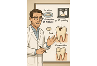
The emergence of stem-cell-derived enamel organoids and dentin-producing dental pulp stem cell constructs presents new possibilities for restoring carious lesions using autologous enamel–dentin inlays. This overview outlines the biological and technological advances supporting this approach and proposes a workflow oriented toward clinical application. The benefits of tissue-based inlays, including inherent biomechanical compatibility, aesthetic accuracy, and potential for biological integration, are contrasted with those of purely artificial materials. Significant regenerative developments include the formation of human enamel organoids and odontoblast-lineage cells in vitro, 3D bioprinting of tooth-shaped constructs with demineralised dentin matrix and poly(ε‑caprolactone) scaffolds, and fibre-guiding periodontal ligament scaffolds that restore Sharpey’s fibres in vivo. The mechanical performance of adhesive resin cements, with bond strengths of approximately 4–7 MPa to enamel and dentin, and their durability in reattaching natural tooth fragments, supports the feasibility of bonding biological inlays. Practical considerations include controlling the slow degradation and hydrophobicity of poly(ε-caprolactone) through the use of ceramic or natural polymer additives, employing multi-material 3D printing to co-print mineralized enamel and cell-laden dentin layers, and achieving the desired shade, microstructure, and mechanical properties, exemplified by a compressive strength of approximately 677 MPa for 3D-printed zirconia crowns. Despite regulatory and translational challenges, the integration of digital dentistry, bioprinting, and stem cell science points toward future “grow and glue” restorations that may replace traditional drill-and-fill methods.
The emergence of stem-cell-derived enamel organoids and dentin-producing dental pulp stem cell constructs presents new possibilities for restoring carious lesions using autologous enamel–dentin inlays. This overview outlines the biological and technological advances supporting this approach and proposes a workflow oriented toward clinical application. The benefits of tissue-based inlays, including inherent biomechanical compatibility, aesthetic accuracy, and potential for biological integration, are contrasted with those of purely artificial materials. Significant regenerative developments include the formation of human enamel organoids and odontoblast-lineage cells in vitro, 3D bioprinting of tooth-shaped constructs with demineralised dentin matrix and poly(ε‑caprolactone) scaffolds, and fibre-guiding periodontal ligament scaffolds that restore Sharpey’s fibres in vivo. The mechanical performance of adhesive resin cements, with bond strengths of approximately 4–7 MPa to enamel and dentin, and their durability in reattaching natural tooth fragments, supports the feasibility of bonding biological inlays. Practical considerations include controlling the slow degradation and hydrophobicity of poly(ε-caprolactone) through the use of ceramic or natural polymer additives, employing multi-material 3D printing to co-print mineralized enamel and cell-laden dentin layers, and achieving the desired shade, microstructure, and mechanical properties, exemplified by a compressive strength of approximately 677 MPa for 3D-printed zirconia crowns. Despite regulatory and translational challenges, the integration of digital dentistry, bioprinting, and stem cell science points toward future “grow and glue” restorations that may replace traditional drill-and-fill methods.
DOI: https://doi.org/10.37349/ebmx.2026.101359
This article belongs to the special issue Innovations in Biomaterials for Dentistry and Oral Surgery
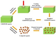
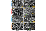
Ultra-high molecular weight polyethylene (UHMWPE) is widely used as a key material in biomedical implants such as artificial joints due to its exceptional wear resistance, high impact strength, and good biocompatibility. However, its inherent bio-inertness, hydrophobicity, risk of osteolysis induced by wear debris, and insufficient mechanical and processing properties severely limit its long-term clinical performance. This review systematically summarizes recent advances in the functional enhancement of UHMWPE via hybrid strategies, including surface modifications (e.g., coatings, chemical grafting, laser processing, plasma treatment) and bulk blending modifications (involving both organic and inorganic composites). These approaches have been shown to significantly improve wear resistance, bioactivity, hydrophilicity, and mechanical properties, while effectively suppressing oxidative degradation and inflammatory responses. The current challenges in modification technologies, such as balancing multiple properties, ensuring long-term biosafety, and achieving clinical translation, are also discussed. Finally, future directions toward multifunctional integration, intelligent responsiveness, and personalized customization of implants are outlined, providing critical insights for the development of next-generation high-performance and long-lasting biomedical materials.
Ultra-high molecular weight polyethylene (UHMWPE) is widely used as a key material in biomedical implants such as artificial joints due to its exceptional wear resistance, high impact strength, and good biocompatibility. However, its inherent bio-inertness, hydrophobicity, risk of osteolysis induced by wear debris, and insufficient mechanical and processing properties severely limit its long-term clinical performance. This review systematically summarizes recent advances in the functional enhancement of UHMWPE via hybrid strategies, including surface modifications (e.g., coatings, chemical grafting, laser processing, plasma treatment) and bulk blending modifications (involving both organic and inorganic composites). These approaches have been shown to significantly improve wear resistance, bioactivity, hydrophilicity, and mechanical properties, while effectively suppressing oxidative degradation and inflammatory responses. The current challenges in modification technologies, such as balancing multiple properties, ensuring long-term biosafety, and achieving clinical translation, are also discussed. Finally, future directions toward multifunctional integration, intelligent responsiveness, and personalized customization of implants are outlined, providing critical insights for the development of next-generation high-performance and long-lasting biomedical materials.
DOI: https://doi.org/10.37349/ebmx.2026.101358
Aim:
Although polytetrafluoroethylene (PTFE) is more hydrophobic than polyvinylidene fluoride (PVDF) in fluorocarbon polymer (FCP) membrane filters, it has been reported that the rate of amyloid fibril formation is faster on PVDF than on PTFE. To clarify whether the effect is due to the membrane’s chemical structure or its hydrophobicity at the membrane interface, studies on amyloid fibril formation were conducted using both hydrophobic and hydrophilic PVDF and PTFE membranes.
Methods:
Heat-treated insulin (INS) was adsorbed onto the FCP membrane filters. Gaussian integrals were employed to determine the amounts of β-sheet and their abundance ratios by curve fitting of attenuated total reflection Fourier transform infrared spectra.
Results:
Adsorbed heat-treated INS onto the FCP membrane filters showed a β-sheet form, with a similar or higher affinity in comparison with that of the β-rich concanavalin A. The adsorption followed a sigmoidal curve with a 2-hour lag time, reaching a plateau after 4–5 hours. The spectral patterns of the adsorbed INS indicated the β-sheet form, demonstrating that INS transformed into β-sheet and then, or simultaneously, adsorbed onto the FCP membrane filters.
Conclusions:
The results regarding the rate and strength of amyloid fibril formation for each FCP membrane filter suggest that, beyond the membrane’s surface hydrophobicity or hydrophilicity, other factors, such as the electron affinity of hydrogen in the PVDF membrane, also influence nucleation. This study provides insight into the role of INS in amyloid fibril formation within FCP membrane filters.
DOI: https://doi.org/10.37349/ebmx.2026.101357
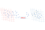
Hydrogels are among the most intensively studied biomaterials for controlled drug delivery, yet translation to routine clinical practice has been limited by rapid diffusion of small molecules and instability of biologics. In their recent report in Nature Nanotechnology (Pogostin et al., 2025, DOI: 10.1038/s41565-025-01981-6), a team from Rice University and collaborators present a nanofibrous supramolecular peptide hydrogel system that addresses these challenges through the incorporation of dynamic covalent chemistry. The SABER (Self-Assembling Boronate Ester Release) platform introduces reversible boronate ester bonds between engineered peptide fibers and boronic acid modified therapeutics, creating a tunable and long-acting drug release system. Proof-of-concept applications included tuberculosis therapy, diabetes management, and prolonged antibody delivery, demonstrating both versatility and clinical relevance. In this Commentary, I situate this advance within the broader trajectory of hydrogel research, highlight the conceptual novelty of dynamic supramolecular interactions, and discuss the opportunities and challenges for clinical translation. I argue that this platform signals a paradigm shift in drug delivery, moving hydrogels from passive depots to dynamic partners in medicine.
Hydrogels are among the most intensively studied biomaterials for controlled drug delivery, yet translation to routine clinical practice has been limited by rapid diffusion of small molecules and instability of biologics. In their recent report in Nature Nanotechnology (Pogostin et al., 2025, DOI: 10.1038/s41565-025-01981-6), a team from Rice University and collaborators present a nanofibrous supramolecular peptide hydrogel system that addresses these challenges through the incorporation of dynamic covalent chemistry. The SABER (Self-Assembling Boronate Ester Release) platform introduces reversible boronate ester bonds between engineered peptide fibers and boronic acid modified therapeutics, creating a tunable and long-acting drug release system. Proof-of-concept applications included tuberculosis therapy, diabetes management, and prolonged antibody delivery, demonstrating both versatility and clinical relevance. In this Commentary, I situate this advance within the broader trajectory of hydrogel research, highlight the conceptual novelty of dynamic supramolecular interactions, and discuss the opportunities and challenges for clinical translation. I argue that this platform signals a paradigm shift in drug delivery, moving hydrogels from passive depots to dynamic partners in medicine.
DOI: https://doi.org/10.37349/ebmx.2026.101356
DOI: https://doi.org/10.37349/ebmx.2025.101354
This article belongs to the special issue Bioinspired Material for Regenerative Medicine
Luminescent markers have been widely used in medicine, biology, agrotechnology, and for marking nuclear wastes and consumer goods. The high sensitivity and selectivity of the markers/labels allow the detection of various substances and the obtaining of valuable information about the distribution of constituents in specific media. This review describes the state of the art in luminescent marking/labeling of various cellulose forms, including nanosized ones, cellulose derivatives, and cellulose-containing materials. The importance of this consideration is explained by the role of cellulose and its derivatives in human life and their overall impact on mankind’s development. The structure and luminescence properties of cellulose and other related materials and cellulose derivatives are discussed from the viewpoint of cellulose luminescent “self-labeling”. It is shown that dyes, organic molecules, and organic-inorganic complexes, as well as inorganic dielectric and semiconductor micro/nanoparticles, can be effectively applied for the purposes of cellulose luminescent marking/labeling. This review discusses various application examples and explains the performance and mechanisms of various systems labeling (e.g., dye-cellulose, quantum dot-cellulose complex) in these applications. The review not only comprehensively summarizes existing approaches to luminescent labeling of cellulose-containing materials. It also highlights problematic issues that arise for developers of new luminescent markers (quenching of luminescence in an aqueous environment, the need to functionalize the luminescent marker material, etc.). At the same time, this work demonstrates the prospects for luminescent labeling data in modern digital technologies, particularly in the Internet of Things (IoT).
Luminescent markers have been widely used in medicine, biology, agrotechnology, and for marking nuclear wastes and consumer goods. The high sensitivity and selectivity of the markers/labels allow the detection of various substances and the obtaining of valuable information about the distribution of constituents in specific media. This review describes the state of the art in luminescent marking/labeling of various cellulose forms, including nanosized ones, cellulose derivatives, and cellulose-containing materials. The importance of this consideration is explained by the role of cellulose and its derivatives in human life and their overall impact on mankind’s development. The structure and luminescence properties of cellulose and other related materials and cellulose derivatives are discussed from the viewpoint of cellulose luminescent “self-labeling”. It is shown that dyes, organic molecules, and organic-inorganic complexes, as well as inorganic dielectric and semiconductor micro/nanoparticles, can be effectively applied for the purposes of cellulose luminescent marking/labeling. This review discusses various application examples and explains the performance and mechanisms of various systems labeling (e.g., dye-cellulose, quantum dot-cellulose complex) in these applications. The review not only comprehensively summarizes existing approaches to luminescent labeling of cellulose-containing materials. It also highlights problematic issues that arise for developers of new luminescent markers (quenching of luminescence in an aqueous environment, the need to functionalize the luminescent marker material, etc.). At the same time, this work demonstrates the prospects for luminescent labeling data in modern digital technologies, particularly in the Internet of Things (IoT).
DOI: https://doi.org/10.37349/ebmx.2025.101353
This article belongs to the special issue Nature-Based Biomaterials for Biomedical Applications
Aim:
Osimertinib’s clinical application is limited by poor aqueous solubility and systemic toxicity. Nano-niosomal formulations can address these challenges by providing controlled release and enhancing delivery. To develop and systematically evaluate nano-niosomal formulations of osimertinib using different surfactants, focusing on physicochemical characteristics, release kinetics, and cytotoxic activity.
Methods:
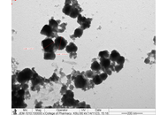
Four niosomal formulations were prepared using Span 60, Tween 60, Pluronic F-127, and Brij 52 (each at a 1:1 cholesterol-to-surfactant ratio). Particle size, zeta potential, and entrapment efficiency were measured. In vitro drug release was analyzed using Franz diffusion cells and fitted to standard kinetic models. Cytotoxicity was assessed by MTT assay in KAIMRC-2, MDA-MB231, and HCT-116 cell lines. Vesicle morphology was visualized by transmission electron microscopy.
Results:
All nano-niosomal formulations showed nanoscale particle sizes (47–292 nm), negative zeta potentials (−18.7 to −26.5 mV), and high entrapment efficiencies (69.8%–76.2%). Release studies indicated Span 60, Tween 60, and Pluronic F-127 followed diffusion-controlled kinetics (Higuchi/Korsmeyer–Peppas model, R2 up to 0.97), while Brij 52 provided a sustained zero-order release (R2 = 0.98). Compared to free osimertinib, all niosomal systems significantly prolonged release. Cytotoxicity studies demonstrated that all formulations enhanced anti-cancer effects, with Span 60-based niosomes exhibiting the greatest potency across cell lines.
Conclusions:
Optimized nano-niosomal encapsulation of osimertinib enables sustained and controlled drug release, improved cellular uptake, and enhanced cytotoxicity in vitro. Differences in surfactant composition critically influence formulation performance, supporting the further development of niosomal osimertinib as a promising strategy for oncological drug delivery applications.
Aim:
Osimertinib’s clinical application is limited by poor aqueous solubility and systemic toxicity. Nano-niosomal formulations can address these challenges by providing controlled release and enhancing delivery. To develop and systematically evaluate nano-niosomal formulations of osimertinib using different surfactants, focusing on physicochemical characteristics, release kinetics, and cytotoxic activity.
Methods:
Four niosomal formulations were prepared using Span 60, Tween 60, Pluronic F-127, and Brij 52 (each at a 1:1 cholesterol-to-surfactant ratio). Particle size, zeta potential, and entrapment efficiency were measured. In vitro drug release was analyzed using Franz diffusion cells and fitted to standard kinetic models. Cytotoxicity was assessed by MTT assay in KAIMRC-2, MDA-MB231, and HCT-116 cell lines. Vesicle morphology was visualized by transmission electron microscopy.
Results:
All nano-niosomal formulations showed nanoscale particle sizes (47–292 nm), negative zeta potentials (−18.7 to −26.5 mV), and high entrapment efficiencies (69.8%–76.2%). Release studies indicated Span 60, Tween 60, and Pluronic F-127 followed diffusion-controlled kinetics (Higuchi/Korsmeyer–Peppas model, R2 up to 0.97), while Brij 52 provided a sustained zero-order release (R2 = 0.98). Compared to free osimertinib, all niosomal systems significantly prolonged release. Cytotoxicity studies demonstrated that all formulations enhanced anti-cancer effects, with Span 60-based niosomes exhibiting the greatest potency across cell lines.
Conclusions:
Optimized nano-niosomal encapsulation of osimertinib enables sustained and controlled drug release, improved cellular uptake, and enhanced cytotoxicity in vitro. Differences in surfactant composition critically influence formulation performance, supporting the further development of niosomal osimertinib as a promising strategy for oncological drug delivery applications.
DOI: https://doi.org/10.37349/ebmx.2025.101355
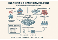
The challenges of conventional animal models and two-dimensional (2D) in vitro cell cultures in effectively forecasting human toxicity have prompted a significant shift towards New Approach Methodologies (NAMs). This development centers on advanced humanized in vitro co-culture models that offer improved physiological relevance for toxicological assessment. This perspective highlights the critical role of biomaterials in the creation of complex microenvironments. This study demonstrates how biomaterials effectively mimic the original extracellular matrix (ECM) through controlled compositional, structural, mechanical, and biochemical signals, thereby enabling the development of sophisticated 3D spheroids, organoids, and Organ-on-Chip systems. These biomaterial-enhanced platforms are essential for precise evaluation of immunotoxicity, as they promote human-specific immune responses and targeted immunomodulation, and for carcinogenicity, as they accurately replicate the tumor microenvironment, affect cancer cell behavior, and enable patient-derived models. Moreover, we underscore the synergistic amalgamation of these biomaterial-based models with omics technologies and computational methodologies (QSAR, AI/ML) for thorough molecular insights and rational design. Despite ongoing challenges in standardization and high-throughput compatibility, the strategic utilization of biomaterials is set to transform predictive toxicology, expedite drug discovery, and promote personalized medicine, thereby diminishing dependence on animal testing and improving human safety.
The challenges of conventional animal models and two-dimensional (2D) in vitro cell cultures in effectively forecasting human toxicity have prompted a significant shift towards New Approach Methodologies (NAMs). This development centers on advanced humanized in vitro co-culture models that offer improved physiological relevance for toxicological assessment. This perspective highlights the critical role of biomaterials in the creation of complex microenvironments. This study demonstrates how biomaterials effectively mimic the original extracellular matrix (ECM) through controlled compositional, structural, mechanical, and biochemical signals, thereby enabling the development of sophisticated 3D spheroids, organoids, and Organ-on-Chip systems. These biomaterial-enhanced platforms are essential for precise evaluation of immunotoxicity, as they promote human-specific immune responses and targeted immunomodulation, and for carcinogenicity, as they accurately replicate the tumor microenvironment, affect cancer cell behavior, and enable patient-derived models. Moreover, we underscore the synergistic amalgamation of these biomaterial-based models with omics technologies and computational methodologies (QSAR, AI/ML) for thorough molecular insights and rational design. Despite ongoing challenges in standardization and high-throughput compatibility, the strategic utilization of biomaterials is set to transform predictive toxicology, expedite drug discovery, and promote personalized medicine, thereby diminishing dependence on animal testing and improving human safety.
DOI: https://doi.org/10.37349/ebmx.2025.101351
This article belongs to the special issue Bioinspired Material for Regenerative Medicine
Aim:
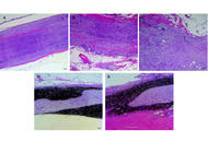
Peripheral nerve injuries (PNIs) often result in a diminished quality of life for those affected and are the most common nervous system injury, with limited treatment options. Regenerative medicine presents novel biomaterial and cell-based therapies to repair the damaged tissue. Graphene oxide (GO), and mesenchymal stem cells (MSCs) have the potential to serve as components to treat PNI. This study evaluates the systemic toxicity of GO and xenogenic human MSCs by analyzing the peripheral blood immune phenotype when a novel nerve guidance conduit (NGC) is implanted in a rat model for six months.
Methods:
A 10-mm long sciatic nerve defect model was created in 8–10-week-old Lewis rats. Four treatment groups were generated: autograft (positive control), poly (lactic-co-glycolic acid) (PLGA) NGC, PLGA NGC with 0.25% GO, and PLGA/GO NGC seeded with 1 × 10⁶ human adipose-derived MSCs. Tail blood was collected before surgery, and at 24 hours, 2 weeks, 2, 3, 5, and 6 months after surgery. Hematological analyses were carried out to evaluate systemic changes, if any, in peripheral immune cell types, namely, T lymphocytes, B lymphocytes, natural killer cells, and macrophages. The treated and contralateral sciatic nerves were excised, paraffin embedded, sectioned, and H&E stained, to identify any local foreign body rejection.
Results:
Treatment groups with GO and MSCs displayed percent total values of peripheral immune cells equivalent to the autograft at each time point. There was no evidence of an inflammatory response in the histological samples.
Conclusions:
The lack of changes in immune phenotype demonstrates a lack of nanotoxicity of the graphene nanoparticles and no evidence of adverse effects due to the MSCs. This was further supported by a lack of local foreign body response at the site of implantation. Overall, the PLGA/GO NGC + MSCs construct is biocompatible for six months in a rat PNI model, exhibiting a potential for clinical translation.
Aim:
Peripheral nerve injuries (PNIs) often result in a diminished quality of life for those affected and are the most common nervous system injury, with limited treatment options. Regenerative medicine presents novel biomaterial and cell-based therapies to repair the damaged tissue. Graphene oxide (GO), and mesenchymal stem cells (MSCs) have the potential to serve as components to treat PNI. This study evaluates the systemic toxicity of GO and xenogenic human MSCs by analyzing the peripheral blood immune phenotype when a novel nerve guidance conduit (NGC) is implanted in a rat model for six months.
Methods:
A 10-mm long sciatic nerve defect model was created in 8–10-week-old Lewis rats. Four treatment groups were generated: autograft (positive control), poly (lactic-co-glycolic acid) (PLGA) NGC, PLGA NGC with 0.25% GO, and PLGA/GO NGC seeded with 1 × 10⁶ human adipose-derived MSCs. Tail blood was collected before surgery, and at 24 hours, 2 weeks, 2, 3, 5, and 6 months after surgery. Hematological analyses were carried out to evaluate systemic changes, if any, in peripheral immune cell types, namely, T lymphocytes, B lymphocytes, natural killer cells, and macrophages. The treated and contralateral sciatic nerves were excised, paraffin embedded, sectioned, and H&E stained, to identify any local foreign body rejection.
Results:
Treatment groups with GO and MSCs displayed percent total values of peripheral immune cells equivalent to the autograft at each time point. There was no evidence of an inflammatory response in the histological samples.
Conclusions:
The lack of changes in immune phenotype demonstrates a lack of nanotoxicity of the graphene nanoparticles and no evidence of adverse effects due to the MSCs. This was further supported by a lack of local foreign body response at the site of implantation. Overall, the PLGA/GO NGC + MSCs construct is biocompatible for six months in a rat PNI model, exhibiting a potential for clinical translation.
DOI: https://doi.org/10.37349/ebmx.2025.101352
Aim:
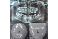
To evaluate the precision of computer-assisted surgery simulation in mandibular condyle reconstruction using a costochondral graft.
Methods:
Ten patients (mean age: 14.5 years) with temporomandibular joint (TMJ) pathology and associated pain were included in the study. All patients underwent TMJ reconstruction using costochondral grafts planned through computer-assisted surgical simulation. Preoperative assessment included mouth opening, facial asymmetry, and the differences between planned and actual mandibular positioning.
Results:
Postoperative mouth opening was significantly improved in all patients, and facial profile modifications were enhanced. The site of the costochondral graft relative to the glenoid fossa was found to be satisfactory in postoperative radiographs, computed tomography images, and quantitative analysis.
Conclusions:
The results of this study demonstrate that virtual surgical planning combined with 3D-printed guiding templates enhanced treatment planning, provided precise osteotomy guidance, facilitated accurate repositioning of bony segments, and improved the contouring of mandibular anatomy in the management of TMJ deformities (ClinicalTrials.gov identifier: NCT06811415).
Aim:
To evaluate the precision of computer-assisted surgery simulation in mandibular condyle reconstruction using a costochondral graft.
Methods:
Ten patients (mean age: 14.5 years) with temporomandibular joint (TMJ) pathology and associated pain were included in the study. All patients underwent TMJ reconstruction using costochondral grafts planned through computer-assisted surgical simulation. Preoperative assessment included mouth opening, facial asymmetry, and the differences between planned and actual mandibular positioning.
Results:
Postoperative mouth opening was significantly improved in all patients, and facial profile modifications were enhanced. The site of the costochondral graft relative to the glenoid fossa was found to be satisfactory in postoperative radiographs, computed tomography images, and quantitative analysis.
Conclusions:
The results of this study demonstrate that virtual surgical planning combined with 3D-printed guiding templates enhanced treatment planning, provided precise osteotomy guidance, facilitated accurate repositioning of bony segments, and improved the contouring of mandibular anatomy in the management of TMJ deformities (ClinicalTrials.gov identifier: NCT06811415).
DOI: https://doi.org/10.37349/ebmx.2025.101350
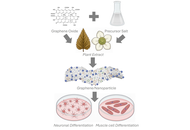
Graphene-based nanomaterials are promising candidates for neuromuscular regeneration due to their electrical conductivity, mechanical strength, and functionalizability. In this perspective, reduced graphene oxide (rGO) nanocomposites decorated with gold nanoparticles (AuNPs) or silver nanoparticles (AgNPs) were synthesized via a one-step green process using Camellia sinensis (tea) extracts. The extracts acted as reducing and stabilizing agents and left bioactive catechins and polyphenols adsorbed on the graphene surface. The resulting nanocomposites combined structural support, electrical conductivity, and bioactive molecular modulation. rGO can provide scaffolding for cell growth, while the retained plant metabolites contributed antioxidant and anti-inflammatory effects. Incorporation of metallic nanoparticles enhanced mechanical strength, surface reactivity, and antimicrobial properties. These multifunctional graphene-metal nanocomposites offer a sustainable and biocompatible platform for guiding neuromuscular regeneration and represent a promising basis for future clinical translation.
Graphene-based nanomaterials are promising candidates for neuromuscular regeneration due to their electrical conductivity, mechanical strength, and functionalizability. In this perspective, reduced graphene oxide (rGO) nanocomposites decorated with gold nanoparticles (AuNPs) or silver nanoparticles (AgNPs) were synthesized via a one-step green process using Camellia sinensis (tea) extracts. The extracts acted as reducing and stabilizing agents and left bioactive catechins and polyphenols adsorbed on the graphene surface. The resulting nanocomposites combined structural support, electrical conductivity, and bioactive molecular modulation. rGO can provide scaffolding for cell growth, while the retained plant metabolites contributed antioxidant and anti-inflammatory effects. Incorporation of metallic nanoparticles enhanced mechanical strength, surface reactivity, and antimicrobial properties. These multifunctional graphene-metal nanocomposites offer a sustainable and biocompatible platform for guiding neuromuscular regeneration and represent a promising basis for future clinical translation.
DOI: https://doi.org/10.37349/ebmx.2025.101349
This article belongs to the special issue Green Nanoparticles for Biomedical Applications
