 Review

Affiliation:
Oswaldo Cruz Foundation, Fiocruz, René Rachou Institute, Fiocruz Minas, Bioactive Natural Products Chemistry Group, Belo Horizonte 30190-002, Brazil
Email: betania.cota@fiocruz.br
ORCID: https://orcid.org/0000-0002-0041-2043
Explor Drug Sci. 2026;4:1008164 DOI: https://doi.org/10.37349/eds.2026.1008164
Received: January 16, 2026 Accepted: April 12, 2026 Published: June 17, 2026
Academic Editor: Mozaniel Santana de Oliveira, Federal University of Pará, Brazil
The article belongs to the special issue Discovery and development of new antibacterial compounds
Brazil harbors remarkable biological and cultural diversity, reflected in a rich body of traditional knowledge regarding the medicinal use of plants. This study synthesized ethnobotanical evidence on plants traditionally used for skin and wound healing in Brazil and examined their convergence with available antibacterial data. An integrative literature review identified twenty ethnobotanical studies, mainly involving rural populations and local residents, reporting 51 plant species traditionally used for skin and wound healing across 22 genera, predominantly native and mainly documented in the Northeastern and Northern regions. The most frequently cited species included Aloe vera (L.) Burm.f. and Anacardium occidentale L., followed by Stryphnodendron adstringens (Mart.) Coville. Fabaceae and Anacardiaceae concentrated the highest number of species with confirmed antibacterial activity, followed by Piperaceae and Euphorbiaceae, which also showed a high proportional representation of active species. A meaningful convergence between ethnobotanical use and experimental antibacterial evidence was observed for more than half of the plants, frequently against Staphylococcus aureus, a key pathogen in wound infections. Antibacterial data were predominantly derived from in vitro assays using non-standardized extracts, and only a limited number of studies reported possible mechanisms of action, such as membrane disruption and biofilm inhibition. Furthermore, few investigations evaluated antibacterial activity in infected wound models or quantified bacterial load reduction in vivo. Future studies should prioritize chemically standardized extracts, testing against resistant clinical strains and mature biofilm models, and validation of safety and therapeutic efficacy in clinical investigations. These findings reveal a gap between traditional use and clinically validated applications, underscoring the urgent need for standardized research approaches and reinforcing Brazil’s potential as a strategic reservoir of bioactive plant resources for primary health care. Addressing these limitations is essential to strengthening the translational basis for the rational use of medicinal plants in primary health care and public health contexts.
Brazil, a country of remarkable biological and cultural diversity, is home to multiple biomes such as the Amazon, Atlantic Forest, Cerrado, Caatinga, and Pantanal, as well as a rich flora with significant therapeutic potential. This biodiversity, combined with a historical formation marked by the presence of Indigenous, African, and European peoples, has resulted in a wide repertoire of traditional knowledge related to the medicinal use of plants. Across different regions of the country, ethnobotanical studies have documented the knowledge of traditional communities, including Quilombola, Indigenous, riverside, and “raizeiros” (traditional Brazilian herbal healers) populations, on plant species used in healthcare practices [1, 2].
The use of medicinal plants in these contexts is often associated with the remote location of these populations and their limited access to healthcare services and industrialized medicines. Furthermore, economic factors and the appreciation of culturally inherited practices contribute to the preservation and transmission of this knowledge [3]. Among the various therapeutic purposes, the traditional application of plant species for the treatment of burns, wounds, and bacterial skin infections stands out as a common practice in the daily lives of these communities, frequently managed with the use of natural resources available in the local environment. Because infection is a major factor that can delay or impair wound healing, the investigation of the antibacterial properties of these traditionally used species becomes particularly relevant.
After an injury, bacterial invasion is frequent, either through migration of the skin microbiota to deeper tissues or exposure to external microorganisms, potentially resulting in infection. Biofilm formation on the wound bed protects bacteria, promotes their proliferation, and increases their tolerance to antibacterial treatments, contributing to lesion persistence and delayed healing, particularly in the context of rising antimicrobial resistance [4].
Even mild wounds and burns can serve as entry points for pathogenic microorganisms and increase the risk of secondary infections. Colonization by Staphylococcus aureus (S. aureus), including methicillin-resistant strains, is associated with increased lesion severity and a higher risk of local and systemic complications. A study conducted in primary healthcare units of the Brazilian Unified Health System found a prevalence of 51.5% for S. aureus and 8.7% for methicillin-resistant S. aureus among patients with infected wounds [5], highlighting the relevance of these infections even outside the hospital setting.
Wounds and burns are considered an important public health concern due to their frequency, possible complications, and socioeconomic impact [6, 7]. Chronic wounds, such as pressure injuries, diabetic foot ulcers, and venous or arterial ulcers, result from disordered healing mechanisms and represent a significant burden on healthcare systems [8, 9]. Burns represent an important cause of morbidity and mortality in Brazil, particularly among vulnerable populations such as children and older adults [6, 9].
The management of skin lesions imposes a significant economic burden and affects patients’ quality of life. Aging, chronic diseases, biofilm formation, and microbial resistance contribute to lesion persistence and increased treatment costs [4, 7, 10].
Although severe infections such as sepsis [9, 11] may occur as a consequence of burns or poorly managed or infected skin lesions, most of these conditions are less severe and are managed outside the hospital setting, within the community, generally involving limited clinical manifestations [12]. In such situations, the use of medicinal plants represents a widely used, accessible, and culturally rooted therapeutic alternative, especially in resource-limited contexts. In addition to their traditional role, several medicinal plants and their secondary metabolites have demonstrated proven antibacterial activity, including against strains resistant to antimicrobials used in clinical practice, with many of their mechanisms of action already characterized [13]. Therefore, integrating ethnobotanical data with experimental antibacterial evidence is essential to critically assess the therapeutic plausibility of these species, validate their traditional use, and identify priority candidates for further pharmacological investigation and potential public health applications.
Although a previous review has addressed the use of medicinal plants for wound healing in Brazil, its scope was limited to the southern region of the country [14]. This review is particularly relevant as it bridges traditional ethnobotanical knowledge with pharmacological evidence, providing a critical synthesis of medicinal plants most frequently reported in Brazilian communities for the treatment of skin conditions with potential bacterial etiology. By systematizing data on antibacterial activity obtained from standardized assays, such as minimum inhibitory concentration (MIC) determinations, this study not only highlights the diversity of traditionally used species but also identifies a restricted group of taxa consistently cited across studies. These taxa represent priority candidates for subsequent investigations aimed at elucidating antibacterial mechanisms of action or exploring their relevance in the context of antimicrobial resistance.
This integrative review followed methodological elements adapted from the Joanna Briggs Institute [15].
To be included in the study, publications had to meet the following criteria: (i) ethnobotanical surveys conducted in Brazilian communities; (ii) reports of plants explicitly indicated for the treatment of wounds, burns or skin problems, with scientific names provided for each species; (iii) articles written in Portuguese or English and published within the last ten years; and (iv) peer-reviewed original research articles. Reviews, book chapters, books, theses, dissertations, monographs, letters to the editor, and case reports were excluded.
The literature search was conducted in three electronic databases: the Virtual Health Library (VHL/BVS), Science Direct, and Scopus. Database searches were updated up to June 2025. The search strategy was adapted to the syntax of each database, using controlled descriptors and free-text keywords combined with Boolean operators (‘OR’ and ‘AND’).
1: Virtual Health Library (VHL/BVS): (“levantamento etnobotânico” OR “estudo etnobotânico” OR “uso tradicional” OR “plantas medicinais”) AND (feridas OR cicatrização OR úlcera* OR erisipela OR “doenças de pele” OR pele).
2: Science Direct: (wound OR wound healing) AND (ethnobotany OR ethnobotanical survey) AND Brazil.
3: Scopus: TITLE-ABS (“ethnobotanical survey” OR “ethnobotanical study” OR “traditional use” OR “medicinal plants”) AND TITLE-ABS (wounds OR healing OR ulcer* OR erysipelas OR “skin diseases” OR dermatological) AND (LIMIT-TO (AFFILCOUNTRY, “Brazil”)) AND (LIMIT-TO (DOCTYPE, “ar”) OR LIMIT-TO (LANGUAGE, “English”) OR LIMIT-TO (LANGUAGE, “Portuguese”)).
Initially, titles and abstracts were evaluated and selected according to the inclusion and exclusion criteria. The papers were retrieved, and after reading, they were evaluated again according to the same criteria.
The Population–Concept–Context (PCC) strategy was applied, where the population refers to Brazilian communities, the concept to medicinal plants, and the context to wounds and skin conditions related to antibacterial use.
This review was designed as a narrative and critical synthesis of the literature addressing two main questions: (i) which medicinal plants are most frequently reported in Brazilian ethnobotanical surveys for the treatment of wounds and skin conditions; and (ii) which of these plants have demonstrated antibacterial activity in scientific studies and which remain unexplored.
The extracted information included the scientific names of plants associated with skin-related indications such as wounds, wound healing, cicatrization, burns, abscesses, furuncles, erysipelas, and skin infections or disorders potentially linked to antibacterial activity. The results were organized according to database source, study location, and region in Brazil, study population, and author with year of publication.
Plant genera represented by more than three species citations across the ethnobotanical studies were then identified, regardless of whether these citations occurred within a single study or across multiple studies, excluding generic records (sp., spp., cf.). The scientific names and plant origins (native or exotic) were verified using the Tropicos (https://www.tropicos.org/) and Reflora (https://reflora.jbrj.gov.br) databases. When synonymous names were identified across studies, records were grouped and harmonized under the currently accepted name to ensure taxonomic consistency.
Additional searches were conducted in SciFinder®, complemented by Google Scholar and the CAPES Journals Portal, using the scientific name of each species in combination with two sets of terms: (i) antibacterial activity (e.g., antibacterial, antimicrobial, MIC) and (ii) wound-healing–related uses (e.g., wound healing, burns, skin). This step aimed to identify representative pharmacological studies related to the reported ethnobotanical uses.
For antibacterial activity, the main focus of this review, only studies reporting MIC values were included, since these values provide a more standardized and comparable measure of antibacterial activity across studies. Although MIC values below 100 µg/mL are often considered indicative of strong activity for crude extracts [16], no universally accepted threshold exists for plant-derived products. Therefore, an inclusive cutoff of ≤ 350 µg/mL was predefined and applied consistently across studies, reflecting the aim of prioritizing species supported by both traditional use and measurable antibacterial activity rather than identifying immediate drug leads.
Studies evaluating wound-healing activity using nanoparticle-based formulations derived from plant extracts, as well as in vitro wound-healing assays (e.g., cell migration or scratch assays), were excluded, as these approaches rely on mechanistic pathways distinct from those of crude plant extracts.
A total of 620 studies were initially identified, and twenty ethnobotanical studies were included in this survey, retrieved from BVS (n = 5), Scopus (n = 6), and ScienceDirect (n = 9) (Table 1, Figure 1). Although the search was updated until June 2025, most of the included studies were published in 2015 and 2016. These studies were predominantly conducted in the Northeastern and Northern regions of Brazil, particularly in the states of Bahia and Ceará. The study populations were mainly composed of local residents, predominantly from rural communities [1, 3, 17–34].
Synthesis of selected ethnopharmacological studies reporting medicinal plants for antibacterial-related skin conditions in Brazil.
| ID | Source | Study location (Brazil) | Brazil region | Study population* | Author(s) |
|---|---|---|---|---|---|
| 1 | BVS | Mato Grosso do Sul | Central-Western | Local residents | [1] |
| 2 | Science Direct | Ceará | Northeastern | Local specialists | [17] |
| 3 | Scopus | Bahia | Northeastern | Specific cultural/professional groups | [18] |
| 4 | Scopus | Ceará | Northeastern | Local specialists | [19] |
| 5 | Science Direct | São Paulo | Southeastern | Local residents | [20] |
| 6 | Scopus | Ceará and Pernambuco | Northeastern | Local specialists | [21] |
| 7 | Science Direct | Mato Grosso | Central-Western | Specific cultural/professional groups | [22] |
| 8 | Scopus | Minas Gerais | Southeastern | Local residents | [23] |
| 9 | Science Direct | Ceará | Northeastern | Local residents | [24] |
| 10 | Scopus | Rio Grande do Sul | Southern | Local residents | [25] |
| 11 | BVS | Alagoas | Northeastern | Herbal vendors from street markets | [26] |
| 12 | Scopus | Roraima and Amazonas | Northern | Local residents | [27] |
| 13 | Science Direct | Bahia | Northeastern | Local residents | [28] |
| 14 | BVS | Paraíba | Northeastern | Herbal vendors from street markets | [3] |
| 15 | Science Direct | Paraná | Southern | Local residents | [29] |
| 16 | Science Direct | Mato Grosso | Central-Western | Local residents | [30] |
| 17 | BVS | Rio Grande do Sul | Southern | Local residents | [31] |
| 18 | Science Direct | Pernambuco | Northeastern | Local residents | [32] |
| 19 | Science Direct | Santa Catarina | Southern | Local residents | [33] |
| 20 | BVS | Maranhão | Northeastern | Local specialists | [34] |
* Local residents (including local population, residents, and rural communities). Local specialists (including informants or local experts, and rural informants). Herbal vendors from street markets (including both terms referring to “raizeiros”). Specific cultural/professional groups (including Quilombola community, families of farmers, riverine experts, and traditional healers).
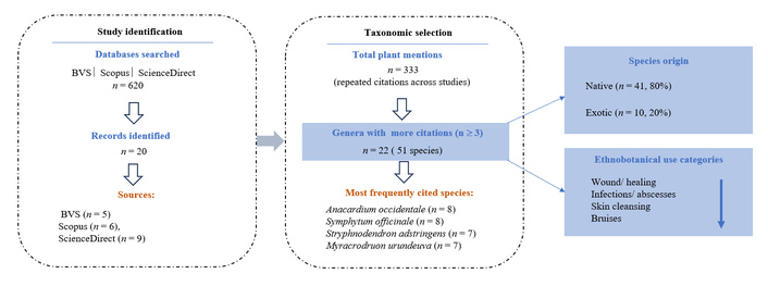
An ethnobotanical evidence schematic summarizing the selection of medicinal plants used for skin conditions in Brazil. A total of 620 records were identified through database searches in BVS, Scopus, and ScienceDirect, from which 20 ethnobotanical studies were included. These studies reported 333 records of plant species related to skin conditions. After applying the selection criterion of the most frequently cited genera (n ≥ 3), 51 species distributed across 22 genera were retained for analysis. Most species were native to Brazil (n = 41), while 10 were exotic. Ethnobotanical uses were grouped into four categories associated with skin conditions of potential bacterial relevance.
In total, 333 mentions of plant species (data not shown) were reported in ethnobotanical studies addressing skin conditions of potential bacterial etiology, including repeated citations of the same species across different ethnobotanical studies for skin-related treatments. After applying the selection criterion of the most frequently cited genera (n ≥ 3), 51 valid species were identified (Supplementary material), distributed across 22 genera and 15 families, of which 41 were native, and 10 were exotics. These results indicate that ethnobotanical knowledge, although diverse, tends to be concentrated in a smaller subset of taxa, many of which are part of the Brazilian flora with recognized medicinal importance.
The most frequently cited species (Supplementary material) were Aloe vera (L.) Burm.f. and Anacardium occidentale L. (A. occidentale L., 8 reports each), followed by Symphytum officinale L. and Stryphnodendron adstringens (Mart.) Coville (7 reports each), Myracrodruon urundeuva Allemão (6 reports), Alternanthera brasiliana (L.) Kuntze and Dysphania ambrosioides (L.) Mosyakin & Clemants (5 reports each), and Ximenia americana L., Psidium guajava L., and Calendula officinalis L. (4 reports each). This pattern shows that in some cases, such as Symphytum and Stryphnodendron, citations were strongly concentrated in a single species, while in others, such as Alternanthera, Croton, Piper, and Sida, references were spread across different species, indicating that their cultural importance is recognized more at the genus level than in a single dominant taxon.
The ethnobotanical indications reported for the 51 selected species were grouped into four main categories. Although the search strategy retrieved a broad range of skin-related uses, the present analysis focused on conditions of potential bacterial relevance. Most species were associated with wounds and healing (37 species). Smaller proportions referred to skin conditions and cleansing (17 species), infections and abscesses such as furuncles and erysipelas (14 species), burns (6 species), and bruises (1 species), with some species reported for more than one indication.
Most surveys were conducted in Northeastern and Northern Brazil, where native species such as M. urundeuva and S. adstringens were frequently reported. In contrast, exotic and widely cultivated species like A. vera and Symphytum officinale were more common in studies from other regions, such as the Southeastern and Southern regions. These findings reflect the geographic distribution of the surveys rather than nationwide patterns of use.
This section explores the alignment between ethnobotanical indications for skin wounds and skin disorders and experimental evidence of wound-healing and antibacterial activities. Traditional uses and regional occurrence of the selected plant species are first presented, followed by in vivo wound-healing studies and in vitro antibacterial evaluations using broth microdilution assays, considering MIC values ≤ 350 µg/mL. Additional data and extended analyses are available in the Supplementary material.
The three Alternanthera species cited in the selected reports, Alternanthera brasiliana (L.) Kuntze (“Benzetacil”), A. dentata (Moench) Stuchlík ex R.E. Fr. (“penicilina”), and A. ramosissima (Mart.) Chodat (“ampicilina”) are native species reported for the treatment of skin conditions, mainly using their leaves [18, 22, 28–31]. An experimental study in Wistar rats supports the wound-healing potential of A. brasiliana, as the topical application of a 20% hydroalcoholic leaf extract incorporated into a 2% carbopol gel significantly accelerated re-epithelialization and collagen deposition when compared to vehicle control. Despite this healing activity, the same extract exhibited a relatively high MIC value (2,000 µg/mL) against S. aureus [35]. Similar healing effects were reported with a 5% (w/w) ointment containing the methanolic leaf extract [36]. Regarding antibacterial activity, the most significant effect was observed against Mycobacterium smegmatis, with an MIC of 15.6 µg/mL [37].
Dysphania ambrosioides (L.) Mosyakin & Clemants (Chenopodium ambrosioides syn.) is native to Central and South America. All parts of this plant, popularly known in Brazil as “erva-de-santa-maria” or “mastruz”, are traditionally used to treat skin conditions such as burns and bruises, as well as for wound healing, in the Central-Western, Northeastern, and Southeastern regions [1, 18, 20, 22, 28]. In rat excisional wound models, topical application of C. ambrosioides extracts accelerated wound contraction and tissue repair. Ethanolic extracts (1–5%) reduced lesion area at later healing stages [38], while an aqueous leaf extract promoted faster early wound contraction and improved granulation tissue organization and partial re-epithelialization [39], supporting the traditional use of this species in skin wound care.
Beyond its direct effects on tissue repair, antibacterial activity may contribute to the healing process by reducing microbial burden at the wound site. In this context, the hydroethanolic leaf extract inhibits Helicobacter pylori (MIC 200 µg/mL) [40], while the essential oil from aerial parts exhibits broad-spectrum antibacterial activity against wound-associated pathogens, including MDR strains such as S. aureus, Pseudomonas aeruginosa, and Acinetobacter baumannii, with MICs ranging from 90 to 230 µg/mL. Its chemical composition is predominantly monoterpenes (60.75%), with α-cyclogeraniol acetate among the main constituents [41].
The species Anacardium humile A. St.-Hil. and A. occidentale L. are native to Brazil, with A. occidentale being widely used in several regions for wound healing [3, 17–19, 22, 26, 27, 30]. Experimental evidence supports this traditional use, as topical application of hydroalcoholic leaf extracts of A. occidentale (1–2%) promoted wound healing in rat models, enhancing wound contraction and collagen deposition compared with control groups [42, 43].
In parallel, organic extracts from Anacardium species have demonstrated relevant antibacterial activity. Different parts of A. occidentale exhibit consistent antibacterial effects, with MIC values below 350 µg/mL. Methanolic extracts of the root bark inhibited both Gram-positive and Gram-negative bacteria, including S. aureus, Enterococcus faecalis, P. aeruginosa, and Escherichia coli, with MICs ranging from 10 to 40 µg/mL [44]. Ethanolic leaf extracts showed moderate to high activity against multiple bacterial strains (MIC 12.5–50 µg/mL) [45], while acetone extracts from nuts exhibited remarkable potency against S. aureus (MIC 0.00188–0.00375 µg/mL) [46]. In addition, acetone leaf extracts were active against Mycoplasma mycoides (MIC 310 µg/mL) [47], and hexane and methanolic extracts from aerial parts inhibited S. aureus and E. faecalis, with lower MICs observed for methanolic preparations [48].
Although less extensively studied, A. humile also exhibits antibacterial potential. Ethanolic leaf extracts inhibited the growth of S. aureus (MIC 4.1 µg/mL) and P. aeruginosa (MIC 8.2 µg/mL) [49], while tannins obtained from acetone extracts showed activity against P. aeruginosa (MIC 4.1 µg/mL) and E. faecalis (MIC 2 µg/mL) [50].
Myracrodruon urundeuva Allemão (Anacardiaceae), popularly known as “aroeira-do-sertão”, “aroeira-preta”, or “aroeira-do-campo”, is traditionally used to treat skin injuries, wounds, and furuncles, mainly in Northeastern Brazil [3, 17–19, 22, 26]. Experimental evidence supports this traditional use, as a carbopol gel containing stem bark extract promoted greater wound contraction and collagen formation after 14 days in an excisional wound healing model in rats [51]. Similarly, a 10% cream prepared from a stem bark decoction reduced wound area and increased collagen deposition in excisional skin wounds of male Wistar rats, demonstrating efficacy in the wound repair process [52].
In addition, antibacterial effects of M. urundeuva are associated with ethanolic extracts and essential oils from the leaves, which inhibited Gram-positive and Gram-negative wound-associated pathogens, including S. aureus, Staphylococcus epidermidis, and P. aeruginosa, with MIC values ranging from 4.1 to 220 µg/mL [49, 53].
Two species of the genus Schinus were reported in association with the treatment of wounds and skin disorders from selected ethnobotanical studies. Schinus molle L., a native species and known as the pepper tree (“periquiteira”), showed use in the Southern region, with flowers employed for wound treatment [30]. Schinus terebinthifolius Raddi (“aroeira-rosa”), also native, presented a higher number of records and a broader range of uses, involving different plant parts and indications such as wounds, furuncles, and skin conditions across distinct regions of the country [18, 22, 28, 29].
The aqueous extract of S. molle L. aerial parts, incorporated into a 5% hydrogel formulation, increased wound closure rates in Wistar rats, promoting re-epithelialization, fibrosis, and neovascularization in the regenerated epidermal and subepidermal tissue. Although the same extract was tested against six microorganisms, including S. aureus and S. epidermidis, antibacterial activity was observed only against Citrobacter freundii and E. faecalis (MIC 1,560 µg/mL), a concentration comparable to gentamicin but considered high in broth microdilution assays [54]. In another study, methanolic extracts from leaves, bark, and flowers were active against S. aureus (MIC 62.5–250 µg/mL) [55]. Hexane extracts from fruits inhibited M. tuberculosis (MIC 125 µg/mL), while extracts from flowers, bark, and fruits were active against Streptococcus pneumoniae (MIC 62.5–250 µg/mL) [56]. Altogether, these findings highlight the antimicrobial potential of S. molle, with bioactive compounds distributed across extracts of different polarities.
Earlier experimental evidence indicates that not all preparations of S. terebinthifolius promote wound repair. In a rat excisional wound model, topical application of a hydroalcoholic bark extract delayed re-epithelialization and resulted in larger wound areas compared with saline-treated controls, despite increased mononuclear cell infiltration at later stages [57]. In contrast, in a murine model of S. aureus—infected wounds, topical administration of an N-acetylglucosamine-binding lectin isolated from S. terebinthifolius leaves at concentrations of 32 and 64 µg/mL accelerated wound healing and reduced inflammatory markers, including IL-6, monocyte chemoattractant protein-1, tumor necrosis factor alpha (TNF-α), and vascular endothelial growth factor (VEGF) [58]. Likewise, the leaf essential oil demonstrated wound-healing activity in excisional wound models, promoting faster lesion closure through modulation of inflammation, angiogenesis, and collagen deposition [59].
Previously, Nunes et al. [58] showed that the lectin isolated from S. terebinthifolius leaves exhibited in vivo antimicrobial activity in a murine model of S. aureus—infected wounds, with a dose-dependent reduction in bacterial load and infection severity, accompanied by decreased exudate formation and local inflammation. Beyond these protein-based preparations, the ethanolic leaf extract inhibited Acinetobacter baumannii, showing 80% growth inhibition at 256 µg/mL, and bioassay-guided fractionation led to the isolation of pentagalloyl glucose. This compound displayed antibacterial activity, likely mediated by iron chelation, inhibiting both carbapenem-resistant and susceptible A. baumannii (MIC 64–256 µg/mL), as well as P. aeruginosa (MIC 16 µg/mL) and S. aureus (MIC 64 µg/mL) [60].
The three Himatanthus species cited in ethnobotanical surveys are native and widely used in Northeastern, Northern, and Central-Western Brazil, particularly through latex and leaf preparations, reflecting a consistent cultural association with wound care across regions [19, 22, 27, 34]. This convergence suggests that wound-healing applications may represent a traditional pharmacological signature of the genus.
Several studies consistently reinforce the traditional wound-healing use of Himatanthus species. In Himatanthus drasticus (Mart.) Plumel (“sucuúba” or “janaguba”), both latex- and leaf-based preparations accelerated wound closure and enhanced fibroblast activity in murine models. Ointments containing 2% soluble latex proteins promoted complete re-epithelialization within 14 days and increased fibroblast density [61], while ethanolic leaf extracts at 50–100 mg/kg similarly improved lesion reduction and collagen deposition after 21 days [62]. Himatanthus sucuuba (Spruce ex Müll. Arg.) latex also showed reproducible regenerative effects, as twice-daily topical application for 10 days stimulated wound healing, and phytochemical profiling revealed 24 constituents, mainly flavonoids, supporting its bioactive potential [63]. Complementary findings from a 15-day treatment with H. sucuuba latex demonstrated early epithelialization, reduced inflammation, and increased collagen deposition and fibroblast abundance compared with zinc oxide cream [64]. Likewise, the ethanolic leaf extract of H. obovatus (Müll. Arg.) Woodson formulated in carbopol gel reduced lesion size and increased collagen content and fibroblast levels by day 21 [62].
Regarding antibacterial activity, only H. sucuuba has demonstrated meaningful inhibition of clinically relevant bacteria in broth microdilution assays. The aqueous latex fraction inhibited Staphylococcus species, including S. aureus, S. epidermidis, and Staphylococcus haemolyticus, at 350 µg/mL [65], whereas gallic acid isolated from this fraction showed markedly greater potency (MIC 31 µg/mL against S. aureus and S. epidermidis). Earlier studies of the methanolic root extract led to the isolation of the iridoids allamandin and plumericin, which exhibited broader antibacterial activity, with MICs ranging from 10 to 40 µg/mL against S. aureus, P. aeruginosa, and E. coli [66]. Collectively, these findings indicate that H. sucuuba may contribute to its traditional use in infected or inflamed wounds.
The species of the genus Aloe recorded in ethnobotanical surveys, all exotic, are also mentioned for the care of burns, infections, and cleansing, with a predominance of leaf use. Aloe arborescens Mill. is associated with wound and burn care in the Southern region [32]. Aloe vera (L.) Burm.f. (synonym Aloe barbadensis Mill.) accounts for a total of nine ethnobotanical records, distributed across the Central-Western, Northeastern, Southeastern, and Southern regions of Brazil [1, 3, 19, 22, 23, 25, 26, 31, 33].
In vivo studies demonstrate that preparations obtained from the leaves of Aloe arborescens, when applied topically, exhibit relevant wound-healing activity in Wistar rats, both in models of third-degree burns and surgical wounds [67, 68]. Consistently, these studies indicate promotion of re-epithelialization, acceleration of wound closure, increased angiogenesis, and improved organization of collagen fibers during the tissue repair process. Extracts prepared using a dichloromethane: methanol (1:1) mixture, as well as acetone leaf extracts, demonstrated activity against H. pylori, with MIC values ranging from 130 to 250 µg/mL [69, 70], and against different enteric and Gram-positive pathogens, notably S. aureus (MIC 18 µg/mL) and Shigella flexneri (MIC 18 µg/mL) [71]. Additionally, alcoholic leaf extracts exhibited relevant antibacterial activity against S. aureus (MIC 70 µg/mL) and E. faecalis (MIC 140 µg/mL) [72].
Although several systematic reviews and meta-analyses have evaluated the efficacy of A. vera in wound healing among patients with second-degree burns [73], no meta-analyses specifically addressing the healing of non-burn cutaneous wounds were identified. In contrast, evidence from veterinary clinical studies indicates that the topical application of A. vera leaf gel in dogs and cats accelerates wound shrinkage, reduces healing time, and decreases lesion severity when compared with silver sulfadiazine [74].
Based on the selected studies, simple preparations obtained from the leaf gel and ethanolic extracts of Aloe vera showed antibacterial activity against oral pathogens, including Streptococcus mutans, Aggregatibacter actinomycetemcomitans, and Porphyromonas gingivalis, as well as against representative Gram-positive and Gram-negative bacteria, with MIC values generally below 60 µg/mL [75]. Notably, activity was also reported against multidrug-resistant P. aeruginosa and Bacillus isolates, with most of which were inhibited at concentrations ≤ 200 µg/mL [76, 77].
Calendula officinalis L., an exotic species, appears in four ethnobotanical records associated with the treatment of skin lesions, burns, and scarring, with predominant use of the flowers across Northeastern, Southeastern, and Southern Brazil [19, 23, 31, 33].
The wound-healing activity of C. officinalis has been demonstrated in both clinical and experimental models. In a clinical trial involving patients with acute traumatic wounds healing by secondary intention, the topical application of a standardized 2% extract prepared from the flowers of C. officinalis significantly increased the rate of wound closure and reduced the time to complete epithelialization [78]. Complementarily, the oral administration of capsules containing 2 g of C. officinalis extract, also derived from the flowers of the species, for 14 days in patients with second-degree burns, resulted in significantly greater improvements in wound-healing scores compared with placebo [79].
Although several studies report antibacterial activity for C. officinalis, including investigations targeting periodontal pathogens [80], only the study by Larçin et al. [81] presented MIC values within the range established in this review. In that study, the methanolic flower extract exhibited an MIC of 256 µg/mL against Erwinia amylovora, a Gram-negative bacterium responsible for fire blight in apples and pears. The authors identified chlorogenic acid, caffeic acid, rutin, and salicylic acid as constituents common to both methanolic and aqueous flower extracts, with the higher relative amount of rutin in the methanolic extract correlating with its greater antibacterial activity.
Ethnobotanical studies indicate that Symphytum officinale L., an exotic species in Brazil and popularly known as confrei, is mentioned in seven surveys conducted across the Central-Western, Southeastern, Northeastern, and Southern regions of the country. The leaf is the most frequently used plant part, and the species is even reported for the treatment of infected wounds [3, 22, 23, 30, 31, 33, 34].
Preclinical studies in animal models and humans demonstrate that extracts of S. officinale accelerate wound healing through modulation of inflammatory infiltrate, significant enhancement of collagen deposition [82], and improved tissue organization associated with antioxidant activity [83]. In humans, standardized creams containing 10% of the aerial-part extract reduced abrasion healing time by nearly 3 days [84]. A more recent study using creams prepared with an ethanolic root extract (10% and 20%) accelerated regeneration in Wistar rats and identified polyphenolic compounds by LC-ESI+-MS analysis, such as salvianolic acids, rosmarinic acid, and caffeic acid, in addition to quantifying allantoin. The extract demonstrated antimicrobial activity against Gram-positive and Gram-negative bacteria; however, the MIC values reported were comparatively high, ranging from 765.38 to 6,123.01 µg/mL [85]. These findings are consistent with the limited number of studies reporting pronounced antibacterial activity for S. officinale. Although Thibane et al. [86] described lower MIC values, 98 µg/mL for methanolic and aqueous leaf extracts against E. coli, such results appear uncommon in the available literature and should be interpreted with caution until more comprehensive searches are undertaken.
Maytenus ilicifolia Mart. ex Reissek (syn. M. officinalis) and Maytenus rigida Mart., known respectively as “Espinheira-santa” [87] and “Bom-nome” [26], are native Brazilian species traditionally used for wound healing, particularly their leaves and stem barks [24, 25, 30]. Maytenus ilicifolia (syn. Maytenus officinalis) and Maytenus rigida Mart., known respectively as “espinheira-santa” [87] and “bom-nome” [26], are native Brazilian species traditionally used in wound healing, particularly through leaf and stem bark preparations [25, 26, 31]. Topical application of a hydroalcoholic leaf extract of M. ilicifolia, rich in phenolic compounds and tannins, accelerated skin wound closure in BALB/c mice at a 4% concentration, using 100 mg of ointment per wound after 3 and 7 days of treatment [88]. In addition, the polar methanolic leaf extract inhibited B. cereus (MIC 156 µg/mL) [87]. In contrast, experimental evaluation of the ethanolic bark extract of M. rigida in a rat excisional wound model did not demonstrate significant wound-healing or anti-inflammatory effects, indicating that cicatrizing activity may depend on species, plant part, or preparation method [89]. Despite these findings, antibacterial activity reported for both species against pathogens relevant to wound infections generally involved high MIC values, frequently exceeding 1,000 µg/mL [90, 91].
In this review, four Croton species emerged as being associated with skin problems: Croton heliotropiifolius Kunth [23, 31], Croton salutaris Casar. [1], Croton urucurana [22, 23] and Croton zehntneri Pax & K. Hoffm. [32] are used in different Brazilian regions for wound healing, skin cleansing, and the treatment of furuncles, with applications involving especially latex.
Experimental studies support the wound-healing properties traditionally attributed to Croton species as medicinal agents. Topical application of a formulation containing 20% essential oil from C. zehntneri leaves enhanced wound closure in mice by increasing fibroblast activity, angiogenesis, and collagen deposition, an effect largely attributed to its major constituent trans-anethole [92]. The ethanolic extract of C. urucurana stem bark also displays consistent regenerative effects: ointments containing 5–10% extract accelerated healing in IL-10 knockout mice, promoting fibroblast proliferation, neovascularization, and deposition of type I/III collagen [93]. In a complementary venom-induced injury model in Swiss mice, higher concentrations of the stem bark ethanolic extract (10, 20, and 40%) markedly increased lesion contraction and led to early re-epithelialization and granulation tissue formation, further confirming its pro-angiogenic and fibroblast-stimulating activity [94].
Among the evaluated species, the essential oil from aerial parts of C. heliotropiifolius was tested against several Gram-positive and Gram-negative bacterial strains; however, it exhibited antibacterial activity only against B. cereus, with a MIC of 62.5 µg/mL [95]. The ethanolic leaf extract of C. zehntneri inhibits S. aureus with a MIC of 64 µg/mL, reinforcing the traditional use of this species and the presence of phenolic metabolites, such as tannins and flavonoids, associated with antibacterial activity [96]. Its essential oil has high content of the monoterpenoid estragole (≥ 80%), which inhibits E. coli, S. aureus, Streptococcus β-haemolyticus, and S. flexneri, with MIC values below 50 µg/mL [97]. In contrast, C. urucurana exhibited a broader spectrum of action. The fresh latex, as well as the 75% ethanolic and chloroform extracts of the stem bark, showed activity against various clinically important bacteria, with MICs ranging from 125 to 250 µg/mL, including P. aeruginosa and different Staphylococcus species [98]. A recent study demonstrates the antibiofilm activity of the hexane extract, and the isolated compound α-costol from the species [99]. On the other hand, no antibacterial activity has been identified for C. salutaris.
Two native Brazilian species of Anadenanthera, Anadenanthera colubrina (Vell.) Brenan and Anadenanthera peregrina (L.) Speg., and their respective varieties (Anadenanthera colubrina var. cebil and Anadenanthera peregrina var. falcata) were associated with skin care and wound healing. The stem bark, particularly the inner bark, was the most frequently used plant part, although roots and resin were also reported for A. peregrina. Most citations were concentrated in the Central-West region, with only one additional record from the Northeast [1, 22, 26].
The 5% ethanolic bark extract of A. colubrina shows evidence of wound-healing activity in an animal model by topical application, with increased collagen deposition in the lesions, neutrophil infiltration, enhanced macrophage infiltration, and elevated IL-10 levels [100, 101]. The strongest evidence for the genus comes from studies demonstrating anti-S. aureus activity. The crude ethyl acetate extract from the leaves of A. colubrina var. cebil showed an IN50 of 312.5 µg/mL, and two of its isolated constituents, hyperoside and proanthocyanidin, displayed even stronger activity, with values of 62.5 µg/mL [102]. In addition, the aqueous leaf extract of A. peregrina also exhibited activity against S. aureus, with a MIC of 310 µg/mL [103].
The only species cited was Caesalpinia ferrea C. Mart., a native Brazilian species popularly known as “jucá or pau-ferro”, whose bark, fruits, and seeds are traditionally used in the Central-Western and Northeastern regions of the country [3, 22, 26]. Experimental studies consistently demonstrate that topical treatment with different preparations of C. ferrea, including a 12.5% ethanolic fruit extract [104], 24% pod powder [105], bark extracts and bark powder mixed with petrolatum (1:2, w/w) [106], as well as bark and pod extracts enriched with 0.1% polysaccharides (also reported as Libidibia ferrea), accelerates wound closure, reduces inflammation, and enhances collagen deposition and fibroblast proliferation in goat, rabbit, and Wistar rat wound models [107, 108].
In the study using an ointment prepared with bark mixed into petroleum jelly, the microbiological analysis performed on day 14 revealed the absence of S. aureus in comparison to the control group [105], suggesting activity against S. aureus. Consistently, methanolic and ethanolic extracts from fruits or pods showed activity against oral and clinically important bacteria, including S. mutans and E. faecalis, with MIC values within the 40–125 µg/mL range [109–111].
The Copaifera species cited for wound treatment are native. Copaifera cearensis Huber ex Ducke (“copaíba”) is used in the Northeastern region for healing wounds [3], while Copaifera langsdorffii Desf. (“copaíba or “pau-d´oleo, podoi”) shows more diverse uses, including erysipelas, with different plant parts employed in the Northeastern [20] and Central-Western regions [22, 26]. Copaifera multijuga Hayne (“copaíba”), recorded in the Northern region [27], is mentioned as a skin healer based on the stem bark and stem oil.
Both C. langsdorffii oleoresin (4%) [112] and hydroalcoholic extract 10% of the leaves and 10% oleo-resin creams [113] demonstrate a beneficial effect on wound healing in Wistar rats, with the latter preparation demonstrating an anti-inflammatory activity boost in reepithelialization, angiogenesis, cell proliferation, and extracellular matrix remodeling. Subsequently, both the 10% hydroalcoholic extract and the 10% oil-resin creams exhibited wound-healing potential in horse skin wounds after 14 days [114]. In addition, the carbopol gel containing 1% C. langsdorffii accelerated wound healing in BALB/c mice [115]. In contrast, although the oleoresin of C. multijuga contributed positively to second-intention skin wound healing in Wistar rats, it was less effective than the reference agent (nitrofurazone) [116]. Nevertheless, a double-blind, randomized, and controlled clinical trial using a commercial formulation based on C. multijuga (CopaibaPolyHy-2) demonstrated significantly faster healing time, increased granulation and epithelial tissue formation, and reduced exudate [117].
The antimicrobial evidence for C. langsdorffii includes relevant activity of trunk oleoresin (MIC 200 and 125 µg/mL, respectively) [118, 119] and the ethyl acetate leaf extract (MIC 32 µg/mL) against S. aureus [120]. Furthermore, the trunk oleoresins, including those for C. multijuga, demonstrated efficacy against M. tuberculosis, with MIC values ranging from 62.5 to 250 µg/mL [121]. These findings, together with studies reporting the isolation of diterpenes from the species with activity against several Gram-positive and Gram-negative bacteria and M. tuberculosis, reinforce its broad antimicrobial potential, particularly against clinically relevant pathogens [122–124].
Hymenaea courbaril L., a native species popularly known as “jatobá” in Brazil, is traditionally used in the Northeast, where fruits, stem bark, and inner bark are applied for wound care [21, 26], and in the Center-West, where the resin is also employed alongside the bark [22]. The species’ sap accelerated wound closure, promoting fibroblast proliferation and migration and re-epithelialization by day 14 [125]. Additionally, seed-derived xyloglucan enhanced wound healing in diabetic mice by advancing re-epithelialization, improving dermal organization, and increasing type I collagen deposition [126].
The methanolic extract of the leaves exhibited substantially higher potency, with an MIC of only 16 µg/mL against S. aureus and P. aeruginosa [127]. For the stem bark, both methanolic and hexane extracts showed MIC values of 200 µg/mL against M. tuberculosis, while the hexane and dichloromethane extracts presented values ranging from 25–200 µg/mL [128]. In addition, the essential oil extracted from the fruit peels exhibited MIC values around 200 µg/mL [129]. In contrast, the hydroalcoholic extracts of the bark and starchy pulp of H. courbaril fruits exhibited moderate antibacterial activity, with MIC values of 350 µg/mL against S. aureus, E. coli, and P. aeruginosa [130]. Notably, during wound-healing assays with seed-derived xyloglucan, microbiological evaluation showed no contamination of the wound area throughout the 12-day treatment period, suggesting a protective antimicrobial effect in the wound environment [126].
Stryphnodendron adstringens (Mart.) Coville, frequently referred to by its synonym Stryphnodendron barbatiman, is a native plant cited across three different regions of Brazil for the treatment of skin problems, particularly using its bark [1, 3, 18, 22, 23, 26, 28].
Multiple studies consistently demonstrate that bark extracts of S. adstringens exert a range of effects related to tissue repair in Wistar rat models, regardless of the extraction solvent employed. These include aqueous crude extracts formulated as gels [131], hydroethanolic extracts incorporated into 5% gels [132], ethyl acetate fractions obtained from acetone:water (7:3) extracts formulated as ointments or 1% gels [133, 134], and standardized formulations such as the 50% dry extract Fitoscar™ [135]. Overall, the findings indicate mechanisms that act concomitantly on the inflammatory, proliferative, and remodeling phases of wound healing, including enhanced migration of fibroblasts, stimulation of angiogenesis, and increased collagen deposition. Notably, a high concentration of proanthocyanidins was identified by mass spectrometry in the acetone:water (7:3) extract, compounds known to modulate oxidative pathways, stimulate fibroblasts, and regulate key angiogenic proteins such as VEGF [133]. In contrast, the hydroalcoholic extract was shown to be rich in phenolic constituents such as tannins and flavonoids, including gallic acid, caffeic acid, and rutin, all recognized for their anti-inflammatory potential [132].
Different preparations obtained from the bark demonstrated relevant antibacterial activity, especially against Staphylococcus spp. and bacteria associated with oral infections. The aqueous and ethanolic bark extracts showed expressive activity against pathogens such as S. aureus, S. mutans, and Actinobacillus actinomycetemcomitans, with MIC values below 60 µg/mL [136]. Subsequently, a similar potency was demonstrated in the work of Cruz et al. [137] for both the hydroalcoholic extracts of bark and leaves. On the other hand, the ethanolic bark extract and the acetone/water (7:3) bark extract, as well as the aqueous and ethyl acetate fractions obtained from the acetone/water extract, inhibited S. aureus with MIC values between 125 and 250 µg/mL [138, 139].
Among the four Sida species referenced for skin treatment, all are native and recorded in the Southeastern and Northeastern regions of Brazil, with predominant use of the leaves. Two of them, Sida planicaulis Cav. and Sida rhombifolia L., are traditionally employed for the treatment of furuncles [18, 20].
For S. cordifolia L., the 10% methanolic extract of the aerial parts in hydrogel improved wound contraction, collagen deposition, tensile strength, and epithelialization in diabetic Wistar rats, whereas the 10% ethanolic whole-plant ointment produced similar effects in excision, incision, and burn wound models [140, 141]. For S. rhombifolia, ointments containing ethanolic extract at 25–50% and the aqueous preparation of the leaves, applied directly, also accelerated wound healing and induced marked fibrosis and collagenization in mice [142].
The ethanol extract of S. cordifolia leaves showed antibacterial activity against B. subtilis, with a MIC of 98 µg/mL [143]. In contrast, for Sida rhombifolia, the methanol extract of the aerial parts displayed weak activity (> 500 µg/mL), and only the enriched ethyl acetate fraction demonstrated lower MIC values, such as 64 µg/mL [144]. Agar-diffusion assays reported for S. rhombifolia suggest a preliminary antibacterial trend that deserves further investigation using standardized microdilution assays; however, such findings must be interpreted cautiously given the methodological limitations inherent to diffusion-based methods [145].
P. guajava L. (Myrtaceae) is widely distributed worldwide, and in Brazil, leaves, bark, and shoots are used to treat skin lesions and wound healing [17, 19, 22, 23]. An emulgel formulation with 1% of guava leaf oil, containing D-limonene, β-caryophyllene, and 1,8-cineole, enhanced wound healing in nondiabetic and diabetic Male Wistar rats. It promoted the production of collagen type I and increased superoxide dismutase, and a decrease in the expression of inflammatory cytokines and enzymes such as TNF-α, IL-1β, and IL-6. According to the authors, the 1% leaf oil emulgel formulation was considered active against Gram-positive and Gram-negative microorganisms in agar plate assays [146].
Previous reports have shown that organic leaf extracts of P. guajava obtained with a methanol–chloroform mixture (1:1) exhibited antibacterial activity against B. subtilis, with a MIC of 250 µg/mL. In the same study, a 10% (w/w) carbopol gel formulation promoted wound area contraction and faster epithelialization, achieved within 9 days [147]. Additionally, ethyl acetate and acetone leaf extracts inhibited E. coli biofilm formation, with BI50 values of approximately 60 µg/mL, and promoted complete wound closure in BALB/c mice by day 14 at doses particularly for the acetone extract [148].
The bark and inner bark of Ximenia americana L. (“ameixa, ameixeira”) have been consistently cited in ethnobotanical studies as being traditionally used for the treatment of burns and wound healing in the Northeastern region of Brazil [3, 5, 16, 33]. Experimental evidence supports these traditional uses, as hydroalcoholic extracts (2.5%) prepared from leaves, wood, and stem bark promoted the healing of surgically induced skin wounds in Wistar rats, characterized by reduced inflammatory cell infiltration and increased fibroblast proliferation [149]. In a complementary experimental model, a hydroalcoholic extract prepared from the branches of X. americana and incorporated into a 10% Lanette-based cream significantly increased the number of fibroblasts, collagen fibers, and blood vessels, accelerating the wound-healing process [150]. In the study reported by Palma et al. [149], the stem bark extract exhibited the most pronounced fibroplastic response, and phytochemical analyses revealed the presence of flavonoids, saponins, and steroids, with tannins suggested as the main compounds potentially responsible for the observed biological activities.
Notably, the most potent antibacterial activity reported for root-derived extracts was observed against enteric pathogens, including Proteus mirabilis, Shigella boydii, S. flexneri, and Salmonella typhi, with low MIC values (25–50 µg/mL) [151]. In the same study, these root fractions were also evaluated in antidiarrheal assays, supporting the relevance of their antibacterial activity against gastrointestinal pathogens. In addition, the dichloromethane root extract exhibited activity against M. tuberculosis, with a MIC value of 125 µg/mL [152].
Five native species of the genus Piper, Piper aduncum L., Piper amalago L., Piper gaudichaudianum Kunth, Piper peltatum L., and Piper umbellatum L., have been ethnobotanically reported as treatments for wounds, skin infections, erysipelas, and related conditions in the Northern, Northeastern, and Southern regions of Brazil [19, 27, 29].
For P. aduncum L. (“pimenta de macaco”), ointments prepared from ethanolic leaf extracts at concentrations of 5, 10, and 15% promoted wound healing in mice, reducing wound size, epithelialization time, and improving collagen fiber deposition and fibroblast scores [153]. In addition, hexane extracts obtained from the leaves and inflorescences exhibited broad-spectrum antibacterial activity against Gram-positive and Gram-negative bacteria, with notable activity of the inflorescence extract against E. coli (MIC 20 µg/mL) [154, 155] and multidrug-resistant S. aureus (MIC 16 µg/mL) [156].
Regarding P. amalago L., popularly known in Brazil as “jaborandi-manso,” a case report described the successful topical use of an aqueous leaf extract in the healing of a lacerated wound in a patient with type 2 diabetes mellitus after 15 days of treatment [157]. More recently, oral administration of an ethanolic leaf extract (100 mg/kg) in Wistar rats reduced the extent of cutaneous necrosis and improved the wound-healing process [158]. From an antimicrobial perspective, the leaf essential oil showed activity against B. cereus (MIC 313 µg/mL), while the chloroform extract inhibited Alicyclobacillus acidoterrestris with a MIC of 62.3 µg/mL, a Gram-positive bacterium of relevance to the food industry [159, 160].
P. gaudichaudianum Kunth, popularly known as “pariparoba” or “jaborandi,” lacks direct evidence supporting its use for skin wound healing. However, essential oils obtained from its aerial parts inhibited neutrophil migration in an antichemotactic assay in rats, indicating anti-inflammatory potential [161]. Although the leaf essential oil showed limited intrinsic antibacterial activity, it potentiated the effects of norfloxacin [162] and gentamicin [163] against antibiotic-resistant S. aureus, as well as gentamicin and amikacin against E. coli [164]. Additionally, it promoted biofilm disaggregation of S. aureus ATCC 25923 at concentrations ranging from 25 to 100 µg/mL [162].
P. peltatum L., popularly known in Brazil as “mão-de-macaco” or “caapeba,” demonstrated antibacterial activity against B. subtilis (MIC 31.25 µg/mL) and S. aureus (MIC 125 µg/mL) [164], as well as against A. acidoterrestris (MIC/MBC 15.62 µg/mL) [165]. However, despite its traditional use for treating erysipelas [26], no experimental evidence has been reported for activity against Streptococcus pyogenes, the classical etiological agent of this condition.
Finally, although hydroethanolic leaf extracts of P. umbellatum L. (syn. Pothomorphe umbellata), known in Brazil as “pariparoba,” have demonstrated healing activity in chronic ulcer models [166], studies evaluating its wound-healing activity are lacking. Nevertheless, its essential oils exhibited antibacterial activity against S. aureus (MIC 156 µg/mL) [167], and a benzene extract yielded the alkaloid N-benzoylmescaline, which showed remarkable activity against H. pylori (MIC 2.5 µg/mL) [168].
Three species of the genus Plantago were cited: Plantago major L. (“tansagem”), Plantago sparsiflora Michx. (“tanchagem”), and Plantago tomentosa Lam. (“tansagem-silvestre, língua-de-vaca”), for the treatment of wounds and skin conditions in Brazil. P. major L., an exotic species, showed the highest number of records, with leaf use reported in the Northeastern and Southeastern regions [19, 23], whereas P. sparsiflora Michx. was cited in the Central-Western region with the use of different plant parts [21]. P. tomentosa Lam., a native species, showed a more localized use in the Southern region, restricted to the leaves [33].
A topical 10% (v/v) hydroalcoholic extract of P. major was reported to reduce wound size in patients with chronic ulcers, including diabetic foot and pressure ulcers [169]. Similarly, a 10% ointment prepared from aqueous leaf and branch extracts, applied to patients with second-degree burn injuries, resulted in wound recovery comparable to the control group; notably, by day seven, all wound cultures were negative, suggesting an antiseptic contribution to the healing process [170].
For P. tomentosa, an ethanolic leaf extract demonstrated wound-healing activity in a postoperative model in female dogs, with reduced incision length and improved epithelialization, accompanied by fewer inflammatory signs such as pain, secretion, and erythema [171]. These effects were associated with the presence of flavonoids, compounds known for their anti-inflammatory and regenerative properties.
In contrast, antibacterial evidence for P. tomentosa remains scarce, and no studies reporting MIC values within the established cutoff (≤ 350 µg/mL) were identified. Conversely, robust antibacterial activity has been reported for P. major, whose hydroethanolic leaf and root extracts, rich in phenolic compounds such as quercetin 7-rutinoside and dicaffeoylquinic acid, were active against E. coli and Klebsiella pneumoniae, with MIC values ranging from 2 to 4 µg/mL, as well as notable antibiofilm activity (MIC 2 µg/mL) [172].
Of the five Solanum species associated with wound care and skin problems, the native Solanum americanum (“erva-moura”), Solanum aculeatissimum (“joá or rebenta-boi”), and Solanum scapsicoides (“arrebenta-cavalo”) stand out, whereas the exotic Solanum lycopersicum (“tomate”) and S. tuberosum are mentioned only occasionally for erysipelas and burns, with uses ranging from the fruits to the whole plant [18, 20, 23, 27, 29, 31].
Consistent experimental evidence of wound-healing activity was identified for S. lycopersicum and S. tuberosum. In the case of S. tuberosum, experimental studies were conducted in murine models of excisional wounds and burns, as well as in human clinical trials involving burn injuries. These studies generally employed preparations derived from the tubers, typically ethanolic extracts incorporated into 1–2% ointments [173, 174], or boiled or triturated potato peel applied directly as a biological dressing on burns [175, 176]. The studies demonstrated a significant acceleration in wound closure accompanied by reduced inflammation, with indications that steroidal glycoalkaloids may be involved in this response [176]. Additionally, greater fibroblast organization, enhanced collagen deposition, and faster epithelialization were observed when compared with both negative and positive controls. These effects have been attributed to the high concentration of phenolic and flavonoid compounds in tubers, such as chlorogenic acid [174]. In burn models, similar outcomes were observed, including reorganization of epidermal layers, increased granulation tissue, and improved alignment of collagen fibers [173].
For S. lycopersicum, in vivo models using cherry tomato extracts incorporated into topical gels showed a significant increase in wound contraction, particularly with 16% formulations, as well as improved histological organization of the skin in Wistar rats, with lycopene, an intense antioxidant compound, being suggested as the active constituent [177].
In contrast, S. americanum, a species occurring in Brazil, showed significant wound-healing activity of the aqueous extract from its aerial parts in open wound models in rabbits and calves, characterized by reduced inflammation, accelerated wound contraction, and increased collagen deposition. In addition, the extract was evaluated against Gram-positive and Gram-negative bacteria, and an antiseptic effect was suggested, attributed to the presence of glycoalkaloids and saponins, although antibacterial activity was observed only at relatively high concentrations (> 1,000 µg/mL) [178].
The antibacterial activity was observed for aqueous and ethanolic fruit extracts of S. lycopersicum against B. cereus, with an MIC of 130 µg/mL isolated from wound patients in hospitals in Nigeria [179]. In the case of S. tuberosum, the methanolic extract of the tubers showed MIC values of 312 µg/mL against E. coli, P. aeruginosa, and S. aureus [180]. A more recent study also identified antimicrobial activity of anthocyanins extracted from pigmented tubers against both Gram-positive and Gram-negative bacteria, with MICs ranging from 16 to 250 µg/mL [181].
The synthesis conducted in this review, based on ethnobotanical surveys selected from different regions of Brazil, revealed a consistent set of 22 genera traditionally used in the treatment of wounds, burns, and other cutaneous conditions of possible bacterial etiology.
In absolute numbers (Figure 2), Fabaceae and Anacardiaceae accounted for the largest concentration of species with confirmed antibacterial activity, followed by Piperaceae and Euphorbiaceae, suggesting a broader taxonomic representation of antibacterial evidence within these families. When proportional distribution was considered, Fabaceae (80%), Anacardiaceae and Euphorbiaceae (75%) maintained a high representation of active species, while Piperaceae exhibited consistent activity across all evaluated taxa (100%). Intermediate proportional representation of species demonstrating antibacterial activity was observed for Malvaceae and Amaranthaceae (50% each), Solanaceae (40%), and Plantaginaceae (33.3%). These patterns may also reflect the phytochemical diversity characteristics of these families, particularly their richness in major classes of bioactive natural products, such as phenolics, alkaloids, and terpenoids. However, the considerable heterogeneity in extraction procedures, experimental models, antimicrobial assays, and extract composition limits the possibility of drawing definitive taxonomic conclusions.
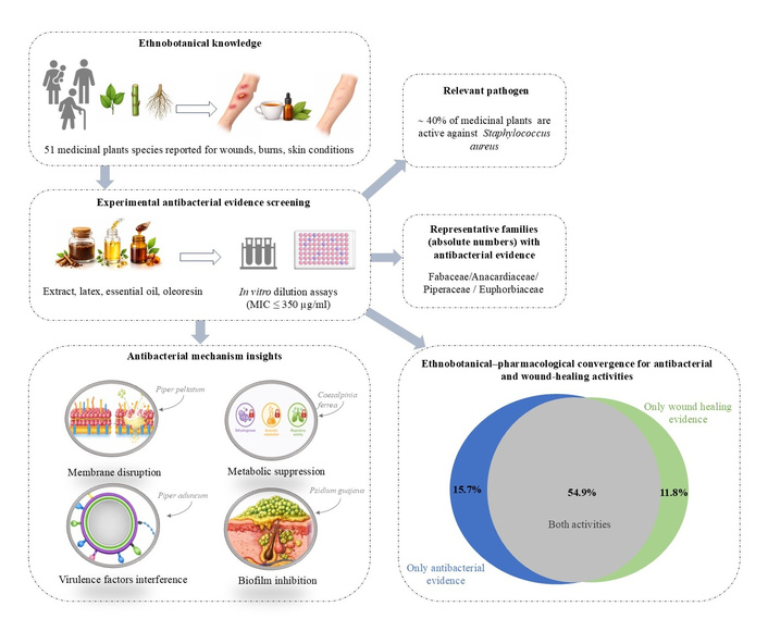
Conceptual representation linking ethnobotanical knowledge to pharmacological evidence. A total of 51 medicinal plant species traditionally used in Brazil for wounds, burns, and other skin conditions were identified from ethnobotanical surveys. Literature analysis of antimicrobial dilution assays using crude extracts, essential oils, or oleoresins (MIC ≤ 350 µg/mL) indicates that approximately 40% of the species exhibit activity against S. aureus, with notable representation in Fabaceae, Anacardiaceae, Piperaceae, and Euphorbiaceae. Experimental studies further report antibacterial mechanisms including biofilm inhibition, membrane disruption, virulence factor interference, and metabolic suppression.
Regarding antibacterial activity (Figure 2), approximately 40% of the species selected from the ethnobotanical studies exhibited MIC values against S. aureus, a classical pathogen associated with skin and wound infections, and/or E. coli. In general, nonpolar extracts or essential oils obtained mainly from leaves and bark, belonging to the families Anacardiaceae, Fabaceae, and Piperaceae, showed the lowest MIC values (Supplementary material).
Among the studies providing experimental evidence of antibacterial mechanisms (Figure 2), disruption of bacterial membrane integrity was mainly reported for hydroalcoholic and ethanolic leaf extracts of Aloe arborescens and P. peltatum [72, 165], whereas suppression of bacterial metabolic activity was demonstrated for hydroalcoholic extracts of A. arborescens, organic leaf extracts of P. guajava, and hydroalcoholic extracts of C. ferrea [72, 109, 148]. Inhibition of biofilm formation was also recurrent, particularly for crude oleoresin of Copaifera langsdorffii, hydroalcoholic extracts of Croton spp., organic leaf extracts of Psidium guajava, and the essential oil of Piper gaudichaudianum [99, 123, 148, 162]. Additional mechanisms, such as interference with bacterial virulence factors, including adhesion and acidogenicity, were reported for ethanolic leaf extracts of Piper aduncum [155], while molecular recognition involving interactions with bacterial cell wall components was demonstrated for leaf-derived lectins obtained from aqueous protein extracts of Schinus terebinthifolius [58].
From a methodological perspective, this review prioritized studies evaluating the antibacterial activity of crude extracts, as these preparations are more consistent with traditional forms of use, such as infusions and tinctures. Although investigations involving fractions or isolated compounds provide relevant mechanistic insights, they were not central to the primary objective of this review and were therefore considered only as complementary evidence supporting plausible antibacterial mechanisms.
Cultural transmission and traditional methods of plant preparation and collection may significantly influence the pharmacological outcomes observed in experimental studies. The intergenerational transmission of traditional knowledge may function as an empirical selection process for species with therapeutic potential, contributing to the recurrence of certain plants in local practices. Many ethnobotanical reports describe preparations such as teas, infusions, decoctions, and tinctures, as well as topical applications such as poultices and macerations, which primarily extract polar or moderately polar compounds from medicinal plants. Antibacterial susceptibility assays, in turn, are conducted in aqueous media, requiring that crude extracts exhibit a certain degree of hydrophilicity to demonstrate activity. However, experimental studies do not always employ preparations obtained with solvents compatible with traditional modes of use. In addition, factors such as season of collection, harvesting time, and plant part used may influence chemical composition and, consequently, pharmacological outcomes. Therefore, closer alignment between traditional practices and experimental designs may enhance the translational relevance of ethnopharmacological research.
Analysis of wound-healing studies from the medicinal plants included in this review indicates that most investigations assessed antibacterial potential indirectly through broth microdilution assays for determination of MIC, particularly for essential oils or ethanolic extracts, with emphasis on activity against S. aureus [51, 115]. In contrast, relatively few studies performed functional evaluations considering bacterial load control or the absence of wound-bed contamination throughout treatment. Notably, H. courbaril, as well as topical formulations containing C. officinalis and C. ferrea, demonstrated maintenance of infection-free wounds or significant reduction of bacterial colonization during the experimental period, even in the absence of direct MIC determination [105, 126, 169].
It was observed that, among the 51 plant species analyzed (Figure 2), more than 50% (n = 28) presented evidence for both antibacterial and wound healing activities, such as Alternanthera brasiliana, A. occidentale, Myracrodruon urundeuva, Aloe vera, Calendula officinalis, Caesalpinia ferrea, Hymenaea courbaril, and Psidium guajava. Traditional uses converge with pharmacological expectations, as wound infection remains one of the main complications associated with impairment of the healing process.
On the other hand, a smaller group of species presented restricted evidence for antibacterial activity (15.7%) (Figure 2), such as Anacardium humile, Croton heliotropiifolius, Anadenanthera peregrina, Piper peltatum, or exclusively to wound-healing activity (11.8%), such as Himatanthus drasticus, Sida rhombifolia, and Plantago tomentosa, when considering the MIC value criteria adopted in this review.
Finally, approximately 17.6% of the species (Figure 2), such as Alternanthera dentata, Alternanthera ramosissima, Croton salutaris, Copaifera cearensis, Sida planicaulis, Plantago sparsiflora, Solanum aculeatissimum, and S. capsicoides, lacked experimental evidence for either antibacterial or wound-healing activity. This highlights significant gaps in pharmacological validation despite their recurrent citation in ethnobotanical surveys.
Considering the evidence discussed above, although studies on these plants suggest convergence of biological effects, relevant gaps persist in the investigation of the wound-healing and antibacterial potential of medicinal plants. There is a predominance of exploratory and screening-based studies relying on non-standardized extracts, often lacking detailed phytochemical characterization, which limits comparability across studies and compromises the reproducibility of findings related to antibacterial activity, even when such approaches are considered appropriate for early stages of investigation. In this context, the limited adoption of standardized extracts and marker-oriented phytochemical approaches represents a critical barrier to advancing the field, as the identification of chemical or biological markers associated with antibacterial activity and key wound-healing outcomes would enable a more robust linkage between extract composition and biological effects. Moreover, in vivo models that simultaneously evaluate the healing of infected wounds remain scarce, restricting the understanding of the actual contribution of antibacterial activity to tissue repair processes.
As integrative reviews can summarize heterogeneous study designs, they are inherently susceptible to selection and interpretative biases. The search was limited to three databases, and data extraction was performed by a single author. In addition, methodological differences among the included studies, such as variations in experimental models and extract preparations, may limit comparability across findings. Furthermore, publication bias and language restrictions may have influenced the representativeness of the included evidence [182].
This review demonstrates that Brazil holds a culturally rich and functionally coherent body of traditional knowledge related to the use of medicinal plants for the treatment of wounds, burns, and other cutaneous conditions of possible bacterial etiology. By mapping ethnobotanical studies from different regions of the country, a total of 51 plant species were identified, most of them native and associated with locally transmitted knowledge. Beyond reflecting botanical diversity, these findings highlight the existence of a consolidated cultural repertoire focused on skin-related therapeutic practices, suggesting the relevance of the country as a source of traditional knowledge systems regarding the management of wounds and cutaneous infections.
The results support a coherence between traditional use and the available pharmacological evidence when assessed under the criteria adopted in this review, particularly regarding experimental conditions and cutoff values for antibacterial activity. Moreover, although the search strategy adopted in this review was not specifically directed toward the combined evaluation of antibacterial and wound-healing activities.
Future investigations should prioritize the use of chemically standardized extracts with defined bioactive markers to ensure reproducibility and pharmacological consistency. Antibacterial evaluation should encompass panels of both susceptible and resistant clinical strains, combined with mechanistic studies aimed at elucidating modes of action and targeting bacterial adaptive resistance mechanisms, including those associated with biofilm formation, as well as testing in mature multispecies biofilm models. Achieving meaningful translational progress will require the integration of in vivo infected wound models capable of linking antimicrobial effects to measurable wound-healing outcomes. Additionally, the development of advanced topical delivery systems may enhance clinical applicability, particularly through sustained and localized release strategies, such as the incorporation of nanostructured lipid carriers within gel-based matrices to optimize drug stability, tissue penetration, and accelerated tissue repair.
Considering that skin infections represent frequent health problems in primary care and that medicinal plants remain widely used in Brazil, this review provides a structured evidence base that may support research prioritization and evidence-informed discussions in public health contexts. By delineating which traditionally used species present convergent antibacterial evidence, the findings inform future translational perspectives related to the rational use of phytotherapy in primary health care.
A. occidentale L.: Anacardium occidentale L.
MIC: minimum inhibitory concentration
S. aureus: Staphylococcus aureus
TNF-α: tumor necrosis factor alpha
VEGF: vascular endothelial growth factor
The supplementary table for this article is available at: https://www.explorationpub.com/uploads/Article/file/1008164_sup_1.pdf.
BBC: Conceptualization, Writing—original draft, Writing—review & editing. The author read and approved the submitted version.
The author declares that there are no conflicts of interest.
Not applicable.
Not applicable.
Not applicable.
Not applicable.
Not applicable.
© The Author(s) 2026.
Open Exploration maintains a neutral stance on jurisdictional claims in published institutional affiliations and maps. All opinions expressed in this article are the personal views of the author(s) and do not represent the stance of the editorial team or the publisher.
Copyright: © The Author(s) 2026. This is an Open Access article licensed under a Creative Commons Attribution 4.0 International License (https://creativecommons.org/licenses/by/4.0/), which permits unrestricted use, sharing, adaptation, distribution and reproduction in any medium or format, for any purpose, even commercially, as long as you give appropriate credit to the original author(s) and the source, provide a link to the Creative Commons license, and indicate if changes were made.

View: 275
Download: 8
Times Cited: 0
Irina Tiganova ... Yulia Romanova
Ebenezer Aborah ... Manuel F. Varela
Daniel Buldain ... Laura Marchetti
Ayuba Olanrewaju Mustapha ... Yusuf Oloruntoyin Ayipo
Monika I. Konaklieva ... Balbina J. Plotkin
Yunuo Zhou ... Heng Zheng
