 Review


Affiliation:
1Neuro-Oncology Program and Brain Tumor Institute, University Hospitals of Cleveland Medical Center, Seidman Cancer Center & UH Neurological Institute, Cleveland, OH 44106, USA
2Molecular Oncology Program, Case Comprehensive Cancer Center, Cleveland, OH 44106, USA
Email: herbert.newton@uhhospitals.org
ORCID: https://orcid.org/0009-0002-1582-2990


 ,
,
Affiliation:
3Department of Neurology, University Hospitals of Cleveland Medical Center, Cleveland, OH 44106, USA
ORCID: https://orcid.org/0000-0002-9953-2175
Explor Neurosci. 2026;5:1006150 DOI: https://doi.org/10.37349/en.2026.1006150
Received: December 23, 2025 Accepted: July 27, 2026 Published: September 19, 2026
Academic Editor: Katrin Sak, Independent senior researcher, NGO Praeventio, Estonia
The article belongs to the special issue Current Approaches to Malignant Tumors of the Nervous System
Targeted cancer therapies for cancer treatment are at the forefront of cancer treatments. The identification of pathologic cell signaling pathways in cancer cells via precision medicine enables the development of new therapies to target the underpinning molecular changes within tumor cells. Applying these techniques to brain tumors is no different, except for the added necessity that these therapeutics penetrate the blood-brain barrier. Besides cell signaling alterations seen in other cancers outside of the brain (such as receptor tyrosine kinase pathways, Raf/MEK/Erk pathway, PI3K/AKT/mTOR pathway, to name a few), there are several cell-signaling pathway aberrations unique to central nervous system tumors (for example isocitrate dehydrogenase mutations or O6-methylguanine DNA methyltransferase methylation status) that allow for therapies to specifically target brain tumors. Herein, we describe the various molecular pathway aberrations that have been identified and used to develop targeted molecular therapies for brain tumors.
The molecular biological revolution in recent decades has allowed numerous advances in medicine, especially in the Oncology field. Recent progress in the understanding of the molecular underpinnings of oncogene mutation, loss of tumor suppressor genes, tumor initiation, growth, and metastasis has spurred the potential promise of “individualized” treatment for patients—the idea of “personalized or precision medicine” [1, 2]. Personalized or precision medicine is still on the horizon for most tumor types, but significant advances have been made in patients with non-small cell lung cancer, melanoma, breast cancer, and several others. For patients with brain tumors, the progress has been much slower, although there is cautious optimism in the field of Neuro-Oncology that we are now entering a new era, where molecular phenotyping of tumor types will begin to guide pathological diagnosis and treatment paradigms [3].
Molecular features of brain tumors are now a predominant diagnostic criterion for brain tumors, as evident in the most recent WHO 2021 Classification of Tumors of the Central Nervous System [4]. For example, in the classification of diffuse astrocytic tumors, the most important initial diagnostic feature is the presence or absence of a mutation in the enzyme isocitrate dehydrogenase 1 (IDH1) (see Figure 1). IDH1 mutation status plays a pivotal role in differentiating IDH1 mutant diffuse astrocytoma grades 2, 3, and 4 from IDH1 wildtype grade 4 glioblastoma (GBM; see Table 1) [4]. This updated classification removed low-grade IDH1 wild-type tumors with low-grade histologic features since these tumors usually have additional mutations and molecular alterations [e.g., endothelial growth factor receptor (EGFR) amplification, TERT promoter mutations, CDKN2A/B loss] that enhance the degree of malignancy to that similar to GBM. Additionally, oligodendroglial tumor diagnosis now strictly requires molecular features analysis to demonstrate the presence of IDH1 mutation, co-deletion of chromosomes 1p and 19q, and a lack of an ATRX mutation [4]. Molecular features now trump histologic appearance in the diagnosis of oligodendroglioma even in tumors that were previously classified as astrocytoma based on histology.
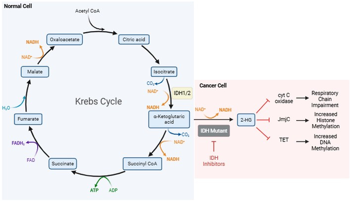
Krebs cycle and IDH metabolism. The Krebs cycle is depicted. One of the intermediaries, alpha-ketoglutarate, is converted to 2-HG if IDH is mutated. This results in inhibition of cyt C oxidase, JmjC, and TET. 2-HG: 2-hydroxyglutarate; ADP: adenosine diphosphate; ATP: adenosine triphosphate; CoA: coenzyme-A; cyt C: cytochrome C; IDH: isocitrate dehydrogenase; JmjC: Jumonji C domain; FAD/FADH2: flavin adenine dinucleotide; NAD+/NADH: nicotinamide adenine dinucleotide; TET: ten eleven translocation. Created in BioRender. Allen, B. (2026) https://biorender.com/eodm3es.
WHO 2021 classification: astrocytic tumors.
| Astrocytic tumors | |
|---|---|
| Diffuse astrocytoma | Astrocytoma, IDH-Mutant, Grade 2 |
| Astrocytoma, IDH-Mutant, Grade 3 | |
| Astrocytoma, IDH-Mutant, Grade 4 | |
| Glioblastoma Multiforme, IDH-Wildtype, Grade 4 | |
| Circumscribed astrocytoma | Pilocytic Astrocytoma |
| High-Grade Astrocytoma with Piloid Features | |
| Pleomorphic Xanthoastrocytoma | |
| Subependymal Giant Cell Astrocytoma | |
| Chordoid Glioma | |
| Astroblastoma, MN1-altered | |
Data were adapted with permission of the Corresponding Author and Elsevier Book Publishers: Newton HB, Loughan AR. Chapter 8: Pathology and treatment of primary and metastatic brain tumors. In: Neuropsychological and Psychosocial Foundations of Neuro-Oncology. Elsevier; 2024. pp. 103–126.
This article reviews the most state-of-the-art information regarding signal transduction pathways, receptor tyrosine kinases (RTKs), oncogene mutations, tumor suppressor gene loss, and other molecular features that contribute to the phenotype of various brain tumors, with a focus on gliomas (See Table 2). These pathogenic signaling pathways are discussed as they are highlighted by the European Association of Neuro-Oncology as potential targets for targeted therapy of brain tumors [5]. For each molecular feature or pathway, we will then update the status of targeted therapy. For further guidance on the level of evidence supporting the recommendation of a given targeted therapy for brain tumor treatment, we will be using the European Society for Medical Oncology “ESMO Scale of Clinical Actionability for Molecular Targets” (ESCAT) whenever possible [6]. The ESMO Scale is a clinical benefit-centered system attributing six levels of clinical evidence based on implications of the respective alteration on patient management. In addition, ESMO has also more recently published detailed recommendations for the use of next generation sequencing (NGS) data in oncology daily practice [7].
Summary of common molecular alterations in GBM & primary brain tumors.
| Mutation | GBMwt | LGGwt | Astrocytoma | Oligo | Medullo |
|---|---|---|---|---|---|
| IDHm | 100% | 100% | Rare | ||
| EGFR | 40–50% | 27% | < 10% | ||
| EGFRvIII | 20% | ||||
| 1p/19qdel | 100% | ||||
| MGMTm | 30% | 70% | 88% | ||
| SMO | 14% | ||||
| PDGFR | 13% | ||||
| MET | 4% | 13% | |||
| MET-ex-14 | 14% | ||||
| FGFR | 3% | ||||
| NTRK | 1–2% | 4% | |||
| ROS1 | 0.29% | Rare | |||
| ALK | 1.9% | Rare | |||
| BRAF | 1.7% | Rare | Rare | ||
| NF1 | 10% | 20% | |||
| PIK3CA | 13–17% | 20% | |||
| PIK3R1 | 8% | 9% | |||
| PTEN | 41% | 23% | |||
| p53 | 28% | 14% | 94% | ||
| MDM2 | 7.6% | 13% | |||
| CDK4/6 | 15% | 7% | |||
| CDKN2A | 50% | 45% | |||
| RB1 | 10% | 27% | |||
| TERT | 50–74% | 15–25% | > 70% | ||
| KIT | 20% | 20% | 25% | ||
| ATRX | 6% | 86% |
Astrocytoma: diffuse astrocytoma IDH mutant; GBMwt: glioblastoma IDH wildtype; LGGwt: low-grade glioma IDH wildtype; Medullo: medulloblastoma; MGMTm: methyl-guanine methyltransferase methylated; Oligo: oligodendroglioma; SMO: smoothened. Data were adapted with permission of the Corresponding Author and Elsevier Book Publishers: Newton HB, Pinho M. Chapter 7 - Next-generation sequencing: overview and application to systemic cancer and brain tumors. In: Newton HB, editor. Handbook of Brain Tumor Chemotherapy, Molecular Therapeutics, and Immunotherapy (Third Edition). Academic Press; 2026. pp. 107–138.
During the initiation and progression of the transformed phenotype, numerous molecular alterations can occur within tumor cells, including different types of genetic mutations (e.g., point mutations, deletions, frameshifts), gene amplification, gene overexpression, deletions of tumor suppressor genes, gene fusions, and epigenetic alterations [8, 9]. In the following sections, the specific genetic alterations that are most important for brain tumors will be reviewed, along with appropriate clinical and therapy related information.
RTKs are a subclass of tyrosine kinases that mediate cell-to-cell communication and control many complex biological activities, including cell growth, motility, differentiation, and metabolism [9–13]. There are 19 different RTK sub-families and 58 known RTKs in humans, many of which are active in glioma cells. The 5 RTK families that have been most studied in high-grade gliomas include the EGFR, platelet-derived growth factor receptor (PDGFR), fibroblast growth factor receptor (FGFR), vascular endothelial growth factor receptor (VEGFR), and Met (see Figure S1). All RTKs share an analogous protein structure that includes an N-terminal extracellular ligand binding domain, a single transmembrane helical domain, and an intracellular region that contains a juxtamembrane regulatory region, a tyrosine kinase domain (TKD), and a carboxyl terminal tail region. Under normal physiological conditions, RTKs are activated by receptor-specific ligands. Growth factor ligands bind to the extracellular domains of RTKs, thereby inducing activation by receptor dimerization/oligomerization and autophosphorylation [10–13]. RTK autophosphorylation activates signaling molecules that contain Src homology-2 (SH2) or phosphotyrosine-binding (PTB) domains, which interact with receptor phosphotyrosine residues to promote downstream signaling pathways (see Figure 2). These docking proteins are a nidus for recruiting additional signaling molecules that contain the SH2 and PTB domains and further enhance downstream signaling. For example, growth factor receptor-bound protein 2 (Grb2) within the SH2 domain will bind the TKD receptor, recruit RAS activator proteins like Sos-1, which functions as a RAS guanine-nucleotide exchange factor (GEF), and promotes further RAS downstream signaling pathways [e.g., RAF, phosphatidylinositol 3-kinase (PI3K), Rac, phospholipase C (PL-C); see below].
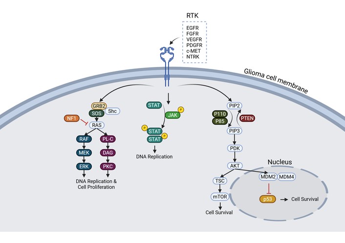
RTK signaling pathways. Overview of the various intracellular signaling pathways involving RTKs. These pathways include the RAS/RAF/MEK/ERK pathway, PKC pathway, JAK-STAT pathway, PI3K pathway leading to mTOR, and MDM2/4 pathways. AKT: protein kinase B; c-MET: mesenchymal-epithelial transcription factor; DAG: diacylglycerol; EGFR: endothelial growth factor receptor; ERK: extracellular signal-regulated kinase; FGFR: fibroblast growth factor receptor; Grb2: growth factor receptor-bound protein 2; JAK: Janus kinase; MDM2/4: mouse double minute 2/4; MEK: mitogen-activated protein kinase kinase; mTOR: mammalian target of rapamycin; NTRK: neurotrophic tropomyosin receptor kinase; PDGFR: platelet-derived growth factor receptor; PDK: pyruvate dehydrogenase kinase; PIP2: phosphatidylinositol-4,5-biphosphate; PIP3: phosphatidylinositol-3,4,5-triphosphate; PKC: protein kinase C; PL-C: phospholipase C; PTEN: phosphatase and tensin homolog; p53: p53 tumor suppressor protein; p85: p85-α protein; p110: p110-α protein; RAF: rapidly accelerated fibrosarcoma protein; RAS: rat sarcoma protein; RTK: receptor tyrosine kinase; Shc: Src homology and collagen protein; SOS: son of sevenless; STAT: signal transducer and activator of transcription; TSC: tuberous sclerosis complex; VEGFR: vascular endothelial growth factor receptor. Created in BioRender. Allen, B. (2026) https://BioRender.com/nr8mxav.
In normal physiologic states, the level of RTK activity is strictly regulated, including inhibitory control through tyrosine phosphatase activity. In cancer cells, RTKs acquire transformational capabilities through several mechanisms [10–14]. RTK gain-of-function mutations lead to aberrant and overactive downstream signal transduction. Some of these mutations can be considered “driver mutations,” which provide tumor cells a physiologic advantage for growth over surrounding cells. Furthermore, somatic mutations to RTK occur commonly in evolutionarily conserved residues that impact adenosine triphosphate (ATP) binding and other catalytic activity. Finally, any RTK domain can be impacted, including the extracellular, transmembrane, or juxtamembrane domains.
Overexpression of RTKs has been described in many human cancers (e.g., EGFR in GBM). Overexpression results in an increased local concentration of receptors, increasing RTK signaling, and thereby overwhelming inhibitory regulatory feedback. RTK overexpression primarily occurs via gene amplification, though additional mechanisms include transcriptional and translational enhancement, loss of phosphatase activity, and loss of other negative regulators. Gene amplification increases the copy number of specific gene sequences in extra-chromosomal genetic material (i.e., double minutes), single-site repeats, or repeat genetic sequences throughout the genome (i.e., distributed insertions). Chromosomal rearrangements can produce oncogenic tyrosine kinase fusion proteins. Small molecule inhibitors (i.e., BCR-ABL) can target these aberrant fusion proteins at either the N- or C-terminal domain. One recently described mechanism of enhanced RTK activation is via intragenic partial duplication, which creates new protein isoforms of enhanced function [14]. Furthermore, tumor cell autocrine signaling generates a positive feedback loop in which tumor cells secrete the needed ligand to further stimulate RTK pathway activity. Lastly, microRNA is also capable of modulating RTK signaling. All told, there are abundant therapeutic targets available given the landscape of pathogenic RTK signaling.
After the binding of ligands to growth factor RTKs and activation of the TKD, many downstream signaling molecules are subsequently activated, in particular those involving the RAS pathway [9–14]. RAS proteins are the founding members of the RAS superfamily of GTPases, which has in excess of 150 members in humans. RAS proteins function as a molecular switch by pairing their membrane-bound small GTPase function to the cell surface receptor, thereby enabling intracellular signal transduction upon extracellular ligand binding. RAS proteins subsequently regulate cell proliferation, membrane trafficking, cytoskeletal organization, migration, differentiation, and apoptosis [12, 13, 15]. The four distinct RAS proteins (H-RAS, N-RAS, K-RAS4A, and K-RAS4B) are inactive when GDP-bound and active when GTP-bound. While RAS pathogenic variants occur in approximately 30% of human cancers in gliomas [12, 16]. Overall, RTK overexpression and hyperactivation contribute to increased RAS signaling, which current data suggest is common in malignant gliomas.
RAS undergoes several C-terminus post-translational modifications that allow it to bind to the inner cell membrane and subsequently activate [12, 15, 16]. RAS is initially synthesized as an inert cytosolic pro-peptide (proRAS), which then undergoes modification by farnesylation of the cysteine residues, proteolytic cleavage of the AAX peptide, and carboxymethylation of the new C-terminal carboxylate. After ligand binding to the RTK, adaptor proteins with SH2 domains such as Grb2 bind to the TKD and then recruit Sos-1 and other GEF’s, which then facilitate the exchange of GDP for GTP, thereby activating RAS at the membrane. Once RAS binds GTP, a conformational change occurs, mainly in the switch I and II regions, allowing RAS to interact with downstream effectors and promote signal transduction. These downstream effectors include mitogen-activated protein kinases (MAPKs) [i.e., Raf-MEK-extracellular signal-regulated kinase (ERK)], PI3K, and the Ral pathways (described below; see Figure 2). Other effectors that are now known to interact with RAS-GTP include Rho-GTPases, Af6, PL-C, and YAP.
For over three decades, researchers have attempted to directly inhibit the RAS signaling pathway, with inadequate results [12, 17]. To date, there is a lack of RAS oncoprotein-targeted therapies, and many consider RAS proteins to be “undruggable”. However, several small molecule strategies have been explored in recent years and remain active today: inhibitors of the enzymes involved in the post-translational modification of RAS, and compounds that bind directly to the RAS protein. In terms of interrupting the post-translational modification of RAS using farnesyltransferase inhibitors (FTIs), farnesylated derivatives, peptidomimetic inhibitors, and non-peptide inhibitors have been investigated [12, 17]. Although these compounds have significant activity in the in vitro setting, including against glioma cells, they have been unsuccessful at improving the progression-free survival (PFS) or overall survival (OS) in phase II and III clinical trials of advanced solid tumors, including GBM (ESCAT Tier IV).
EGF is a 6.5-kDa polypeptide that is composed of 53 amino acids and functions as a monomeric ligand; the EGF gene is located on chromosome 4q25 [9, 12, 13]. EGF binds to the EGFR, inducing receptor dimerization, conformational changes, activation, and autophosphorylation of the TKD. Other monomeric ligands that can bind to and activate EGFR include TGF-α, amphiregulin, heparin-binding EGF, betacellulin, and epiregulin. As noted earlier, EGFR is a 170-kDa transmembrane glycoprotein of the growth factor TRK family and arises from the EGFR gene on chromosome 7p12. After EGFR activation, it recruits adaptor molecules to the phosphorylated tyrosine residues, including Grb2 and Sos-1, which subsequently activate other pathways as described previously, including RAS-GAP, PI3K, PLC, protein kinase B (AKT), Src, Src homology and collagen protein (Shc), and Janus kinase (JAK)-STAT (see Figure 2) [12, 13]. The signaling molecules that associate with EGFR depend on which C-terminal region is autophosphorylated, and this in turn depends on which heterodimer molecule is bound to EGFR (e.g., HER1, HER2, or HER4). Therefore, the specificity and potency of the signaling output from activated EGFR will vary depending on the identity of the co-receptor.
In normal non-transformed cells, EGFR concentration and signaling activity are tightly controlled and regulated. Glioma cells can express more EGFR with higher signaling activity [9, 12, 13]. Overexpression of EGF, TGF-α, and EGFR is an established finding in high-grade glioma cells, especially GBM, consistent with autocrine and paracrine stimulatory loops. IDH wildtype GBM commonly have EGFR amplification, noted in 40–50% of tumors, while EGFR amplification does not occur in IDH mutant lower grade gliomas. In GBM, amplification of the wild-type gene is often a precursor event to subsequent mutations of EGFR, which frequently involve intragene deletions (see Figure S2). The vIII variant of EGFR (also known as ΔEGFR or del2-7EGFR), which is present in 67% of EGFR-positive tumors and involves a 5’ deletion of codons 6–273, is the most common EGFR mutation. This mutation results in constitutive activity of EGFR due to removal of the ligand-binding domain. Unlike wild-type EGFR, the constitutively active mutant receptors do not undergo downregulation or processing for lysosomal degradation. The C-958 mutant EGFR occurs in 15% of EGFR mutations and is characterized by a 3’ deletion altering kinase activity via a structural change to the intracellular receptor region. Other mutations in EGFR are known, including in the transmembrane domain (15% of EGFR mutations) and missense mutations or insertions (less commonly encountered). In GBM cells with EGFR gene mutations, 33% will have multiple EGFR gene alterations. In addition, in 10–20% of EGFR genes that are overexpressed, there is no amplification. Amplification and mutation of EGFR, with subsequent overexpression and hyperactivation of receptor activity, have numerous effects on GBM cells that promote growth and survival [9, 12, 13]. The cells develop an increased proliferative capacity and reduced tendency to undergo apoptosis. They also become more invasive and infiltrative, with upregulation of matrix metalloproteinases (MMPs) and serine proteases. The presence of mutant EGFR also confers increased resistance to radiotherapy and chemotherapy.
As a result of the importance of the EGFR signaling pathway in the biology of solid tumors, including gliomas, numerous targeted therapies were developed [9, 12, 13, 18–22]. Small molecule inhibitors have been the most intensively studied and applied to solid tumors thus far. EGFR TKI’s can be classified as reversible or irreversible, depending on how the TKD is inhibited. In general, reversible inhibitors compete for the ATP binding site in the EGFR TKD through non-covalent mechanisms such as electrostatic, hydrogen-bonding, and hydrophobic interactions. In addition, EGFR TKI’s can also be classified based on their target kinases: monotherapy for EGFR, dual EGFR inhibitors, or multi-kinase inhibitors. First-generation EGFR inhibitors (Gefitinib, Erlotinib, Lapatinib) were designed to orthosterically and reversibly block the ATP/substrate-binding pocket of EGFR in the TKD. Although these drugs showed potential to inhibit growth and improve survival in pre-clinical models, they were not effective in phase II clinical trials in patients with newly diagnosed or progressive GBM, either as monotherapy or in combination therapy (ESCAT Tier IIIA/IV) [18, 21–24].
The failure of first-generation EGFR inhibitors was at least partially due to suboptimal dosing within the GBM cells, with only 15% target inhibition of p-EGFR by Lapatinib, suggesting poor blood-brain barrier (BBB) entry. Second-generation EGFR inhibitors were designed to irreversibly bind to the TKD of EGFR, and include Afatinib, Dacomitinib, Brigatinib, and Neratinib [20–22]. In pre-clinical studies, both Afatinib and Dacomitinib were active against glioma cells in vitro and in GBM animal models. Afatinib was well tolerated in a phase I/randomized phase II study but demonstrated minimal activity in GBM patients as monotherapy. The addition of TMZ to Afatinib did not improve the 6-month PFS (PFS-6) rate or the median PFS of GBM patients. Dacomitinib was able to inhibit the growth of tumors exhibiting EGFR amplification in GBM xenograft models, but as a single agent had limited efficacy in clinical trials of recurrent patients with GBM that contained EGFR amplification. Thus, the clinical activity of second-generation EGFR inhibitors had limited efficacy like the first-generation drugs (ESCAT Tier IIIA/IV).
Third-generation EGFR inhibitors were developed in response to the resistance of EGFR secondary mutations while minimizing adverse side effects, and include Osimertinib, Rociletinib, and Olmutinib [18–22]. These are all irreversible inhibitors designed for tumors with the EGFR T790M mutation and have little activity against wild-type EGFR. Osimertinib is the first TKI with a non-quinazoline core and is the only third-generation EGFR inhibitor currently on the market. In contrast to first- and second-generation EGFR TKI’s, Osimertinib has excellent BBB penetration. Pre-clinical testing of Osimertinib in EGFRvIII-positive GBM stem cells demonstrated significant ability to reduce constitutive activity of the EGFRvIII TKD with high potency, as well as inhibit downstream signaling [9, 22, 25]. Further testing in heterotopic and orthotopic xenograft models suggested that Osimertinib was active in vivo as well. Application of Osimertinib to patients with progressive high-grade gliomas and GBM is limited, although a few small retrospective series and a case report have demonstrated some limited efficacy, including several patients with partial responses on MRI (ESCAT Tier IIIA/IV) [26–28]. In contrast, for patients with EGFR mutant non-small lung cancer and brain metastases, Osimertinib seemed to be very active in the brain [29, 30]. In both studies, there was a significant objective response rate (ORR; > 50%) and intracranial PFS, including patients who were pre-treated with other TKI’s.
Several anti-EGFR antibodies have also been developed and applied to GBM, including Cetuximab, Panitumumab, and Nimotuzumab [9, 12, 20–22]. All three antibodies block ligand binding to EGFR and/or EGFR dimerization by interacting with the L2 domain. Because EGFRvIII lacks the ligand-binding domain, it is not responsive to treatment by these antibodies. Cetuximab binds to EGFR with higher affinity than EGF or TFG-α, prevents ligand binding, blocks ligand-induced tyrosine kinase activation, and stimulates receptor internalization. It was also able to inhibit EGFR-overexpressing GBM cells in vitro and in vivo. However, when Cetuximab was used in clinical trials of patients with EGFR+ recurrent GBM, it was tolerated well but had minimal clinical activity as monotherapy (ESCAT Tier IIIA/IV). In a combination trial with Bevacizumab and Irinotecan, the use of Cetuximab was well tolerated but did not prove superior to single-agent Bevacizumab or Bevacizumab plus Irinotecan. Nimotuzumab has also demonstrated the ability to inhibit EGFR-overexpressing GBM cells in vitro and in animal models. However, it has also demonstrated minimal clinical efficacy as monotherapy and in combination regimens in clinical trials of newly diagnosed malignant gliomas and GBM in children and adults [9, 22, 31, 32]. In one phase II trial, Nimotuzumab was added to standard radiotherapy and TMZ for GBM [32]. The ORR at the end of radiotherapy was 72.2%, while the median PFS and OS were 11.9 months and 24.5 months, respectively. In a randomized double-blind trial, Nimotuzumab or placebo was used in 70 evaluable patients with newly diagnosed high-grade glioma (anaplastic astrocytoma = 41; GBM = 29) [31]. The median OS for the Nimotuzumab and placebo groups was 17.76 months and 12.63 months, respectively (HR = 0.64; p = 0.032). The median PFS was also in favor of the Nimotuzumab cohort vs. placebo: 15.73 months vs. 6.5 months (ESCAT Tier II).
Anti-EGFR vaccines have also been developed, with the focus on GBM tumors with the EGFRvIII mutation [9, 21, 22]. Rindopepimut (CDX-110) is a 14-mer peptide that spans the mutation site of EGFRvIII, conjugated to the immune adjuvant keyhole limpet hemocyanin (KLH). A phase I clinical trial demonstrated that CDX-110 safely eliminates tumor cells that express EGFRvIII. In a multicenter phase II trial of newly diagnosed EGFRvIII-positive GBM patients, in combination with TMZ, CDX-110 treatment significantly improved OS in comparison to historical controls. However, in a randomized, double-blind, international phase III trial (“ACT IV”) of EGFRvIII-expressing newly diagnosed GBM patients, CDX-110 was unable to improve the OS in comparison to control patients (median OS 20.1 months vs. 20.0 months; HR 1.01, p = 0.93) (i.e., ESCAT Tier IIIA/IV) [31]. The negative results of this phase III trial were very disappointing, after more than a decade of positive results [32]. More recent data suggest that in 57–59% of the GBM tumors in the trial, there was a loss of EGFRvIII expression, regardless of whether or not Rindopepimut or control treatment was administered. This suggests that EGFRvIII loss of expression may not necessarily be due to immunization with Rindopepimut, and instead may be an intrinsic aspect of the natural molecular evolution of GBM progression. Lessons from this study include the issue that early phase clinical trials might not provide predictive power for wide-scale benefit in GBM patients, and the importance of executing earlier trials with randomization in this patient population.
Chimeric antigen receptors (CARs) are engineered immune cell receptors (i.e., T cells, NK cells) comprised of a single-chain variable fragment (scFv) derived from a monoclonal antibody (mAb) [9, 21, 33]. CARs enable the immune cell to bind to and respond to cells of interest expressing the CAR target. EGFR-specific and EGFRvIII-specific CARs have been developed and have been applied to EGFR-expressing GBM, high-grade gliomas, medulloblastoma [21, 33–35]. Pre-clinical studies and early clinical trials have begun; in vitro studies and animal models have demonstrated positive results (ESCAT Tier IV). However, the results of EGFR-targeted CAR T cell therapy in brain tumor patients has demonstrated limited efficacy thus far, due to tumor antigen heterogeneity, difficulty of CAR T cells to traffic from the blood to the tumor site, and to the severely immunosuppressive tumor microenvironment of GBM.
There are numerous clinical trials for EGFR-positive GBM, including the combination of Gefitinib, Erlotinib, and Afatinib (NCT03239015), BDTX 1535 (NCT05256290), MCLA-129 (NCT04868877), Bevacizumab (NCT05271240), Tofacitinib (NCT05326464), Pembrolizumab & Olaparib (NCT05463848), MCLA-129 (NCT04930432), CAR-T cell therapy for EGFRvIII-positive GBM wild type tumors (NCT03941626), and many others.
PDGF is a 30-kDa protein comprised of four chains (A, B, C, D), each encoded by a different gene that exists as disulfide-bonded heterodimers or homodimers [9, 12, 13, 22, 36]. Each gene has a similar organizational structure, consisting of seven exons. PDGFR also exists in two forms, α and β, each with a unique gene and forms heterodimers or homodimers (αα, αβ, and ββ). The PDGF ligand and receptors have preferential binding affinities. PDGFR-α selectively binds the ligands PDGF-A and PDGF-C, while PDGFR-β preferentially binds the PDGF-D ligand. PDGF-B has equal affinity for the α and β forms of PDGFR. PDGFR is a transmembrane glycoprotein that is a member of the RTK family of growth factor receptors. The receptor has a similar organization to EGFR, with an extracellular ligand-binding region, a hydrophobic transmembrane-spanning region, and an internal TKD region. Binding of the PDGFR receptor to divalent PDGF induces receptor dimerization, thereby initiating the signaling cascade by activating internal membrane protein complexes. The intracellular TKDs become approximated by this dimerization, allowing for the catalytic domains to autophosphorylate tyrosine residues on the TKDs, which form SH2 signaling molecule binding sites (see Figure 3). These signal transduction molecules include Src, Shc, Grb2, RAS GAP, PI3K, STATs, and PL-C. Second messenger phosphorylation by the TKDs promotes downstream pathway activation.
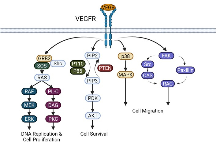
VEGF signaling pathway. A depiction of VEGF activating the various pathways, including the RAS/RAF/MEK/ERK pathway, PKC pathway, PI3K pathway, MAPK pathway, and FAK/paxillin pathways. AKT: protein kinase B; CAS: Crk-associated substrate; DAG: diacylglycerol; ERK: extracellular signal-regulated kinase; FAK: focal adhesion kinase; Grb2: growth factor receptor-bound protein 2; MAPK: mitogen-activated protein kinase; MEK: mitogen-activated protein kinase kinase; PDK: pyruvate dehydrogenase kinase; PIP2: phosphatidylinositol-4,5-biphosphate; PIP3: phosphatidylinositol-3,4,5-triphosphate; PKC: protein kinase C; PL-C: phospholipase C; PTEN: phosphatase and tensin homolog; p38: p38 protein; p85: p85-α protein; p110: p110-α protein; RAC: RAS-related C3 botulinum toxin substrate; Raf: rapidly accelerated fibrosarcoma protein; Ras: rat sarcoma protein; Shc: Src homology and collagen protein; Src: proto-oncogene tyrosine kinase; SOS: son of sevenless; VEGF: vascular endothelial growth factor; VEGFR: vascular endothelial growth factor receptor. Created in BioRender. Allen, B. (2026) https://BioRender.com/b7amfyo.
Evidence indicates that glial tumorigenesis relies upon PDGF and PDGFR as oncogenic catalysts [9, 12, 13, 22, 36]. In glioma cell lines, PDGF-AA is highly expressed, while PDGF-AB and PDGF-BB are less expressed. PDGFR is also concomitantly expressed in variable amounts in glioma cell lines. However, amplification or rearrangement of the PDGF or PDGFR genes seems to be infrequent, indicating that the enhanced expression is secondary to aberrancies in gene regulation. Evaluations of human glioma biopsies demonstrated the presence of PDGF and PDGFR in all tumor grades. Early transformations in the oncogenic pathway include PDGFR-mediated autocrine stimulation. PDGFR-α was expressed at a consistent level in all tumor grades, while PDGFR-β expression was generally exceptionally low. PDGFR-α gene amplification is uncommon in glioma tissue specimens overall. It is not present in low-grade tumors and is documented in less than 10% of high-grade tumors. When amplification of PDGFR-α is present, no EGFR gene amplification will co-occur.
Molecular approaches to targeting the PDGF/PDGFR signaling pathway have focused on small molecule inhibitors of the receptor [9, 12, 13, 22, 36]. SU101 was the first small molecule inhibitor of PDGFR for neuro-oncology applications. SU101 is an isoxazole derivative that disrupts PDGFR-mediated tyrosine phosphorylation, DNA synthesis, cell cycle progression, and cellular proliferation. Phase I and II trials demonstrated sufficient tolerability and possible glioma activity. Despite early promise, subsequent comparative analysis of SU101 vs. procarbazine in recurrent glioma treatment in a phase III trial ended early due to increased early deaths in the SU101 treated patients (ESCAT Tier IV). Another small molecular inhibitor of PDGFR is Imatinib mesylate, which blocks the ATP binding site of tyrosine kinase receptors. It has significant in vitro activity against PDGFR, c-kit, and bcr-abl, and is highly active against chronic myelogenous leukemia and gastrointestinal stromal tumors. Imatinib has been studied in several phase I and II clinical trials as monotherapy for newly diagnosed or progressive GBM and other recurrent gliomas [36–40]. All of these studies consistently demonstrated that Imatinib monotherapy had very limited or negligible efficacy for newly diagnosed or progressive GBM, as well as other lower grade recurrent gliomas (ESCAT Tier IIIA/IV). Several small studies were also undertaken to assess if the combination of Imatinib and Hydroxyurea would be a more effective treatment for patients with progressive and recurrent GBM and low-grade gliomas [41, 42]. Although the series evaluating Imatinib and Hydroxyurea suggested some possible minor benefit in high-grade gliomas, the combination was not active at all for patients with progressive low-grade gliomas.
Ongoing clinical trials for PDGFRA are limited (i.e., unspecified solid tumors) but include Nilotinib & Pazopanib (NCT02029001), Regorafenib (NCT04116541), and Sunitinib (NCT02693535).
The FGF family includes 19 different polypeptides that are involved in a wide range of biological activities, including differentiation, mitogenesis, and angiogenesis [9, 12, 13, 22, 43]. The acidic FGF1 and basic FGF2 are the best characterized FGF’s. FGF1 is an 18-kDa polypeptide consisting of 154 amino acids that are encoded on chromosome 5q. FGF2 is encoded on chromosome 4q26-q27 and is expressed in four forms, ranging in size from 18 kd to 24 kd. FGFRs exist in four types—FGFR1–4—and specifically bind FGF. The genes for FGFR-1, -2, -3, and -4 are located on chromosomes 8p, 10q, 4p, and 5q, respectively. The FGFRs belong to the protein RTK family of receptors, like EGFR and PDGFR. After binding of FGF to an FGFR, there is subsequent dimerization, activation of the TKD, autophosphorylation, and subsequent phosphorylation of FGFR substrate 2 alpha (FRS2α) and FRS2β. Activated FRS2α and FRS2β then recruit Grb2/Sos complexes and Grb2/Gab1 complexes, resulting in activation of several signal transduction pathways, including RAS, PI3K, protein kinase C, and RAF (see Figures 2 and 3).
Abnormalities affecting the FGFR1–4 genes are common in human cancer, affecting between 5–10% of all cancer patients [13, 22, 43]. In some cancers (e.g., urothelial, intrahepatic cholangiocarcinoma), the rate can be much higher, with a frequency of 10–30%. One common genomic alteration that affects FGFRs is single nucleotide variants (SNVs), which occur in 26–65% of FGFR-altered cancer specimens. Activating SNVs can cause the receptor to be constitutively active through several mechanisms, including increased dimerization, increased activity of the TKD, or enhanced affinity for FGF ligands. FGFR SNVs can occur anywhere along the different regions of the receptor, affecting the extracellular domain, transmembrane domain, and the intracellular kinase domain. SNVs most commonly occur in FGFR2 but can also involve the other receptor subtypes. Gene fusions are common FGFR aberrations that occur via chromosomal rearrangements or translocations, and lead to increased receptor dimerization, increased receptor activation, and dysregulation of expression of FGFR and/or its fusion partner gene. There are many potential oncogenic partners for FGFR, including TACC3 (transforming acidic coiled-coil containing protein 3), CCDC6, KIAA1976, CASP7, and many others. FGFR fusions are also quite variable among different cancer types, and have a frequency ranging from 8% to 36%. Pathogenic FGFR fusion variants typically occur due to in-frame fusion. The most frequent genomic alteration of FGFRs involves copy number amplification (CNAs), which can occur in up to 66% of specimens in some studies. FGFR1 and FGFR4 have the highest frequency of gene amplification, and are often noted in breast cancer, non-small cell lung cancer, and urothelial cancer.
Expression of FGF and FGFR has been noted in gliomas for decades—in low-grade and high-grade tumors [9, 12, 13, 22, 44]. The frequency and density of expression has been correlated with tumor grade, so that high-grade gliomas and GBM often have elevated expression of FGFRs. This is consistent with the histological appearance of these tumors, which are highly vascular. The presence of high expression and density of FGFR in a glial tumor is usually correlated with a poor prognosis. Overall, the presence of FGFR amplification and SNVs is rare in glial tumors and GBM. The most common FGFR gene alteration is the presence of fusion events—almost exclusively FGFR-TACC fusions, which are noted in roughly 3% of IDH wildtype GBM specimens, as well as lower grade IDH wildtype gliomas [22, 24]. FGFR-TACC fusions do not occur in IDH mutant lower-grade gliomas. The most commonly noted fusion subtype is FGFR3-TACC3, which is located only 48 kb apart on chromosome 4p16, and joins FGFR3 exon 17 to TACC3 exon 11 “in frame”, with the entire FGFR3 TKD upstream of the TACC-coding sequences (including the intact coiled-coil TACC domain). The other less common fusion subtypes are FGFR1-TACC1 and FGFR2-TACC2, which have similar biological and oncogenic properties to FGFR3-TACC3. The FGFR-TACC fusion proteins are constitutively active as RTKs, with excessive activation of the downstream signaling pathways (e.g., RAS, PI3K). In addition, the presence of the TACC coiled-coil domain increases the likelihood that the fusion proteins will form dimers; after dimerization they can then autophosphorylate and activate the FGFR TKD.
The frequent presence of overexpressed and constitutively active FGFRs has led to significant research into targeted treatment options, including monoclonal antibodies to FGFR, antisense strategies, FGF ligand traps, and small molecule receptor inhibitors [9, 12, 13, 22, 43–45]. Numerous phase I, II, and III trials have been performed with small molecule inhibitors and anti-FGFR antibodies; small molecule approaches have shown the most activity thus far [43]. Non-selective and selective small molecule TKI drugs have been developed, since both groups are able to bind to and block the ATP-binding pocket in the TKD of FGFR1–4. The non-selective drugs were initially employed in clinical trials, including Dovitinib and Ponatinib, with severe side effects and limited efficacy (ESCAT Tier IV). The non-selective TKI’s are now considered more applicable for overcoming secondary mutations in FGFRs that have become resistant to primary selective FGFR inhibitors. Numerous selective TKIs of FGFR have been developed, including several that have been advanced to phase III trials (e.g., Erdafitinib, Infigratinib, Rogaratinib, Pemigatinib, Futibatinib); only Erdafitinib and Pemigatinib are currently FDA-approved. The majority of these drugs are pan-inhibitors that reversibly target the ATP-binding domain; however, they are often more effective at reducing activity of FGFR1–3, with less ability to block FGFR4. More selective FGFR pan-inhibitors that irreversibly bind the ATP-binding domain with a covalent bond are now in development and early clinical testing.
Several selective and non-selective FGFR inhibitors have been tested in GBM and other gliomas in recent years [13, 22, 44]. Non-selective inhibitors evaluated in phase I and II trials include Dovitinib, Ponatinib, and Anlotinib [22, 44, 46–50]. Treatment with Ponatinib was used in 15 patients with recurrent GBM that had failed Bevacizumab, speculating that it would be effective against VEGFR and FGFR pathways [46]. The 3-month PFS rate was 0, with a median PFS and OS of only 28 days and 98 days, respectively; no objective responses were noted (ESCAT Tier IV). In a similar study of Dovitinib, 33 patients with recurrent GBM received treatment [47]. The median Time to Progression was only 1.8 months, with a 6-month PFS rate of roughly 12%; there were no OR noted (ESCAT Tier IV). Anlotinib has been applied to newly diagnosed and recurrent high-grade gliomas [48, 50]. It is a multi-kinase inhibitor with anti-FGFR, VEGFR, PDGFR, and c-Kit activity. In a series of 31 patients with recurrent high-grade gliomas (median KPS of 60), 17 were treated with Anlotinib monotherapy while 14 received Anlotinib plus dose-dense TMZ (50 mg/m2/day) [49]. The median PFS and OS for the entire cohort were 4.5 months and 7.7 months, respectively. The authors concluded that Anlotinib impeded recurrent high-grade gliomas as monotherapy and combined with TMZ (ESCAT Tier IIIA/IV). In another study of Anlotinib monotherapy for newly diagnosed and recurrent patients with high-grade gliomas, Anlotinib seemed to have activity when used in combination with chemo-radiotherapy [48]. The median PFS was 8.9 months, with a median OS of 12 months; the ORR was 73.1%. A phase II trial of anlotinib alone or in combination with bevacizumab in 25 patients with recurrent high-grade glioma showed anlotinib was safe, resulting in a median OS of 13.6 months and PFS of 5.0 months [51]. Overall, anlotinib appears to slow high-grade glioma progression and increase survival.
There is limited published data using selective FGFR inhibitors in gliomas thus far [13, 22, 44]. A phase II study of 26 patients with recurrent or progressive gliomas containing aberrant FGFR was administered Infigratinib on days 1 to 21 of 28-day cycles [52]. The median PFS was 1.7 months, with a 6-month PFS rate of 16.0%; the ORR was 3.8% (ESCAT Tier IV). Four patients had greater than 1 year disease control, including 3 that had activating point mutations of FGFR1 or FGFR3, and another that had an FGFR3-TACC3 fusion. There has also been a case report of an adult with a progressive juvenile pilocytic astrocytoma, who had an FGFR1N546K mutation and responded to treatment with Pemigatinib [53]. He was enrolled in a dose-escalation phase I/II trial of Pemigatinib in advanced malignancies with FGFR mutations (NCT02393248). He developed a substantial PR for 18 months before mild progression of the tumor, while remaining clinically stable through 27 cycles of treatment.
There is an ongoing clinical trial for FGFR fusion-positive IDH wild-type GBM patients using Pemigatinib (NCT05267106).
The VEGF family includes VEGF-A, VEGF-B, VEGF-C, VEGF-D, and placental growth factor (PlGF) [9, 22, 54, 55]. Eight exons with seven introns comprise the VEGF-A gene on chromosome 6p21.3. While the other VEGF proteins are structurally similar to VEGF-1, their genes are on different chromosomes. (e.g., VEGF-B is located on 11q13; VEGF-C is on 4q34). Hypoxia, cell surface receptors, transcription factors, inflammatory mediators, mechanical forces, and oncogenes regulate VEGF gene expression. The most important mechanism involves ambient oxygen tension, with increased expression of VEGF in conditions of hypoxia. Hypoxia induces HIF-1α dimerization with the constitutively active HIF-1β, which binds the VEGF gene promoter sequence at the hypoxia-responsive element (HRE) location and promotes VEGF expression. von Hippel-Lindau (VHL) tumor suppressor protein impacts VEGF expression by way of controlling HIF-1α concentrations. Other VEGF regulatory mechanisms include growth factors and receptors (e.g., EGF/EGFR, PDGF/PDGFR, FGF), internal signal transduction pathways (e.g., RAS, PI3K/AKT), and inflammatory cytokines (e.g., interleukin-1α and -1β, interleukin-6, tumor necrosis factor-α).
Functional VEGF exists as a basic, heparin-binding homodimeric glycoprotein comprised of two antiparallel, identical 23 kd subunits [55]. Alternative splicing produces the various VEGF protein forms. Six VEGF-A polypeptide isoforms exist, each with a different amino acid length beyond the signal sequence (VEGF-A121, VEGF-A145, VEGF-A165, VEGF-A183, VEGF-A189, VEGF-A206). VEGF-A165 is the most common isoform produced by neoplastic and normal cells alike. VEGF-A121 and VEGF-A189 are also commonly present in most cells and tissues that express the VEGF-A gene. The longer VEGF-A isoforms (e.g., VEGF-A189, VEGF-A206) remain sequestered in the extracellular matrix (ECM) and proximal to the cell surface due to high heparin and heparin-like moiety binding affinity. These sequestered VEGF-A isoforms may constitute a reserve of available growth factor released by plasmin or MMPs. Once VEGF is released and activated, it binds to various receptor types, including VEGFR-1, VEGFR-2, VEGFR-3, Neuropilin-1, and Neuropilin-2. Numerous experiments in animal models and human glioma specimens have demonstrated that VEGF is the major endothelial mitogen for glial tumors and GBM.
There are three known VEGFRs—VEGFR-1 (Flt-1), VEGFR-2 (KDR), and VEGFR-3 [9, 22, 54, 55]. All three receptors have a similar structure comprising an extracellular region comprised of seven immunoglobulin-like domains, a single transmembrane region, and an intracellular TKD sequence domain (see Figure S3). There is a soluble VEGFR-1 form (sVEGFR-1) comprised only of the extracellular domain. VEGF binds the second immunoglobulin-like domain of both VEGFR-1 and VEGFR in which deletion of this domain completely abolishes VEGF binding. VEGFR-1 expression in macrophages, pericytes, monocytes and endothelial cells enables binding to VEGF-A, VEGF-B, and PlGF. Homodimeric VEGFR-1 transmits weak mitogenic signals to endothelial cells, but heterodimerization with VEGFR-2 induces a stronger signal. Angiogenesis, hypoxia, and VEGF transcription factors upregulate VEGFR-1 expression. High-affinity binding of sVEGFR-1 to VEGF inhibits VEGF-induced mitogenic signaling. In endothelial cells, VEGFR-2 is the primary VEGFR and binds VEGF-A, VEGF-C, and VEGF-D, with a lower affinity than VEGFR-1. The VEGFR-2 pathway mediates vasodilation, endothelial cell migration, and endothelial cell proliferation via VEGF. During development, VEGFR-3 expressed on endothelial cells binds to VEGF-C and VEGF-D. In adulthood, VEGFR-3 becomes restricted to the endothelial cells of lymphatic vessels and selected fenestrated vasculature.
After binding with VEGF, the VEGFR’s undergo dimerization, forming VEGFR-1 homodimers or VEGFR-1/VEGR-2 heterodimers, which results in TKD autophosphorylation, similar to other TKRs (e.g., PDGFR, EGFR). Once activated, VEGFR dimers induce various signaling pathways, including the PI3K/AKT and MAPK pathways, RAS/Raf/MEK/Erk, focal adhesion kinase (FAK), Paxillin, and protein kinase C (see Figure 3). These signaling cascades then impact various endothelial cell functions. PI3K and AKT pathways enhance cell survival, while RAS/Raf/MEK/Erk and MAPK pathways facilitate DNA replication and cell proliferation. FAK and Paxillin enhance cell migration, which is also promoted by PI3K/AKT and MAPK pathways.
Numerous drugs were developed to inhibit angiogenesis in solid tumors, most of which have targeted the VEGF/VEGFR signaling axis [9, 22, 54–57]. The most effective of these drugs to date is Bevacizumab (Avastin; BEV), which has been studied intensively for over 20 years. BEV is an IgG1 isotype mAb that is derived from a murine anti-VEGF antibody (i.e., A4.6.1) but has been “humanized” for better tolerance. The humanized mAb comprises the six complementarity-determining region (CDR) amino acid sequences from the murine anti-VEGF antibody grafted onto a consensus human scaffold of disulfide-linked heavy and light chains with associated variable and constant regions (see Figure 4). The recombinant BEV mAb is comprised of 93% consensus human IgG1 framework and antigen-binding regions and 7% complement-determining regions from mAb A4.6.1. BEV neutralizes all VEGF-A isoforms in a similar manner to A4.6.1, with dissociation constants (Kd) of 1.1 and 0.8 nmol/L, respectively.
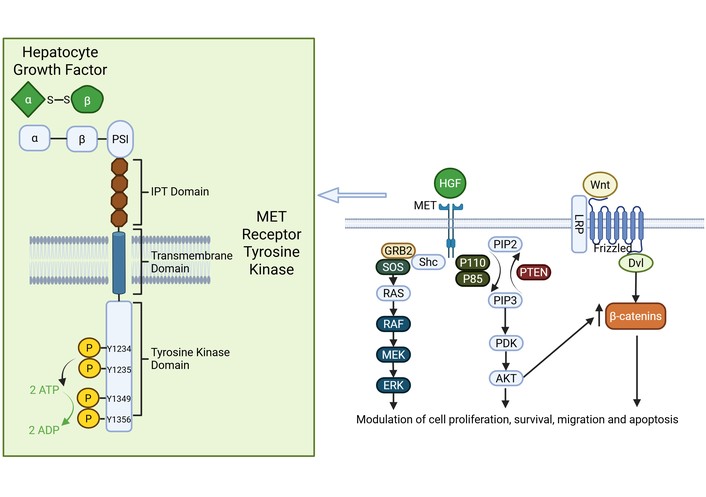
Hepatocyte growth factor/mesenchymal-epithelial transition signaling pathway. HGF binding to the MET tyrosine kinase receptor. HGF is comprised of an alpha and beta domain connected by a disulfide bond. HGF binds to the alpha and beta regions of the SEMA domain of the MET receptor. The next domain is a PSI region followed by four immunoglobulin-like domains connected to the transmembrane domain. The intracellular portion of the receptor comprises a tyrosine kinase domain with several phosphorylated tyrosine residues. HGF causes activation of the RAS/RAF/MEK/ERK pathway, PI3K pathway, and β-catenin pathway. ADP: adenosine diphosphate; ATP: adenosine triphosphate; AKT: protein kinase B; Dvl: dishevelled; ERK: extracellular signal-regulated kinase; Grb2: growth factor receptor-bound protein 2; HGF: human growth factor; IPT: immunoglobulin-like, plexin, transcription factor; LRP: LDL receptor-related protein; MEK: mitogen-activated protein kinase kinase; MET: mesenchymal-epithelial transcription factor; PDK: pyruvate dehydrogenase kinase; PIP2: phosphatidylinositol-4,5-biphosphate; PIP3: phosphatidylinositol-3,4,5-triphosphate; PSI: plexin-semaphorin-integrin domain; PTEN: phosphatase and tensin homolog; p85: p85-α protein; p110: p110-α protein; Raf: rapidly accelerated fibrosarcoma protein; Ras: rat sarcoma protein; Shc: Src homology and collagen protein; SOS: son of sevenless. Created in BioRender. Allen, B. (2026) https://BioRender.com/2nx41jw.
BEV was first applied to recurrent and progressive brain tumors by Stark-Vance in 2005, in combination with Irinotecan; the ORR was 43% [9, 22, 54–58]. Further work by Vredenburgh and co-workers at Duke applied BEV (10 mg/kg every 2 weeks) and Irinotecan to patients with recurrent malignant gliomas in a phase II trial [59]. The MRI ORR was 59%, with 2 CR and 38 PR. For the GBM cohort, the 6-month PFS rate was 43%, with a median PFS of 23 weeks, with other studies showing similar results. These favorable preliminary results led to the execution of the BRAIN trial, which was a phase II, multicenter, open-label, non-comparative trial evaluating the efficacy of BEV alone or in combination with Irinotecan [60]. The 6-month PFS rates for BEV alone and BEV plus Irinotecan were 42.6% and 50.3%, respectively. ORRs by MR imaging were 28.2% for the BEV alone cohort and 37.8% for the BEV plus Irinotecan subgroup. Median PFS for the BEV alone and BEV plus Irinotecan cohorts were 4.2 months and 5.6 months, respectively. The OS times were also comparable between the BEV alone and BEV plus Irinotecan groups—9.2 months vs. 8.7 months. BEV has also been used in combination with other drugs for treatment of recurrent high-grade gliomas, including Carboplatin, Carmustine, oral Etoposide, and Lomustine, with variable success. Overall, BEV alone and in combination with traditional chemotherapy drugs has efficacy when used for treatment of patients with recurrent malignant gliomas, and appears to improve PFS and OS (ESCAT Tier II) [58]. BEV has been FDA approved for the treatment of recurrent GBM since 2009.
BEV has also been applied to patients with newly diagnosed high-grade gliomas and GBM [9, 22, 54–57, 61]. Early pilot studies and phase II trials suggested that Standard of Care treatment (i.e., chemo-radiotherapy with concomitant TMZ, followed by adjuvant TMZ) in combination with BEV was active, with encouraging PFS and OS rates. These preliminary studies prompted two large phase II trials to explore adding BEV to standard chemoradiation with TMZ in patients newly diagnosed with GBM. The subsequent European AVAglio trial evaluated PFS and OS in 921 patients receiving concomitant TMZ and radiotherapy in addition to either BEV (10 mg/kg every 2 weeks) or placebo [62]. Upon completion of radiotherapy, patients continued receiving BEV or placebo every two weeks, along with adjuvant TMZ (150–200 mg/m2/day) for up to 6 cycles. Once adjuvant TMZ was completed, patients continued single-agent BEV (15 mg/kg every 3 weeks) or placebo until disease progression or intolerable toxicity. The bevacizumab group experienced statistically significantly longer median PFS compared to the placebo group (median 10.6 months vs. 6.2 months; HR = 0.64; p < 0.001). However, there was no significant difference in OS between the groups (median 16.8 months vs. 16.7 months; HR = 0.88; p = 0.10). The OS rates of the BEV and placebo groups were 72.4% and 66.3% at 1 year (p = 0.049) and 33.9% and 30.1% at 2 years (p = 0.24), respectively.
The RTOG 0825 study was a phase III trial of 637 patients with newly diagnosed GBM in which patients were randomized in a similar design to AVAglio, with the exception that patients could continue TMZ for up to 12 cycles during the adjuvant phase [63]. OS was not significantly different between the BEV and placebo groups (median 15.7 months vs. 16.1 months; HR = 1.13). While PFS was longer in the BEV cohort (median 10.7 months vs. 7.3 months; HR = 0.79), PFS did not reach the pre-specified improvement target; therefore, this was not a significant difference. Over the course of the study, the BEV cohort experienced increased symptom burden, worse QoL, and a decline in neurocognitive function in comparison to the placebo group. Based on the findings from these two phase III trials, the overall ESCAT rating for BEV in the setting of newly diagnosed GBM was assigned Tier II.
Small-molecule VEGFR inhibitors were developed for GBM treatment, including Cediranib and Sunitinib [9, 22, 57]. Cediranib is a potent ATP-competitive inhibitor that targets VEGFR2, with additional activity against PDGFRβ and c-Kit. Cediranib demonstrated significant activity in pre-clinical vascular models, as well as in xenograft animal experiments. A phase II clinical trial of Cediranib monotherapy for patients with recurrent GBM showed promising PFS-6 rates and MRI radiographic responses; however, there was no improvement in OS (ESCAT Tier IIIA/IV) [64]. Sunitinib is another multikinase inhibitor designed for anti-angiogenic therapy that is also targeted against VEGFR, as well as PDGFRα and β. Preclinical data and mouse models of orthotopic GBM suggested activity. However, similar to Cediranib, a phase II clinical trial of patients with recurrent GBM did not demonstrate any extension of PFS or OS (ESCAT Tier IIIA/IV) [65].
The HGF and MET signaling pathway involves paracrine cellular signaling and mediates proliferation, motility, angiogenesis, and tissue regeneration under normal conditions [13, 66, 67]. The HGF (or Scatter Factor) gene on chromosome 7 encodes a 728 amino acid protein. The mature HGF heterodimer consists of a disulfide-linked α chain and β chain. HGF binds to MET through a serine protease analog domain on the β chain. The MET gene on chromosome 7 (7q21-q31) contains 21 exons and 20 introns and encodes an RTK protein that is roughly 120 kDa in size. The translated MET protein forms a heterodimer linked to the extracellular α chain and the transmembrane β chain. The transmembrane β chain consists of a sema homology region (SEMA), a plexin-semaphorin-integrin domain (PSI), four immunoglobulin-like domains, a transmembrane domain, a juxtamembrane domain, a TKD, and a C-terminal docking site region. HGF binds to MET at the SEMA site, and the PSI domain stabilizes this interaction. After binding of HGF to MET, Tyr-1234 and Tyr-1235 in the intracellular TKD undergo autophosphorylation, promoting further autophosphorylation of Tyr-1349 and Tyr-1356 in the C-terminal docking site region. These autophosphorylation events allow for recruitment of GRB2, Src, PI3K, and GAB1 that subsequently promote downstream signaling pathways, including RAS, Raf/MEK/Erk, PI3K/AKT/mammalian target of rapamycin (mTOR), and Wnt/β-catenin (see Figure 4).
Previous analyses of TCGA data noted that roughly 30% of GBMs overexpress HGF and MET, suggesting an autocrine activating mechanism [9, 22, 68, 69]. Further review of this dataset demonstrated that genetic gain and amplification of MET at chromosome 7q31-34 was noted in 47% of primary GBM and 44% of secondary GBM. In addition, activating MET mutations are involved in the progression of low-grade gliomas to secondary GBM. Overexpression of MET in diffuse astrocytoma has also been associated with reduced OS (median 43.0 vs. 70.7 months; p = 0.004). In clinical specimens, 4% of GBM are known to display an amplification of MET that results in overexpression and constitutive activation of the kinase. The auto-activating METΔ7-8 mutation, in which exons 7 and 8 are deleted, has been detected in 6% of high-grade gliomas, including roughly 3.3% of GBM. Another MET mutation—METex14, is noted in 14% of secondary GBM and “skips” over exon 14, thereby removing the juxtamembrane region of MET, creating a cytosolic MET that is constitutively active in a ligand-independent manner. MET fusion transcripts have also been noted, including PTPRZ1-MET, TFG-MET, and CLIP2-MET—all of which activate MET and are associated with a poor prognosis, and have been demonstrated in up to 15% of secondary GBM. Finally, it has also been demonstrated that inhibition of the angiogenesis pathway through sequestration of VEGF results in activation of the MET pathway, converting gliomas to a more aggressive and invasive phenotype.
Numerous clinical trials have explored monoclonal antibodies and small molecule inhibitors targeting MET in GBM and other solid tumors [9, 13, 22, 66–69]. Monoclonal antibodies to HGF or MET inhibit activation of the MET TKD and reduce downstream signaling activity. The first antibody was targeted against HGF—Rilotumumab (AMG102), which had antitumor activity in pre-clinical GBM animal models. However, Rilotumumab was ineffective in a clinical trial as a monotherapy in patients with recurrent GBM (ESCAT Tier IV) [70]. When Rilotumumab was added to BEV for patients with recurrent malignant gliomas, the combination did not substantially increase the ORRs in comparison to BEV alone (ESCAT Tier IV) [71]. Onartuzumab (MetMAb) is a monoclonal, monovalent antibody targeting MET; it binds MET to form a complex with the SEMA-PSI domain, thereby impairing access of the α-chain of HGF to the receptor [13, 66, 67]. Onartuzumab was also able to reduce growth of GBM in pre-clinical testing. When it was applied to patients in a randomized, placebo-controlled, double-blind phase II trial in combination with BEV, however, it proved ineffective vs. BEV plus placebo [72]. The median PFS was 3.9 months vs. 2.9 months (HR 1.06; p = 0.7444), with a median OS of 8.8 months vs. 12.6 months (HR 1.45; p = 0.1389), respectively (ESCAT Tier IV). It is theorized that anti-HGF and anti-MET monoclonal antibodies lacked efficacy due to poor penetrance of the BBB and low concentrations in the CNS around the tumor cells.
Small-molecule inhibitors of MET are much smaller than antibodies and often hydrophobic, enabling improved penetration of the BBB and access to the CNS and brain tumors [9, 13, 22, 66–69]. Numerous small molecule MET inhibitors underwent evaluation in phase I and II trials, including Crizotinib, Cabozantinib, PLB-1001, INCB28060, SGX523, and others. Crizotinib is primarily an inhibitor of MET but also inhibits anaplastic lymphoma kinase (ALK) and ROS1, which are structurally similar. It has significant activity against MET-positive glial cells in vitro and improves survival in mouse xenograft models [13, 66]. However, to date there have only been two ongoing phase I clinical trials to evaluate the safety and tolerability of Crizotinib (ESCAT Tier IV); in adult patients, it was added to TMZ and radiotherapy for newly diagnosed GBM (NCT02270034), while in pediatric patients it was used in combination with Dasatinib for newly diagnosed pontine gliomas and high-grade gliomas (NCT01644773). Cabozantinib is another potent inhibitor of MET, as well as VEGFR2, and has anti-angiogenic, anti-proliferative, and anti-invasive activity in mouse E98-xenograft models.[13, 66, 67].
The MET pathway is intrinsically linked to GBM pathogenesis and has been implicated in the development of resistance during treatment with BEV. This led to a phase II trial using the small molecule inhibitor of MET, Cabozantinib, in patients with GBM that previously failed anti-angiogenic therapy [73]. However, in this population Cabozantinib did not demonstrate significant activity. In the setting of recurrent GBM patients naïve to anti-angiogenic therapy, Cabozantinib demonstrated evidence of clinical activity, but did not meet the predefined statistical target for success in the clinical trial (ESCAT Tier IIIA/IV) [74]. Capmatinib (INC280) is a potent inhibitor of MET and has shown significant efficacy against GBM in pre-clinical models when used in combination with Buparlisib, an inhibitor of PI3K [13, 66, 67]. A phase Ib/II clinical trial of Capmatinib used alone or in combination with Buparlisib was undertaken in patients with recurrent phosphatase and tensin homolog (PTEN)-deficient GBM [75]. The results did not support any significant activity of Capmatinib used as monotherapy or in combination with Buparlisib in this GBM cohort (ESCAT Tier IV). Since secondary GBM was noted to have an increased incidence of METex14, PTPRZ1-MET fusions, and MET amplification, Hu et al. [76] used PLB-1001, a potent MET kinase inhibitor, in a phase I clinical trial of 18 patients with recurrent high-grade gliomas. PLB-1001 has a higher binding affinity and is a more potent inhibitor of MET than Crizotinib, and also has excellent BBB penetration. In addition, it also demonstrated significant activity in pre-clinical studies and mouse GBM tumor models. In the phase I trial, PLB-1001 was well tolerated and able to achieve two partial responses in a group of chemo-resistant high-grade glioma patients (ESCAT Tier IIIA/IV).
Ongoing clinical trials targeting tumors with MET amplification include SPH3348 and Osimertinib (NCT05088070) and Savolitinib (NCT03598244), while trials against tumors with MET fusions include Bozitinib (NCT03175224), Elzovantinib (NCT03993878), and Glumetinib (NCT03457532).
Neurotrophic tropomyosin receptor kinase 1-3 (NTRK1-3) receptors comprise a group of RTKs with ligands that include nerve growth factor (NGF), brain-derived neurotrophic factor (BDNF), and neurotrophin-3 and 4 (NT-3 and NT-4) [77]. Ligand binding results in NTRK1-3 dimerization and subsequent activation of the MAPK, PI3K/AKT, and PL-C-gamma pathways that result in neuronal cell differentiation and survival (see Figure 5) [77]. NTRK fusion proteins commonly involve the 3’ kinase domain binding to the 5’ end of an unrelated protein [78]. These fusion proteins result in aberrant constitutive activation of NTRK pathways, thereby functioning as a proto-oncogene. In a review of 13,467 tumor samples, 0.31% of adult tumors and 0.34% of pediatric tumors demonstrate NTRK fusions [79]. NTRK fusions occur in 3.97% of pediatric gliomas and 0.8% of adult gliomas, demonstrating a higher prevalence in children. The presence of NTRK fusions in brain tumors is variable, with one study showing that of 356 diffuse adult gliomas only 5 had NTRK fusions, while in 127 high-grade neonatal gliomas in infants, about 40% had NTRK fusions [80, 81].
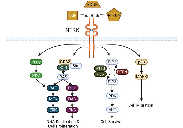
NTRK signaling pathway. Ligand binding of NGF, BDNF, or NT-3/NT-4 to NTRK results in activation of the PKC pathway, the RAS/RAF/MEK/ERK pathway, PI3K pathway, and the MAPK pathway. AKT: protein kinase B; BDNF: brain-derived neurotrophic factor; DAG: diacylglycerol; ERK: extracellular signal-regulated kinase; Grb2: growth factor receptor-bound protein 2; MAPK: mitogen-activated protein kinase; MEK: mitogen-activated protein kinase kinase; NGF: nerve growth factor; NTRK: neurotrophic tropomyosin receptor kinase; NT-3/4: neurotrophin-3/4; PDK: pyruvate dehydrogenase kinase; PIP2: phosphatidylinositol-4,5-biphosphate; PIP3: phosphatidylinositol-3,4,5-triphosphate; PKC: protein kinase C; PL-C: phospholipase C; PLCγ: phospholipase C-gamma; PTEN: phosphatase and tensin homolog; p38: p38 protein; p85: p85-α protein; p110: p110-α protein; Raf: rapidly accelerated fibrosarcoma protein; Ras: rat sarcoma protein; Shc: Src homology and collagen protein; SOS: son of sevenless. Created in BioRender. Allen, B. (2026) https://BioRender.com/2i3is6t.
Two pharmaceutical agents have received FDA approval for treatment of cancers expressing NTRK fusions. Larotrectinib is an oral ATP-competitive inhibitor of NTRK that was evaluated in a phase 1 study that enrolled infants, children, and adolescents with metastatic or advanced solid or CNS tumors. This study demonstrated anti-tumor activity in all NTRK fusion tumors, with evidence for decreased tumor burden (ESCAT Tier II). Only four of the 24 patients experienced grade 3 treatment-related adverse events [82]. Entrectinib is a pan-NTRK, ROS1, and ALK inhibitor that was evaluated in two phase 2 studies (ALKA-371-001 and STARTRK-1) in patients with advanced or metastatic tumors, including CNS tumors [83]. These studies showed that Entrectinib had a favorable safety profile with mostly Grade 1 or 2 adverse events, in which dose modification reverses the adverse effects. In these trials, only 8 of the 25 patients (32%) had a primary or metastatic intracranial tumor, but 5 of these patients did have a treatment response (only one patient had an NTRK1 fusion) (ESCAT Tier II). One patient had NSCLC with an NTRK1 fusion with 15–20 metastases to the brain, and experienced a complete treatment response with remission at 15 months.
A large follow-up phase I/II study of Entrectinib was performed by Desai and colleagues [84] in children and young adults with extracranial or CNS tumors that harbored NTRK, ROS1, or ALK gene fusions. Entrectinib was evaluated in 26 patients with NTRK fusion-positive tumors (16 with primary CNS tumors; 15 of which had NTRK fusions). In the cohort of CNS tumors with NTRK fusions, there was an ORR of 60% noted in a heterogeneous group of high-grade and low-grade gliomas. The median time to response was 1.9 months with a 56% PFS (95% CI: 38–74) and 85% OS (95% CI 71–99). Based on this and other studies of NTRK fusion-positive high- and low-grade gliomas for treatment of pediatric and young adult patients, Entrectinib is highly recommended (ESCAT Tier I).
NCT04655404 is the clinical trial of Larotrectinib for newly diagnosed, NTRK fusion-positive high-grade gliomas (NCT04655404).
The ROS1 gene, on chromosome 6q22.1, encodes an RTK expressed in brain and lung tissue [85]. It is a proto-oncogene that was first identified in 1987 within a GBM multiforme cell line [86]. The oncogenic fusion of ROS1 results in accelerated tyrosine kinase activity with subsequent downstream activation of the JAK/STAT, MAPK, and PI3K pathways within neoplastic cells. Various studies have shown that ROS1 fusions exist in various primary brain tumors, including rare cases in adult GBM (0.29%) and pediatric low-grade gliomas [87–89]. Pre-clinical testing of ROS1 inhibitors against GBM and pediatric glial tumors suggests activity, but clinical data is lacking (ESCAT Tier IV).
Initially approved ROS1 TKIs, including Crizotinib and Entrectinib, faced the challenge of rapidly acquired resistance mutations in tumors treated with these drugs [90, 91]. The recent TRIDENT-1 registrational phase 1–2 study evaluated the efficacy of a ROS1 tyrosine kinase inhibitor, Repotrectinib, in patients with ROS1 fusion-positive non-small cell lung cancer [92]. This TKI was designed to thwart the resistance mutations that occurred in ROS1 fusion-positive tumors treated with earlier generation TKI’s including Crizotinib, Entrectinib, and Lorlatinib [90, 91, 93]. Repotrectinib was also designed to have superior BBB penetration to improve utility in brain metastasis treatment. The phase 2 portion of the trial treated patients with ROS1 fusion-positive tumors with 150 mg daily for 14 days, followed by 160 mg BID. In patients with brain metastases, 8 of 9 patients (89%) experienced an intracranial response in those previously on an earlier-generation ROS1 TKI and in 5 of 13 patients (38%) who previously had been on a ROS1 TKI but had never been on chemotherapy. Of these patients, an estimated 83% and 60%, respectively, had a durable intracranial treatment response at 12 months (ESCAT Tier II) [92].
The ongoing clinical trial treating newly diagnosed ROS1 or ALK fusion-positive high-grade gliomas with Lorlatinib is NCT06333899.
The RTK, ALK, is involved in the JAK/STAT, RAS/MAPK, PI3K/AKT, and PLC-gamma pathways [94]. ALK overexpression, ligand interaction, mutation, and fusion can contribute to gliomagenesis via various cellular proliferation pathways and anti-apoptosis pathway activation [95–97]. Per EANO guidelines, consideration of ALK-specific treatments is indicated in prospective registries and clinical trials, once standard treatments have been exhausted [5]. Various primary CNS cancers overexpress oncogenic ALK, including neuroblastoma, neuroectodermal tumors, and GBM [94, 98].
In one study of 371 GBM cases reported by Blandin and co-workers [95], 45 (12%) had aberrations in ALK expression. Other studies have shown ALK can be overexpressed in 43–70% of GBM cases [99, 100]. In the Blandin et al.’ study [95], they demonstrated that ALK-fusion-positive GBM cellular and mouse models were responsive to the ALK inhibitor Ceritinib, including mice expressing PP1CB-ALK-NIH3T3 aberrant tumors, which had prolonged survival. Use of the third generation inhibitor, Lorlatinib, in vivo with mice expressing LRRF1P-ALK tumors also showed benefit. This provides evidence that the use of ALK inhibitors in GBM patients with ALK aberrant expression has the potential to provide some benefit (ESCAT Tier IIIA/IV).
There is one clinical trial of Lorlatinib in diagnosed ROS1 or ALK fusion-positive high-grade gliomas (NCT06333899).
Mutated and aberrant proteins within the RAS/Raf/MEK/Erk pathway are part of the RTK signaling cascade that regulates gene expression and prevents apoptosis [101]. Various proteins within this signaling cascade can experience tumorigenic aberrancy leading to cancer. The MAPK signaling cascade comprises three tiers. The first tier involves RTK and RAS activation of RAF. In the second tier, RAF activates and phosphorylates MEK through various mechanisms. In the final tier, the cascade of phosphorylation events results in the formation of the ternary RAF-MEK-ERK complex that drives cell proliferation, mobility, and survival [102]. Genetic, transcriptional, and post-translational errors in any of the essential steps in this RAS/RAF/MEK/ERK pathway are associated with nearly every type of human cancer [102]. In terms of CNS tumors, RAS/RAF/MEK/ERK pathway aberrancies occur in pilocytic astrocytoma, low-grade glioma, pilomyxoid astrocytoma, and ganglioglioma [102].
First-generation RAF inhibitors, such as Vemurafenib, Dabrafenib, and Encorafenib, have been used as monotherapy to treat BRAFV600E-harboring cancers or in conjunction with MEK inhibitors [103–105]. These drugs were stymied by tumor development of resistance genes. These first-generation RAF inhibitors also paradoxically induced secondary malignancies [106]. Therefore, numerous second-generation RAF inhibitors and pan-RAF inhibitors were developed and underwent clinical trials [106]. Trametinib and Cobimetinib, MEK inhibitors, are currently approved for BRAFV600E-harboring cancers as single agents or in combination with V-RAF murine viral oncogene homolog B1 (BRAF) inhibitors [106, 107]. ERK inhibitors are currently undergoing clinical trials, and none are currently approved for clinical use.
The RAF1 serine/threonine protein kinase, BRAF, is activated by RAS, a small GTPase, which causes homo- or heterodimerization, which subsequently activates the MAP kinases MEK1 and MEK2 [108]. These MAP kinases go on to stimulate ERK, ERK1 and ERK2, which promote cell survival, proliferation, and differentiation [108]. BRAF is an oncogene associated with several primary CNS tumors, including the pediatric tumors pilocytic astrocytoma, diffuse leptomeningeal glioneural tumor, ganglioglioma, desmoplastic infantile ganglioglioma, dysembryoplastic neuroepithelial tumor and the adult tumors epithelioid GBM (> 50%), low-grade glioma, pleomorphic xanthoastrocytoma (60–70%), and astroblastoma; BRAF mutations are rare in the other types of GBM (1.7%) [108, 109].
BRAF mutations are stratified as class I–III. Class I mutations are point mutations, in which 44% of BRAF mutations are secondary to V600E in CNS tumors [108, 109]. Class II mutations are non-V600E mutations, in-frame deletions, or fusions (e.g., KIAA1549::BRAF) that lead to RAS-independent dimerization, which causes increased ERK activation and low RAS activation [109, 110]. Class III is the least common mutation type and is RAS-dependent, with increased affinity and response to RAS activation [109].
At this time, there is no data on the effective concentration of BRAF inhibitors in glioma patients, but various studies have explored BRAF-targeted therapies. The BRAF inhibitors Vemurafenib and Dabrafenib have been used independently or combined with the MEK inhibitor Trametinib in high-grade glioma cases with BRAFV600E mutations in various studies [108]. Vemurafenib and Dabrafenib are not effective against tumors that harbor BRAF fusions. More robust evidence from the VE-BASKET trial evaluated Vemurafenib in GBM with BRAFV600E mutations and demonstrated a 25% ORR, 5.5 month PFS, and median OS of 28.2 months (ESCAT Tier IB) [111]. Another study of 206 adults and 90 children with BRAF-altered glioma demonstrated that in children, BRAF fusion and class I mutations are the most common while a variety of BRAF alterations are common in adults, with V600E being the most common SNV in both adults and pediatric gliomas [109]. In this study, 13 patients received BRAF targeted therapy with median time to progression of 5 months on therapy [109]. The type II RAF inhibitor Tovorafenib was used to treat patients with relapsed, refractory pediatric low-grade gliomas and demonstrated an ORR of 51% with median duration of response of 13.8 months [112]. More recent studies with Tovorafenib have more clearly established its efficacy in patients with relapsed or recurrent pediatric low-grade gliomas that have BRAF alterations (mutations or fusions) [112, 113]. The phase 2 Firefly-1 study enrolled 137 patients and had an ORR of 67% (using RANO high-grade glioma criteria), with a median duration of response of 16.6 months and median time to response of 3.0 months (ESCAT Tier IB) [112]. The ongoing Firefly-2 study is a phase 3, randomized trial of Tovorafenib vs. physician’s choice chemotherapy in pediatric and young adult patients experiencing newly diagnosed low-grade gliomas that have with active BRAF mutations [113]. This study has the potential to change the current paradigm for initial treatment of a primary CNS tumor harboring a BRAF activating alteration. Based on the above data, BRAF inhibitors are active in the treatment of pediatric and adult patients with primary brain tumors with BRAF mutations and fusions.
A clinical trial of RX208 for treatment of gliomas with BRAF mutations is now available (NCT05092802).
The tumor suppressor gene, NF1, is located on chromosome 17q11.2 and produces neurofibromin, which negatively regulates the RAS cellular pathway [114]. Neurofibromin is primarily expressed in nerve cells, oligodendrocytes, and Schwann cells, existing in two isoforms—type 1 and type 2 [114, 115]. Type 1 is primarily expressed in the brain, exhibiting significant RAS-promoting pathway regulatory activity, while Type 2, also called domain II-related GAP (GRD2), is expressed mainly in Schwann cells [114]. Sporadic mutations in the NF1 gene can lead to neurofibromatosis type 1, one of the more common genetic disorders worldwide, typified by a predisposition to cutaneous neurofibromas, plexiform neurofibromas, cafe-au-lait spots, and Lisch nodules [116, 117].
The attributed tumor suppression function of NF1 comes from the protein domain that is structurally similar to the RAS-guanosine-triphosphate (GTP) activation proteins (GAPs), called the GAP-related domain (GRD). GAPs enhance active RAS-GTP conversion to the inactive guanosine diphosphate (GDP)-bound form, resulting in RAS inactivation, which subsequently prevents downstream MAPK and PI3K/AKT/mTOR proliferation and differentiation [118–120]. Other NF1 functions include positive regulation of adenylyl cyclase (AC) to alter RAS activity, cell adhesion and motility regulation, promotion of apoptosis, epithelial mesenchymal transition inhibition, and heat shock factor inhibition [121–125]. Common tumors in neurofibromatosis type 1 include optic glioma, malignant peripheral nerve sheath tumors, neuroblastoma, pheochromocytoma, and carcinoid tumors [126, 127]. Numerous gliomas are associated with neurofibromatosis type 1, including the hallmark optic pathway glioma, WHO grade I pilocytic astrocytoma, pilomyxoid astrocytoma, and diffuse astrocytoma (WHO grade II, III, and IV). Gliomas tend to be more aggressive in adults with neurofibromatosis type 1, which is posited to be related to less frequent IDH mutations.
Therapeutics for tumors with pathogenic variants of NF1 target the downstream RAS pathway, which is pathologically upregulated. Additional therapeutics under exploration include mTOR inhibitors, MEK inhibitors (e.g., Trametinib, Cobimetinib), pegylated interferon, and pomalidomide or lenalidomide, which target angiogenesis in the tumor microenvironment [128, 129]. Selumetinib, a MEK inhibitor, is currently approved for treating inoperable plexiform neurofibromas in pediatric populations [130]. In a phase I/II trial of inoperable plexiform neurofibromas in children with neurofibromatosis type 1 (N = 74), Selumetinib was noted to have a confirmed partial response rate of 70%, with 59% of the responses lasting ≥ 12 cycles (ESCAT Tier IB). There was significant improvement in tumor-related pain intensity and pain interference that was durable out to 5 years of follow-up. For inoperable and symptomatic plexiform neurofibromas in pediatrics and adults with neurofibromatosis type 1, Selumetinib is also approved.
Another MEK inhibitor, Mirdametinib, has recently been approved for use in adults and children with symptomatic NF1-associated plexiform neurofibromas [131, 132]. Initial studies of Mirdametinib in this patient population noted partial responses in target tumors in 8 of 19 patients (42%) by cycle 12, while 10 (53%) had stable disease [131]. There were also significant and durable improvements in tumor-related pain. In the pivotal ReNeu phase IIb trial, Mirdametinib (2 mg/m2 twice daily, maximum 4 mg twice daily) was used to treat adults (N = 58) and children (N = 56) with neurofibromatosis type 1 and symptomatic plexiform neurofibromas (ESCAT Tier IB) [132]. The ORR was 41% in adults and 52% in children, with significant durable improvements in tumor-related pain.
Numerous ongoing clinical trials in NF1-positive solid tumors, including Abemaciclib and Temuterkib (NCT02454283), Cobimetinib (NCT04185831), Trametinib (NCT04116541), MRTX0902 (NCT05578092), and OKI-179 & Binimetinib (NCT05340621).
Alterations to the phosphatidylinositol-4,5-bisphosphate 3-kinase catalytic subunit alpha (PIK3CA) gene are present in about 11% of GBM [8]. This gene produces the catalytic subunit, p110α, of the class IA PI3K lipid kinases within the PI3K/AKT pathway that is responsible for promoting cell proliferation, angiogenesis, growth, motility, and survival [133]. Abnormal activation of the PI3K/AKT pathway occurs via loss of PTEN function/inactivation (see below) or EGFR/PIK3CA overactivation [134, 135]. PIK3CA mutation is associated with worse OS in astrocytoma and diffuse astrocytic glioma [136]. Furthermore, in some tumors with glioma PIK3CA mutations, EGFR amplification and PTEN inactivation can all co-exist [137]. In addition, PIK3CA mutations are maintained in some recurrent gliomas, demonstrating that PIK3CA is a potential therapeutic target in selected recurrent gliomas.
An additional pathogenic variant that causes PI3K signaling dysregulation is an alteration to the PIK3R1 gene. PIK3R1 encodes p85α, p55α, and p50α class 1A PI3K regulatory subunits [138]. P85α is a regulatory subunit that associates with the catalytic p110-alpha unit (encoded by PIK3CA), which forms the Class 1A PI3K heterodimer. P85α subsequently inhibits the catalytic subunit p110α by binding to the N-terminus SH2 domain [139]. Therefore, mutations of PIK3R1 disinhibit p110α, allowing for unchecked catalytic activity within the PI3K pathway and tumorigenesis. PIK3R1 is associated with a poor clinical outcome in IDH mutant astrocytomas [140].
PI3K pathway dysregulation is present in 90% of GBM, but only 11% of GBM have PIK3CA gene mutations [141, 142]. PI3K pathway inhibition is a therapeutic target for tumors with PIK3CA mutations. Isoform-selective PI3K inhibitors include Eganelisib, Idelalisib, and Alpelisib; however, these inhibitors have not entered clinical trials in gliomas since the isoform targets are not consistently altered in GBM [143, 144]. PI3K therapeutics for GBM have focused on the pan-PI3K inhibitors Taselisib, Pilaralisib, Buparlisib, and Copanlisib [145–148]. Regarding pan-PI3K inhibitors in glioma, Buparlisib is the most studied and was shown at low concentration to inhibit migration and invasion of GBM cells, with apoptosis achieved at higher concentrations and longer exposure (ESCAT Tier IV) [148, 149]. Challenges with developing PI3K inhibitors have been poor drug tolerance, intrinsic drug resistance, and acquired drug resistance [150].
Paxalisib is undergoing clinical trial in CNS tumors with PI3K/AKT/mTOR pathway mutations (NCT03994796).
PTEN is an essential tumor suppressor gene in the PI3K pathway [151]. PTEN protein removes the 3’ phosphate from the phospholipid secondary messenger phosphatidylinositol-3,4,5-triphosphate (PIP3) [151]. This produces PIP2, thereby counteracting the mitogenic signaling of class 1 PI3K and impairing PI3K signaling [152, 153]. Similarly, SH2-containing phosphatases SHIP1 and SHIP2 dephosphorylate the 5’ position of PIP3, producing PI(3,4)P2. PIP3 accumulates due to the loss of PTEN, activating various downstream oncogenic signaling cascades such as phosphatidylinositol-dependent kinases, serine/threonine kinase AKT/protein kinase B, S6 kinase, and mTOR (see Figure 6) [153].
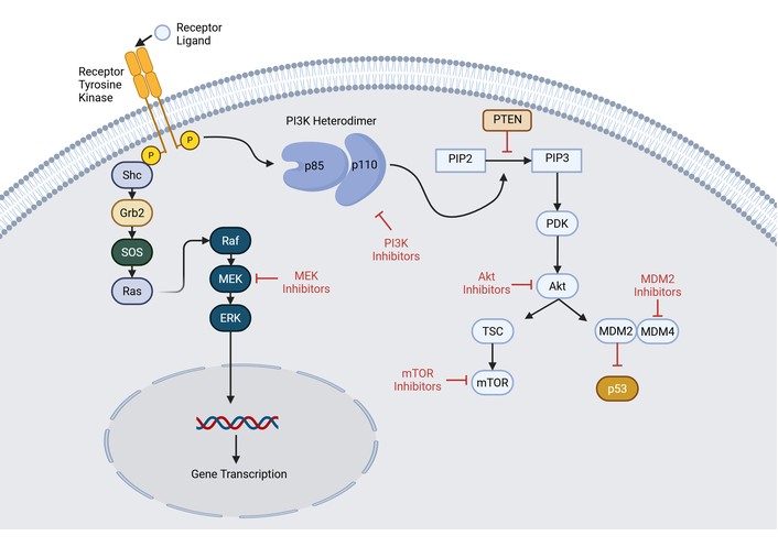
Phosphoinositide 3-kinase signaling pathway. Ligand binding to the tyrosine kinase receptor causes activation of upstream regulators of the RAS/RAF/MEK/ERK pathway, which leads to ERK activation and subsequent gene transcription. An additional pathway results in the conversion of PIP2 to PIP3. This results in activation of AKT, which subsequently leads to mTOR activation and inhibition of p53 by way of MDM2 activation. AKT: protein kinase B; ERK: extracellular signal-regulated kinase; Grb2: growth factor receptor-bound protein 2; MDM2/4: mouse double minute 2/4; MEK: mitogen-activated protein kinase kinase; mTOR: mammalian target of rapamycin; PDK: pyruvate dehydrogenase kinase; PIP2: phosphatidylinositol-4,5-biphosphate; PIP3: phosphatidylinositol-3,4,5-triphosphate; PI3K: phosphatidylinositol 3-kinase; PTEN: phosphatase and tensin homolog; p53: p53 tumor suppressor protein; p85: p85-α protein; p110: p110-α protein; Raf: rapidly accelerated fibrosarcoma protein; Ras: rat sarcoma protein; Shc: Src homology and collagen protein; SOS: son of sevenless; TSC: tuberous sclerosis protein. Created in BioRender. Allen, B. (2026) https://BioRender.com/efcfohw.
An additional proposed mechanism of PTEN is via control of oncogenic gene expression. PTEN was shown to regulate the loading of H3.3 (a histone variant) onto chromatin. H3.3 interacts with a histone chaperone protein DAXX to regulate gene expression [154, 155]. In the absence of PTEN, DAXX removes H3.3 from chromatin, allowing for oncogenic expression [152]. This was corroborated by a study using genomic display of genome-wide DAXX-knockdown/PTEN-deficient GBM cells, demonstrating genome-wide changes in H3.3 expression that resulted in increased tumor suppressor gene expression and decreased oncogene expression [152]. Therefore, DAXX-inhibition in PTEN-deficient tumor cells is a potential therapeutic target for preventing oncogene expression.
Numerous preclinical therapeutics have been studied in PTEN-deficient tumor cells. Promotion of synthetic lethality, the phenomenon in which two gene aberrations cause cell death, in PTEN-deficient cancer cells leads to cell death when knockdown of the genes Nemo-like kinase (NLK), Polo-like kinase 4 (PLK4), and monopolar spindle 1 (MLK) occurs [156]. Exome-mediated PTEN exportation into PTEN-deficient cells is an additional therapeutic option [157]. A variant of PTEN called “PTEN-long” is capable of permeating through lipid membranes; therefore, it can enter cancer cells to inhibit PI3K signaling [158]. Statins upregulate PTEN mRNA levels via increasing the transcriptional factor peroxisome proliferator-activated receptor-gamma [159]. Demethylating agents are another therapeutic option that may alter PTEN gene methylation and potentially reverse epigenetic silencing of PTEN [160].
Specific to PTEN-deficient GBM cells, several preclinical therapies have been identified (all ESCAT Tier IV). Icaritin, a glycolysis-impairing agent used in the treatment of GBM, suppresses the IL-6/STAT3 axis and the EMT pathway by impairing AKT/HIF-1 alpha [161]. Icaritin increases PTEN levels while suppressing AKT/HIF-1 alpha [162]. Tangeretin is another agent that increases PTEN while suppressing EMT [163]. Silibinin increases TMZ sensitivity in PTEN-deficient GBM [164]. And finally, curcumin induces PTEN activity and was shown to suppress AKT/mTOR signaling to impair GBM progression [165]. While no PTEN-specific therapies are currently on the market, numerous therapeutic targets have been studied.
There are several ongoing PTEN-specific clinical trials investigating Copanlisib and hormonal therapy in unspecified solid tumors (NCT05082025) and Talazoparib in PTEN-positive IDH mutant and IDH wildtype high-grade gliomas (NCT04740190).
Aspects of the PI3K/AKT/mTOR pathway were previously reviewed in the PIK3CA/PIK3R1 and PTEN sections, with the role of AKT and mTOR reviewed here further. PI3K has 4 classes and various isoforms, each with unique catalytic subunits. Class I is the most common in cancers [151, 166]. Briefly, PI3K recruitment to the plasma membrane after the G-protein coupled receptor activation results in subsequent catalysis of PIP2 into PIP3 [167]. PIP3 acts as a lipid secondary messenger that performs PDK1-mediated phosphorylation of AKT and mechanistic target of rapamycin C2 (mTORC2)-induced AKT1 phosphorylation (see Figure 6) [168, 169]. AKT has three isoforms, with AKT1 playing the major role in this pathway [170]. Subsequent AKT1 activation results in cell growth, survival, and metabolism through further phosphorylation of downstream effectors, including TSC2 [167]. TSC2 has a down-regulatory effect on RAS homolog enriched in brain (Rheb) GTPase, which subsequently recruits mTORC1 for lysosomal activation and results in protein, nucleotide, and lipid synthesis while suppressing autophagy [167]. The mTORC1 and mTORC2 complexes are two prominent mammalian mTOR complexes with mTORC1 containing a unique Raptor subunit and mTORC2 containing Rictor and Sin1 subunits [167]. AKT mediates signal transduction from mTORC2 to mTORC1, while mTORC1 can be regulated independently [167].
Pathogenic changes in PI3K signaling are common in high-grade gliomas, with dysregulation in up to 90% of GBM. PI3K-specific treatment modalities include isoform-specific PI3K inhibitors, pan-PI3K inhibitors, and dual PI3K/mTOR inhibitors. PI3K inhibitors were discussed previously, with the only FDA-approved PI3K inhibitors being Idelalisib, Copanlisib, Duvelisib, and Alpelisib (ESCAT Tier IV in gliomas) [171–173]. Copanlisib is the only currently approved pan-PI3K inhibitor to inhibit the four isoforms of PI3K. Numerous other PI3K inhibitors are undergoing clinical trials, with AZD6482, a PI3K beta isoform-specific inhibitor, demonstrating anti-proliferation and promotion of apoptosis in glioma cells [174].
AKT overexpression has been documented in lower-grade glioma, GBM, and meningiomas [175–177]. There currently are no approved AKT inhibitors. SC66 was studied in GBM and was able to impede GBM cell growth and migration via tumor cell arrest in G1 phase, and also caused apoptosis via AKT/beta-catenin signaling [178]. Various clinical trials of AKT inhibitors have tested Capivasertib, Uprosertib, and Afuresertib, to name a few [146, 179, 180]. The clinical benefit of AKT inhibitors in glioma needs further exploration (ESCAT Tier IV).
Inhibition of mTOR has documented some of the best clinical evidence in patients with neurofibromatosis and tuberous sclerosis. The mTOR pathway-specific therapies include ATP-competitive inhibitors and allosteric inhibitors of mTORC1 and mTORC2 [167]. There are two allosteric mTORC1 inhibitors approved: Everolimus and Temsirolimus; both are rapamycin derivatives [167]. These agents bind and suppress mTORC1 via binding to the FKBP12 protein, which disrupts the Raptor/mTOR interaction [181]. Everolimus does not inhibit mTORC2, which can lead to AKT upregulation [142]. Temsirolimus, a prodrug of sirolimus, inhibits activation and sensitivity to IL-2 in T and B cells by inhibiting mTORC2; most glioma treatment trials have used temsirolimus [182]. Everolimus is ESCAT Tier IB in patients with tuberous sclerosis-related subependymal giant cell astrocytoma (SEGA).
The paradoxical upregulation of upstream PI3K/AKT pathways by mTOR inhibitors led to the development of dual PI3K/mTOR inhibitors to address this shortcoming. Voxtalisib is a pyridopyrimidinone derivative that inhibits class-I PI3Ks, mTORC1, and mTORC2 and was studied in addition to TMZ in a phase 1 study for the treatment of high-grade glioma with a response in only 4% of patients [183]. Bimiralisib is a 4,6-dimorpholino-1,3,5-triazine-based dual pan-PI3K/mTOR inhibitor which has demonstrated the ability to cross the BBB [184]. GDC-0084 is an oral dual pan-PI3K/mTOR inhibitor that penetrates the BBB, in which a phase 1 trial in recurrent high-grade glioma demonstrated a 40% response rate [185]. And finally, NVP-BEZ235 given systemically demonstrated BBB penetrance and the ability to overcome TMZ resistance in glioma [186]. All in all, there is a growing body of research for treatment of the aberrations of the PI3K/AKT/mTOR pathway in brain tumors.
The p53 protein is encoded by TP53, a tumor suppressor gene on chromosome 17; the p53 protein binds DNA and prevents cell cycle progression, especially in the event of DNA damage [187]. Therefore, pathogenic variants of p53 predispose to cancer (see Figure 7). Mutations and pathogenic variants of p53 commonly occur in glial brain tumors, representing 28% of GBM tumors, 14% of lower-grade IDH wild-type gliomas (likely molecular GBM in the new WHO classification), and 94% of IDH mutant gliomas. Trials evaluating mutant p53 as a molecular target via treatment with small molecules aimed at refolding mutant p53 in GBM or other gliomas have all been unsuccessful and ineffective [5, 9, 22, 188–193]. Resultantly, inhibition of mouse double minute 2 (MDM2) and mouse double minute 4 (MDM4), the negative regulatory proteins of p53, represents the current treatment approach. One ongoing clinical trial combined anti-PD-1 therapy with TP53-EphA-2-CAR-DC—an autologous EphA-2 targeting CAR dendritic cell vaccine that contains p53 mutant peptides—in solid tumors harboring p53 273H mutations and EphA-2 expression (NCT05631886).
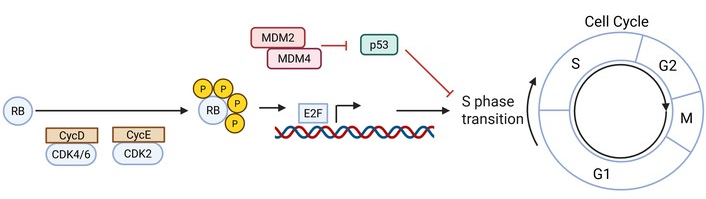
DNA damage signaling pathway. CycD and CycE sequentially phosphorylate RB which leads to activation of the EF transcription factor in which the gene products result in the S phase transition of the cell cycle. The MDM2/MDM4 pathway results in inhibition and inactivation of the tumor suppressor protein, p53 (which inhibits the S phase transition), thereby releasing p53 inhibition and allowing the S phase transition to proceed. CDK2/4/6: cycling dependent kinase 2/4/6; CycD: cyclin D; CycE: cyclin E; E2F: early transcription factor 2; G1: G1 phase; G2: G2 phase; M: M phase; MDM2/4: mouse double minute 2/4; p53: p53 tumor suppressor protein; RB: retinoblastoma protein; S: S phase. Created in BioRender. Allen, B. (2026) https://BioRender.com/u5pquat.
MDM2 downregulates the P53 tumor suppressor via E3 ligase activity; dysregulation of MDM2 prevents normal p53-mediated cell death and survival signaling [194, 195]. MDM4 is a protein that forms a heterodimer with MDM2 to reduce MDM2-mediated E3 ligase activity, thereby increasing levels of p53 [196, 197]. Pathologic amplification of MDM2 and MDM4 occurs in high-grade gliomas. 7.6% of GBM demonstrated MDM2 amplification, while MDM4 amplification occurred in 7.2% of GBM and 13% of lower-grade IDH wild-type gliomas. Recently, MDM2-specific inhibitors became clinically available (ESCAT IV), thus, clinical trials evaluating ASTX295 (NCT03975387), BI 907828 (NCT03449381), and BI 1703880 in combination with Ezabenlimab (NCT05471856) for treatment of unspecified solid tumors with MDM2 amplification and wild-type p53 are underway [5, 9, 188–192].
D-cyclins are regulatory subunits of cyclin-dependent kinase 4 and 6 (CDK4/6), which promote cell proliferation via phosphorylation of retinoblastoma protein-1 (RB1) and RB-like proteins (RBL1 and RBL2), thereby advancing into S-phase of the cell cycle [198, 199]. 15% of GBM and 7% of lower-grade IDH wild-type gliomas involve CDK4/6 amplification. Mutations of RB1 occur in 10% of GBM and 27% of lower-grade IDH wild-type gliomas. Monotherapy with Ribociclib and Palbociclib, two CDK inhibitor drugs, in GBM treatment showed no benefit (ESCAT IIIA/IV) [5, 9, 22, 188–193].
Ongoing clinical trials evaluating targeted therapies against CDK4 and/or CDK6 amplification include Abemaciclib (NCT03310879), Palbociclib and Abemaciclib (NCT02693535), Siremadlin and Ribociclib (NCT04116541), and Palbociclib (NCT03239015).
IDH1 and IDH2 are Krebs cycle enzymes that catalyze reversible decarboxylation of isocitrate to α-ketoglutarate (αKG) [200, 201]. IDH1 is in the cytosol and peroxisomes, while IDH2 is present in the mitochondria (see Figure 1). Mutations of IDH1 (e.g., R132H; and to a lesser extent, IDH2, R172K) are early and primary events in gliomagenesis, leading to an abnormal accumulation of 2-hydroxyglutarate (2-HG) in tumor cells. Increased levels of 2-HG are permissive to malignant transformation and involve epigenetic modification leading to DNA and histone hypermethylation. In particular, 2-HG seems to function as a potent inhibitor of Jumonji-class histone demethylases, as well as ten eleven translocation (TET)-family 5-methylcytosine hydroxylases, which mediate DNA demethylation. IDH mutations occur in approximately 80% of glial tumors with histological grade 2 and grade 3 features, as well as a small percentage of tumors with grade 4 histology [200].
Researchers have investigated several treatments that target the presence of IDH mutations, including small-molecule inhibitors of mutant IDH1 and IDH2, as well as vaccines directed towards mutant IDH1/2 [200, 202, 203]. IDH inhibitors bind inside the active catalytic site of mutant IDH1/2, thereby blocking the enzyme conformational change required to convert αKG to 2-HG. In vitro, in vivo, and animal studies have confirmed the ability of these drugs to reduce intracellular and serum levels of 2-HG, which results in reversal of epigenetic alterations. Numerous IDH inhibitors have been developed, including AG-120 (Ivosidenib), AG-221 (Enasidenib), AG-881 (Vorasidenib), FT-2102 (Olutasidenib), AGI-6780, BAY-1436032, and many others; Ivosidenib, Enasidenib, and Olutasidenib have now been FDA-approved for use in AML [202, 203].
Ivosidenib, Vorasidenib, and Olutasidenib have recently been applied to IDH1 mutant glioma patients [200, 203]. A phase I open-label dose escalation and expansion trial of Ivosidenib enrolled 66 patients with progressive IDH1 mutant gliomas, demonstrating good tolerability without dose-limiting toxicities (DLTs); 500 mg daily was used in the subsequent expansion cohort [204]. In the non-enhancing group, the median PFS was 13.6 months, and 1.4 months for the enhancing subgroup. In an exploratory imaging analysis, Ivosidenib was noted to reduce the volume and growth rates of non-enhancing tumors. Several case reports have suggested that Ivosidenib use in IDH1 mutant oligodendroglioma may improve seizure control [200, 203].
Vorasidenib, a dual inhibitor of IDH1 and IDH2, was demonstrated in animal models to have excellent penetrance into brain tissue [205]. In a recent multicenter, open-label, phase I dose escalation study, 52 patients with progressive gliomas underwent treatment with Vorasidenib [206]. Well-tolerated, Vorasidenib treatment conferred an ORR of 18% (1 PR, 3 MR) among patients with non-enhancing glioma, with a median PFS of 36.8 months for 22 patients with non-enhancing glioma and 3.6 months for 30 patients with enhancing glioma. Imaging analysis revealed sustained tumor shrinkage in multiple patients. In a comparative perioperative phase I trial of Ivosidenib and Vorasidenib, Mellinghoff and co-workers administered one of the drugs prior to surgery, and then measured 2-HG concentrations in resected tumor tissue in 49 patients with IDH1 mutant non-enhancing gliomas [207]. Tumor 2-HG concentrations were reduced by 92.6% with Vorasidenib (50 mg/day) and 91.1% with Ivosidenib (500 mg/day). Vorasidenib showed superior BBB penetration and more consistent 2-HG suppression; therefore, it was advanced to phase III testing in patients with IDH1 mutant low-grade gliomas (i.e., the INDIGO trial).
The INDIGO trial demonstrated unequivocally positive results in patients with IDH1 or IDH2 mutant low-grade gliomas who had only undergone surgical resection and were eligible for a “watch and wait” strategy (ESCAT Tier IA) [208]. The study randomized 331 patients to Vorasidenib (40 mg/day) or placebo. Comparing the Vorasidenib cohort to the placebo group, the median PFS increased to 27.7 months vs. 11.1 months, respectively (HR 0.39; p < 0.001). In addition, significant improvement was noted in the time to the next treatment intervention in the Vorasidenib group vs. the placebo group (HR 0.26; p < 0.001). The Vorasidenib group was more likely to not require treatment intervention by 18 months compared to the placebo group, 85.6% vs. 47.4%, respectively. Olutasidenib is a selective inhibitor of mutant IDH1 with high potency, oral bioavailability, and BBB penetrance, that was evaluated in a phase Ib/II open-label trial of IDH1 mutant gliomas [209]. Twenty-six patients were enrolled and received Olutasidenib 150 mg bid; the drug was well tolerated without DLTs. The imaging disease control rate was 48%; 2 patients had partial responses, while another 10 patients had stable disease for a median duration 5.0 (95% CI 4.2–14.6) months (ESCAT II). Thus far, Vorasidenib is the only IDH inhibitor that has FDA-approval for treatment of brain tumors (August 2024). Additional clinical studies are ongoing, including a trial of patients with progressive IDH mutant enhancing gliomas that will receive a combination of Ivosidenib and Nivolumab (NCT04056910), as well as a similar trial of patients with progressive enhancing IDH mutant astrocytoma that will receive Vorasidenib and Pembrolizumab (NCT05484622).
Other investigators have focused on the development of an IDH1 mutant-specific peptide vaccine. After development of a 20-mer peptide vaccine that was derived from the IDH1-R132H protein, animal studies in mice demonstrated induction of specific antitumor immune responses and reduction of growth of IDH mutant glial tumors. This data led to the single-arm, open-label phase I NOA16 clinical trial by the German Cancer Consortium, using the IDH1 peptide vaccine [210]. This trial treated 33 patients experiencing newly diagnosed WHO grade 3 and 4 IDH1 mutant astrocytoma with the peptide vaccine. Vaccine-related adverse events were restricted to grade 1. In 93.3% of the cohort, vaccine-induced immune responses were noted. The 3-year PFS and death-free rates were 0.63 and 0.84, respectively. In the subgroup of patients with immune responses, the 2-year PFS was 0.82. A high rate of pseudo-progression was noted and theorized to suggest intratumoral inflammatory reactions.
O6-methylguanine DNA methyltransferase (MGMT) is a DNA repair protein that is encoded by the MGMT gene on chromosome 10q26.3, and performs a guanine-to-cysteine methyl group transfer to repair damaged guanine nucleotides [9, 211]. Removing the methyl groups allows the cell to avoid gene mutations, cell death, and tumorigenesis; cellular MGMT is depleted during this process. The expression of the MGMT gene, and thereby the cellular concentrations of the MGMT protein, are mainly regulated by epigenetic modifications. It now appears that the degree of methylation of the MGMT promoter impacts expression activity—in particular, gene-specific core CpG island sites within the promoter—e.g., differentially methylated region 1 (DMR1) and DMR2 [211]. Methylation of CpG island sites leads to heterochromatinization and rearrangement of nucleosomes, thereby obscuring transcription initiation sites. With methylation of the promoter and reduced concentrations of MGMT, tumor cells become more sensitive to radiotherapy and alkylating chemotherapy drugs [212]. This relationship has been clearly demonstrated by Hegi and others during large multicenter studies of TMZ for treatment of GBM, as well as in the treatment of lower grade gliomas [213, 214]. This has led to MGMT methylation status becoming an important predictor of therapy responsiveness.
Recent estimates and meta-analyses of clinical trials of chemo-radiotherapy would suggest that the median OS for adult patients with unmethylated GBM was 14.11 months, while for patients with methylated GBM the OS was 24.59 months—a very significant difference [214]. Similarly, the PFS for adult patients with unmethylated GBM was only 4.99 months, while for methylated GBM patients the PFS was 9.51 months. Based on the powerful effect that MGMT methylation status has on the effectiveness of TMZ in terms of OS and PFS, many investigators have begun to devise strategies to target the MGMT protein and abrogate its effect in tumor cells. One of the most active approaches has been to use analogs of MGMT, such as O6-benzylguanine (O6-BG) and O6-(4-bromothenyl) guanine (O6-BTG), which are pseudo-substrates that can inactivate MGMT through alkyl group transfer [212]. O6-BG is able to pass through the BBB and has demonstrated the ability to inhibit MGMT and sensitize glioma cells to TMZ in pre-clinical studies, as well as a few preliminary clinical trials. There are many ongoing clinical trials utilizing O6-BG in brain tumor patients, including a study of young patients with recurrent gliomas or brainstem tumors treated with O6-BG and TMZ (NCT00257002), TMZ and O6-BG in children with recurrent brain tumors (NCT00052780), and O6-BG, TMZ, and Carmustine along with hematopoietic stem cell rescue for adult patients with recurrent GBM (NCT05052957). The major DLT of O6-BG and other MGMT analogs has been severe myelosuppression.
Other strategies that are under investigation to reduce the impact of MGMT in tumor cells include RNA Interference and the use of oncolytic viruses [215, 216]. These studies are all at very early stages of exploration in cell cultures and other in vitro systems.
Histone proteins provide critical protection and access regulation to DNA undergoing replication, repair, and transcription [217]. The four core histones are well-conserved basic proteins that initially form dimers (H2A/H2B) and tetramers (H3/H4), which then create a histone octamer that forms a nucleosome by interacting with roughly 147 base pairs of negatively charged DNA. Formation of a nucleosome, separating histones from DNA, and chromatin remodeling dynamically regulate gene expression mechanisms by adjusting chromatin accessibility.
Histone gene alterations are common in many different forms of cancer, and include mutations, structural variants, and copy number modifications [217]. They are noted in 7% of H1 genes, 11% of H2A genes, 9% of H2B genes, 10% of H3 genes, and 6% of H4 genes. Cancer patients who carry any form of histone genomic alteration have a lower OS in comparison to cancer patients without histone mutations or alterations. The most common type of histone mutation is of the missense variety (85%). Roughly 25% of all histone missense mutations occur in histone H3, mainly affecting two critical amino acids: K27 and G34 [217]. H3 K27M mutations are frequently present in pediatric GBM and diffuse midline glioma (DMG), including diffuse intrinsic pontine glioma. The presence of H3 K27M disrupts normal patterns of histone methylation via 3 main mechanisms: reduced trimethylation of H3 at position 27, suppressed histone methyltransferase activity, and polycomb repressive complex 2 sequestration.
ONC201 (Dordaviprone) is a newly approved, first-in-class imipridone, oral formulation with BBB penetration, small-molecule antagonist of dopamine receptor D2/3, as well as an allosteric agonist of the mitochondrial protease caseinolytic mitochondrial matrix peptidase proteolytic subunit (ClpP) [218, 219]. A review of numerous studies involving a total of 50 patients with recurrent H3 K27M-mutant DMG treated with Dordaviprone demonstrated an ORR using RANO-HGG criteria of 20.0%, with a median time to response of 8.3 months [219]. The median duration of response was 11.2 months. When using a combined RANO-HGG/LGG criteria for review, the ORR was 30.0%. A greater than or equal to 50% reduction in use of corticosteroids was noted in 7 of 15 evaluable patients (46.7%). Based on these results, ONC201 has an ESCAT Tier rating = II.
The Hedgehog (HH) signaling pathway regulates cell growth and patterning via intermediates including HH ligands, Patched protein (PTCH), smoothened (SMO) signaling regulators, and GLI transcription factors (GLI-1, GLI-2, and GLI-3) [220, 221]. Activation of SMO at the plasma membrane enables signal transduction via suppressor of fused homolog (SUFU), kinesin 7 (KIF7), and full-length glioma-associated oncogenes (GLiFL) protein complexes. SUFU dissociation from the complex releases GLIs that transfer to the nucleus to activate gene expression involved in cancer cell invasion, cell growth, and stem cell activity (see Figure 8) [222–225]. The off-state of the HH pathway is characterized by the PTCH protein binding to SMO, causing SMO inhibition via translocation prevention [226]. HH ligands can bind to PTCH and remove SMO inhibition, activating the HH pathway [227]. SMO is therefore a cancer treatment target for tumors with overactivation of the HH pathway.
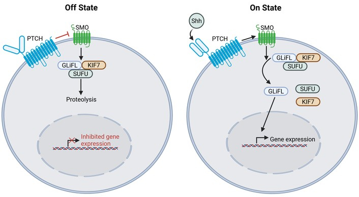
Hedgehog and SMO signaling pathway. In the Off State, PTCH remains unbound and inhibits SMO which prevents gene expression. Shh binding to PTCH, as seen depicted in the On State, allows for SMO activation, which catalyzes GLiFL release and subsequent gene expression. GLiFL: full-length glioma-associated oncogenes; KIF7: kinesin 7; PTCH: Patched protein; Shh: sonic hedgehog protein; SMO: Smoothened; SUFU: suppressor of fused homolog. Created in BioRender. Allen, B. (2026) https://BioRender.com/f314o62.
Early studies demonstrated that loss-of-function pathogenic variants in PTCH1 lead to constitutive overactivation of HH signaling in medulloblastoma [228]. In GBM, recombinant sonic HH N-terminal peptide (rhSHH), increases MMP-2 and MMP-9 expression, thereby enhancing HH signaling. Protein expression of GLI1 increases with increased MMP-2 and MMP-9 expression, demonstrating the stimulation of the HH signaling pathway [229]. Furthermore, HH pathway overactivation is caused by multiple mutations, including PTCH1 loss of function, SMO gain of function, and loss of function of SUFU [230–233]. For example, in medulloblastoma, SMO mutations are noted in 14% of patients. GLI1 amplification occurs rarely in most cancers, including GBM, but can be used as a marker of HH signaling pathway overactivation [234].
Studies have demonstrated that in GBM tumors with HH signaling overactivation with high GLI1 expression, SMO inhibitors were effective in these tumors, which led to SMO as a therapeutic target for inhibition in cancers with abnormal HH signaling activity [235, 236]. Vismodegib, an SMO inhibitor, either as monotherapy or in combination, is used for treatment of medulloblastoma (ESCAT Tier II) or GBM [237, 238]. Sonidegib demonstrated efficacy against medulloblastoma in a phase II trial, but is not currently used in GBM [239]. A recent trial evaluated the SMO inhibitor GDC-0449 in patients with GBM [240]. GDC-0449 was administered in the 7 days before GBM resection vs. placebo in patients without known HH signaling pathway abnormalities. After resection, both groups received the SMO inhibitor until intolerance, progression, death, or withdrawal of consent. While GDC-0449 was safe, well-tolerated, and felt to reach clinically relevant plasma concentrations, there was no difference in PFS between the two groups. Efficacy of GDC-0449 may have been limited by selecting GBM patients without known HH signaling pathway abnormalities. Furthermore, one of the main challenges with SMO inhibitors is the various means in which resistance develops [241]. Addressing the above challenges is an ongoing area of research.
The era of Molecular Oncology and Precision Medicine has finally arrived, especially in recent years with the development of 2nd Generation and 3rd Generation NGS platforms, which can rapidly analyze tumor-related DNA, RNA, and proteins [1, 2]. However, the clinical impact of Precision Medicine has mainly affected systemic Oncology, especially in patients with non-small cell lung cancer (NSCLC), where there are a significant number of ESCAT Tier I and Tier II molecular therapy options. Since there are fewer genetic variants amenable to molecular therapy in GBM, targeted NGS panels are recommended for GBM and other primary brain tumors to identify these potential targets [3, 8, 9].
There are many obstacles that preclude placing a GBM/glioma patient on a targeted therapy [192, 242]. The most critical obstacle is the paucity of actionable molecular alterations in GBM and other gliomas, in comparison to the options available for NSCLC and other systemic tumors. In addition, the activity for many of the available targeted drugs is insignificant or modest for GBM and other high-grade gliomas. It will be imperative moving forward to better understand and evade the molecular mechanisms of acquired therapy resistance, so that the drugs we do have can achieve more prolonged responses. There is also the issue that many medical centers that are not Comprehensive Cancer Centers and/or “academic” research hospitals may not have the oncological infrastructure to handle the necessary NGS and related testing required for Neuro-Oncology Precision Medicine in GBM/glioma patients. Another obstacle is how fragile patients with progressive GBM are, with many of them having rapid clinical progression so that they cannot tolerate further active treatment. In addition, many insurance companies will not reimburse targeted therapies in patients with GBM and progressive gliomas, due to insufficient GBM-specific evidence of efficacy.
In spite of the limitations noted above, the application of Precision Medicine to Neuro-Oncology has still seen advances in recent years, with more ESCAT Tier I and II targeted drugs now available for consideration in GBM and other brain tumor patients [3, 9, 192, 242]. For example, the presence of a BRAFV600E mutation in a brain tumor has an ESCAT Tier 1B rating for therapy with Dabrafenib/Trametinib or Vemurafenib. Similarly, for patients with CNS tumors that contain BRAF fusions and mutations, Tovorafenib has an ESCAT Tier 1B rating. In grade 2 IDH1/2 mutant astrocytoma and oligodendrogliomas that have progression after initial surgical resection, the use of Vorasidenib has an ESCAT Tier 1A rating. Bevacizumab has an ESCAT Tier II rating for progressive GBM and other malignant gliomas with significant nodular enhancement, edema, and mass effect. For pediatric and young adult patients with NTRK fusion-positive high-grade and low-grade gliomas, Entrectinib now has an ESCAT Tier I rating. In patients with Neurofibromatosis Type 1 that have NF1 gene mutations and symptomatic and unresectable plexiform neurofibromas, the use of Selumetinib or Mirdametinib have ESCAT Tier 1B ratings. In tuberous sclerosis patients with a related SEGA, treatment with Everolimus is rated as ESCAT Tier 1B. And finally, in newly diagnosed patients with an H3 K27M mutant Diffuse Midline Glioma, the use of Dordaviprone has a Tier II rating.
In spite of the limitations noted above, the application of Precision Medicine to Neuro-Oncology patients has still seen improvements in recent years, with more ESCAT Tier I and II targeted drugs now available for consideration in GBM and other CNS tumor varieties [3, 9, 192, 242]. The change to molecular classification of brain tumors has thus enabled the identification of more precise treatments for numerous brain tumor pathogenic variants such as Dabrafenib/Trametinib or Vemurafenib in BRAFV600E mutations, Tovorafenib for BRAF fusions and mutations, Vorasidenib in grade 2 IDH1/2 mutant astrocytomas and oligodendrogliomas, Entrectinib in NTRK fusion-positive high and low-grade gliomas, Mirdametinib in certain NF1 gene mutant CNS tumors, and Dordaviprone in H3 K27M mutant DMG, to name a few.
Additional molecular alterations are discovered each year, with the opportunity to continue identifying new therapeutic targets for the treatment of various brain cancers. As previously noted, currently there are numerous clinical trials exploring targeted therapies for the various pathogenic variants in GBM and other gliomas. There is hope that soon there will be new targeted treatments available for the common mutations and fusions occurring in brain tumors, including EGFR, MET, PIK3CA, PIK3R1, NF1, and others. That said, the ability of GBM cells to evade treatment by adjusting cell signaling to utilize non-targeted cell signaling pathways may be contributing to treatment resistance in various targeted therapies. This raises the possibility that more effective treatment modalities in the future will include multiple targeted molecular therapies utilized in tandem. Furthermore, future adaptive clinical trials may help the field progress more rapidly. For example, numerous ongoing clinical trials are using an innovative design to focus on molecular biomarkers, such as the Bayesian adaptive randomization platform, which is being used in GBM-AGILE (NCT03970447) and the Individualized Screening Trial of Innovative Glioblastoma Therapy (INSIGhT) trial (NCT02977780) [243, 244]. In these two trials, if a specific biomarker treatment arm demonstrates a low probability of improved PFS, adaptive randomization occurs in which these trial arms are removed from the study. Leveraging these types of adaptive trials may help to more quickly identify effective, molecular-targeted GBM treatments in the future.
2-HG: 2-hydroxyglutarate
AKT: protein kinase B
ALK: anaplastic lymphoma kinase
ATP: adenosine triphosphate
BRAF: V-RAF murine viral oncogene homolog B1
CARs: chimeric antigen receptors
CDK4/6: cyclin-dependent kinase 4 and 6
DLTs: dose-limiting toxicities
DMG: diffuse midline glioma
EGFR: endothelial growth factor receptor
ERK: extracellular signal-regulated kinase
ESCAT: ESMO Scale of Clinical Actionability for Molecular Targets
FAK: focal adhesion kinase
FGF: fibroblast growth factor
FGFR: fibroblast growth factor receptor
GBM: glioblastoma
Grb2: growth factor receptor-bound protein 2
HGF: human growth factor
HH: hedgehog
IDH1: isocitrate dehydrogenase 1
JAK: Janus kinase
mAb: monoclonal antibody
MAPKs: mitogen-activated protein kinases
MDM2: mouse double minute 2
MDM4: mouse double minute 4
MET: mesenchymal-epithelial transcription factor
MGMT: O6-methylguanine DNA methyltransferase
MMPs: matrix metalloproteinases
mTOR: mammalian target of rapamycin
NGS: next generation sequencing
NTRK: neurotrophic tropomyosin receptor kinase
O6-BG: O6-benzylguanine
ORR: objective response rate
OS: overall survival
PDGF: platelet-derived growth factor
PDGFR: platelet-derived growth factor receptor
PDK: pyruvate dehydrogenase kinase
PFS: progression-free survival
PI3K: phosphatidylinositol 3-kinase
PIK3CA: phosphatidylinositol-4,5-bisphosphate 3-kinase catalytic subunit alpha
PIP2: phosphatidylinositol-4,5-biphosphate
PIP3: phosphatidylinositol-3,4,5-triphosphate
PL-C: phospholipase C
PlGF: placental growth factor
PSI: plexin-semaphorin-integrin domain
PTB: phosphotyrosine-binding
PTCH: Patched protein
PTEN: phosphatase and tensin homolog
RB1: retinoblastoma protein-1
RTK: receptor tyrosine kinases
SEGA: subependymal giant cell astrocytoma
SH2: Src homology-2
Shc: Src homology and collagen protein
SMO: smoothened
SNVs: single nucleotide variants
SUFU: suppressor of fused homolog
TKD: tyrosine kinase domain
VEGF: vascular endothelial growth factor
VEGFR: vascular endothelial growth factor receptor
αKG: α-ketoglutarate
The supplementary figures for this article are available at: https://www.explorationpub.com/uploads/Article/file/1006150_sup_1.pdf.
HBN: Conceptualization, Writing—original draft, Writing—review & editing. BCA: Writing—original draft, Writing—review & editing. Both authors read and approved the submitted version.
The authors declare that there are no conflicts of interest.
Not applicable.
Not applicable.
Not applicable.
Not applicable.
Not applicable.
© The Author(s) 2026.
Open Exploration maintains a neutral stance on jurisdictional claims in published institutional affiliations and maps. All opinions expressed in this article are the personal views of the author(s) and do not represent the stance of the editorial team or the publisher.
Copyright: © The Author(s) 2026. This is an Open Access article licensed under a Creative Commons Attribution 4.0 International License (https://creativecommons.org/licenses/by/4.0/), which permits unrestricted use, sharing, adaptation, distribution and reproduction in any medium or format, for any purpose, even commercially, as long as you give appropriate credit to the original author(s) and the source, provide a link to the Creative Commons license, and indicate if changes were made.



View: 448
Download: 13
Times Cited: 0
Zohreh Khosravi Dehaghi
Maria Ciscar-Fabuel ... Andreu Gabarros-Canals
Maria Ciscar-Fabuel ... Andreu Gabarros-Canals
Julius Mulumba ... Yong Yang
Adam H. Lapidus, Malaka Ameratunga


