Review


Affiliation:
School of Molecular and Cell Biology, University of the Witwatersrand, Johannesburg 2000, South Africa
†These authors contributed equally to this work.
Email: Oluwatobi.otun@wits.ac.za; sarahholabamiji@gmail.com
ORCID: https://orcid.org/0000-0002-5408-695X


 ,
,
Affiliation:
School of Molecular and Cell Biology, University of the Witwatersrand, Johannesburg 2000, South Africa
†These authors contributed equally to this work.
Explor Neurosci. 2026;5:1006149 DOI: https://doi.org/10.37349/en.2026.1006149
Received: June 09, 2026 Accepted: August 21, 2026 Published: September 16, 2026
Academic Editor: Marcello Iriti, Milan State University, Italy
The article belongs to the special issue Medicinal Plants and Bioactive Phytochemicals in Neuroprotection (Vol II)
The protein Mitogen-activated protein kinase 9 (MAPK9) (c-Jun N-terminal kinase 2 [JNK2]) is a critical signalling molecule involved in cellular survival, inflammation, oxidative stress, and the programming of cell death. This is because there has been emerging evidence of the effects of the abnormal activation of MAPK9 in the pathogenesis and progression of neurodegenerative disorders like Alzheimer’s disease, Parkinson’s disease, and Huntington’s disease. However, many medicinal plants possess multiple phytoconstituents with neuroprotective activity that can modulate the MAPK9 signalling cascade and protect neurons against injury. Thus, this review focuses on the biology and signalling pathways of MAPK9, its function in neurodegeneration, and medicinal plants and phytochemicals targeting MAPK9, together with their biological mechanism(s) of action. Recent advancements in drug design and discovery, involving computational techniques such as molecular docking, network pharmacology, and artificial intelligence, were also discussed. Additionally, current problems, limitations, and possibilities in the clinical translation of these natural compounds were highlighted. Therefore, knowledge of how these phytochemicals modulate the MAPK9 signalling pathway may offer safer, more effective preventive and therapeutic approaches for neurodegenerative disorders.
Neurodegenerative diseases remain one of the major public health problems affecting many parts of the world due to their characteristic gradual degradation of neuronal structure and function, causing cognitive impairment, loss of mobility, and eventually, severe disability [1]. Some of the common neurodegenerative diseases include Alzheimer’s disease (AD), Parkinson’s disease (PD), Huntington’s disease (HD), and amyotrophic lateral sclerosis (ALS), which affect their respective patients in terms of morbidity and even death in many cases [2, 3]. AD is mainly known for causing dementia, with major hallmarks of amyloid β (Aβ) plaques, neurofibrillary tangles, synaptic dysfunction, and cognitive decline [3]. Similarly, the pathogenesis of PD involves the loss of dopaminergic neurons from the substantia nigra along with the formation of proteinaceous aggregates called α-synuclein, producing symptoms such as tremor, rigidity, and dyskinesias [4]. Furthermore, HD involves inherited genetic mutations that cause cytosine-adenine-guanine (CAG) repeat expansions in the huntingtin gene, resulting in neuronal malfunction and the development of motor and neuropsychiatric symptoms [5]. Likewise, in ALS, there is degeneration of both upper and lower motor neurons, leading to muscle weakness and ultimately death due to respiratory insufficiency [6].
However, the burden from neurodegenerative disorders remains on the rise due to the increase in the elderly population and life expectancy globally [1]. Such diseases present significant socio-economic and health care problems, not only in terms of patient suffering but also for their caregivers and the health system. Currently, therapeutic measures for neurodegenerative disorders are mainly symptomatic, failing to prevent the disease process and even reverse it [7]. As such, acetylcholinesterase inhibitors and N-Methyl-D-aspartate (NMDA) antagonists that form part of treatment options for AD are only able to temporarily improve the symptoms, with dopamine agonists used in PD becoming less and less effective with time [8, 9]. On the same note, currently available treatments for HD and ALS only yield little improvement and are unable to address the cause of neuronal cell damage [2, 5].
In recent years, significant advances in molecular neuroscience have revealed that intracellular signal transduction pathways are essential to the pathophysiology of neurodegenerative diseases. Of these signal transduction pathways, the mitogen-activated protein kinase (MAPK) pathway is crucial in regulating cellular responses to stress, inflammation, apoptosis, and survival [10]. MAPK9, also known as c-Jun N-terminal kinase 2 (JNK2), plays a significant role in the induction of neuronal injury and neurodegeneration [11]. It is induced by oxidative stress, inflammatory cytokines, protein misfolding, and mitochondrial dysfunction, thereby activating downstream transcription factors and pro-apoptotic signalling pathways. However, dysfunction in MAPK9 signalling has been associated with neuronal cell death, tau hyperphosphorylation, amyloid toxicity, and neuroinflammation seen in many neurodegenerative conditions [12].
While the expression of JNK3 (MAPK10) is largely confined to the brain and has been studied intensively as a potential therapy against neuron-specific diseases [13], recent findings suggest that JNK2 (MAPK9) too has several roles to play in neurodegenerative diseases. In contrast to JNK3, which is selectively expressed, JNK2 is expressed in all cell types and, besides contributing to neuronal apoptosis, also helps with oxidative stress responses, microglial activation, astrogliosis-mediated neuroinflammation, and signalling from peripheral immunity that affect disease progression [14, 15]. Therefore, targeting MAPK9 alongside JNK3 can be of significant advantage in treating such diseases. Hence, this review is focused on MAPK9.
Consequently, the use of medicinal plants has attracted increased attention due to their ability to exert antioxidative, anti-inflammatory, anti-apoptotic, and mitochondrial-protective effects. Natural compounds such as curcumin, resveratrol, quercetin, epigallocatechin gallate (EGCG), and berberine have been shown to modulate MAPK-related signalling cascades and reduce neuronal injury in experimental studies [16–18]. Given the rising interest in plant-based treatment modalities and the pivotal role of MAPK9 in the development of neurological disorders, the present paper provides an overview of the existing literature on the interaction between plant bioactives and MAPK9 signalling cascades. Besides, the paper will focus on the opportunities, barriers, and prospects of phytochemical treatment of neurological conditions.
MAPK9, also known as JNK2, is a member of the MAPK family, which forms an evolutionarily conserved protein signalling pathway that modulates various physiological functions in cells [19]. The MAPK superfamily includes multiple subfamilies, including the extracellular signal-regulated kinases (ERKs), p38 MAPKs, and the JNKs [20–22]. While ERKs are mainly activated by growth factors and other stimuli associated with cell proliferation, JNK kinases are activated by various forms of cellular stress, including oxidative stress, pro-inflammatory cytokines, ultraviolet (UV) irradiation, DNA damage, and protein aggregation [23]. As such, they play a key role in the development of certain chronic conditions.
The JNK protein kinase family consists of three isoforms having unique tissue expression profiles and biological roles. JNK1 (or MAPK8) and JNK2 (or MAPK9) have wide tissue expression profiles, while JNK3 (or MAPK10) is mainly found in the brain, heart, and testis [23]. JNK3 has been considered to be the primary target for central nervous system (CNS) drug design due to its predominant expression in neurons and the expectation that selective blockade of JNK3 will decrease neuronal damage without causing systemic toxicity. However, recent evidence indicates that JNK2 is a major player in neurodegeneration via oxidative stress, inflammatory signalling, mitochondrial dysfunction, and glia activation. The diverse biological role of MAPK9 differentiates it from JNK3 and justifies consideration of MAPK9 as an independent target for therapeutic intervention [11, 24].
Structurally, MAPK9 encodes a serine-threonine protein kinase comprising an adenosine triphosphate (ATP)-binding site along with a catalytic site that is critical for phosphorylation of substrates (Figure 1). Like other MAPKs, JNK2 has a dual phosphorylation motif consisting of three amino acids: threonine-proline-tyrosine (TPY) [25]. Phosphorylation of these sites is required for kinase activation and subsequent signal transduction. In addition, the protein has docking sites that are required for interaction with scaffolding proteins, transcription factors, and regulators. Alternative splicing produces several JNK2 isoforms, such as All and p46, that can differ in substrate specificity, localisation, and function. Chromosomally, the MAPK9 gene resides on chromosome 5q35 [26].
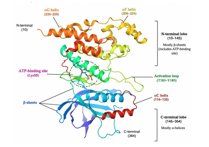
Crystal Structure of JNK2. The three-dimensional structure of JNK2 highlights its unique kinase domain, which includes α-helices, β-sheets, and the ATP-binding site. JNK2 is an important molecule that participates in the control of cellular stress response, inflammation, cell death, and neurodegeneration. Source: UniProt: P45984 · MK09_HUMAN MAPK9: PDB: 8ELC.
The cellular localisation of MAPK9 is dynamic and directly correlates with its biological function [22]. In a basal state, inactive JNK2 is localised to the cytoplasm; however, once activated, it translocates to the nucleus and phosphorylates multiple transcription factors that regulate stress-induced genes. In neurons, activated JNK2 has also been found localised to mitochondria, synapses, and axons, thereby emphasising its diverse physiological and pathological functions. In the mitochondria, JNK2 plays an important role as it is responsible for causing mitochondrial dysfunction, release of cytochrome C, and the activation of intrinsic apoptosis pathways [27]. For example, mitochondrial JNK activation in response to oxidative stress has been shown to cause the death of dopaminergic neurons in PD models [28].
The activity of MAPK9 occurs in an organised manner within a signalling cascade that involves phosphorylation steps [20]. MAPK pathway activation occurs primarily through upstream kinases called MAP kinase kinase kinases (MAP3Ks), such as apoptosis signal-regulating kinase 1 (ASK1), MLKs, TAK1, and MEKK1-4. MAP3K activation occurs in response to cellular stimuli, including oxidative stress, endoplasmic reticulum stress, inflammatory cytokines, and excitotoxicity (Figure 2). Afterwards, MAP3K phosphorylates the downstream kinases known as MAP kinase kinases (MAP2Ks), such as MKK4 and MKK7, and these proteins phosphorylate the TPY motif of JNK family proteins, causing JNK2 activation [29].
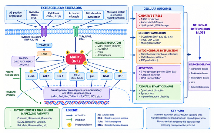
MAPK9/JNK signalling pathway in neurodegeneration. A schematic representation of the MAPK9/JNK2 signalling pathway in neurodegeneration. Extracellular stressors activate MAPK9 (JNK2) via upstream kinases, leading to phosphorylation of transport factors and induction of pro-apoptotic, inflammatory, and oxidative pathways. This results in neuronal dysfunction and loss. Negative regulators and phytochemicals can also inhibit MAPK9 signalling and confer neuroprotection.
The involvement of the stress-activated pathway via MAPK9 is critical in influencing cell fate in disease states. Oxidative stress constitutes one of the most powerful activators of JNK2 signalling. The accumulation of excessive amounts of reactive oxygen species (ROS) causes the disruption of the cell redox balance and initiates the phosphorylation cascades mediated by ASK1, which in turn results in the constant activation of JNK [30]. Furthermore, inflammatory cytokines like tumour necrosis factor-alpha (TNF-α) and interleukin (IL)-1β activate JNK signalling in response to receptor-mediated signals [31]. In neurodegenerative disorders, the activation of the MAPK9 pathway leads to continuous neuroinflammation and neurological dysfunction. Overexpression of c-Jun activation correlates strongly with the development of neuronal cell death and neurodegeneration. Increased phosphorylation of c-Jun in degenerating neurons under conditions of oxidative stress and excitotoxicity has been reported in experimental models [32].
From a physiological perspective, MAPK9 has several vital functions in maintaining cellular homeostasis and neural health. When activated under normal conditions, JNK2 triggers various adaptive stress responses to protect cells from damage and support their recovery [33]. The temporary activation of JNK signalling might increase antioxidant levels and facilitate the disposal of damaged cellular structures via autophagy. On the contrary, prolonged activation usually triggers JNK-mediated apoptotic cascades. In general, JNK-induced apoptosis occurs via two major processes: nuclear activation of AP-1, which induces pro-apoptotic genes, and the mitochondrial pathway, involving caspase activation and cytochrome c release [34].
In addition to the apoptotic pathway, MAPK9 has been found to play a role in neuronal development and differentiation. Signalling through JNK affects cytoskeletal remodelling, axon growth, and neuronal migration during early brain development [35]. JNK signalling has been shown to affect neuritogenesis and synapse formation, processes crucial to the formation of functional neural circuits. In one instance, JNK-dependent phosphorylation of microtubule-binding proteins plays a critical role in the formation of neuronal architecture and intracellular transport. Also, MAPK9 is involved in synaptic plasticity, a vital process essential for learning and memory [36]. Regulated JNK signalling influences the dynamics of synaptic protein turnover, receptor trafficking, and long-term potentiation (LTP). On the other hand, dysregulated JNK signalling leads to deterioration of synaptic function and impaired cognition, especially in AD patients, where increased JNK signalling activity is associated with synaptic loss and memory impairment. There have been reports that aberrant JNK signalling activity is implicated in the phosphorylation of tau protein. The combination of MAPK9/JNK2 forms a vital stress-response kinase that mediates signals from the external environment and within the cell to control neuronal survival, inflammatory response, and cell death [37].
Neurodegenerative diseases refer to a group of chronic diseases that are progressive in nature and cause deterioration of neurons through structural changes and impaired function, ultimately resulting in cognitive and movement disabilities [38]. Several studies have shown that MAPK9 (JNK2), a kinase in the JNK family, is an important regulator of the biological mechanisms underlying such pathologies [21, 24, 39]. The activation of MAPK9 is driven by stress, inflammation, oxidative stress, and protein aggregation, factors characteristic of neurodegeneration. The involvement of MAPK9 dysregulation in neuronal cell death has been demonstrated in neurodegenerative diseases like AD, PD, and HD, which are further discussed.
Neuroinflammation is considered an important feature of several neurodegenerative disorders, in which microglial cells and astrocytes become chronically activated, resulting in the continuous production of pro-inflammatory factors that lead to neuronal death [40, 41]. However, the importance of MAPK9 (JNK2) lies in its ability to regulate neuroinflammatory pathways in response to cellular and oxidative stress. After activation, the MAPK9 pathway increases the expression of pro-inflammatory factors such as TNF-α, IL-1β, and IL-6 [31, 42].
Numerous studies have shown the importance of JNK signalling in neuroinflammation. For instance, genetic depletion or inhibition of JNK2 significantly reduces α-synuclein production and inflammatory responses in neuronal models of PD [43], indicating that JNK2 is a key factor in the neuroinflammation associated with the pathology. Additionally, studies of neuroinflammation in AD models have found elevated activation of MAPK pathway components in microglia, and this increase, in turn, corresponded to the induction of inflammation. Therefore, MAPK9 serves as a mediator between cellular stress and inflammation [44]. Hence, blocking MAPK9 activity may be a useful approach for treating neuroinflammation.
Programmed cell death, also known as neuronal apoptosis, is a significant pathophysiological hallmark of neurodegeneration, as it underlies the progressive loss of neurons seen in conditions such as AD, PD, and HD [45, 46]. MAPK9 (JNK2) is involved in inducing apoptosis by transducing intracellular signals in response to cellular stress from oxidative stress, inflammation, mitochondrial dysfunction, and accumulation of abnormal proteins [47]. Following its activation, MAPK9 phosphorylates transcription factors such as c-Jun, thus activating pro-apoptotic genes. Several studies have also shown that modulation of JNK signalling results in better autophagy-mediated degradation of aggregated Aβ and α-synuclein [48, 49].
For example, in PD models, treatment with the neurotoxin 6-hydroxydopamine (6-OHDA) led to an increase in JNK pathway activity and c-Jun phosphorylation, followed by apoptosis [50]. JNK inhibition showed a decrease in neuronal death and enhanced survival. Likewise, in AD models, Aβ peptides were found to activate the JNK signalling pathway, which led to mitochondrial dysfunction as well as neuronal death [51]. Inhibition of JNK caused a reduction in the activation of caspases, thus protecting neurons from amyloid neurotoxicity.
MAPK9 also plays a vital role in the pathogenesis of tau in AD. Tau proteins normally play a significant role in stabilising microtubules within the neurons [52]. However, in cases of hyperphosphorylation, tau leads to its aggregation, resulting in the formation of neurofibrillary tangles [53]. JNK2 phosphorylates tau, resulting in microtubule destabilisation. Increased levels of phospho-JNK and tau proteins in postmortem AD brain samples indicate a link between MAPK9 activity and AD pathogenesis.
Synaptic dysfunction in AD may be related to JNK signalling. Continuous activation of MAPK9 disrupts synaptic plasticity and neurotransmission, subsequently resulting in memory problems. Research shows that continuous activation of JNK prevents LTP, a physiological phenomenon necessary for learning and memory [54]. Therefore, MAPK9-induced synaptic damage in AD is a contributing factor towards impaired cognition.
Another factor linked to the aggregation of alpha-synuclein, a hallmark feature of PD, is MAPK9. The misfolding of α-synuclein leads to the stimulation of inflammation and oxidative stress, which then activates the JNK pathway [55]. This, in turn, causes increased protein aggregation and cell death, forming a vicious cycle. Furthermore, the experimental model that uses 6-OHDA, a neurotoxin, to produce a Parkinson-like degeneration of dopamine neurons has been extensively used to investigate this mechanism. In these studies, 6-OHDA induces the activation of JNK signalling, including MAPK9, that results in c-Jun phosphorylation and apoptosis gene expression [56]. The pharmacological inhibition of JNK signalling increases cell survival by preventing neuronal cell death.
Furthermore, a widely used model of induced Parkinsonism consists of treating mice with the toxin 1-methyl-4-phenyl-1,2,3,6-tetrahydropyridine (MPTP). It was found that exposure to MPTP increases JNK activation in dopaminergic neurons. Moreover, pharmacological inhibition of JNK or the knockout of JNK2 decreases neurodegeneration and maintains mouse motor function [57]. Also, according to Ruan et al. [58], the protective effect of nicotine against PD was studied using an MPTP-induced mouse model, which induces loss of dopaminergic neurons. The results demonstrated that nicotine administration prior to MPTP exposure improved motor activity, reduced anxiety-related behaviours, and protected dopaminergic neurons in the substantia nigra and striatum. In addition, it inhibited neuronal cell death, reduced phosphorylated α-synuclein levels, and also suppressed neuroinflammation-related microglial activation. Mechanistically, nicotine was observed to regulate MAPK signalling pathways by inhibiting the JNK pathway and increasing ERK signalling, without altering the p38 pathway. This resulted in a decrease in degeneration and neuroinflammation processes in the brain.
The relevance of MAPK9 (JNK2) as a kinase associated with various stresses that contribute to neuronal death has recently been elucidated and is considered alongside classical experimental models of neurodegenerative diseases such as PD and AD. It has been found that MAPK9 is a major mediator of oxidative stress, excitotoxic injury, and inflammatory damage, each of which is prevalent in many neurological disorders [20, 21]. Activation of JNK signalling has been implicated in delayed neuronal cell death following cerebral ischemia, with increased activation of JNK2 and JNK3 observed after ischemic injury [59].
The activation of MAPK9 cascades is initiated in traumatic brain injury (TBI) when mechanical injury affects brain tissue. Such injury induces neuronal cell death via c-Jun phosphorylation, followed by upregulation of pro-apoptotic gene expression. Animal experiments indicate that blocking JNK signalling decreases lesion size and enhances recovery after injury. The involvement of MAPK9 has also been found in multiple sclerosis (MS), in which demyelination induced by inflammation is linked to increased JNK activity in glia and neurons. In experimental demyelination, JNK suppression was shown to reduce inflammatory damage and maintain neuronal structural integrity. Collectively, these studies reveal the involvement of MAPK9 in several types of neurodegenerative disorders in which neuronal damage results from cellular stress, thus making it a valuable candidate for developing neuroprotective therapies [60].
Furthermore, recent evidence has also shown that MAPK9 is involved in vascular changes that contribute to neurodegeneration [61, 62]. Chronic cerebral hypoperfusion, dysfunctional endothelium, and neurovascular unit damage lead to cognitive impairment because of increased oxidative stress, inflammatory response, and blood-brain barrier (BBB) damage [62, 63]. The activation of the JNK pathway in cerebrovascular endothelial cells leads to increased synthesis of pro-inflammatory factors, increased vascular permeability, and more severe neurotoxic effects after ischemia. Therefore, the development of vascular inflammation can potentiate the effect of MAPK9 on neuronal apoptosis and promote the development of vascular dementia and mixed neurodegenerative diseases.
Moreover, the common pathway in both traumatic and vascular neurodegeneration includes the MAPK9 signalling pathway, which could also play an important role in chronic traumatic encephalopathy (CTE), which is characterised by progressive neuroinflammation and brain injuries resulting from repetitive mild brain injuries. Contemporary studies on CTE emphasise the possible value of various diagnostic modalities, including diffusion tensor imaging (DTI), functional magnetic resonance imaging (fMRI), magnetic resonance spectroscopy (MRS), susceptibility weighted imaging (SWI) and positron emission tomography (PET), together with cerebrospinal fluid biomarkers, including phosphorylated tau, TREM2, CCL11, NfL and GFAP [64]. Importantly, both stroke and neurodegenerative disease have similar secondary pathological mechanisms such as cerebral hypoperfusion, dysfunction of the endothelium, disruption of the BBB, oxidative stress, neurovascular inflammation, mitochondrial dysfunction, and secondary neuron death. These overlapping secondary pathological mechanisms can lead to persistent activation of MAPK9/JNK signalling and exacerbate neuronal damage after the primary insult. Thus, the targeting of MAPK9-induced secondary injury mechanisms could be relevant for treatment of both conditions [65]. This implies that neuroprotection targeting MAPK9 might be applicable to other diseases as well, given similarities in secondary injury mechanisms. MAPK9/JNK is a stress-signalling pathway, not a disease-specific one. The role of MAPK9 in neurodegeneration may vary in patients with AD, PD, stroke-induced neurodegeneration, and CTE since there will be different stresses acting upstream [64, 65]. Thus, treatment of MAPK9-mediated neurodegeneration needs to be aetiology-based. Table 1 summarises some neurodegenerative diseases associated with MAPK9 dysregulation.
Neurodegenerative diseases associated with MAPK9 dysregulation.
| Neurodegenerative disease | Role of MAPK9 (JNK2) | Major pathological mechanisms | Key outcomes |
|---|---|---|---|
| Alzheimer’s disease (AD) | Increased MAPK9 activation in response to amyloid β accumulation and oxidative stress | Amyloid β toxicity, tau hyperphosphorylation, neuroinflammation, synaptic dysfunction | Cognitive decline, neuronal loss, memory impairment [51] |
| Parkinson’s disease (PD) | MAPK9-mediated apoptosis of dopaminergic neurons | α-Synuclein aggregation, mitochondrial dysfunction, oxidative stress, and neuroinflammation | Dopaminergic neuronal degeneration, motor dysfunction |
| Huntington’s disease (HD) | Activation by mutant huntingtin-induced cellular stress | Oxidative stress, mitochondrial impairment, apoptotic signalling | Striatal neuron degeneration, cognitive and motor deficits [66] |
| Ischemic brain injury | Rapid activation following cerebral ischemia and reperfusion | Excitotoxicity, oxidative stress, inflammatory cytokine release, apoptosis | Neuronal death and neurological impairment [67] |
| Vascular dementia | MAPK9 contributes to inflammation and ischemic neuronal damage | Chronic cerebral hypoperfusion, endothelial dysfunction, blood-brain barrier disruption, oxidative stress, neurovascular inflammation, and MAPK9-mediated apoptosis | Cognitive impairment and neuronal dysfunction [68] |
| Multiple sclerosis (MS) | MAPK9 participates in inflammatory and demyelinating processes | Immune cell activation, cytokine production, oxidative stress | Demyelination and neurodegeneration [69] |
| Frontotemporal dementia (FTD) | Dysregulated MAPK9 signalling is associated with protein aggregation and neuronal loss | Tau pathology, neuroinflammation, apoptotic signalling | Behavioural and cognitive dysfunction [70] |
MAPK9: mitogen-activated protein kinase 9; JNK2: c-Jun N-terminal kinase 2.
Medicinal plants are considered rich in secondary metabolites, called phytochemicals, that exhibit remarkable neuroprotective effects [71]. Natural products have become more popular in neurodegenerative studies owing to their antioxidant, anti-inflammatory, anti-apoptotic, and mitochondrial-protective properties. Important classes of neuroprotective phytochemicals include polyphenolic, flavonoid, alkaloid, and terpenoid compounds. One of the most studied groups of phytochemicals is polyphenols because of their strong antioxidant nature. These molecules have more than one phenolic group, which can scavenge free radicals or reduce oxidative stress. Polyphenols are abundant in fruits, vegetables, teas, and herbal medicines [72]. For instance, curcumin is a polyphenol extracted from Curcuma longa that exhibits neuroprotective effects against oxidative stress, inflammation, and apoptotic pathways [41].
Resveratrol, another polyphenol found in grapes and berries, also acts as an antioxidant and anti-inflammatory agent against neuronal injury in AD and PD [73, 74]. Flavonoids are an example of sub-groups of polyphenols defined on the basis of a shared flavone structural backbone. Flavonoids occur in abundance in medicinal plants, fruits, and beverages, including green tea. Flavonoids exhibit potent antioxidant activity and control several cellular signalling mechanisms involved in neuronal survival. Quercetin, luteolin, kaempferol, and EGCG have received extensive attention due to their potential in neuroprotection. Research has demonstrated that EGCG, isolated from Camellia sinensis, exhibits anti-aggregation activity toward Aβ and exerts neuroprotection by inhibiting neuroinflammation and oxidative stress [75].
Alkaloids are nitrogen-containing chemical compounds with diverse biological actions in living organisms, including neuroprotective effects. There are several alkaloids extracted from medicinal herbs that have anti-inflammatory and anti-apoptotic functions useful in neurodegenerative disorders [76]. One example is berberine, which is an isoquinoline-type alkaloid obtained from species like Berberis vulgaris [77]. The compound reduces oxidative stress, neuroinflammation, and neuroapoptosis. Huperzine A, which is an acetylcholinesterase inhibitor obtained from Huperzia serrata, has been found effective in treating AD by enhancing cognitive abilities and preventing neuronal degeneration [78].
Terpenoids represent another type of neuroprotective agents obtained from isoprenoids. The compounds have antioxidant, anti-inflammatory, and neuroregulatory properties. Examples of terpenoids include ginsenosides from Panax ginseng and withanolides from Withania somnifera, both of which possess neuroprotective properties. They have the ability to modulate apoptosis, mitochondrial activity, and neuroinflammation [79]. For instance, withanolides have been noted to lower oxidative stress and increase neuron survival [80].
The protective effect of phytochemicals from medicinal plants against neurological damage occurs through several mechanisms. The first mechanism involved in the neuroprotective properties of phytochemicals is their antioxidant activity. Oxidative stress is one of the main causes of neurological disorders, as it leads to the oxidation of macromolecules and impairment of neurons’ function [40]. Phytochemicals can protect neurons from oxidative damage by donating electrons to free radicals and strengthening the antioxidant system in cells (Figure 3). Some phytochemicals, such as curcumin, quercetin, and resveratrol, stimulate activation of the nuclear factor erythroid 2-related factor 2 (Nrf2) and increase production of such enzymes as superoxide dismutase, catalase, and glutathione peroxidase [81].
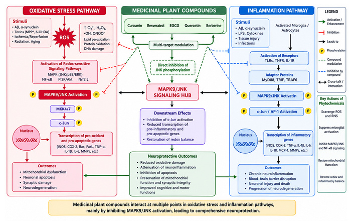
Interaction of medicinal plant compounds with oxidative stress and inflammation pathways. Overview of how medicinal plant bioactives modulate oxidative stress and inflammatory signalling. Phytochemicals suppress ROS generation, inhibit MAPK9/JNK and NF-κB activation, reduce pro-inflammatory cytokine production, restore redox balance, and ultimately protect neurons from degeneration. EGCG: epigallocatechin gallate; ROS: reactive oxygen species.
Another essential mechanism underlying the neuroprotective action of phytochemicals is their anti-inflammatory effects. Neuroinflammation plays an important role in neuronal death through microglial activation and the subsequent release of inflammatory cytokines. A number of phytochemicals have been found to block inflammation via NF-κB, MAPKs, and JNK pathways. EGCG and curcumin, for example, have been found to block microglial activation and down-regulate TNF-α, IL-1β, and IL-6 [82]. In addition, resveratrol has been found to suppress inflammation induced by the release of inflammatory cytokines in models of PD and cerebral ischemia.
Phytochemicals also possess anti-apoptotic properties by regulating both the intrinsic and extrinsic apoptotic pathways. Diseases associated with neurodegeneration are characterised by progressive neuronal cell death caused by the activation of apoptotic cascades. Various phytoconstituents can prevent apoptosis by modulating Bcl-2 proteins, caspases, and mitochondrial-related mechanisms. It has been found that curcumin and berberine can downregulate Bax gene expression and upregulate the expression of anti-apoptotic Bcl-2 proteins, thereby preventing mitochondrial membrane depolarisation. Quercetin also prevents the activation of caspase-3 [83].
A number of medicinal herbs have been explored to discover their neuroprotective actions and usefulness in neurodegenerative disorders. The plant Curcuma longa, popularly known as turmeric, is one of the most commonly studied medicinal herbs because of the presence of curcumin, which has excellent antioxidant and anti-inflammatory activities [84]. Curcumin has proven capabilities for inhibiting the formation of Aβ and reducing inflammation and oxidative stress in AD.
The other medicinal plant under discussion is Camellia sinensis (Green tea), which contains catechins such as EGCG. The major effects of EGCG include reducing oxidative stress, inhibiting protein aggregation, and attenuating the inflammatory response in neurodegeneration models. There are reports indicating that green tea catechins enhance cognitive function and protect dopaminergic neurons in PD models [85].
Ginkgo biloba is yet another well-known medicinal plant used for cognitive enhancement and neuroprotection. The active components of Ginkgo biloba are flavonoids and terpenoids, which enhance cerebral blood flow and provide protection against apoptosis and oxidative stress. Studies show promising results in improving memory and cognitive functions in mild cognitive impairment (MCI) and AD [86].
Withania somnifera (Ashwagandha) is one of the valuable medicinal herbs used in Ayurveda. Active phytochemicals present in the plant, called withanolides, possess antioxidant, anti-inflammatory, and anti-apoptotic activities. In experiments, it was found that Ashwagandha exerts its protective effect on neurons in AD, PD, and cerebral ischemia through anti-oxidation [87]. Bacopa monnieri is an Ayurvedic herb famous for its ability to enhance cognition and protect nerve cells. Bacosides are the main active components of Bacopa monnieri; they stimulate synaptic activity, decrease oxidative stress, and improve memory. Laboratory studies suggest that Bacopa monnieri prevents neurons from damage due to Aβ exposure and positively affects cognitive functions in neurodegenerative diseases [88].
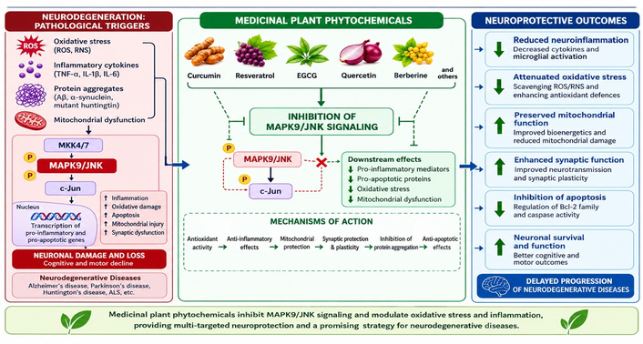
In totality, the medicinal properties of plants and their bioactive phytochemicals are strong therapeutic candidates for neurodegenerative diseases because of their multitarget mechanisms. The modulation of oxidative stress, neuroinflammation, apoptosis, and mitochondrial dysfunction by these plants makes them strong candidates in the development of neuroprotective drugs focusing on the MAPK9 pathway.
Phytochemicals from medicinal plants have been of interest for their use in the treatment of neurodegenerative diseases based on their ability to influence stress-activated signalling pathways, which include MAPK9/JNK2. Many natural products have been found to have neuroprotective activity based on their ability to inhibit oxidative stress, neuroinflammation, mitochondrial dysfunction, and apoptosis, processes that are highly correlated with MAPK9 activity (Figure 4). Some phytochemicals have been shown to influence JNK pathways in models of neurodegenerative disorders such as AD, PD, HD, and ischemia.
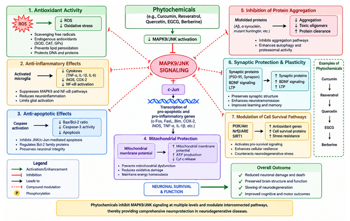
Mechanisms of phytochemical-mediated neuroprotection. An illustration of the neuroprotective actions of phytochemicals, including antioxidant, anti-inflammatory, anti-apoptotic, and mitochondrial protective effects. Bioactive compounds inhibit MAPK9/JNK signalling, reduce oxidative stress and protein aggregation, preserve synaptic function, and promote neuronal survival in neurodegenerative conditions. EGCG: epigallocatechin gallate.
Neuroprotection mediated through the preservation of mitochondria is yet another key role played by phytochemicals. Mitochondria are extremely sensitive to oxidative stress and are key organelles in the generation of ATP and the regulation of apoptosis. Deterioration of mitochondria results in increased ROS formation, calcium imbalance, and ultimately leads to neuronal cell death in neurodegeneration. There are several phytochemicals that aid in the preservation of mitochondrial function by increasing the stability of mitochondria, enhancing ATP synthesis, and protecting mitochondria from oxidative stress [39].
Curcumin, the main polyphenol extracted from Curcuma longa, also known as turmeric, is one of the best-studied natural agents used in neurodegenerative disorders. The ability of curcumin to act as an antioxidant, anti-inflammatory, and antiapoptotic compound has been demonstrated experimentally, and many of its actions can be attributed to the inhibition of the MAPK9/JNK pathway.
For instance, a study carried out by Wang et al. [89] demonstrated that curcumin reduced JNK-3 expression and phosphorylation and attenuated neuronal apoptosis in the hippocampus of rats with Aβ-induced AD. Curcumin has also been found to inhibit activation of NF-κB and pro-inflammatory cytokines, including TNF-α and IL-1β, which could explain its anti-inflammatory effects. Moreover, curcumin prevents mitochondrial depolarisation and cytochrome c release, protecting the mitochondria [90]. As for PD, curcumin showed protective effects when administered to patients suffering from rotenone-induced and MPTP-induced neurodegeneration. In particular, curcumin inhibited JNK-mediated apoptosis, resulting in decreased loss of dopaminergic neurons and improved motor function [91].
Several in vitro and in vivo studies have reported that curcumin attenuates JNK phosphorylation and reduces neuronal apoptosis induced by oxidative stress or Aβ exposure. However, these effects vary across experimental models, doses, and formulations, and may also involve indirect regulation through upstream antioxidant and anti-inflammatory pathways, including Nrf2 and NF-κB, rather than direct inhibition of MAPK9.
Resveratrol is a polyphenolic compound naturally occurring in grapes, berries, and red wine. This compound possesses strong antioxidant and anti-inflammatory activities, along with exhibiting neuroprotective effects in multiple models of neurodegeneration [92]. The most prevalent pathway through which resveratrol exerts neuroprotective activity is its antioxidant and anti-apoptotic actions by inhibiting oxidative stress and JNK signalling pathways. Inhibitory effects of resveratrol on JNK signalling pathways involve decreased production of ROS and improved antioxidant defense by activation of sirtuin 1 (SIRT1) and Nrf2 signalling pathways. Although resveratrol can decrease the activation of JNK in various models of neurodegenerative disorders [93], the effectiveness of the treatment is determined by many factors, including the disease model used, dosage, and the duration of the treatment, as well as the decrease in oxidative stress and SIRT1/Nrf2 activation rather than MAPK9 inhibition.
Furthermore, resveratrol modulates neuroprotective pathways involving neuron survival and synaptic transmission. In models of ischemic stroke, resveratrol alleviated neuronal injury by decreasing the inflammatory response and inhibiting phosphorylation of JNK [94]. Moreover, it increased the synthesis of ATP through promoting mitochondrial biogenesis in neurons. Based on multiple actions described above, resveratrol is considered an efficient phytochemical capable of modulating MAPK9-related neurodegeneration.
Quercetin is a type of flavonoid found abundantly in fruits, vegetables, onions, apples, and various medicinal herbs. Quercetin is notable due to its strong antioxidant properties and ability to regulate cellular signalling pathways involved in oxidative stress and inflammation. Quercetin’s protective actions on the nervous system involve ROS scavenging and regulation of MAPK signalling pathways, including JNK activation.
In vitro experiments have established that quercetin can reduce neuronal injury induced by oxidative stress due to its ability to remove free radicals and increase the activity of antioxidant enzymes [95]. In neurons exposed to hydrogen peroxide, quercetin has been found to decrease JNK phosphorylation and apoptosis of neurons under oxidative stress conditions. Yet, clinical studies have not been conducted, and its neuroprotective action most probably involves the regulation of more than one signalling pathway, both antioxidative and anti-inflammatory [96]. Quercetin also acts as an anti-inflammatory agent by suppressing NF-κB activation and the secretion of pro-inflammatory cytokines. In AD animal models, quercetin has been demonstrated to reverse Aβ-mediated neuronal damage and enhance cognitive function. Moreover, quercetin provides protection against mitochondrial dysfunction by stabilising mitochondrial membranes and reducing ROS generation. In PD animal models, quercetin caused a decrease in dopaminergic neuron degeneration and improvement of motor dysfunction caused by oxidative stress-induced MAPK signalling pathways. The aforementioned evidence highlights the potential of quercetin in treating MAPK9-mediated neurological disorders.
EGCG, the main catechin in Camellia sinensis (green tea), is a widely studied phytochemical known for its neuroprotective properties through interactions with the MAPK pathway. The first mechanism underlying the neuroprotective action of EGCG is its ability to inhibit protein aggregation. It was found that EGCG prevents the accumulation of Aβ protein and reduces protein aggregation. Moreover, EGCG can reduce JNK activation in response to exposure to Aβ and induce a decrease in cell apoptosis [97]. Another effect of EGCG is the inhibition of hyperphosphorylation of the tau protein, thereby inhibiting the development of neurofibrillary tangles.
Another neuroprotective property of EGCG is mitochondrial protection. It reduces ROS generation, stimulates ATP production, and maintains mitochondrial membrane stability. EGCG protects dopaminergic neurons from oxidative stress and mitochondria-mediated apoptosis in animal models of PD. Moreover, EGCG can inhibit microglial activation and inflammatory cytokine production. Thus, these studies have shown a decreased activation of JNK due to treatment with EGCG in animal models of AD and PD. However, such observations are not always consistent, and the effect might be a consequence of decreased oxidative stress, mitochondrial preservation, and neuroinflammation suppression.
The isoquinoline alkaloid berberine, isolated from herbs such as Berberis vulgaris, has great potential as a neuroprotective agent due to its anti-inflammatory and antioxidant properties. Specifically, berberine regulates several pathways related to neuronal survival, including the MAPK and NF-κB pathways. Several studies have found that berberine can inhibit JNK phosphorylation in neuroinflammatory and ischemic models. Nevertheless, it appears from existing studies that such an effect is part of a more complex regulation of MAPKs and NF-κB signalling pathways, which complicates ascribing neuroprotective effects only to MAPK9 inhibition [98].
Moreover, berberine alleviates oxidative stress through increased antioxidant enzyme activity and inhibition of ROS production. For instance, berberine enhances cognitive outcomes and reduces Aβ levels in Alzheimer’s models. Additionally, the phytochemical acts on mitochondria by boosting ATP synthesis and protecting mitochondrial membranes.
Other new phytochemicals that can regulate MAPK9-mediated pathways include luteolin, kaempferol, and ginsenosides. Particularly, luteolin is a flavonoid found in celery and green peppers that can inhibit the activation of microglia and block JNK-induced inflammatory signalling. On the other hand, kaempferol reduces oxidative stress and neuronal death in ischemia models. Ginsenosides are compounds derived from Panax ginseng that induce neuroprotection by modulating MAPK and mitochondrial pathways. Medicinal plant bioactives constitute novel candidates in MAPK9-related neurodegenerative diseases. This is attributed to their ability to regulate numerous pathological processes, including neuroinflammation, oxidative stress, apoptosis, and mitochondrial dysfunction. Table 2 highlights some reported medicinal plants and active phytochemicals targeting MAPK9 (JNK2) in neurodegenerative diseases.
Medicinal plants and active phytochemicals targeting MAPK9 (JNK2) in neurodegenerative diseases.
| Medicinal plant | Active phytochemical(s) | Neurodegenerative model/Disease | Effect on MAPK9/JNK signalling | Neuroprotective mechanism | Key references |
|---|---|---|---|---|---|
| Curcuma longa (Turmeric) | Curcumin | Alzheimer’s disease (AD), Parkinson’s disease, cerebral ischemia | Inhibits JNK phosphorylation and c-Jun activation | Anti-inflammatory, antioxidant, anti-apoptotic | [84] |
| Camellia sinensis (Green Tea) | Epigallocatechin gallate (EGCG) | AD, Parkinson’s disease | Suppresses JNK activation and downstream inflammatory signalling | Inhibits amyloid aggregation, mitochondrial protection | [85, 97] |
| Vitis vinifera (Grape) | Resveratrol | Parkinson’s disease, cerebral ischemia, AD models | Reduces oxidative stress-mediated JNK activation | Activates SIRT1, antioxidant, anti-inflammatory | [3, 99] |
| Berberis vulgaris | Berberine | Neuroinflammation, AD models | Attenuates MAPK/JNK and NF-κB signalling | Reduces reactive oxygen species (ROS) production and cytokine release | [98, 100] |
| Ginkgo biloba | Ginkgolides, Bilobalide, Flavonoids | AD, vascular dementia | Modulates MAPK-mediated inflammatory pathways | Antioxidant, anti-apoptotic, improves cerebral blood flow | [86, 101] |
| Withania somnifera (Ashwagandha) | Withanolides | AD, Parkinson’s disease | Downregulates stress-induced MAPK activation | Anti-inflammatory, mitochondrial protection | [80, 102] |
| Bacopa monnieri | Bacosides A and B | Cognitive impairment, AD models | Suppresses oxidative stress-associated JNK signalling | Enhances synaptic function and antioxidant defences | [103] |
| Panax ginseng | Ginsenosides (Rg1, Rb1, Rd) | Parkinson’s disease, ischemic injury | Regulates MAPK/JNK-mediated apoptosis | Mitochondrial stabilisation and neuronal survival | [104] |
| Scutellaria baicalensis | Baicalein, Baicalin | Parkinson’s disease, neuroinflammation | Inhibits JNK phosphorylation and microglial activation | Antioxidant and anti-inflammatory activity | [105] |
| Allium cepa (Onion) | Quercetin | AD, oxidative stress models | Suppresses JNK/c-Jun signalling | ROS scavenging and apoptosis inhibition | [96] |
| Glycyrrhiza glabra (Licorice) | Liquiritigenin, Glycyrrhizin | Neuroinflammation and ischemic injury | Modulates MAPK-mediated inflammatory responses | Anti-inflammatory and antioxidant effects | [17] |
| Centella asiatica | Asiaticoside, Madecassoside | Cognitive impairment, AD models | Reduces stress-induced MAPK activation | Neurogenesis promotion and antioxidant activity | [18] |
While there are many reports on the ability of several phytochemicals to inhibit JNK phosphorylation, care should be taken when drawing conclusions from these results. The majority of these data are based on in vitro and animal experiments, in which various disease models and doses were used. Furthermore, most of the time, the decrease in JNK phosphorylation was due to a reduction in oxidative stress, improvement in mitochondrial function or alteration of upstream signalling pathways instead of inhibition of MAPK9 itself.
While JNK3 has been found to be the most studied isoform in relation to neurodegenerative disorders due to its primary neuronal expression, inhibition of JNK3 alone may not be enough to inhibit the complicated inflammatory and oxidative responses involved in the pathogenesis of such disorders. In contrast to JNK3, MAPK9 (JNK2) has been found to be expressed in neurons, astrocytes, microglial cells, and immune cells [14]. This enables MAPK9 to regulate not only neuronal apoptosis but also neuroinflammation. From experimental evidence, it has been seen that JNK2 participates in cytokine secretion, oxidative stress enhancement, mitochondrial damage, and protein aggregation [106]. However, the widespread occurrence of JNK2 has its own dangers, as the side effects in the system after long-term inhibition need to be considered. Thus, future research efforts should be concentrated on the creation of brain-specific inhibitors for MAPK9 that could provide maximum protection to the neurons without causing any toxic effects outside the nervous system. JNK2 and JNK3 proteins can thus be considered complementary members of the JNK pathway rather than competing therapeutic targets.
Bioactive molecules extracted from medicinal plants that could act on MAPK9/JNK2, a MAPK pathway, have shown considerable neuroprotective effects against neurodegeneration in animal studies. Nevertheless, the clinical efficacy of these drugs is often determined by the permeability of the molecules through the BBB. The BBB acts as a natural barrier that regulates the transport of foreign particles into brain tissue while protecting it from harm caused by external sources [107]. As such, several neuroprotective phytochemicals fail to enter the brain due to the BBB.
Most natural bioactives have physicochemical constraints, including low solubility, poor membrane permeability, and rapid metabolic degradation, resulting in minimal BBB permeation [108]. In addition to acting as a barrier to drug delivery, BBB dysfunction is a key pathology associated with neurodegenerative diseases. It has been observed that MAPK9-mediated inflammatory signalling leads to endothelial cell damage, loss of tight junction protein function, and dysfunction of neurovascular units, and thus facilitates immune cell entry and chronic neuroinflammation. This means that therapy targeting MAPK9 can serve a double purpose.
There is substantial evidence for curcumin’s anti-inflammatory and MAPK9-inhibitory actions, although its clinical application is hindered by limited brain uptake and poor pharmacokinetics [41, 90, 109]. Furthermore, the antioxidant and anti-apoptotic actions of quercetin and resveratrol are significant in mitigating cellular damage, although they exhibit poor CNS bioavailability when administered orally [110]. Despite these challenges, phytochemicals such as EGCG and berberine have exhibited BBB penetration and protective effects in experimental models of neurodegenerative conditions. Knowledge of the physiological factors that determine BBB permeation of compounds is vital to the design of therapies based on plant derivatives.
Another key drawback is that poor bioavailability poses a significant constraint in the use of medicinal plants’ phytochemicals for therapeutic purposes. Many phytochemicals get degraded and metabolised quickly, thereby posing a challenge to their therapeutic efficiency due to reduced bioavailability. Curcumin is one such compound, which, despite having excellent neuroprotective effects, shows poor bioavailability due to its poor absorption rate, metabolic rate, and short plasma half-life [111]. Another such compound is resveratrol, which shows a high first-pass effect [112]. For example, Almeida et al. [113] conducted a randomised, double-blind, placebo-controlled clinical study assessing the pharmacokinetics and safety profile of trans-resveratrol in a randomised, double-blind and placebo-controlled study among healthy human volunteers. They were administered doses ranging from 25 to 150 mg, six times per day. The study indicated that trans-resveratrol is highly absorbed and achieves maximum plasma levels within 0.8 to 1.5 hours following dosing. While the drug showed high tolerance and produced only minor side effects, its plasma levels were still low because of low bioavailability and quick clearance from the body [113].
Some approaches used to address the above-mentioned barriers to the effective delivery of phytochemicals include structural modification, formulation optimisation, and various encapsulation techniques. Nano-curcumin (NCMN) has demonstrated improved bioavailability, enhanced ability to cross the BBB, and more pronounced neuroprotective effects in AD [107, 108].
Although many phytochemicals have been found to display significant activity towards pathways associated with MAPK9, care must be taken in the interpretation of their positive results from biochemical and cellular assays. A number of polyphenols commonly used in research, including curcumin, display structural features typical of PAINS [114]. These compounds might create biological activity by non-specific mechanisms, such as redox cycling, protein aggregation, fluorescence interference, covalent modification, or metal chelation. Hence, the observed biological activity does not automatically translate into the selectivity of a certain MAPK9 pathway. This problem is especially important in kinase drug discovery, since false positives resulting from interference in the assay might occur and increase the overestimation of the therapeutic value of certain compounds [115]. However, the classification of a phytochemical as a PAINS compound does not mean it lacks biological activity. Instead, it means the necessity of rigorous validation with orthogonal biochemical assays, target engagement studies, cellular mechanistic assays, and pharmacology in vivo. The future study should involve computational modelling, biophysical binding assays, selective kinase profiling, and proper experimental validation of the results.
Development of novel drug delivery systems has greatly increased the potential of neuroprotective phytochemicals. Nanotechnological formulations, including nanoparticles, liposomes, polymeric micelles, dendrimers, and solid lipid nanoparticles, serve as efficient tools to increase BBB penetration and targeted delivery to neuronal cells. Such systems protect phytochemicals against degradation, improve their solubility, and facilitate controlled drug delivery [108].
The use of nanoparticle delivery of curcumin led to higher brain uptake and decreased neurotoxicity caused by Aβ deposition in AD models [116, 117]. Also, delivery of EGCG via nanoparticles was effective in protecting mitochondria and inhibiting dopaminergic neuronal loss in PD models [118]. The development of intranasal drug delivery systems is gaining popularity as it does not require passage across the BBB and uses the olfactory route to deliver drugs directly into the brain. Such an approach can increase the efficiency of therapy for MAPK9-dependent neurodegenerative diseases.
Another potentially effective approach to enhancing neuroprotective efficacy is combination therapy. Given that neurological diseases such as AD and PD have several interrelated pathological pathways, monotherapies are unlikely to deliver significant benefits. A combination of phytochemicals from medicinal plants with pharmaceutical drugs or other biologically active compounds could offer improved efficacy through synergistic effects. A number of studies have shown the benefits of using combination therapies of phytochemicals. Combination therapy of curcumin and donepezil was more effective in improving cognitive function and neuroinflammation than each agent alone in AD [119]. Beltagy et al. [119] examined the therapeutic value of NCMN, both alone and in combination with donepezil, in a rat model of AD. The findings revealed that NCMN significantly enhanced cognitive function, locomotor ability, and neuronal protection through antioxidant, anti-inflammatory, anti-tau hyperphosphorylation, and anti-cholinesterase properties. It should be noted that NCMN also acted as an active agent in modulating the PI3K/AKT/GSK-3β signalling pathway, which is very important in neuronal protection and survival. The use of NCMN along with donepezil showed better results than either alone [119].
Similarly, Resveratrol combined with levodopa (LD) was more effective at increasing dopaminergic neuronal survival and reducing oxidative stress in PD. For example, Liu et al. [120] studied the synergistic effect of LD and resveratrol on rotenone-induced neurodegeneration in PD using cellular and animal models. Their study results revealed that the combination of LD and resveratrol inhibited ROS generation, improved mitochondrial function, and prevented cell death. Additionally, they inhibited inflammation by downregulating the toll-like receptor (TLR)-2 and TLR-4 signalling pathways, thereby reducing the secretion of inflammatory cytokines. In rotenone-exposed rats, there were improvements in motor function, enhanced superoxide dismutase (SOD) activity, and diminished malondialdehyde (MDA) levels [120]. Thus, combination therapies can reduce doses of pharmaceutical drugs and lower their side effects. In addition, combination therapies targeting MAPK9 and other related signalling pathways could provide more effective neuroprotection.
Precision medicine holds promise for advancing personalised neuroprotective strategies against MAPK9. Neurodegenerative disorders are characterised by significant heterogeneity in terms of their pathogenesis due to genetic predispositions, differences in inflammation profiles, disease progression, and responsiveness to therapy among different patients [121]. Modern advances in genomics, proteomics, transcriptomics, and computational biology have enabled the identification of biomarkers that correlate with MAPK9 dysfunction and neuronal stress responses. These biomarkers can help classify patients and apply an individualized phytochemical approach. For instance, individuals exhibiting signs of increased oxidative stress or activation of the MAPK9 pathway are likely to respond positively to antioxidant compounds such as curcumin, quercetin, or EGCG [122]. Moreover, innovative computational techniques, such as molecular docking, artificial intelligence (AI), and network pharmacology, can help identify novel plant bioactives with selective activity against MAPK9.
On balance, bioactive compounds from medicinal plants targeting MAPK9 hold great promise for the treatment of neurodegenerative conditions (Table 3). Despite the considerable difficulties posed by issues such as the penetration of these drugs across the BBB, insufficient bioavailability, and limited clinical translatability, several potential solutions are emerging.
Advantages and limitations of phytochemical therapeutics in neuroprotection.
| Aspect | Advantages | Limitations |
|---|---|---|
| Multi-target activity | Simultaneously modulate multiple pathological pathways, including MAPK9/JNK, oxidative stress, neuroinflammation, mitochondrial dysfunction, and apoptosis. | Multi-target effects may complicate mechanistic interpretation and target validation. |
| Safety profile | Generally exhibit lower toxicity and better tolerability compared to many synthetic drugs. | Long-term safety data are often lacking, particularly at therapeutic doses. |
| Natural origin | Derived from medicinal plants with extensive historical use in traditional medicine systems. | Variability in plant source, cultivation conditions, and harvesting practices can affect quality and efficacy. |
| Antioxidant activity | Efficiently scavenge reactive oxygen species (ROS) and enhance endogenous antioxidant defences. | Antioxidant effects observed in vitro may not always translate into clinical efficacy. |
| Anti-inflammatory effects | Suppress pro-inflammatory cytokines and signalling pathways such as MAPK9 and NF-κB. | Limited understanding of optimal dosing regimens required to achieve sustained anti-inflammatory effects in humans. |
| Neuroprotective potential | Protects neurons from apoptosis, mitochondrial dysfunction, excitotoxicity, and protein aggregation. | Most evidence is derived from preclinical studies rather than large-scale clinical trials. |
| Blood-brain barrier (BBB) penetration | Certain compounds, such as berberine, resveratrol, and epigallocatechin gallate (EGCG), demonstrate partial BBB permeability. | Many phytochemicals exhibit poor BBB penetration, limiting therapeutic concentrations within the brain. |
| Bioavailability | Some compounds can be optimised through formulation technologies and structural modifications. | Poor solubility, rapid metabolism, and low oral bioavailability remain major challenges for compounds such as curcumin and quercetin. |
| Drug development potential | Serve as valuable lead compounds for the design of novel neuroprotective drugs targeting MAPK9. | Isolation, purification, and large-scale production may be costly and technically challenging. |
| Combination therapy | Can be combined with conventional drugs to enhance efficacy and reduce adverse effects through synergistic mechanisms. | Potential herb-drug interactions may influence pharmacokinetics and treatment outcomes. |
| Precision medicine applications | It may be tailored to individual molecular profiles and disease mechanisms in future personalised therapies. | Biomarkers for patient stratification and treatment monitoring remain inadequately validated. |
| Computational drug discovery | Integration with molecular docking, AI, machine learning, and network pharmacology accelerates candidate identification. | Computational predictions require extensive experimental and clinical validation before translation. |
| Standardisation and quality control | Advances in phytochemical characterisation improve consistency and reproducibility. | Lack of universal standards for extraction, formulation, and quality assessment remains a significant challenge. |
| Clinical translation | Growing interest from academia and industry supports further development. | Significant translational gaps exist between promising laboratory findings and successful clinical outcomes. |
Phytochemical therapeutics offer a unique advantage in neuroprotection by simultaneously targeting multiple disease mechanisms, including MAPK9-mediated oxidative stress, neuroinflammation, and neuronal apoptosis. However, challenges related to bioavailability, blood-brain barrier penetration, standardisation, and limited clinical evidence must be addressed before these compounds can be widely adopted as effective therapies for neurodegenerative diseases.
While having shown promising preclinical efficacy, the clinical development of phytochemicals that target MAPK9 faces challenges due to low aqueous solubility, fast metabolism, inability to penetrate the BBB, and inconsistency in pharmacokinetic profiles. Thus, nanoformulation technology has been recognised as an appropriate way to overcome these obstacles rather than merely being used as carriers for the drugs. For instance, polymeric nanoparticles, solid lipid nanoparticles, nanostructured lipid carriers, liposomes, dendrimers, and nanoemulsions enhance the stability of phytochemicals, enable prolonged circulation in the bloodstream, control release, and increase BBB passage via receptor-mediated or adsorptive endocytosis [123]. Similarly, NCMN has significantly higher oral bioavailability and accumulation in the brain compared with curcumin, leading to better antioxidant, anti-neuroinflammatory, and anti-apoptotic effects in AD mouse models [124]. Furthermore, nanoencapsulated EGCG and resveratrol have an increased ability to enter neurons and protect mitochondria [125].
Yet, there are some limitations associated with the use of nanoformulations as well. The effectiveness of these formulations will be determined by factors such as the particle size, surface charge, entrapment efficiency, stability of the formulation, repeatability, and safety of the manufacturing process, which need to be optimised to facilitate successful transition to the clinical setting [126]. In addition, there are no examples of the nanoformulations targeting MAPK9 that have passed preclinical testing; thus, comparative analysis of various nano-carriers under the same conditions is still very rare.
Effective translation of the MAPK9 phytochemicals in the clinic will necessitate the development of innovative imaging and biomarker approaches in order to optimise patient stratification, therapeutic response monitoring and pharmacological drug development. Advanced neuroimaging modalities such as structural MRI, DTI, fMRI, and PET can be used to assess the presence of brain atrophy, white matter pathology, neuroinflammation, and brain metabolism non-invasively [127]. Imaging techniques involving the use of radiolabeled ligands against Aβ, tau, and activated microglia might be considered as an approach to elucidate whether targeting MAPK9 signalling leads to a decrease in the disease-related burden. Fluid biomarkers, including p-tau, NfL, GFAP, inflammatory cytokines, and markers of oxidative stress, can serve as a quantitative measure of neuronal injury and disease progression [128]. As MAPK9 modulates oxidative stress, apoptosis, and neuroinflammation, integration of imaging with biomarkers will allow early detection of treatment responses and personalised medicine strategies.
An equally important element to consider here is the utilisation of an extended set of biomarkers, since there are multiple pathological mechanisms which interact in order to bring about neurodegenerative disorders. As opposed to a single biomarker, a multimodal panel should be used in this case, consisting of neurofilament light chain, total and phosphorylated tau, glial fibrillary acidic protein, TNF-α, IL-1β, IL-6, MDA, glutathione, superoxide dismutase, as well as BBB integrity markers. Furthermore, incorporation of transcriptomic, proteomic and phosphoproteomic signatures that indicate the activation of MAPK9 would make the procedure even more efficient. Altogether, with the help of neuroimaging in combination with an extended biomarker panel, it will be possible to implement precision medicine that will enable early detection of disease progression, as well as a more accurate objective measurement of MAPK9-targeted therapies.
Due to the increased complexity of the pathologies associated with neurodegenerative disorders, as well as the complexity of the active components of medicinal plants, there has been an urgent need to incorporate computational methodologies in neuropharmacology studies. Experimental studies of natural products have proven to be costly and time-consuming processes. Therefore, computational techniques have proved highly useful for the identification, analysis, and optimisation of phytochemicals targeting MAPK9/JNK2 (Figure 5). Techniques include molecular docking, molecular dynamics (MD) simulations, AI, machine learning (ML), and network pharmacology.
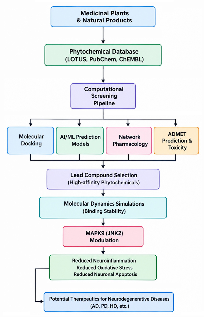
Computational and emerging approaches for identifying MAPK9-targeting phytochemicals in neurodegenerative diseases. Identification of MAPK9-targeting phytocompounds through computational and novel techniques. Lead identification of natural products extracted from medicinal plants through molecular docking, network pharmacology, artificial intelligence, machine learning, ADMET predictions, and molecular dynamics to determine their ability to target MAPK9 and reduce neurodegeneration, including inflammation, oxidative stress, and neuronal apoptosis. AD: Alzheimer’s disease; ADMET: Absorption, Distribution, Metabolism, Excretion, and Toxicity; HD: Huntington’s disease; PD: Parkinson’s disease.
Molecular docking remains one of the most utilised methods in structure-based drug design. The technique predicts the best way that a compound docks in the receptor site of a protein and also measures the strength of interaction between a protein and a ligand [129]. In neurodegenerative diseases, molecular docking has emerged as an important technique for identifying bioactive compounds from medicinal plants that can inhibit MAPK9 activity. Furthermore, with the availability of high-resolution crystal structures of MAPK9, molecular docking becomes possible and enables the identification of the binding energy of phytochemicals to crucial catalytic and allosteric sites of the protein [130]. Molecular docking studies often assess hydrogen-bond formation, hydrophobicity, electrostatic attraction, and binding energy to establish the feasibility of the interaction [131].
Among several phytochemicals, curcumin, quercetin, resveratrol, luteolin, kaempferol, and berberine exhibit good docking potential towards MAPK9/JNK proteins. For instance, molecular docking studies have revealed that curcumin can form stable hydrogen bonds with residues in the ATP-binding site of JNK enzymes, suggesting potential inhibition of their activity [132]. Likewise, quercetin can strongly interact with catalytic residues taking part in the phosphorylation process, thereby possibly preventing the subsequent activation of c-Jun and inflammatory pathways [133].
Virtual screening can be used in addition to docking to quickly test a large number of chemicals against the target protein. Phytochemical libraries, such as ZINC, IMPPAT, NPASS, and Traditional Chinese Medicine Libraries, may be screened for MAPK9 inhibitors [134–136]. This technique saves time by eliminating the need to test a large pool of compounds in the laboratory. Some recent studies have successfully identified new types of flavonoids and polyphenols with high affinity for JNK proteins using virtual screening approaches [137, 138]. For example, dos Santos Maia et al. [139] assessed the possibility of utilising lignans as multitarget drugs for AD by applying the QSAR, docking, and Absorption, Distribution, Metabolism, Excretion, and Toxicity (ADMET) approaches. As a result, 139 lignans were found to exhibit activity against several targets implicated in neurodegenerative processes, including MAPK9-linked JNK-3, PTP1B, NOX1, NQO1, PDE5, Nrf2, COX-2, and iNOS. Six lignans, including austrobailignan 6, anolignan C, 7-epi-virolin, compound 64, ococymosin, and mappiodoinin B, exhibited high neuroprotective and antioxidant properties. It was concluded that lignans are suitable candidates for multitarget drug development against AD.
Although docking gives a static view of interactions between the protein and ligand, MD studies give a dynamic view by observing the interactions that occur during a period of time under physiological conditions [140]. It is now mandatory to validate docking results using MD simulation and also to determine the stability of plant secondary metabolite-MAPK9 complexes. Thus, using Newtonian mechanics, the MD simulation technique helps researchers analyse the atomic movement of protein-ligand interactions. Various properties like root mean square deviation (RMSD), root mean square fluctuation (RMSF), radius of gyration (Rg), hydrogen bonding occupancy, solvent accessible surface area (SASA), and binding energy calculations help evaluate the stability and functional aspects of protein-ligand interactions [141].
For example, studies investigating the complex between curcumin and JNKs have exhibited protein-ligand interactions that were stable over 100 ns of MD simulation trajectories, indicating that curcumin can stabilise itself at the kinase’s active site under physiological conditions [142]. Likewise, EGCG and resveratrol have been found to adopt stable conformations upon binding to JNKs in MD simulations, making them likely candidates for neuroprotective inhibitors [143, 144]. Therefore, MD simulation is extremely useful for studying the induced-fit effect on MAPK9 induced by ligand binding, as it may help identify new allosteric binding sites and thus develop more selective inhibitors. Additionally, free energy calculations using MM/PBSA or MM/GBSA provide quantitative data on binding energy, making computational predictions more reliable before proceeding to experimentation.
AI and ML techniques are transforming modern pharmaceutical discovery through fast screening and analysis of large biological and chemical datasets. They show promise in significantly accelerating the identification of medicinal plant compounds that inhibit MAPK9 and other protein kinases linked to neurodegenerative diseases.
ML methods could be used with available data on kinase inhibitors to predict biological activities, toxicity, pharmacological characteristics, and BBB penetration capacity of phytochemicals [145]. Random forests, support vector machines, gradient boosting, and deep learning methods achieved strong performance in predicting compound-target interactions [146].
In the case of natural product discovery, AI techniques could be applied to select phytochemicals with the highest likelihood of inhibiting MAPK9 before any docking or laboratory experiments. Moreover, ML techniques can reveal hidden structural features associated with kinase inhibition, helping to discover novel chemical scaffolds in medicinal plants [147, 148].
These advances in generative AI technology have further increased opportunities to discover neuroprotective drugs. New phytochemical compounds designed using GANs, VAEs, and transformer architectures could be developed with enhanced pharmacokinetic profiles to offset problems arising from their low bioavailability and poor target selectivity. Moreover, AI-driven predictions of compound permeability across the BBB and of their ADME/Tox properties could reduce the likelihood of failure and increase the likelihood of success in the discovery of treatments for neurological disorders.
Whereas traditional medicines act on a single target site at a time, bioactive compounds from medicinal plants often exert their therapeutic effects through multiple molecular targets. Network pharmacology has become an increasingly popular field of systems biology that studies the complex mechanism of action of plants [149]. Network pharmacology combines bioinformatics, pharmacology, genomics, proteomics, and systems biology to study network interactions among natural compounds, proteins, signalling pathways, and diseases. It is especially useful for neurodegenerative conditions because of the presence of several interrelated pathological factors, including oxidative stress, neuroinflammation, mitochondrial dysfunction, protein aggregation, and apoptosis.
Network pharmacology techniques help determine key molecular hubs and signalling pathways modulated by phytochemicals from medicinal plants. Studies done on the compounds curcumin and resveratrol have shown involvement in the MAPK9, NF-κB, PI3K/Akt, Nrf2, and apoptotic pathways, among others [150]. This shows the importance and the wide spectrum of activity of these compounds. Similarly, studies on the plants Withania somnifera and Bacopa monnieri have shown regulation of multiple neuronal targets, including those governing neuronal survival and synaptic transmission [151, 152].
By integrating information from transcriptomics and proteomics datasets, researchers will be able to establish how phytochemicals modulate cellular signalling pathways in their entirety, rather than individual proteins. The application of molecular docking, MD simulations, AI, ML, and network pharmacology is revolutionising the screening of bioactive compounds from medicinal plants for MAPK9 targeting.
However, despite the considerable body of work supporting the neuroprotective effects of plant-derived, MAPK9/JNK2-specific bioactive compounds, many factors still make their effective utilisation in a clinical setting quite difficult. Although many plant-derived compounds show great promise for their antioxidant, anti-inflammatory, and anti-apoptotic properties in both cell and animal models, several issues, including pharmacokinetics, clinical validation, and extraction challenges, remain.
Poor pharmacokinetics: A problem related to phytochemicals as potential therapeutic agents is their poor pharmacokinetic properties. Many plant-derived drugs are poorly soluble in water, absorbed through the intestine, quickly metabolised, and have poor bioavailability; thus, they cannot be used effectively. For instance, curcumin has proven highly effective at inhibiting the MAPK9-mediated inflammatory and apoptotic pathways, but it is difficult to use in clinical practice due to poor bioavailability and rapid systemic clearance [84, 109]. The same is true for resveratrol, which undergoes extensive first-pass effect; hence, it has a low plasma level despite its neuroprotective effects [73, 99].
Limited clinical trials: While there is ample research demonstrating the benefits of natural bioactives, few of these agents have undergone extensive clinical testing. Most information on the potential benefits of MAPK9-targeting phytochemicals comes from preclinical experiments conducted in cells or animals. Although the information gained from these experiments may be informative, it does not always translate to a positive effect when the same compound is tested on humans. Clinical studies involving curcumin, EGCG, resveratrol, and Ginkgo biloba extracts, for example, have yielded inconclusive results in part because of disparities in dosages, treatment durations, target populations, and methodologies [86, 94, 153].
Variability in plant extracts: The other important issue that must be mentioned in relation to natural products is their intrinsic variability. The chemical makeup of plants can be affected by various factors, including geography, climate, soil, harvest timing, farming techniques, and even the extraction process itself. Consequently, different extracts from the same plant type could vary greatly in their biochemical concentrations. For instance, curcuminoids and catechins can have varying quantities when taken from Curcuma longa and Camellia sinensis, respectively [84, 154].
Standardisation issues: As such, it can be seen that closely related to the issue of extract variability is that of standardisation. While synthetic drugs have well-defined chemical structures due to consistent synthesis methods, natural medicinal extracts typically include complex arrays of chemicals. Without consistent standards in their extraction process, quality control, and characterisation of compounds present, it is possible to see large inconsistencies in their biochemical makeup [155]. This is especially important when studying the effects of MAPK9-targeted compounds, given variations in their biochemical composition.
Translational gaps: Although some interesting discoveries have been made in the lab setting, there are several obstacles that impede translation from research results to therapeutic applications. Animal models of neurodegenerative disorders may lack features such as disease progression, individual variations, and the influence of the environment, which complicate human pathologies. Therefore, drugs effective in experimental models may not yield the same results when applied clinically [91, 100]. Moreover, while most investigations concentrate on single target molecules, neurodegeneration is characterised by intricate processes involving signalling pathways for oxidative stress, inflammation, mitochondrial malfunction, protein aggregation, and more.
Therefore, closing these gaps requires an interdisciplinary approach that combines methods from computational biology, pharmacology, biomarker discovery, drug delivery, and rigorous clinical trials.
Increasing awareness of the vital role of MAPK9/JNK2 in promoting neurodegeneration opens possibilities for designing more efficient next-generation neuroprotective agents. Research efforts ought to focus on isolating MAPK9-specific phytotherapeutics that modulate abnormal MAPK9 activity while leaving the physiological activities of other MAPK proteins unaltered. Technological advances in phytochemical extraction, identification, and medicinal chemistry can greatly assist in identifying highly specific and effective plant-derived compounds.
Also, nanotechnology-based approaches for drug delivery have great potential to address poor pharmacokinetic properties and the limited ability of phytochemicals to cross the BBB. Nanoparticles, liposomes, dendrimers, and other drug carriers are likely to enhance the efficacy of phytotherapeutic agents due to their superior pharmacodynamics [156]. Future clinical studies should incorporate standardised neuroimaging and validated molecular biomarkers to evaluate target engagement, disease progression, and therapeutic efficacy of MAPK9-targeted interventions.
Thus, enhanced understanding of the indispensable role of MAPK9/JNK2 in driving neurodegeneration enables the development of better next-generation neuroprotective agents. Research should focus on extracting plant-derived phytotherapeutics that act on the MAPK9 pathway without interfering with the functions of other MAPK proteins. The technological advancements in phytochemistry and medicinal chemistry will play an important role in achieving such specificity and effectiveness. Drug delivery using nanotechnology holds immense promise for overcoming poor pharmacokinetics and the inability of phytochemicals to cross the BBB. Drug delivery techniques, including nanoparticles, liposomes, and dendrimers, among others, would make phytotherapeutic drugs more effective by enhancing pharmacodynamics.
Even though JNK3 is still the main neuron-specific drug target, there is evidence suggesting that MAPK9 acts through complementary roles in integrating neuronal damage with oxidative stress, neuroinflammation, and immune signalling from the periphery. This underscores the importance of investigating MAPK9 as a separate drug target and, at the same time, calls for the development of isoform-specific inhibitors. Hence, these factors make MAPK9 a prospective therapeutic target, while bioactive agents derived from medicinal plants (e.g., curcumin, resveratrol, quercetin, EGCG, berberine) show great ability to regulate MAPK9-related pathways and provide significant neuroprotection. Future studies should prioritise rigorous target validation and account for potential assay interference associated with highly reactive phytochemicals to facilitate the identification of truly selective MAPK9 modulators.
It should be acknowledged that integrating natural product studies with advanced technologies is one of the most fascinating directions in this field. For example, thanks to recent advances in molecular modelling, AI, and network pharmacology, new approaches to identifying phytochemicals targeting MAPK9 may emerge. However, successful implementation of such an approach would face several obstacles, including issues regarding bioavailability, standardisation, and clinical evaluation. Thus, interdisciplinary research would continue being critical in this context.
6-OHDA: 6-hydroxydopamine
AD: Alzheimer’s disease
AI: artificial intelligence
ALS: amyotrophic lateral sclerosis
ASK1: apoptosis signal-regulating kinase 1
ATP: adenosine triphosphate
Aβ: amyloid β
BBB: blood-brain barrier
CNS: central nervous system
CTE: chronic traumatic encephalopathy
DTI: diffusion tensor imaging
EGCG: epigallocatechin gallate
ERKs: extracellular signal-regulated kinases
fMRI: functional magnetic resonance imaging
HD: Huntington’s disease
IL: interleukin
JNK2: c-Jun N-terminal kinase 2
LD: levodopa
LTP: long-term potentiation
MAP3Ks: MAP kinase kinase kinases
MAPK: mitogen-activated protein kinase
MD: molecular dynamics
MDA: malondialdehyde
ML: machine learning
MPTP: 1-methyl-4-phenyl-1,2,3,6-tetrahydropyridine
NCMN: nano-curcumin
NF-κB: nuclear factor kappa B
Nrf2: nuclear factor erythroid 2-related factor 2
PAINS: Pan-Assay Interference Compounds
PD: Parkinson’s disease
PET: positron emission tomography
ROS: reactive oxygen species
SIRT1: sirtuin 1
TLR: toll-like receptor
TNF-α: tumour necrosis factor-alpha
TPY: threonine-proline-tyrosine
The authors would like to acknowledge the University of the Witwatersrand’s Research Office for awarding Dr. Otun a postdoctoral fellowship.
SO: Investigation, Writing—original draft. IA: Conceptualization, Supervision, Writing—review & editing. Both authors read and approved the submitted version.
The authors declare no conflicts of interest.
Not applicable.
Not applicable.
Not applicable.
Not applicable.
Open access funding provided by the University of the Witwatersrand, and the authors would like to acknowledge the National Research Foundation (NRF) of South Africa for awarding Prof. Achilonu the CPRR research grant (Grant number CPRR23042899244). The funders had no role in study design, data collection and analysis, decision to publish, or preparation of the manuscript.
© The Author(s) 2026.
Open Exploration maintains a neutral stance on jurisdictional claims in published institutional affiliations and maps. All opinions expressed in this article are the personal views of the author(s) and do not represent the stance of the editorial team or the publisher.
Copyright: © The Author(s) 2026. This is an Open Access article licensed under a Creative Commons Attribution 4.0 International License (https://creativecommons.org/licenses/by/4.0/), which permits unrestricted use, sharing, adaptation, distribution and reproduction in any medium or format, for any purpose, even commercially, as long as you give appropriate credit to the original author(s) and the source, provide a link to the Creative Commons license, and indicate if changes were made.



View: 366
Download: 13
Times Cited: 0
Odunayo M. Agunloye ... Ganiyu Oboh
Jamil A. Chahrour ... Akram Hijazi