 Review


Affiliation:
Independent Researcher, Mountain View, CA 94040, USA
Email: Moawiah.Naffaa@proton.me
ORCID: https://orcid.org/0000-0003-0451-5901


Explor Neurosci. 2026;5:1006144 DOI: https://doi.org/10.37349/en.2026.1006144
Received: April 28, 2026 Accepted: June 25, 2026 Published: August 09, 2026
Academic Editor: Jinwei Zhang, Chinese Academy of Sciences, China
The article belongs to the special issue Advances in Epilepsy Research
Epilepsy is traditionally defined by recurrent seizures arising from abnormal neuronal excitability, yet growing evidence indicates that this view is incomplete. This narrative review examines epilepsy through the framework of neural–glial network instability, an integrative perspective that places neuronal dysfunction within a broader system shaped by glial regulation, calcium dysregulation, neuroinflammatory signaling, and circuit remodeling. The review first reframes epilepsy as a disorder of progressive instability in which failures of excitation–inhibition balance, extracellular homeostasis, inflammatory restraint, and adaptive plasticity shift neural networks from compensated function toward seizure-prone states. It then synthesizes neuronal mechanisms, including excitation–inhibition imbalance, GABAergic dysfunction, and altered intrinsic excitability, together with glial mechanisms involving astrocytic regulation of ionic and neurotransmitter environments, microglial inflammatory and synaptic responses, and oligodendroglial contributions to conduction and network coordination. Calcium dysregulation is considered a cross-cutting mechanism linking excitability, intracellular stress, gliotransmission, inflammation, and long-term remodeling. The review further examines how neuroinflammation and chronic circuit reorganization help convert transient disturbances into persistent epileptic networks. Together, this framework offers a more integrated account of seizure emergence, epileptogenesis, and chronic seizure susceptibility and points toward mechanism-informed therapeutic strategies aimed at restoring durable network stability.
Epilepsy is one of the most common serious neurological disorders and remains a major global cause of recurrent seizures, disability, reduced quality of life, and substantial clinical burden [1–3]. Although epilepsy is often framed primarily as a disorder of abnormal neuronal firing, the biological reality is far more heterogeneous. Diverse genetic, developmental, metabolic, inflammatory, traumatic, and degenerative factors can contribute to seizure susceptibility and epileptogenesis, indicating that epilepsy is not a single mechanistic entity but a broad group of disorders with multiple and partially overlapping pathogenic pathways [4–6]. This mechanistic diversity has contributed to major advances in the field, but it has also highlighted the limitations of explanatory models that center almost exclusively on neurons.
Neuron-centered frameworks have been indispensable for understanding epilepsy. Altered excitation–inhibition balance, impaired GABAergic signaling, aberrant ion channel activity, and hypersynchronous circuit discharge remain fundamental to seizure generation [7, 8]. However, these models do not fully account for the dynamic extracellular, inflammatory, metabolic, and plasticity-related conditions that shape when, where, and how seizures emerge or why certain neural networks transition into chronically epileptic states [6, 9, 10]. Increasing experimental evidence indicates that seizure generation and epileptogenesis are influenced not only by intrinsic neuronal dysfunction but also by reciprocal interactions between neurons and glial cells across molecular, cellular, and circuit scales [11, 12].
Astrocytes, microglia, and, in some contexts, oligodendroglial lineage cells contribute actively to the regulation of network excitability rather than serving merely as passive support elements or secondary responders [11, 13]. Astrocytes control extracellular potassium buffering, glutamate clearance, water homeostasis, metabolic support, and calcium-dependent gliotransmission, all of which can influence seizure threshold and network synchronization [9, 14, 15]. Microglia, through immune surveillance, cytokine signaling, synaptic remodeling, and interactions with both neurons and astrocytes, may amplify or constrain network instability depending on context and disease stage [12, 16, 17]. These glial functions intersect with neuronal mechanisms of excitability, intracellular calcium imbalance, inflammatory signaling, and activity-dependent circuit remodeling, suggesting that epilepsy is best understood as a disorder of disturbed neural–glial system regulation rather than isolated neuronal pathology [6, 10, 18].
This review examines epilepsy through the framework of neural glial network instability, an evidence-based perspective that integrates neuronal dysfunction with glial dysregulation, calcium imbalance, neuroinflammatory signaling, and maladaptive circuit plasticity. Rather than treating these processes as separate themes, this review considers how they converge to destabilize brain networks, lower seizure threshold, and promote epileptogenic remodeling [6, 10, 18]. In doing so, it aims to provide a more integrated account of how seizures arise and persist across different forms of epilepsy.
This review is also intended to address an important gap in the literature. While prior reviews have often examined neuronal excitability, neuroinflammation, astrocyte biology, or microglial activation as relatively separate domains, fewer have synthesized these findings within a unified framework centered on network instability across interacting neural and glial compartments [9, 11, 12]. By drawing together evidence from molecular, cellular, animal, translational, and, where relevant, clinical studies, this article seeks to clarify how glial and neuronal mechanisms cooperate in epileptogenesis and disease progression. Such an integrative view may help refine mechanistic understanding, identify shared points of vulnerability across epilepsy subtypes, and support the development of therapeutic strategies aimed not only at suppressing seizures but also at restoring system-level stability [9–11]. The novelty of this review lies not simply in discussing glia in epilepsy, but in integrating neuronal dysfunction, glial regulation, calcium signaling, inflammation, ionic and extracellular homeostasis, and circuit remodeling into a single instability-based framework for understanding seizure emergence and epileptogenesis. Rather than replacing neuroinflammatory, ionic, computational, or neuron-centered models, this framework organizes them across biological scales and asks how their interactions shift neural–glial systems from compensated function toward seizure-prone and chronically epileptic states.
Epilepsy can be understood not only as a disorder of excessive neuronal firing, but more broadly as a condition in which the mechanisms that normally preserve network stability become progressively disrupted. In the healthy brain, stable function is not maintained passively. It depends on active coordination across multiple levels, including synaptic excitation and inhibition, extracellular ion balance, neurotransmitter clearance, metabolic support, inflammatory restraint, and activity-dependent plasticity [12, 19]. These stabilizing functions arise through continuous interactions between neurons and glial cells, which together maintain the local and distributed conditions required for controlled signaling and resistance to pathological synchronization. From this perspective, epilepsy is usefully reframed as a disorder of network instability, in which the systems that support neural homeostasis and circuit resilience begin to fail [20–22].
Here, network instability refers to a state in which brain circuits become increasingly vulnerable to hypersynchronous activity because the balance between stabilizing and destabilizing forces has shifted. This instability may be transient, as in an acutely provoked seizure, or progressive and persistent, as in epileptogenesis and chronic epilepsy. It does not reflect a single abnormality, but rather a systems-level change produced by interacting disturbances in excitability control, extracellular regulation, inflammatory signaling, and plastic remodeling. Importantly, a network may appear relatively compensated for a period of time while still moving toward instability if protective buffers are weakened, recovery mechanisms are less effective, and thresholds for synchronized discharge are progressively lowered [23–25].
The neuronal contribution to this process remains fundamental. Neurons determine the electrical output of brain circuits, and disturbances in excitation–inhibition balance are central to seizure generation. Reduced inhibitory tone, altered GABAergic signaling, excessive glutamatergic drive, abnormal ion channel activity, and impaired intrinsic firing control can each increase the likelihood of recurrent and synchronized discharge [26]. These neuronal mechanisms form the core of classical models of epilepsy and remain essential for understanding how seizures are initiated. Yet neuronal hyperexcitability alone does not fully explain why seizure thresholds fluctuate over time, why similar insults produce different outcomes across individuals and brain regions, or why some vulnerable networks transition into chronically epileptic states whereas others do not [21–23, 25]. These features point to the importance of the broader cellular environment in which neuronal activity is embedded.
Glial cells are integral components of that environment and are increasingly recognized as active regulators of network stability rather than passive support elements. Astrocytes shape neuronal excitability through potassium buffering, glutamate uptake, water homeostasis, metabolic coupling, and calcium-dependent signaling [9, 19, 27]. Microglia influence network function through immune surveillance, cytokine release, synaptic remodeling, and responses to tissue stress. In some contexts, oligodendroglial and myelin-related changes may further influence conduction properties and network timing. These glial processes help determine whether local disturbances are buffered and resolved or instead amplified into persistent instability [11, 12, 28]. Framing epilepsy in this way shifts attention from isolated neuronal dysfunction to the failure of coordinated neural–glial regulation.
Several biologically grounded processes help explain how this destabilization unfolds. One major axis is disruption of excitation–inhibition balance, which may arise through receptor alterations, interneuron dysfunction, or persistent changes in synaptic strength. A second is impaired ionic buffering, particularly involving extracellular potassium, which can facilitate abnormal depolarization and increase susceptibility to recurrent firing. A third is altered neurotransmitter clearance, especially reduced glutamate uptake, which can prolong excitatory signaling and intensify local network stress. A fourth is inflammatory priming, in which cytokines, danger signals, blood–brain barrier (BBB) dysfunction, and immune-glial activation lower seizure threshold and increase vulnerability to maladaptive remodeling. A fifth is maladaptive plasticity, whereby repeated seizures, injury, or chronic inflammatory exposure drive structural and functional reorganization that stabilizes the epileptic state rather than restoring normal circuit behavior [20, 29]. Together, these processes show that network instability is neither purely electrical nor purely inflammatory. It is a multi-system disturbance that emerges when neural and glial mechanisms of regulation no longer act in a coordinated manner.
This framework is especially useful because it captures the transition from normal function to seizure susceptibility as a dynamic continuum rather than a binary event. Under physiological conditions, neuronal and glial systems cooperate to absorb perturbations, preserve extracellular equilibrium, restrain inflammatory escalation, and prevent runaway synchronization. In vulnerable tissue, however, repeated insults, genetic predisposition, developmental abnormalities, metabolic stress, or chronic inflammatory signaling may progressively erode these stabilizing mechanisms [30, 31]. Early disturbances may remain partially compensated, but once inhibitory restraint, buffering capacity, and inflammatory control are sufficiently weakened, the network can shift toward a seizure-prone state characterized by lower thresholds, greater responsiveness to perturbation, and more permissive conditions for pathological synchrony [23–25]. This transition may be reflected empirically by dynamic changes in electrophysiological activity, network recovery, metabolic state, inflammatory signaling, glial injury markers, and structural or functional imaging patterns. Epilepsy, in this sense, develops when coordination between neurons and glial cells becomes destabilized across molecular, cellular, and circuit levels to the point that recurrent seizures can emerge and persist.
Reframing epilepsy as a network instability disorder therefore provides a conceptual foundation for the mechanistic sections that follow. It preserves the central importance of neuronal hyperexcitability while placing it within a broader biological system shaped by glial regulation, extracellular homeostasis, inflammatory state, and activity-dependent remodeling. This perspective does not replace established models of seizure generation; rather, it expands them by emphasizing that epileptogenesis and chronic seizure susceptibility arise from interacting cellular systems. The relative contribution of inhibitory failure, glial dysfunction, inflammatory activation, calcium dysregulation, and circuit remodeling is likely to vary across temporal lobe epilepsy, developmental epilepsies, genetic epilepsies, and acquired epileptogenic conditions. With this framework in place, the next section turns to the neuronal mechanisms that initiate and propagate instability within epileptic networks.
The major components of this neural–glial instability framework and their interactions in seizure emergence and epileptogenesis are illustrated in Figure 1.
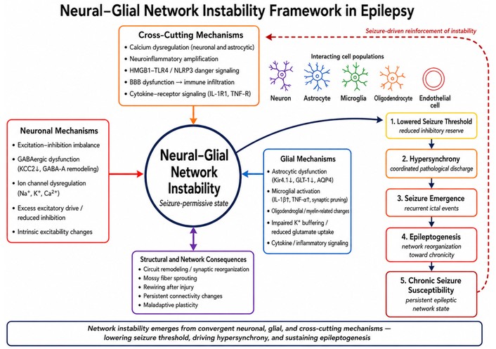
Neural–glial network instability framework in epilepsy. Schematic summary of the proposed neural–glial network instability framework. Neuronal, glial, cross-cutting inflammatory/calcium-related mechanisms, and structural network consequences converge on a seizure-permissive state of neural–glial instability. This instability lowers seizure threshold, promotes hypersynchrony, drives seizure emergence, and supports progression toward epileptogenesis and chronic seizure susceptibility. The dashed feedback arrow indicates that recurrent seizures can further reinforce upstream instability-promoting mechanisms.
A central neuronal mechanism underlying instability in epilepsy is disruption of the normal balance between excitation and inhibition. Under physiological conditions, neural circuits rely on tightly regulated interactions between excitatory and inhibitory neurons to support information processing while preventing uncontrolled synchronization. This balance is not static; it is continuously adjusted across synaptic, cellular, and network levels to permit flexible activity without loss of stability. Seizures emerge when this regulatory equilibrium is disturbed, and excitatory influences begin to outweigh the inhibitory restraints that normally confine neuronal firing in space and time [24, 32].
Excessive excitatory drive is one major route through which this imbalance develops. Glutamatergic transmission is essential for normal synaptic communication, plasticity, and circuit recruitment, but when glutamate release is excessive, postsynaptic responsiveness is enhanced, or excitatory synapses become pathologically strengthened, local activity can be amplified beyond physiological limits [32, 33]. Increased activation of excitatory pathways may facilitate recurrent firing, promote spread to neighboring neuronal populations, and lower the threshold for synchronized discharge. In epileptic networks, such changes can be reinforced by recurrent excitatory loops and activity-dependent synaptic strengthening, allowing transient bursts of excitation to evolve into more persistent and self-amplifying instability [33].
At the same time, reduced inhibition is an equally important determinant of excitatory dominance. Inhibitory interneurons normally constrain principal cell firing, regulate the temporal precision of network activity, and limit the spatial spread of excitation. Their function is therefore not restricted to suppressing discharge frequency; they also help organize oscillatory timing and prevent neuronal ensembles from entering hypersynchronous states [32, 34]. When inhibitory tone is weakened, circuits become less able to dampen activity bursts, restrict recruitment, or restore stable firing patterns after perturbation. This reduction in inhibition may arise through impaired interneuron function, decreased inhibitory drive, altered synaptic responsiveness, or broader changes in inhibitory network organization [34, 35]. Even in the absence of markedly elevated excitatory transmission, a relative failure of inhibition may be sufficient to destabilize the circuit.
Excitation–inhibition imbalance is also shaped by synaptic organization. Epileptic circuits often show altered excitatory coupling, impaired feedforward or feedback inhibition, and changes in synaptic strength that favor amplification over containment [33–35]. These changes do not simply increase neuronal activity in a general sense; they alter how activity is distributed across the circuit, how effectively it is restrained, and how easily it is propagated. Repeated seizures, developmental abnormalities, or prior injury may progressively remodel synaptic architecture in ways that shift signaling from adaptive responsiveness toward recurrent excitability. In this sense, synaptic imbalance is both a driver and a consequence of network instability [33, 35].
Altered firing thresholds further intensify this destabilized state. Neurons within epileptic circuits may become more responsive to synaptic input, more likely to fire repetitively, or more prone to burst discharge in response to stimuli that would not normally generate pathological output [34, 36]. Such changes increase the ease with which local neuronal populations are recruited into synchronized activity. Once thresholds for firing and recruitment are lowered, even modest perturbations may trigger disproportionate network responses, particularly when recurrent excitation is already enhanced and inhibitory containment is weakened [36, 37]. This helps explain why epileptic tissue may become highly sensitive to otherwise minor physiological or environmental fluctuations.
A key point is that excitation–inhibition imbalance should not be understood as a purely local cellular defect. Its pathological significance emerges at the network level, where excessive excitation, inadequate inhibitory restraint, altered synaptic organization, and lowered firing thresholds interact to promote coordinated discharge across interconnected neuronal populations [24, 34, 37]. The same degree of excitatory enhancement may have different consequences depending on circuit architecture, developmental stage, regional connectivity, and the integrity of surrounding regulatory systems. For this reason, excitation–inhibition imbalance is best viewed not simply as “too much excitation” or “too little inhibition”, but as a disturbance in the neuronal organization of circuit stability.
Within the broader framework of this review, excitation–inhibition imbalance represents a foundational neuronal driver of epileptic instability. It defines one of the principal routes by which circuits become vulnerable to recurrent discharge, but it does not by itself explain the full heterogeneity, persistence, or progression of epilepsy. Its functional consequences are shaped by extracellular buffering, glial regulation, inflammatory state, and circuit remodeling, which together determine whether neuronal instability remains limited or evolves into a chronic epileptic condition. The next subsection therefore examines one of the most important mechanistic dimensions of this imbalance in greater detail: GABAergic dysfunction.
Among the neuronal mechanisms that contribute to instability in epilepsy, GABAergic dysfunction is one of the most central and mechanistically informative. In the healthy brain, GABAergic signaling provides the principal inhibitory constraint on excitatory activity and is essential for limiting firing probability, shaping temporal precision, coordinating oscillatory behavior, and restricting the spread of excitation. Through these functions, inhibitory circuits do more than suppress neuronal output in a general sense; they actively organize when, where, and how neural ensembles can fire without entering pathological synchrony [38, 39]. Disruption of this inhibitory architecture is therefore a major route through which epileptic networks lose stability.
A common manifestation of GABAergic dysfunction in epilepsy is reduced inhibitory tone. When inhibitory transmission is weakened, neurons become more susceptible to recurrent firing, local activity is less effectively contained, and circuit synchronization becomes easier to initiate and sustain [38, 40, 41]. This reduction may result from interneuron loss, impaired interneuron recruitment, decreased GABA synthesis or release, altered postsynaptic responsiveness, or seizure-associated remodeling of inhibitory circuits [38, 39, 42]. Importantly, inhibitory control is not uniform across the brain. Distinct interneuron populations contribute differently to feedforward inhibition, feedback restraint, oscillatory timing, and spatial confinement of excitation, such that selective dysfunction of particular inhibitory microcircuits may produce different forms of epileptic instability [38, 39]. Thus, reduced inhibitory tone should not be understood simply as a quantitative decline in inhibition, but as a disturbance in the architecture of inhibitory control.
An additional layer of complexity arises from alterations in GABA receptor composition and function. GABAergic inhibition is mediated primarily through GABAA and GABAB receptors, which differ in localization, kinetics, and downstream signaling properties. Within the GABAA receptor family, subtype composition influences channel behavior, pharmacological sensitivity, synaptic targeting, and the balance between rapid and persistent inhibitory signaling [26, 41, 43]. Changes in receptor expression, trafficking, clustering, or subunit composition can therefore alter inhibitory efficacy even when presynaptic GABA release is relatively preserved [42, 44, 45]. Such receptor-level alterations are especially relevant in epilepsy because they may reshape inhibitory strength, timing, and responsiveness in ways that are region-specific, stage-dependent, or linked to prior seizure activity. This subtype-level diversity is especially important because GABA-ρ receptors display distinctive molecular pharmacology and functional properties that can shape inhibitory signaling beyond the classical GABAA/GABAB framework [46]. Pharmacological studies of GABA-ρ2 receptors further support the idea that inhibitory receptor subtypes differ in ligand sensitivity and subtype selectivity, which may be relevant when considering receptor-level modulation of inhibitory tone [47].
A particularly important distinction in this context is that between phasic and tonic inhibition. Phasic inhibition is mediated largely by synaptic GABA release and generates temporally discrete inhibitory currents that constrain moment-to-moment firing. Tonic inhibition, by contrast, arises from persistent activation of extrasynaptic receptors and exerts a more sustained influence over baseline neuronal excitability [40, 48]. Both forms are essential, but they regulate network behavior in different ways. Phasic inhibition contributes strongly to temporal precision and rapid suppression of synaptic excitation, whereas tonic inhibition helps set the background excitability of neurons and their responsiveness to incoming input. Disturbances in either mode can destabilize circuits, but the consequences may differ depending on cell type, receptor distribution, developmental stage, and disease context [40, 48, 49]. In epilepsy, this distinction is important because inhibitory dysfunction may reflect not only an overall loss of inhibition, but also a selective distortion in how inhibitory control is distributed over time and across neuronal populations. At the molecular level, studies of GABA-ρ1 receptor ligand-binding residues show how specific binding-site features can influence ligand effects, reinforcing the broader point that inhibitory signaling depends not only on receptor presence but also on receptor subtype structure and pharmacological responsiveness [50].
Chloride homeostasis adds another critical determinant of GABAergic function. The inhibitory effect of GABAA receptor activation depends on the transmembrane chloride gradient, which is maintained by chloride transport mechanisms and broader ionic homeostasis. When chloride regulation is disrupted, GABAergic currents may become less hyperpolarizing and therefore less effective at constraining excitability [51–53]. This has major implications for epilepsy, because inhibition may be functionally compromised even when GABA release and receptor activation remain present. Such changes can emerge during development, after repeated seizures, or under pathological conditions associated with injury and inflammation, thereby weakening one of the most important restraints on network excitation [52, 54, 55].
This altered chloride environment also helps explain the phenomenon of paradoxical GABA effects. Although GABA is classically inhibitory, its action is context dependent and may vary with developmental stage, chloride equilibrium, receptor distribution, and circuit state. Under certain conditions, GABAergic signaling may become depolarizing or may facilitate synchronization rather than suppress it [54, 56]. In epileptic tissue, this paradox may arise when disturbed chloride gradients reduce the inhibitory value of GABAergic input or when synchronized interneuron activity promotes coordinated discharge in already vulnerable networks. These observations do not challenge the central inhibitory role of GABA; rather, they show that GABAergic signaling must be interpreted within its ionic and circuit context. The presence of GABAergic activity does not automatically guarantee effective inhibitory control.
GABAergic dysfunction also contributes to instability through its effects on timing and network coordination. Inhibitory interneurons shape rhythmic activity, regulate ensemble timing, and determine how excitation is filtered across space and time. A network may therefore remain seizure-prone even when some inhibitory signaling is preserved if inhibitory control is mistimed, unevenly distributed, or unable to constrain recurrent excitation at critical moments [34, 38, 57]. This is an important point because it shifts the discussion away from inhibition as a purely quantitative variable and toward inhibition as an organizer of circuit stability.
Taken together, these findings show that GABAergic dysfunction in epilepsy extends far beyond a simple decrease in inhibition. Reduced inhibitory tone, receptor subtype alterations, disturbed relationships between tonic and phasic inhibition, chloride dysregulation, and paradoxical GABA effects all contribute to a more unstable neuronal environment [34, 51]. GABAergic signaling therefore provides one of the clearest mechanistic windows into how epileptic circuits lose the capacity to restrain excitability while still supporting highly organized pathological synchrony. At the same time, the consequences of GABAergic dysfunction are shaped by extracellular regulation, glial interactions, inflammatory state, and network remodeling, underscoring that inhibitory failure is a core neuronal driver of instability but not a self-sufficient explanation for the disease as a whole. The next subsection considers another major contributor to neuronal instability: alterations in ion channel function and intrinsic excitability.
Alterations in ion channel function and intrinsic excitability represent another major neuronal mechanism through which epileptic networks lose stability. Whereas excitation–inhibition imbalance and GABAergic dysfunction describe abnormalities in synaptic and circuit-level control, intrinsic excitability reflects how individual neurons respond to incoming input, initiate action potentials, sustain repetitive firing, and recover from activity. These properties depend on the coordinated behavior of ion channels that regulate sodium, potassium, and calcium flux across the membrane. When these channel systems are disrupted, neurons may become more easily depolarized, less effectively repolarized, and more likely to enter repetitive or burst firing states [58–60]. Such changes lower seizure threshold and increase the probability that local instability will be converted into synchronized network discharge.
Voltage-gated sodium channels are central to action potential initiation and propagation, and their dysregulation can markedly enhance neuronal excitability. Gain-of-function changes, impaired inactivation, abnormal channel expression, altered subcellular localization, or increases in persistent sodium current may all prolong depolarizing drive and facilitate repetitive firing [61–63]. In epileptic tissue, these changes can increase the responsiveness of neurons to synaptic input and promote more rapid recruitment of neighboring cells into coordinated discharge. Even modest alterations in sodium channel behavior may therefore have major effects when combined with weakened inhibition or recurrent excitatory circuitry [64, 65].
Potassium channels play an equally important stabilizing role by repolarizing the membrane, shaping firing frequency, generating afterhyperpolarization, and supporting spike-frequency adaptation. Mechanistic studies of KCNQ channel activation have shown that voltage sensor–pore coupling and phosphoinositide-related regulation are central to channel opening, illustrating how molecular regulation of potassium conductance can influence neuronal excitability [66]. When potassium channel function is reduced or improperly regulated, neurons may remain depolarized for longer periods, recover too quickly from prior firing, or lose the adaptive mechanisms that normally restrain repetitive discharge [60, 67]. Impaired repolarization and reduced afterhyperpolarization can make membrane behavior less stable and increase the likelihood of burst generation. Because potassium conductances help define whether excitation is self-limited or self-reinforcing, their dysfunction can strongly favor seizure initiation and persistence within vulnerable circuits [68, 69]. Among potassium-channel families, KCNQ/KV7 channels are particularly relevant because they regulate membrane excitability and have been explored as ligand-sensitive targets for reducing neuronal hyperexcitability [70].
Calcium channels introduce another important dimension of intrinsic instability. Calcium influx contributes to dendritic excitability, burst firing, neurotransmitter release, gene regulation, and activity-dependent plasticity, thereby linking immediate membrane behavior to longer-term cellular adaptation [71, 72]. Abnormal calcium channel function may increase excitability directly by enhancing burst propensity or indirectly by altering intracellular signaling pathways that reinforce pathological responsiveness [73, 74]. In epilepsy, this is particularly important because calcium-related changes do not only affect acute discharge patterns; they may also help stabilize hyperexcitable states over time. For this reason, calcium occupies a strategic position between intrinsic neuronal excitability and broader epileptogenic remodeling, a point that will be developed further in a later section.
These channel-level abnormalities converge at the level of membrane behavior. Neurons in epileptic circuits may exhibit lowered firing thresholds, increased input responsiveness, impaired spike-frequency adaptation, greater burst propensity, and reduced ability to maintain stable firing patterns during sustained stimulation [75–77]. Such membrane instability makes neuronal responses more easily amplified and more likely to shift from physiological signaling to pathological discharge. Importantly, intrinsic excitability is not simply a property of isolated neurons. Changes in membrane behavior influence how neurons participate in ensembles, how readily they are recruited into population activity, and how effectively they remain under inhibitory or modulatory control [78, 79]. In this way, intrinsic excitability links single-cell dysfunction to network-level instability.
Firing synchrony is a particularly important consequence of these changes. Seizures require not only heightened excitability, but also coordinated discharge across neuronal populations. Ion channel dysfunction can promote such coordination by increasing burst firing, lowering the threshold for collective recruitment, and favoring rapid propagation of depolarizing activity through vulnerable circuits [64, 75]. The pathological significance of altered intrinsic excitability therefore lies not merely in enhanced firing, but in its capacity to transform local membrane instability into synchronized network instability [64, 75, 78, 79]. This distinction is important because it explains why the effects of channel dysfunction depend on circuit context rather than on single-cell behavior alone.
At the same time, ion channel and intrinsic excitability changes should not be treated as a complete explanation for epilepsy. They provide compelling evidence that neuronal instability is real, biologically meaningful, and often sufficient to create a permissive substrate for seizure generation [64, 71]. Yet the marked heterogeneity of epilepsy indicates that abnormal channel function alone cannot fully explain why seizure susceptibility differs across brain regions, developmental stages, pathological settings, or individual patients. The effects of intrinsic neuronal instability are shaped by extracellular conditions, glial buffering, inflammatory signaling, and activity-dependent circuit remodeling, all of which influence whether hyperexcitable neurons remain contained or become part of a chronically unstable epileptic network [18, 80]. Ion channel dysfunction is therefore best understood as an essential neuronal component of epileptic instability, but one whose consequences are realized within a larger neural–glial system.
Together, excitation–inhibition imbalance, GABAergic dysfunction, and altered intrinsic excitability define a core set of neuronal mechanisms that promote epileptic instability. These mechanisms establish the central importance of neuronal pathology in seizure generation, but they also point toward a broader conclusion: neuronal dysfunction alone does not fully account for the emergence, persistence, and progression of epilepsy. That broader context becomes clearer in the next section, which examines the glial mechanisms that regulate, amplify, or constrain network instability in epileptic brain states.
Astrocytes are central regulators of the extracellular environment in which neuronal signaling occurs and therefore play a major role in determining whether neural networks remain stable or become seizure-prone. Although epilepsy has traditionally been interpreted through neuron-centered mechanisms, it is increasingly clear that astrocytes are not passive support cells or merely secondary responders to seizure activity. Rather, they actively shape the ionic, neurotransmitter, metabolic, and signaling conditions that influence neuronal excitability, synchronization, and recovery after perturbation [9, 14]. Within the framework of neural–glial network instability, astrocytic dysfunction is therefore highly consequential: if neurons generate the electrical output of the circuit, astrocytes help define the extracellular conditions under which that output remains constrained or becomes pathologically amplified.
One of the most important astrocytic functions in this context is potassium buffering. Neuronal activity releases potassium into the extracellular space, and astrocytes play a key role in clearing and redistributing this excess to maintain ionic homeostasis. Efficient potassium buffering helps prevent sustained depolarization of nearby neurons and limits the spread of hyperexcitability across local circuits [81, 82]. Astrocytic networks and spatial buffering mechanisms may further support redistribution of ionic loads across tissue domains, thereby helping to stabilize activity beyond the immediate site of potassium accumulation [14, 83]. When astrocytic potassium handling is impaired, extracellular potassium may accumulate, lowering the threshold for neuronal firing and favoring recurrent discharge. Even modest disruption of this buffering system can destabilize network behavior because potassium homeostasis is closely tied to membrane excitability, firing probability, and the capacity of tissue to recover after intense activity [82, 84]. In epileptic brain states, compromised astrocytic regulation of extracellular potassium may therefore transform transient increases in activity into more sustained and self-reinforcing instability.
A second major stabilizing function of astrocytes is glutamate uptake. Astrocytes normally remove glutamate from the extracellular space through high-affinity transport systems, thereby limiting excessive activation of excitatory receptors and preserving the temporal precision of synaptic transmission [85, 86]. This function is essential because glutamate is not only the principal excitatory neurotransmitter in the brain, but also a potent driver of pathological excitation when clearance mechanisms fail. Reduced astrocytic glutamate uptake can prolong excitatory signaling, increase extrasynaptic spillover, facilitate recruitment of neighboring neurons, and amplify the conditions that favor synchronized discharge [86, 87]. In this way, impaired glutamate handling does not merely intensify neuronal activity; it reshapes the excitatory landscape of the network and weakens one of the key extracellular restraints on seizure propagation.
Astrocytes also contribute importantly to water balance, another factor with direct implications for excitability. Water movement in the brain is closely linked to ionic homeostasis, extracellular volume, and tissue responses to intense neuronal activity. Through channels and membrane transport processes, astrocytes help regulate cellular swelling, extracellular space dynamics, and the movement of water associated with potassium and neurotransmitter clearance [88, 89]. Disturbances in astrocytic water regulation can promote astrocytic swelling and reduce extracellular space volume, thereby concentrating ions and neurotransmitters within a smaller compartment and favoring the conditions under which neuronal synchronization becomes easier to initiate and sustain [89, 90]. Although water balance is sometimes treated as a secondary physiological issue, in epilepsy it may directly alter the physical and ionic microenvironment in which hyperexcitability develops and spreads.
In addition to these homeostatic roles, astrocyte calcium signaling has emerged as an important mechanism through which astrocytes sense and respond to network activity. Astrocytes do not generate action potentials in the neuronal sense, but they exhibit dynamic intracellular calcium signals that reflect local stimulation and can influence neighboring cells and synapses [91–93]. These calcium-dependent responses allow astrocytes to integrate neuronal activity with extracellular regulation, vascular responses, and intercellular communication. In epileptic contexts, abnormal astrocytic calcium signaling may contribute to instability by altering local signaling loops, modulating synaptic function, or promoting conditions that reinforce excitability [94–96]. Astrocyte calcium activity is therefore important not only because it mirrors neuronal firing, but because it links astrocytes to the dynamic regulation of seizure-relevant microenvironments. At the same time, the mechanistic interpretation of astrocytic calcium signaling and its relationship to gliotransmission remains context dependent and is not resolved uniformly across experimental models and seizure settings.
Related to this is the role of gliotransmission, through which astrocytes can influence neuronal activity by releasing signaling molecules that affect synaptic and extrasynaptic function. Although the extent, mechanisms, and physiological relevance of gliotransmission remain actively debated, substantial experimental evidence supports the view that astrocytes can modulate circuit behavior through regulated release of neuroactive substances, including glutamate, ATP, D-serine, and related mediators [9, 92, 97]. In epilepsy, such signaling may become maladaptive if it enhances excitatory tone, alters synaptic responsiveness, or amplifies local synchronization. At the same time, astrocytic signaling may also reflect compensatory or context-dependent responses, suggesting that its effects cannot be interpreted as uniformly protective or pathological [27, 98]. This complexity is important because it underscores that astrocytes are active participants in network regulation, not merely cells that fail in housekeeping roles.
Taken together, these functions show that astrocytes regulate neuronal excitability through multiple, tightly interconnected mechanisms. Potassium buffering, glutamate uptake, water balance, calcium signaling, and gliotransmission are often discussed separately, but in practice they converge to shape the extracellular conditions that determine whether neuronal activity remains controlled or becomes unstable [11, 14]. A disturbance in one astrocytic domain may propagate into others: impaired potassium clearance can influence water movement, altered calcium signaling can affect gliotransmission, and deficient glutamate uptake can amplify depolarization and stress across the local network. Astrocytic dysfunction should therefore be understood not as an isolated support failure, but as a direct contributor to the extracellular permissiveness of epileptic circuits. This contribution is mediated through defined regulatory systems, including Kir4.1-dependent potassium handling, gap junction coupling, GLT-1/EAAT2-mediated glutamate uptake, aquaporin-4-associated water and volume regulation, and ATP–adenosine signaling.
Within the broader framework of this review, astrocytes are best understood as active regulators of seizure threshold and network resilience. Their dysfunction can lower the capacity of tissue to buffer excitation, recover from intense activity, and restrict the spread of pathological synchronization. This places astrocytes at the center of the neural–glial instability model developed here. They do not simply react to seizures after they occur; they help determine whether the extracellular environment favors containment, propagation, or persistence of epileptic activity [9, 27, 99]. The next subsection extends this glial perspective by examining microglia, which act as sensitive responders to tissue disturbance and important amplifiers of inflammatory and synaptic mechanisms of instability.
Microglia are highly responsive regulators of the brain microenvironment and are increasingly recognized as important contributors to epileptic instability. Traditionally viewed primarily as immune sentinels or secondary responders to injury, microglia are now understood to participate actively in the sensing, interpretation, and modulation of changes in neuronal activity and tissue state. In epilepsy, this is especially important because microglia occupy a strategic position at the interface of excitability, inflammation, synaptic remodeling, and cellular stress [28, 35, 100]. Rather than functioning only as downstream markers of seizure-related pathology, they can influence whether local disturbances are contained, amplified, or converted into more persistent forms of network instability.
A central feature of microglial biology in epilepsy is the diversity of their activation states. Microglia do not behave as a uniform cell population, nor do they shift simply between “resting” and “activated” forms. Instead, they adopt context-dependent states shaped by developmental stage, regional identity, local signals, injury history, and disease progression [101–103]. In epileptic tissue, these states may include surveillance-like, reactive, inflammatory, phagocytic, or remodeling-associated phenotypes, each with distinct implications for network behavior. This heterogeneity matters because the consequences of microglial activation depend not only on whether activation is present, but also on its timing, intensity, and functional direction. Accordingly, microglial actions in epilepsy may be protective, maladaptive, or mixed, depending on the stage of disease, the nature of the initiating insult, and the local tissue environment. Early or transient responses may help clear damaged elements, remove debris, and support restoration of local homeostasis, whereas persistent or maladaptive activation may reinforce inflammation, alter synaptic organization, and prolong seizure susceptibility [28, 100]. Microglial activation should therefore be understood as a dynamic component of network regulation rather than a generic sign of pathology.
One major route through which microglia influence epileptic circuits is cytokine release. Activated microglia can produce and respond to a broad array of cytokines, chemokines, and related mediators that directly affect neuronal and glial function [17, 100]. In epileptic settings, these signals may lower seizure threshold by modifying receptor activity, altering ion channel behavior, influencing neurotransmitter systems, and shifting the extracellular environment toward a more excitable state [17, 100]. Cytokine release can also strengthen communication between microglia and other cell types, including astrocytes and neurons, thereby extending the impact of local activation beyond the initial site of disturbance. In this way, cytokine signaling becomes one of the major mechanisms through which microglia translate tissue stress into changes in circuit behavior.
Microglia also regulate network behavior through synaptic pruning and remodeling. Under physiological conditions, they contribute to refinement of synaptic architecture by monitoring synaptic activity, removing weak or damaged elements, and participating in activity-dependent circuit maintenance. In epilepsy, however, these processes may become dysregulated, leading to maladaptive changes in connectivity and microcircuit organization [35, 104, 105]. Excessive or mistargeted pruning may weaken inhibitory control, distort excitatory–inhibitory balance, or favor patterns of connectivity that support recurrent excitation. Conversely, insufficient removal of dysfunctional synaptic elements may allow unstable circuit components to persist. Microglial effects on synapses are therefore important not simply because they alter structure, but because they influence which patterns of connectivity are preserved, weakened, or amplified during epileptogenesis and chronic disease [28, 35].
Beyond the release of individual mediators, microglia contribute to broader inflammatory signaling programs that shape the progression from acute disturbance to more persistent network dysfunction. They respond to danger-associated signals, disrupted homeostasis, cellular debris, and altered neuronal activity with signaling cascades that intersect with BBB dysfunction, oxidative stress, and metabolic disturbance [30, 106]. These pathways can prime tissue for exaggerated responses to subsequent insults, sustain inflammatory amplification after seizures, and promote conditions favorable to maladaptive plasticity [28, 106]. Importantly, this broader inflammatory signaling is not merely a consequence of seizures. In some settings, it may precede overt seizure activity and help create a biologically permissive state for epileptogenesis. This makes microglia especially relevant to the transition from transient disturbance to chronic network instability.
The influence of microglia on epileptic networks is also shaped by their interactions with neurons and astrocytes. Microglia are highly sensitive to neuronal stress signals, altered firing patterns, ATP release, and synaptic dysfunction, allowing them to detect changes in local circuit state at an early stage [12, 107]. Their responses can then feed back onto neurons through cytokines, trophic factors, receptor-modulating signals, and synapse-directed effects. At the same time, microglia interact closely with astrocytes, and this relationship may be especially important in epilepsy. Astrocytes can influence microglial activation through extracellular mediators and inflammatory cues, while microglia can affect astrocytic reactivity, glutamate handling, and broader homeostatic functions [12, 107, 108]. These reciprocal interactions mean that microglial effects cannot be understood in isolation; they are embedded within a wider neural–glial signaling network in which inflammatory and homeostatic processes continuously shape one another.
At the molecular level, these effects may involve IL-1β/IL-1R1 and TNF-α signaling, HMGB1–TLR4 danger signaling, P2X7 receptor activation, NLRP3 inflammasome pathways, complement-mediated synaptic remodeling, and chemokine or fractalkine-related neuron–microglia communication.
A key point is that microglia do not simply amplify disturbance indiscriminately. Their role is context dependent and temporally dynamic. In some phases or models, microglial responses may limit tissue damage, promote clearance, and support short-term recovery after acute insults [28, 100, 109]. In other settings, prolonged or dysregulated activation may intensify inflammatory signaling, promote maladaptive remodeling, and sustain seizure-prone network states. This duality is important because it reinforces a central theme of the present review: glial cells are not secondary epiphenomena of epilepsy, but active regulators of whether network perturbations are resolved or escalated.
Taken together, these observations position microglia as sensitive sensors of network disturbance and potent amplifiers of epileptic instability. Through context-dependent activation states, cytokine release, synaptic pruning and remodeling, broader inflammatory signaling, and reciprocal interactions with neurons and astrocytes, microglia help determine whether local insults remain limited or evolve into chronic circuit dysfunction [12, 35, 100]. Within the neural–glial instability framework, microglia are therefore best understood not as passive inflammatory bystanders, but as dynamic cellular intermediaries linking tissue stress to seizure susceptibility, epileptogenic remodeling, and disease persistence. The next subsection briefly considers oligodendrocytes and myelin-related contributions, which, although less often emphasized, add further depth to the glial architecture of epileptic network dysfunction.
The broad influence of astrocytes and microglia on epileptic networks is mediated through specific molecular pathways that directly regulate excitability, extracellular homeostasis, inflammatory amplification, and synaptic organization. These pathways are important because they move the discussion of glia beyond general support functions and show how neuron–glia communication can actively stabilize or destabilize seizure-prone circuits.
One major astrocytic regulatory pathway involves potassium buffering through inwardly rectifying potassium channels, particularly Kir4.1, together with astrocytic gap junction coupling. During intense neuronal activity, extracellular potassium accumulation promotes depolarization and increases the likelihood of recurrent firing [82, 110]. Astrocytic Kir4.1-mediated potassium uptake and redistribution through coupled astrocytic networks help restrain this process by limiting the spatial and temporal spread of potassium elevation. When Kir channel function or astrocytic coupling is impaired, potassium clearance becomes less efficient, membrane excitability rises, and local activity is more likely to propagate into hypersynchronous discharge. This mechanism illustrates how astrocytes regulate network stability through a defined ionic pathway rather than through nonspecific support.
A second key astrocytic pathway is glutamate clearance through excitatory amino acid transporters, especially GLT-1/EAAT2 and related transport systems. Astrocytic glutamate uptake limits excitatory spillover, prevents excessive activation of synaptic and extrasynaptic glutamate receptors, and helps preserve the temporal precision of excitatory signaling. Reduced GLT-1/EAAT2 function can prolong extracellular glutamate exposure, strengthen excitatory recruitment, and increase the probability that local excitation will become self-amplifying [85, 111, 112]. This pathway is particularly relevant to the neural–glial instability framework because it links astrocytic transporter biology directly to seizure threshold, excitatory spread, and impaired recovery after intense activity.
Astrocytic water and volume regulation provide another molecular route to instability. Aquaporin-4, often functionally coupled to potassium and ion-water movement, contributes to the regulation of extracellular volume and ionic microenvironments. Disturbance of aquaporin-dependent water handling can alter extracellular space, concentrate ions and neurotransmitters, and increase the probability of synchronized neuronal firing [113, 114]. In this way, astrocytic water channels do not simply regulate tissue hydration; they influence the physical and ionic conditions under which epileptic synchronization becomes more or less likely.
Neuron–astrocyte signaling is also shaped by purinergic and adenosinergic mechanisms. ATP released during intense neuronal activity, tissue stress, or glial activation can act on purinergic receptors expressed by neurons, astrocytes, and microglia, thereby linking network activity to glial responses and inflammatory signaling [115, 116]. Extracellular ATP can be converted to adenosine, which generally acts as an endogenous anticonvulsant modulator through suppression of excitatory transmission and stabilization of network activity. Disruption of ATP–adenosine balance may therefore shift purinergic signaling from a homeostatic response toward a pro-excitatory or pro-inflammatory loop. This pathway is important because it shows how activity-dependent neuron–glia communication can either restrain or amplify instability depending on timing, receptor context, and disease stage.
Microglial regulation of network stability is strongly mediated by inflammatory receptor pathways. IL-1β/IL-1R1 signaling, TNF-α signaling, HMGB1–TLR4 activation, P2X7 receptor signaling, and NLRP3 inflammasome activation can each influence seizure susceptibility by modifying neuronal excitability, synaptic transmission, glial reactivity, and cytokine amplification [110, 117, 118]. These pathways provide molecular mechanisms by which tissue stress and seizure activity can be translated into a lower seizure threshold and a more permissive inflammatory environment. Importantly, they also create feedback loops: seizures and cellular stress can activate danger and cytokine pathways, while those pathways can further increase excitability and glial activation.
Microglia also alter network stability through complement-mediated synaptic remodeling. Complement components such as C1q and C3 can tag synaptic elements for microglial interaction and elimination. In physiological contexts, complement-dependent pruning contributes to circuit refinement, but in epilepsy this process may become maladaptive if inhibitory synapses are preferentially weakened, excitatory–inhibitory balance is distorted, or unstable connectivity patterns are preserved [105, 119, 120]. Complement signaling therefore provides a specific mechanism through which microglia can reshape circuit architecture and influence long-term epileptogenic remodeling.
Taken together, these pathways show that neuron–glia communication in epilepsy operates through defined regulatory mechanisms rather than only broad homeostatic support. Astrocytic Kir4.1 and gap junction coupling regulate potassium dynamics; GLT-1/EAAT2 regulates extracellular glutamate; aquaporin-4 helps shape ionic and water microenvironments; purinergic and adenosinergic signaling links activity to glial and neuronal modulation; inflammatory receptor pathways such as IL-1β/IL-1R1, HMGB1–TLR4, P2X7, and NLRP3 regulate inflammatory amplification; and complement pathways contribute to synaptic remodeling. These molecular mechanisms provide concrete biological routes through which glial cells alter seizure threshold, network resilience, and epileptogenic progression.
Although oligodendrocytes are less frequently emphasized than astrocytes or microglia in discussions of epilepsy, their inclusion adds important depth to a network-based view of disease. Oligodendrocytes are best known for generating and maintaining myelin, but their functions extend beyond passive insulation. By supporting axonal conduction, metabolic stability, and the temporal coordination of distributed signaling, oligodendroglial lineage cells contribute to the integrity of neuronal communication across circuits [121–123]. This is highly relevant to epilepsy because seizure generation and propagation depend not only on whether neurons fire, but also on how abnormal activity travels, synchronizes, and is sustained across connected networks. In disorders where epileptic activity extends beyond a single focal population, oligodendrocyte and myelin-related changes may therefore be especially relevant to large-scale coordination and propagation dynamics [29, 123].
Myelin is a major determinant of conduction velocity and spike timing, both of which influence how neuronal ensembles are coordinated across space and time. Disruption of myelin integrity can alter signal propagation, impair temporal precision, and reshape synchrony within distributed circuits [122, 124]. In epilepsy, such effects may be important because changes in conduction properties can modify how excitatory activity spreads through vulnerable pathways and how effectively inhibitory and excitatory signals remain coordinated at the network level [29, 123]. Thus, oligodendrocyte and myelin dysfunction may influence seizure susceptibility not simply by compromising axonal structure, but by changing the timing architecture through which circuit activity is organized.
Oligodendrocytes also provide important metabolic support to axons, helping sustain conduction during repeated firing and maintaining long-range transmission under conditions of high energetic demand. Disturbances in this support relationship may render axons more vulnerable to activity-related stress, particularly in the setting of recurrent seizures or chronic hyperexcitability [121, 125, 126]. Although the evidence base here is less extensive than for astrocytes or microglia, this aspect is conceptually important because it links oligodendroglial function to the energetic burden of unstable circuits rather than to structural insulation alone. In this sense, oligodendrocytes may influence not only how signals travel, but also how well axonal pathways tolerate repeated pathological activity.
In addition, seizure activity has been investigated in relation to oligodendrocyte lineage cells, myelin organization, adaptive myelin plasticity, and broader white matter integrity [127, 128]. Recurrent seizures, inflammatory signaling, developmental abnormalities, or injury-related processes may alter oligodendrocyte maturation, myelin maintenance, or circuit-level connectivity. Evidence suggests that these relationships may be bidirectional in some experimental models, with seizure-driven changes in oligodendrogenesis and myelination contributing to network synchrony and epilepsy progression; however, such alterations were not detected in a GRIN2B-related neurodevelopmental disorder model, indicating that these effects are likely context- and model-dependent [29, 128].
At present, oligodendrocyte-related mechanisms in epilepsy remain less well defined than astrocytic and microglial contributions. Nevertheless, the available evidence supports their inclusion within a broader glial architecture of network instability. Their relevance lies not only in replacing more established mechanisms of excitability or inflammation, but in extending the model to encompass axonal support, white matter organization, conduction dynamics, and myelin-associated regulation of circuit synchrony [122, 123]. This broader view strengthens the central argument of the present review: glial cells are not merely secondary responders to epileptic activity, but active contributors to whether unstable activity remains constrained, becomes seizure-prone, or is efficiently propagated across connected networks.
Taken together, astrocytes, microglia, and oligodendroglial lineage cells represent complementary but interconnected regulators of epileptic network behavior. Astrocytes shape extracellular permissiveness, microglia interpret and amplify tissue disturbance, and oligodendrocytes influence conduction stability, axonal support, and propagation dynamics. Considering these glial populations together provides a more complete understanding of how epilepsy emerges from interacting cellular systems rather than from neuronal dysfunction alone. This glial regulation of excitability leads naturally to calcium dysregulation, which functions as a shared neuronal–glial signaling axis linking acute activity disturbances to broader cellular and network instability.
Calcium dysregulation occupies a uniquely important position in the biology of epilepsy because it links processes that are often analyzed separately but operate together during seizure generation and epileptogenesis. Whereas earlier sections have considered neuronal excitability, glial regulation, and inflammatory signaling as distinct but interacting domains, calcium provides one of the clearest mechanistic bridges among them. It is central to membrane excitability, neurotransmitter release, intracellular signaling, glial responsiveness, mitochondrial function, synaptic plasticity, and cell injury [72, 129, 130]. For this reason, calcium is not simply one mechanism among many in epilepsy; it is a cross-cutting instability signal through which acute activity disturbances can be converted into longer-lasting cellular and circuit consequences. Although the cross-cutting importance of calcium dysregulation is strongly supported, the dominant calcium sources, signaling pathways, and downstream effectors may differ substantially across cell types, experimental models, epileptic contexts, and clinical epilepsy subtypes.
At the neuronal level, calcium overload is a major contributor to excitability and seizure-related stress. Calcium influx influences action potential dynamics, synaptic transmission, burst firing, dendritic integration, and activity-dependent signaling. During excessive neuronal activity, abnormal calcium entry can amplify excitatory output, increase neurotransmitter release, and reinforce network synchronization [131, 132]. This is especially important in epileptic tissue because calcium-dependent processes can convert transient electrical overactivity into sustained intracellular stress. When calcium loading becomes excessive or poorly regulated, neurons may shift from adaptive signaling into states marked by metabolic strain, altered gene expression, impaired structural stability, and increased vulnerability to injury [133, 134]. Prolonged calcium overload may also activate downstream injury-related pathways that contribute to persistent dysfunction rather than recovery [18, 135]. Thus, neuronal calcium dysregulation is relevant not only to seizure initiation, but also to the persistence of hyperexcitable states and the progression toward maladaptive remodeling.
Beyond membrane influx, calcium handling also depends critically on intracellular calcium stores, particularly those associated with the endoplasmic reticulum. The endoplasmic reticulum acts as a major intracellular reservoir and regulator of calcium signaling, helping coordinate release, uptake, and buffering in response to physiological demand. In epilepsy, disturbances in endoplasmic reticulum calcium signaling may destabilize intracellular homeostasis, alter excitability, and interact with cellular stress pathways that promote network vulnerability [130, 136, 137]. Calcium release mediated through receptor-linked pathways, including signaling associated with inositol trisphosphate and related intracellular release mechanisms, can amplify neuronal responses independently of direct membrane depolarization [138, 139]. This is especially important because it extends the impact of seizure-related activity beyond immediate electrical events and links calcium dysregulation to deeper forms of cellular stress involving protein handling, endoplasmic reticulum strain, and susceptibility to long-term dysfunction [136, 140].
Calcium is also a crucial mediator of astrocytic dynamics. As discussed in “Glial mechanisms of instability in epilepsy”, astrocytes do not generate action potentials in the neuronal sense, but they exhibit dynamic intracellular calcium signals that enable them to sense local activity and respond to changes in the extracellular environment. These calcium signals are tied to gliotransmission, vascular coupling, intercellular communication, and broader homeostatic regulation [94, 96]. In epileptic contexts, abnormal astrocytic calcium dynamics may contribute to instability by altering local signaling loops, modulating synaptic function, and indirectly influencing glutamate-related signaling, potassium handling, and water balance [96, 141]. Astrocytic calcium therefore represents an important glial dimension of the broader calcium instability framework, showing that seizure-related calcium dysregulation is not confined to neurons but extends across the neural–glial environment.
Calcium also helps drive inflammatory signaling in epileptic tissue. Calcium-dependent pathways can regulate cytokine production, inflammatory gene expression, immune-glial activation, and stress-responsive signaling cascades [142, 143]. In microglia and astrocytes, abnormal calcium dynamics may shape how inflammatory responses are initiated, sustained, or amplified. In neurons, calcium overload may also function as a signal of dysfunction or injury that recruits glial and inflammatory responses. This bidirectional relationship is important because it positions calcium not only as a downstream consequence of hyperactivity, but also as an upstream regulator of the inflammatory environment that can lower seizure threshold and reinforce epileptogenic progression [144, 145].
Calcium dysregulation is also tightly linked to mitochondrial stress and metabolic vulnerability. Mitochondria play a central role in calcium buffering and energy production, and excessive calcium loading can impair mitochondrial function, increase oxidative stress, and reduce the capacity of cells to sustain normal activity under demanding conditions [146, 147]. In epilepsy, this is especially relevant because repeated seizures create cycles of intense energetic demand, ionic disruption, and intracellular stress. When calcium homeostasis and mitochondrial buffering are overwhelmed, the result may be a feedforward process in which metabolic dysfunction further compromises the stability of neurons and glial cells. In this sense, calcium acts as a mechanistic bridge between electrical instability and bioenergetic failure [135, 146, 147].
A particularly important feature of calcium’s biology in epilepsy is its ability to connect acute seizures with chronic remodeling. Calcium-dependent signaling pathways influence transcriptional responses, synaptic plasticity, structural reorganization, inflammatory priming, and cell survival. This means that seizure-associated calcium disturbances can leave persistent molecular and cellular consequences even after the immediate electrical event has resolved [143, 148]. Repeated episodes of abnormal calcium signaling may therefore contribute to long-term changes in circuit architecture and cellular behavior that stabilize the epileptic state rather than restoring normal function. Calcium thus provides one of the clearest mechanistic routes by which acute network instability can be translated into chronic epileptogenic changes [143, 145].
The contribution of calcium dysregulation to epileptic instability is unlikely to be uniform across epilepsy subtypes. Rather, calcium-related mechanisms may differ in source, timing, cellular localization, and relative importance depending on whether epilepsy is genetic or acquired, focal or generalized, developmental or adult-onset, and whether seizures arise from a discrete structural substrate or a more distributed network disorder [5, 71, 149]. This distinction is important because calcium should be understood as a cross-cutting instability mechanism rather than a single identical pathway operating in all epilepsies.
In genetic epilepsies, calcium dysregulation may be especially relevant when pathogenic variants affect calcium channels, calcium-coupled excitability, synaptic release machinery, or signaling pathways that regulate neuronal firing and network synchronization [132, 150]. In these settings, calcium-related dysfunction may be closer to the initiating mechanism of instability, particularly when altered channel gating, burst propensity, neurotransmitter release, or developmental circuit maturation directly shifts excitability. However, even in genetic epilepsies, calcium does not act in isolation. Its effects are shaped by inhibitory tone, synaptic architecture, developmental stage, and compensatory changes in glial and network regulation.
In acquired epilepsies, including post-traumatic, post-ischemic, infection-associated, tumor-associated, and status epilepticus-related epilepsies, calcium dysregulation may be more strongly linked to injury responses, excitotoxic stress, mitochondrial dysfunction, endoplasmic reticulum stress, neuroinflammation, BBB disruption, and maladaptive remodeling [96, 137, 151]. Here, calcium may not be the primary initiating abnormality, but it can serve as a major mediator through which acute injury or repeated seizures become translated into persistent cellular stress and epileptogenic reorganization. In this context, calcium overload may interact strongly with inflammatory amplification, glial activation, oxidative stress, and metabolic failure.
Differences may also exist between focal and generalized epilepsies. In focal epilepsies, calcium-related instability may be concentrated within a local epileptogenic zone, where lesion-associated gliosis, altered astrocytic calcium signaling, synaptic reorganization, mitochondrial stress, or local excitatory–inhibitory imbalance promotes recurrent discharge. In such cases, calcium dysregulation may interact with region-specific structural pathology and local neuron–glia microenvironments. In generalized epilepsies, calcium mechanisms may be more closely linked to distributed circuit dynamics, including thalamocortical oscillations, widespread excitability regulation, and synchronization across large-scale networks [96, 149, 152]. Thus, the same broad category of calcium dysregulation may operate through different circuit architectures in focal versus generalized seizure disorders.
Developmental epileptic encephalopathies and early-life epilepsies add another layer of complexity. During development, calcium signaling contributes to neuronal maturation, synapse formation, gene expression, and activity-dependent circuit refinement. Calcium dysregulation in these settings may therefore affect both acute excitability and long-term developmental circuit organization [132, 153]. By contrast, in many adult-onset acquired epilepsies, calcium-linked mechanisms may be more strongly associated with injury, metabolic stress, inflammatory signaling, and structural remodeling after the initiating insult.
These distinctions support a subtype-sensitive interpretation of the neural–glial instability framework. Calcium dysregulation is presented here as a convergent mechanism linking excitability, glial signaling, inflammation, mitochondrial stress, and remodeling, but its clinical meaning depends on the epilepsy context. In some patients, calcium-channel dysfunction or calcium-coupled excitability may be a dominant driver. In others, calcium overload may be a downstream mediator of repeated seizures, inflammation, or injury. In still others, astrocytic or microglial calcium signaling may contribute to altered neuron–glia communication and network permissiveness. Recognizing this heterogeneity increases the clinical applicability of the framework by suggesting that calcium-related interventions, biomarkers, and therapeutic priorities should be matched to epilepsy subtype, disease stage, and dominant instability architecture rather than applied as a uniform model across all epilepsies.
Taken together, calcium dysregulation provides one of the most integrative mechanistic frameworks for understanding epilepsy. It links neuronal excitability, neurotransmitter release, intracellular store signaling, astrocytic responses, inflammatory activation, mitochondrial stress, plasticity, and injury within a single multi-level system [18, 143]. This does not mean that calcium is the sole driver of epileptic instability, but rather that it functions as a key mediator through which diverse pathological processes converge and interact. Importantly, the relative contribution of calcium dysregulation should be interpreted in a subtype- and stage-sensitive manner, because calcium-related mechanisms may act as primary drivers in some genetic epilepsies, downstream mediators of injury and inflammation in acquired epilepsies, local amplifiers in focal epileptogenic tissue, or contributors to distributed oscillatory instability in generalized epilepsies. Within the broader argument of this review, calcium dysregulation stands out as a unifying mechanism that helps explain how disturbances at molecular, cellular, and network levels become coupled across time and cell type.
Neuroinflammation is increasingly recognized as a major contributor to the transition from transient network disturbance to more persistent epileptic instability. Although inflammatory responses were once viewed mainly as downstream consequences of seizures or tissue injury, experimental and translational evidence now indicates that they can also participate actively in lowering seizure threshold, reshaping circuit behavior, and promoting epileptogenic progression [144]. This is especially relevant in settings such as status epilepticus, traumatic brain injury, developmental insults, and other seizure-associated pathologies in which inflammatory signaling may persist beyond the initiating event [154]. Within the framework of neural–glial network instability developed in this review, neuroinflammation is therefore not simply an accompanying feature of disease, but a biologically plausible mechanism through which acute disturbances are converted into chronic vulnerability.
A key feature of this relationship is its bidirectionality. Seizures themselves can induce inflammatory signaling through neuronal stress, glial activation, cellular injury, altered extracellular homeostasis, and recruitment of tissue-level danger responses [155]. In turn, inflammatory mediators can feed back onto the network by modifying receptor function, ion channel behavior, neurotransmitter systems, and glial regulation, thereby lowering the threshold for subsequent seizures [156]. This reciprocal relationship creates conditions in which a single seizure or acute insult may not remain an isolated event, but instead initiate signaling cascades that make future instability more likely. Neuroinflammation is therefore relevant not only to seizure expression, but also to the progressive shift toward seizure-prone brain states.
Inflammation acts on epileptic networks at several levels simultaneously. Cytokines and related inflammatory mediators can directly influence neuronal excitability, alter synaptic transmission, and modify the responsiveness of both neurons and glial cells [157]. These effects may increase excitatory drive, weaken inhibitory stability, and interfere with the homeostatic functions that normally help restore network balance after intense activity [158]. In parallel, inflammatory signaling can support structural and synaptic changes that stabilize pro-epileptic circuit reorganization rather than full recovery. This matters because it allows an acute seizure to leave a more durable pro-excitatory and pro-remodeling imprint on tissue.
Danger-associated signaling adds another critical dimension to this process. Cellular stress, ATP release, oxidative injury, membrane disruption, and other tissue perturbations can activate molecular pathways that signal the presence of damage or instability even in the absence of infection [157–159]. In epileptic contexts, such signals may arise after seizures, trauma, ischemic injury, developmental abnormalities, or chronic metabolic strain, and they can initiate inflammatory cascades that reinforce glial reactivity and circuit vulnerability [157, 158]. This is important because it helps explain how sterile inflammatory responses may become embedded within epileptogenic progression. The tissue need not be infected to enter a biologically meaningful inflammatory state that promotes seizure susceptibility; endogenous danger signaling alone may be sufficient to prime further instability.
Another major contributor to epileptogenic transition is BBB disruption. The BBB is a critical regulator of central nervous system homeostasis, and its impairment can expose brain tissue to peripheral mediators, disturb ionic and metabolic balance, and intensify local inflammatory signaling [30, 160]. In epilepsy, barrier dysfunction has been associated with increased tissue vulnerability, altered glial behavior, and conditions favorable to hyperexcitability and maladaptive remodeling [160, 161]. Importantly, BBB changes may both follow seizures and contribute to future seizure generation, making them part of a broader feedback architecture through which inflammation and excitability reinforce one another. This further supports the view that epileptogenesis is not solely a neuronal process, but one shaped by disruption of tissue-level protective boundaries and signaling relationships.
Neuroinflammatory progression also reflects immune crosstalk within and beyond the central nervous system. Microglia, astrocytes, endothelial cells, stressed neurons, and peripheral immune-associated signals can participate in interconnected communication networks that amplify inflammatory tone and reshape circuit conditions [155, 162, 163]. This crosstalk may alter glial homeostatic regulation, sustain cytokine production, influence extracellular signaling, and promote synaptic and structural remodeling that stabilizes the epileptic state. Such processes are especially relevant when inflammatory activity persists beyond the initiating insult, because chronic low-grade activation may maintain networks in a sensitized condition even between overt seizures [155, 160]. In this way, immune crosstalk contributes not only to acute pathology, but also to the maintenance of a biologically permissive environment for recurrent epileptic activity.
These interactive mechanisms help explain epileptogenic transition as a process of progressive destabilization rather than a single event. Following an insult or seizure, the network may initially retain partial capacity for recovery through neuronal adaptation, astrocytic buffering, microglial containment, restoration of extracellular balance, and repair of tissue barriers. However, when inflammatory signaling becomes sustained, amplified, or repeatedly reactivated, these stabilizing systems may be progressively weakened [154, 155, 160]. The result is a shift from transient disturbance toward a more persistent state characterized by a lower seizure threshold, impaired homeostatic recovery, maladaptive plasticity, and greater susceptibility to recurrent synchronization. Neuroinflammation is therefore best understood as one of the mechanisms through which acute perturbations are consolidated into chronic network vulnerability. These inflammatory contributions may be especially prominent in some post-injury, infection-related, and status epilepticus-associated epileptogenic contexts, although they are not restricted to these settings.
A crucial point is that this framework should remain evidence-based and not overgeneralized. Not all seizures produce the same inflammatory consequences, and not all inflammatory responses inevitably drive epilepsy. The effects of neuroinflammation depend on timing, severity, regional context, developmental stage, underlying pathology, and the balance between protective and maladaptive responses [155, 163, 164]. Nevertheless, the accumulated evidence strongly supports the view that inflammatory signaling can do more than accompany epilepsy: it can participate in the transition toward it. This makes neuroinflammation a particularly important bridge between cellular stress, glial dysfunction, circuit remodeling, and epileptogenic persistence.
Taken together, neuroinflammation provides a compelling framework for understanding how transient disturbances evolve into enduring epileptic states. Seizures can induce inflammatory responses, inflammation can lower seizure threshold, and bidirectional feedback loops can drive chronicity through cytokine signaling, danger pathways, BBB disruption, and immune crosstalk [155, 160, 165]. Within the broader neural–glial instability model, neuroinflammation is therefore not a peripheral modifier but a central mechanism of epileptogenic transition. It helps explain how seizure-related events become biologically embedded within tissue and circuit organization, thereby promoting persistent network instability rather than full recovery. These inflammatory transitions set the stage for the structural and synaptic reorganization through which instability is maintained more chronically within epileptic circuits. These interacting neuronal, glial, calcium-linked, and inflammatory mechanisms are summarized in Table 1 as an integrated framework of network instability in epilepsy.
Neural and glial mechanisms of network instability in epilepsy.
| Mechanism/Domain | Principal cell type(s) | Key biological disturbance | Effect on network stability | Contribution to seizure emergence/epileptogenesis | Representative evidence type |
|---|---|---|---|---|---|
| Excitation–inhibition imbalance | Excitatory neurons, inhibitory interneurons | Excessive glutamatergic drive, weakened inhibitory restraint, altered synaptic balance, lowered firing thresholds | Favors recurrent excitation, hypersynchrony, and easier recruitment of neuronal populations into pathological discharge | Creates a foundational permissive state for seizure initiation and promotes network conditions that support recurrent epileptic activity [80, 163] | Molecular, electrophysiological, circuit, animal, and translational studies |
| GABAergic dysfunction | GABAergic interneurons, principal neurons | Reduced inhibitory tone, receptor subtype alterations, disturbed tonic/phasic inhibition, chloride dysregulation, paradoxical GABA effects | Weakens inhibitory containment, disrupts timing control, and reduces the capacity of circuits to restrain synchronized firing | Lowers seizure threshold and contributes to persistent instability by impairing one of the main mechanisms that normally confine excitation [55] | Receptor studies, electrophysiology, animal models, human tissue, and translational studies |
| Ion channel/intrinsic excitability changes | Neurons | Altered sodium, potassium, and calcium channel function; increased burst propensity; impaired repolarization; membrane instability | Increases firing probability, repetitive discharge, and synchrony across vulnerable circuits | Supports seizure generation by making neurons more responsive to input and more likely to enter stable hyperexcitable states [80, 129] | Genetic, molecular, electrophysiological, animal, and clinical studies |
| Astrocytic dysfunction | Astrocytes | Impaired potassium buffering, including Kir4.1/gap junction-related regulation; reduced GLT-1/EAAT2-mediated glutamate uptake; disturbed aquaporin-4-associated water and volume regulation; abnormal calcium signaling; altered ATP–adenosine signaling and gliotransmission | Destabilizes extracellular ionic and neurotransmitter homeostasis and promotes an environment permissive for hyperexcitability and synchronization | Amplifies seizure susceptibility and reduces network resilience by weakening the homeostatic constraints that normally limit propagation and support recovery [27, 82] | Cellular, imaging, animal, human tissue, and translational studies |
| Microglial activation | Microglia | Context-dependent activation states; cytokine and danger signaling, including IL-1β/IL-1R1, TNF-α, HMGB1–TLR4, P2X7, and NLRP3-related pathways; complement-mediated synaptic pruning/remodeling; inflammatory amplification | Converts local disturbance into broader inflammatory and synaptic destabilization and may prolong seizure-prone network states | Contributes to epileptogenic progression by linking tissue stress, immune signaling, and maladaptive remodeling to chronic vulnerability [100] | Cellular, immunological, animal, human tissue, and translational studies |
| Oligodendroglial/Myelin-related changes | Oligodendrocytes, oligodendroglial lineage cells, axons | Altered myelin integrity, disturbed conduction timing, impaired axonal metabolic support, white matter dysregulation | Affects signal propagation, timing precision, and large-scale synchrony across distributed circuits | May influence seizure spread, persistence, and network-level coordination, especially in chronic or distributed epileptic states [11] | White matter studies, animal models, imaging, developmental, and translational studies |
| Calcium dysregulation | Neurons, astrocytes, microglia, mitochondria-associated cellular systems | Excessive calcium influx, disturbed intracellular store signaling, astrocytic calcium abnormalities, calcium-linked stress and inflammatory signaling | Couples excitability to intracellular stress, gliotransmission, metabolic strain, and long-term remodeling | Serves as a cross-cutting mechanism that links acute seizures to chronic epileptogenic change across cell types and timescales [18, 166] | Molecular, imaging, electrophysiological, organoid, animal, and translational studies |
| Neuroinflammatory amplification | Microglia, astrocytes, neurons, endothelial/BBB-associated cells, immune-related signaling systems | Cytokine signaling, danger-associated responses, BBB dysfunction, immune crosstalk, persistent inflammatory priming | Lowers seizure threshold, weakens recovery mechanisms, and reinforces chronic instability through feedback loops | It helps drive the transition from transient disturbance to enduring epileptogenic states and supports persistence of seizure-prone networks [144] | Molecular, immunological, animal, human tissue, and clinical/translational studies |
A defining feature of epilepsy is that instability does not always remain transient. In many forms of the disease, repeated seizures, prior injury, developmental disruption, or prolonged inflammatory and cellular stress are associated with progressive changes in circuit structure and function that allow pathological activity to persist over time [29, 167]. This transition is critical because it marks a shift from mechanisms that trigger instability to mechanisms that maintain it. Within the framework of this review, circuit remodeling provides the structural and functional substrate through which acute disturbances become embedded in long-term network organization. In this sense, remodeling may reflect not successful repair, but a failure of recovery to restore balanced function. It may instead represent incomplete recovery or misdirected plasticity that stabilizes seizure-prone circuitry rather than re-establishing network stability [168, 169].
One major component of this process is synaptic reorganization. Neural circuits are not static, and activity-dependent changes in synapse formation, elimination, strengthening, and weakening are normal features of plasticity. In epilepsy, however, these same adaptive mechanisms may become maladaptive, favoring network architectures that amplify recurrent excitation, weaken inhibitory containment, or stabilize abnormal patterns of synchronization [168, 169]. Repeated seizures or sustained cellular stress can alter the balance of synaptic connectivity across local circuits, changing not only how strongly neurons communicate, but also which neurons become linked in seizure-relevant ensembles. In this way, epileptic activity may reshape the very circuit properties that determine whether future disturbances remain contained or spread more efficiently [170, 171].
A classic and highly relevant example is mossy fiber sprouting, particularly in temporal lobe epilepsy and related hippocampal contexts. In these settings, aberrant growth of mossy fiber projections has been associated with the formation of recurrent excitatory loops that may reinforce pathological synchronization [172, 173]. Although the precise causal role and context-dependence of mossy fiber sprouting remain debated, its significance in the present review lies in what it represents: seizure-associated rewiring can generate circuit motifs that favor recurrent excitation and chronic instability [172, 174]. Mossy fiber sprouting is therefore best understood not merely as a histological marker, but as part of a broader principle in which structural reorganization can support the persistence of epileptic network behavior.
Another major contributor to chronic network reorganization is alteration of interneuron networks. Inhibitory interneurons are essential for timing, spatial confinement of excitation, oscillatory coordination, and protection against runaway synchronization. When interneuron populations are lost, functionally impaired, mistimed, or reorganized after seizures or injury, inhibitory control may become less effective even if excitatory circuitry is not dramatically increased. This is important because chronic seizure susceptibility is not maintained by excitation alone. It also depends on whether inhibitory networks retain the capacity to constrain recruitment, organize timing, and interrupt the spread of pathological activity. Persistent changes in interneuron connectivity, recruitment, or function may therefore become a key mechanism through which initially unstable circuits evolve into chronically permissive epileptic networks [167, 175, 176].
Rewiring after injury provides another route through which instability may become persistent. Brain insults such as status epilepticus, traumatic brain injury, ischemia, infection, or developmental abnormalities can trigger complex repair and plasticity responses involving neurons, glia, extracellular matrix changes, and vascular components [160, 177, 178]. Although some aspects of this rewiring may be compensatory, others may reorganize circuits in ways that favor epileptogenesis. Damaged pathways may be replaced by less stable alternatives, new patterns of connectivity may emerge, and networks may adopt altered modes of synchrony that are more vulnerable to recurrent seizure activity [20, 37, 169]. This helps explain why the post-injury brain may not simply return to its prior functional state, but instead enter a reorganized condition with persistent seizure risk.
A central consequence of these processes is the emergence of persistent changes in connectivity and synchrony. Epileptic circuits often become characterized not only by increased excitability, but by more durable alterations in how neuronal populations are coordinated across time and space. Changes in recurrent connectivity, pathway strength, inhibitory architecture, and long-range communication can create local and distributed networks that are more easily recruited into hypersynchronous states and less able to recover full stability after perturbation [37, 169, 179]. Recurrent propagation pathways and stabilized hypersynchronous network motifs may then reinforce the persistence of abnormal activity across interconnected regions [20, 37, 180]. In this sense, chronic epilepsy reflects a failure of network reorganization to restore balanced function. Instead, remodeling may stabilize patterns of connectivity that favor recurrence, propagation, and temporal persistence of pathological discharge.
These structural and functional changes help explain chronic seizure susceptibility. Once remodeling has progressed beyond a certain point, seizure-proneness may no longer depend entirely on the continued presence of the initiating insult or acute trigger. Instead, the network itself becomes part of the disease substrate [37, 169, 178]. Lowered thresholds for recruitment, recurrent excitatory motifs, weakened inhibitory patterning, altered timing relationships, and maladaptive plasticity can combine to sustain vulnerability even during periods between overt seizures. This is one of the key reasons epilepsy must be understood as a disorder of chronic network organization as well as acute hyperexcitability.
Importantly, circuit remodeling should not be treated as a purely neuronal phenomenon. Although synaptic reorganization, interneuron dysfunction, post-injury rewiring, and persistent changes in connectivity are often described in neuronal terms, these processes are shaped by the broader tissue environment in which they occur. Glial signaling, inflammatory activity, extracellular matrix remodeling, metabolic state, and calcium-dependent plasticity can all influence whether remodeling supports recovery, compensation, or the stabilization of seizure-prone circuitry [11, 160, 181]. In this context, recent evidence that lateral ventricular neural stem cells can provide negative feedback to circuit activation through GABAergic signaling expands the concept of activity regulation beyond mature neuronal circuits alone, suggesting that stem-cell niches may also participate in mechanisms that restrain or reshape excessive network activity [182]. This view is also consistent with evidence that cortical activity can regulate neurogenesis and cell proliferation in the ventral subventricular zone, supporting a broader model in which circuit activity influences stem-cell and progenitor dynamics during plasticity and disease-related remodeling [183]. Together, these observations reinforce the central argument of this review: the maintenance of epilepsy reflects interactions among neurons, glia, inflammatory pathways, plasticity mechanisms, and tissue-level regulatory systems rather than isolated neuronal pathology. Circuit remodeling is therefore the stage at which earlier mechanisms converge and become structurally embedded within chronic epileptic networks.
Taken together, chronic epilepsy can be understood as a state in which instability is maintained by reorganized circuitry rather than by acute disturbances alone. Synaptic reorganization, mossy fiber sprouting, altered interneuron networks, post-injury rewiring, and persistent changes in connectivity and synchrony all contribute to a more durable seizure-prone architecture. Within the neural–glial instability framework, this section marks the transition from mechanisms of instability to mechanisms of maintenance. It shows how repeated perturbation, incomplete recovery, and maladaptive remodeling can transform transient seizure-related events into persistent network susceptibility. This progression from early destabilization to structurally embedded chronic epilepsy is summarized in Table 2 as a staged model of network instability across disease evolution.
From acute disturbance to chronic epilepsy.
| Disease stage/instability phase | Dominant processes | Neural contribution | Glial contribution | Tissue/Circuit consequence | Clinical or translational relevance |
|---|---|---|---|---|---|
| Stable/Compensated network | Balanced excitation and inhibition, preserved extracellular homeostasis, effective buffering and recovery mechanisms, restrained inflammatory tone, adaptive plasticity | Neurons maintain controlled firing, appropriate inhibitory restraint, and normal response to perturbation | Astrocytes, microglia, and other glial cells support potassium buffering, glutamate clearance, inflammatory restraint, metabolic coupling, and synaptic stability | Network remains resistant to hypersynchrony and can absorb transient perturbations without progressing to seizure-prone organization | Represents the physiological reference state and highlights the stabilizing systems that later therapies may seek to preserve or restore [9, 82, 184] |
| Early destabilization | Partial weakening of inhibitory restraint, impaired buffering, altered neurotransmitter clearance, inflammatory priming, reduced recovery efficiency | Increased responsiveness to perturbation, emerging excitability changes, less effective inhibitory containment, fluctuating thresholds for synchronized activity | Reduced astrocytic homeostatic efficiency, early microglial activation, beginning loss of inflammatory restraint, stress-sensitive glial signaling | Network appears partly compensated but becomes more vulnerable to recurrent excitation and less able to return fully to baseline after stress | Identifies a potentially intervention-sensitive phase in which compensatory mechanisms still exist but are becoming less reliable [178] |
| Acute seizure-associated disturbance | Intense neuronal firing, extracellular ionic disruption, neurotransmitter imbalance, calcium overload, acute glial activation, tissue stress signaling | Pathological discharge, hypersynchrony, recruitment of seizure-relevant ensembles, acute excitatory–inhibitory breakdown | Astrocytic buffering stress, microglial activation, inflammatory signaling, altered water and neurotransmitter regulation | Acute instability, local and distributed network disruption, cellular stress, and increased risk that seizure activity will leave a biological imprint | Clinically relevant for seizure termination, injury limitation, and prevention of secondary destabilizing cascades [37, 185, 186] |
| Epileptogenic transition | Sustained inflammatory signaling, danger-associated responses, blood–brain barrier dysfunction, maladaptive plasticity, incomplete recovery, repeated reactivation of destabilizing pathways | Persistent threshold lowering, altered synaptic responsiveness, impaired recovery, reinforcement of pro-excitatory circuit behavior | Chronic or repeated glial activation, inflammatory amplification, impaired homeostatic support, glia-mediated contribution to remodeling and persistent vulnerability | Acute disturbances become consolidated into more durable seizure-prone states with impaired resilience and increased susceptibility to recurrence | Represents a major translational target because it is the phase in which chronic epilepsy may be prevented or attenuated before remodeling becomes more fixed [169, 178] |
| Chronic remodeled epileptic network | Synaptic reorganization, mossy fiber sprouting where relevant, interneuron network alteration, rewiring after injury, persistent connectivity and synchrony changes | Neuronal networks become chronically permissive for recruitment, propagation, and recurrent pathological synchronization | Glial signaling, inflammatory persistence, extracellular matrix remodeling, metabolic and calcium-linked support of maladaptive circuit organization | Seizure-proneness becomes embedded in tissue and circuit architecture, allowing instability to persist even between overt seizures | Highlights why long-standing epilepsy often requires network-level and mechanism-guided therapies rather than acute suppression alone [37, 167, 169] |
This progression from compensated network function to structurally embedded chronic epilepsy is illustrated schematically in Figure 2.
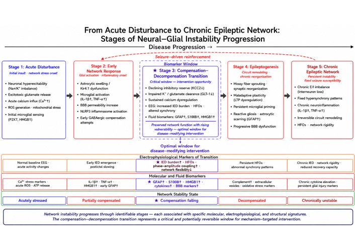
From acute disturbance to chronic epileptic network: stages of neural–glial instability progression. Schematic timeline showing the proposed progression from acute neural–glial disturbance to chronic epileptic network organization. Early neuronal hyperexcitability, glial activation, calcium dysregulation, neuroinflammation, blood–brain barrier dysfunction, and circuit remodeling are organized across five stages. The compensation–decompensation transition is highlighted as a biomarker-rich window marked by increasing epileptiform activity, high-frequency oscillations, altered synchrony, glial injury markers, inflammatory mediators, and declining network flexibility. This stage may represent an optimal opportunity for mechanism-targeted intervention before instability becomes chronically consolidated.
Understanding neural–glial instability in epilepsy requires experimental systems that can capture interactions across molecular, cellular, circuit, and tissue levels. No single model fully reproduces the heterogeneity of human epilepsy, but together animal models, in vitro systems, human tissue studies, and emerging analytical tools provide complementary ways to investigate how excitability, glial signaling, inflammation, calcium dysregulation, and circuit remodeling interact over time [187–189]. This section is important not because methods are secondary to mechanism, but because interpretation of mechanism depends on what kinds of biological relationships a given model can reveal or obscure. A modern understanding of epilepsy increasingly depends on integrating evidence across multiple platforms rather than treating any one system as sufficient on its own.
Animal models remain foundational for studying epileptogenesis, seizure propagation, inflammation, and circuit remodeling in vivo. Genetic models, chemoconvulsant models, and injury- or status epilepticus-based paradigms each provide distinct windows into seizure susceptibility and disease progression [190–192]. Their value lies especially in allowing investigators to examine how seizures emerge within intact neural systems and how neuronal, glial, vascular, and immune-related processes interact across time after developmental abnormalities, induced hyperexcitability, brain injury, or prolonged seizures [188, 193, 194]. These models are particularly informative for studying dynamic transitions into chronic epilepsy, including glial reactivity, interneuron disruption, mossy fiber sprouting, BBB dysfunction, and behavioral outcomes. At the same time, they remain limited by species-specific physiology, restricted representation of human disease heterogeneity, and incomplete translational fit. Their interpretation is therefore strongest when paired with human-relevant systems rather than treated as self-sufficient representations of the disorder.
In vitro systems provide more controlled environments for mechanistic dissection. Primary cultures, acute slices, co-culture models, induced cellular systems, and related preparations offer high experimental manipulability and allow investigators to isolate causal relationships that may be difficult to resolve in intact organisms [19, 187]. These systems are especially useful for examining ion channel behavior, excitation–inhibition balance, astrocyte–neuron interactions, microglial signaling, calcium-dependent responses, and defined inflammatory perturbations with greater precision. Their main limitation is that reduction in complexity often comes at the cost of tissue architecture, long-range connectivity, vascular influences, and full-system interactions. Even so, they remain indispensable for targeted hypothesis testing and mechanistic resolution.
Human tissue studies provide an essential bridge between experimental models and clinical reality. Resected epileptic tissue, postmortem material, and patient-derived samples can reveal features of neuronal loss, glial activation, synaptic reorganization, molecular signaling, and structural remodeling that are difficult to infer fully from model systems alone [188, 193, 195]. These studies are particularly valuable for validating candidate mechanisms, identifying disease-relevant cellular states, and clarifying which features of network instability are most robust in human pathology. Their limitations include variable clinical histories, medication exposure, timing of tissue acquisition, and limited access to early disease stages. Nonetheless, they provide irreplaceable insight into how epilepsy is organized in human brain tissue and remain essential for anchoring mechanistic claims in translational relevance.
Among newer systems, organoids and related stem cell-derived platforms offer a promising way to study aspects of human neural development, cell-type interactions, excitability, and disease-associated signaling in a more human-relevant context [196–198]. Patient-derived induced pluripotent stem cell systems may be especially useful for examining genetically influenced epilepsies, developmental vulnerability, and cell-type-specific dysfunction [199, 200]. Brain organoids and related human stem-cell-derived systems are increasingly valuable for bridging molecular mechanisms with therapeutic innovation, especially when disease mechanisms involve developmental, circuit-level, or patient-specific vulnerabilities that are difficult to capture in conventional animal models [201]. Although organoids do not recapitulate the full architecture, vascularization, sensory input, or long-range systems integration of the intact brain, they can still provide important insight into developmental network formation, calcium signaling, and neural–glial interactions that are otherwise difficult to access. In the context of this review, organoids are important not as replacements for animal or human tissue studies, but as additional systems that may clarify how human cellular programs contribute to neural–glial instability.
A major advance across model systems has been the growing use of calcium imaging to track neuronal and glial dynamics in space and time. Because calcium signals are closely tied to excitability, intracellular signaling, and glial responsiveness, calcium imaging provides an especially informative window into the multi-level instability framework developed throughout this review [96, 198, 202]. It can reveal coordinated activity patterns, seizure propagation, astrocytic responses, and activity-dependent state changes that are not easily resolved through endpoint measurements alone. This approach has been particularly powerful for linking acute activity patterns with longer-lasting changes in circuit and glial behavior, although interpretation must remain attentive to the distinction between calcium dynamics and direct electrical output [55, 198].
Electrophysiology remains equally central because epilepsy is fundamentally a disorder of abnormal excitability and synchronization. Single-cell recordings, field potential analysis, patch-clamp methods, multi-electrode arrays, and related approaches provide direct access to firing properties, synaptic function, oscillatory behavior, network recruitment, and seizure-like discharge [55, 187, 198]. These methods are especially valuable for defining excitation–inhibition balance, membrane instability, firing synchrony, and circuit-level recruitment with temporal precision that complements imaging-based approaches. In many cases, the most informative studies are those that combine electrophysiology with cellular or molecular readouts, allowing investigators to connect electrical behavior with glial state, calcium signaling, or inflammatory activity rather than examining these domains separately [187, 195].
Omics methods now provide a powerful way to map the molecular architecture of epileptic instability. Bulk and single-cell transcriptomics, spatially resolved profiling, proteomic analyses, and related approaches can identify cell-type-specific programs of neuronal stress, astrocyte reactivity, microglial activation, oligodendroglial changes, and inflammatory remodeling [195, 203]. These approaches are especially useful for revealing heterogeneity within epileptic tissue and for uncovering molecular pathways that link excitability to chronic remodeling. Their main limitation is interpretive: molecular association alone does not establish causal contribution, so omics data become most informative when integrated with anatomical, physiological, and histological context [195, 203].
Finally, computational and network modeling offer tools for understanding how diverse cellular and molecular changes scale into emergent circuit behavior. These approaches can help bridge local perturbations in inhibition, glial buffering, cytokine signaling, calcium dysregulation, or rewired connectivity to large-scale effects on synchronization, seizure threshold, propagation, and chronic instability [204–206]. They are especially valuable for testing nonlinear interactions that may be difficult to isolate experimentally and for formalizing hypotheses about how multiple abnormalities combine to destabilize neural–glial systems. Computational models do not replace experimental biology, but they can reveal hidden relationships, sharpen causal reasoning, and clarify which combinations of dysfunction are most likely to produce seizure-prone network states.
The most informative future studies will likely be those that combine physiological recording, imaging-based dynamics, molecular profiling, and human-relevant experimental systems rather than relying on any one modality in isolation. Such integrative approaches are especially important for epilepsy because instability emerges across interacting cellular and circuit scales that cannot be fully resolved by electrophysiology, omics, imaging, or model selection alone.
Taken together, modern epilepsy research depends on the integration of experimental models and analytical tools that capture different levels of neural–glial organization. Animal models, in vitro systems, human tissue studies, organoids, calcium imaging, electrophysiology, omics approaches, and computational modeling each provide distinct but complementary insight into how instability emerges, propagates, and becomes chronic [187, 195, 198, 206]. No individual model captures the full biological, developmental, and clinical complexity of human epilepsy, making cross-platform integration essential rather than optional for mechanistic interpretation. Their combined use strengthens the central argument of this review by showing that neural–glial instability is not simply a conceptual framework, but a tractable biological problem that can be interrogated across increasingly sophisticated experimental platforms. These methodological advances now make it increasingly possible to test mechanism-guided stabilization strategies rather than approaching epilepsy only through broad seizure suppression. The complementary strengths and limitations of the principal experimental models and analytical tools discussed in this section are summarized in Table 3.
Experimental models and tools for studying neural–glial instability.
| Model/Tool | What it captures well | Strength for studying epilepsy instability | Main limitation | Best use of this review framework |
|---|---|---|---|---|
| Animal models | In vivo seizure generation, network propagation, cell-type interactions, inflammatory responses, circuit remodeling, longitudinal disease progression | Allows mechanistic study of how neuronal, glial, inflammatory, and circuit-level changes interact over time within intact tissue and behaviorally relevant systems | Species differences, model-specific bias, incomplete representation of human epilepsy heterogeneity, and variable translational fidelity | Best for studying multi-scale disease evolution, epileptogenic transition, and testing causality across cellular and circuit levels [187, 207] |
| In vitro systems | Controlled cellular interactions, defined perturbations, basic excitability changes, glial responses, and mechanistic manipulation under simplified conditions | Useful for isolating specific pathways such as neurotransmitter imbalance, glial regulation, calcium signaling, and inflammatory responses without full in vivo complexity | Limited tissue architecture, reduced long-range network organization, and incomplete representation of chronic disease progression | Best for mechanistic dissection of defined pathways and rapid hypothesis testing under controlled conditions [187, 208] |
| Human tissue studies | Human-relevant cellular pathology, network features in diseased tissue, glial changes, inflammatory signatures, and structural remodeling | Provides direct relevance to human epilepsy biology and can validate whether mechanisms identified in models are present in human epileptic tissue | Limited availability, variable quality, restricted experimental manipulation, and difficulty capturing dynamic progression over time | Best for translational validation of disease-relevant mechanisms and comparison with model-derived findings [102, 209] |
| Organoids/Stem cell-derived systems | Human developmental context, patient-specific biology, cell-type interactions, emerging network behavior, and genetically tractable human-relevant systems | Valuable for linking neural and glial biology to human-specific developmental, genetic, and patient-specific mechanisms of instability | Incomplete maturation, limited vascular/immune complexity, simplified architecture, and uncertain correspondence to full clinical epilepsy states | Best for studying human-relevant mechanisms, genetic vulnerability, and patient-specific neural–glial interactions in controlled systems [198, 210] |
| Calcium imaging | Spatiotemporal activity dynamics, population-level signaling patterns, glial calcium activity, and propagation of network events across cells | Especially useful for visualizing how neuronal and glial activity evolves across space and time during destabilization and seizure-like states | Limited direct measurement of membrane conductance, variable temporal resolution, and interpretive complexity when linking signals to causality | Best for mapping activity coordination, propagation, calcium-linked instability, and neuron–glia signaling dynamics [93, 96] |
| Electrophysiology | Membrane excitability, synaptic transmission, firing properties, inhibitory/excitatory balance, oscillations, and synchronized network discharge | Remains one of the strongest approaches for defining seizure-relevant excitability and testing causal changes in neuronal and circuit behavior | Often narrower in molecular or spatial context unless combined with imaging, molecular profiling, or anatomical analysis | Best for precise measurement of excitability, synaptic function, and network synchronization underlying epileptic instability [187, 208] |
| Omics approaches | Cell-state programs, transcriptional and proteomic heterogeneity, inflammatory signatures, stress pathways, and molecular remodeling across cell types | Powerful for identifying cell-type-specific pathways linking excitability, glial activation, inflammation, and chronic remodeling | Association does not equal causation; findings require physiological and anatomical context for mechanistic interpretation | Best for mapping molecular architecture, heterogeneity, and candidate pathways that can be integrated with functional studies [207, 209] |
| Computational/Network modeling | Multi-scale interaction logic, network dynamics, synchronization rules, parameter testing, and systems-level prediction | Useful for integrating diverse data and exploring how changes in inhibition, glial regulation, calcium signaling, or connectivity may alter network behavior | Dependent on assumptions, model structure, and input quality; may oversimplify biological heterogeneity if not constrained by empirical data | Best for systems-level synthesis, hypothesis generation, and testing how interacting instability mechanisms might produce emergent epileptic behavior [205, 211] |
The neural–glial network instability framework proposed in this review is not intended to replace established neuroinflammatory, ionic, computational, or neuron-centered models of epilepsy. Rather, it offers a higher-order explanatory layer that organizes these models within a shared systems-level account of how seizure-prone states emerge and become stabilized over time. In this framework, epilepsy is not reduced to a single initiating mechanism. Instead, seizure susceptibility is understood as the outcome of interacting disturbances in neuronal excitability, extracellular ion and neurotransmitter homeostasis, glial regulation, inflammatory signaling, calcium-dependent stress pathways, and circuit remodeling. The novelty of the framework therefore lies less in proposing an entirely new molecular trigger than in clarifying how multiple established mechanisms converge to erode compensation and promote persistent network instability.
This distinction is especially important in relation to neuroinflammatory frameworks of epileptogenesis. Neuroinflammation is often described as a driver of seizure susceptibility and epileptogenic progression, particularly through cytokine signaling, danger-associated pathways, BBB dysfunction, immune–glial activation, and maladaptive synaptic or structural remodeling. The present framework accepts this central role but expands it by positioning inflammation not simply as an upstream pathological trigger or downstream consequence of seizures, but as one component of a broader feedback architecture. Inflammatory mediators can lower seizure threshold by altering receptor function, ion channel behavior, synaptic transmission, astrocytic homeostasis, and microglial remodeling. At the same time, seizures, calcium overload, metabolic stress, extracellular ionic disturbance, and tissue injury can further activate inflammatory pathways. Thus, inflammation is redefined here as an amplifier and stabilizer of network instability: it helps determine whether an acute disturbance is resolved, remains compensated, or becomes biologically consolidated into chronic epileptogenic vulnerability.
The framework also differs from computational and ionic models while remaining compatible with them. Computational and ionic models have shown that changes in extracellular and intracellular ion concentrations, especially K+ and Na+ dynamics, can support nonlinear state transitions, network bistability, seizure initiation, and seizure termination. The neural–glial instability framework does not dispute these mechanisms and does not claim that K+/Na+-dependent transitions are unnecessary. Instead, it places ionic bistability within the biological context that determines how such ionic conditions arise, persist, or recover. Astrocytic potassium buffering, sodium-dependent glutamate uptake, chloride homeostasis, energy metabolism, BBB integrity, inflammatory signaling, and seizure-induced remodeling all influence whether ionic disturbances remain transient or become part of a self-reinforcing epileptic state. In this sense, ionic models describe an important dynamical mechanism of seizure-state transition, whereas the neural–glial instability framework asks how cellular, glial, inflammatory, metabolic, and structural processes create or constrain the conditions under which those transitions become likely.
Therefore, the framework should be understood as a multi-scale integrative model rather than a competing single-mechanism hypothesis. Its contribution is to connect mechanistic explanations operating at different levels: ion concentration dynamics and bistability at the biophysical level; neuronal excitability and inhibition at the circuit level; astrocytic and microglial regulation at the cellular level; inflammatory and calcium-linked stress at the molecular signaling level; and remodeling at the chronic disease level. By linking these layers, the model helps explain why similar ionic or inflammatory perturbations may produce different outcomes depending on tissue context, developmental stage, disease etiology, glial state, metabolic reserve, and prior seizure history. This integrated view also provides a stronger basis for translational reasoning, because it suggests that durable seizure control may require not only suppressing acute hyperexcitability, but restoring the stabilizing mechanisms that prevent networks from entering or remaining in pathological seizure-prone states.
The neural–glial instability framework has important therapeutic implications because it shifts the treatment question from suppressing seizures in the moment to restoring the biological conditions that support durable network stability. Conventional antiseizure therapies have been highly valuable in reducing seizure frequency and severity, yet a substantial proportion of patients continue to experience incomplete control, recurrent seizures, or drug-resistant disease despite treatment [212–214]. This limitation reflects, at least in part, the fact that epilepsy is not simply a disorder of excessive neuronal firing. As the preceding sections have shown, epileptic instability emerges from interacting disturbances in inhibitory regulation, glial homeostasis, calcium signaling, inflammatory amplification, and circuit remodeling. A therapeutic framework built around these interacting mechanisms may therefore offer a more complete translational logic than one centered on acute neuronal suppression alone [155, 202]. Its clinical value lies in linking seizure control to mechanism-informed diagnosis, dynamic risk assessment, patient stratification, and therapeutic strategies aimed at restoring the stabilizing processes that normally prevent seizure-prone network states. Accordingly, therapeutic priorities may differ across syndromes, disease stages, and the dominant architecture of instability present in a given patient or epileptic context.
This broader view does not diminish the importance of established antiseizure treatments. Rather, it places them within a wider mechanistic landscape. Therapies that enhance inhibition, reduce excitability, or interrupt seizure propagation remain essential, but they may be insufficient when network instability is sustained by glial dysfunction, chronic inflammatory signaling, intracellular stress, maladaptive plasticity, or reorganized circuitry [12, 14, 215]. From this perspective, the most effective future strategies may be those that combine symptomatic seizure control with interventions that restore homeostatic regulation and reduce the biological permissiveness of epileptic networks. The therapeutic goal is therefore not only to silence pathological firing, but to re-stabilize the system that allows such firing to emerge and persist.
The translational contribution of the neural–glial instability framework is that it shifts clinical reasoning from seizure suppression alone toward identification of the dominant biological processes that make a given network seizure-prone. Traditional therapeutic logic has often prioritized reduction of excessive neuronal firing, enhancement of inhibition, or interruption of seizure propagation. These remain essential goals, but the present framework suggests that diagnosis and treatment may also benefit from asking why the network remains permissive for recurrent seizures in the first place. In this view, epilepsy may reflect different instability architectures across patients: some may be driven primarily by impaired inhibitory control, others by astrocytic failure of potassium or glutamate homeostasis, others by inflammatory amplification, calcium-linked intracellular stress, BBB dysfunction, maladaptive remodeling, or combinations of these mechanisms.
This perspective opens several translational avenues that are less apparent in models focused mainly on acute hyperexcitability. First, it supports mechanism-informed diagnostic profiling, in which electrophysiological, imaging-based, molecular, inflammatory, metabolic, and clinical features are interpreted together to identify the dominant instability pattern. Second, it supports dynamic monitoring of seizure risk, because a network may move from a partially compensated state toward a decompensated seizure-prone state before recurrent seizures become clinically obvious. Third, it provides a rationale for mechanism-guided treatment selection, where therapies are matched not only to seizure type but also to the biological drivers of instability. For example, a patient with evidence of inflammatory amplification or BBB disruption may require a different adjunctive strategy than a patient whose instability is dominated by ion channel dysfunction, impaired inhibition, or structural network reorganization.
The framework also has implications for disease modification. If epileptogenesis reflects a progressive loss of network compensation, then treatment opportunities may exist before instability becomes structurally embedded. Interventions aimed at preserving astrocytic homeostasis, limiting inflammatory feedback, reducing calcium-linked stress, stabilizing the BBB, or preventing maladaptive circuit remodeling may be most valuable during vulnerable transition periods after status epilepticus, traumatic brain injury, infection, developmental disruption, or other epileptogenic insults. In this sense, the framework does not simply add new therapeutic targets; it reorganizes existing targets according to when and why they may matter during disease evolution.
Finally, this approach supports combination and precision strategies. Because chronic epilepsy often reflects interacting mechanisms rather than a single isolated abnormality, durable stabilization may require coordinated treatment across multiple levels: acute seizure suppression, restoration of inhibitory balance, glial and inflammatory modulation, metabolic or calcium-stress control, neuromodulation, and circuit-directed intervention when appropriate. The clinical value of the neural–glial instability framework therefore lies in providing a structured way to connect mechanistic biology with patient stratification, biomarker development, and rational therapeutic selection, including distinction between patients whose instability is dominated by impaired glial homeostasis, inflammatory amplification, ionic or inhibitory dysfunction, calcium-linked stress, structural remodeling, or mixed mechanisms.
A central translational implication of the neural–glial instability framework is that the transition from compensated network function to decompensated seizure susceptibility may be empirically trackable. Compensation refers to a state in which neural and glial regulatory mechanisms are stressed but still able to preserve sufficient inhibitory restraint, extracellular homeostasis, metabolic support, inflammatory control, and recovery capacity. Decompensation occurs when these stabilizing mechanisms are no longer able to contain perturbations, allowing lower seizure thresholds, increased hypersynchrony, impaired recovery, and progressive epileptogenic remodeling [216–218]. Although no single biomarker is likely to define this transition across all epilepsy syndromes, several dynamic features may help mark movement toward decompensation.
Electrophysiological measures are among the most clinically accessible candidates. A rising burden of interictal epileptiform discharges, increased high-frequency oscillations, abnormal phase–amplitude coupling, increased pathological synchrony, reduced network flexibility, altered functional connectivity, and impaired recovery of background activity after seizures may all indicate declining network resilience [219–221]. EEG and MEG may therefore be useful not only for detecting epileptic activity, but also for estimating whether a network is becoming less able to return to a stable baseline after perturbation. In this context, the most informative measures may be longitudinal and dynamic rather than static: increasing discharge burden, shortening inter-seizure intervals, seizure clustering, or worsening sensitivity to sleep disruption, stress, fever, or metabolic challenge may suggest that compensatory capacity is being progressively eroded.
Metabolic and imaging markers may provide a second layer of information. A transition toward decompensation may be associated with altered glucose utilization, lactate accumulation, mitochondrial stress, impaired perfusion–metabolism coupling, or imaging evidence of regional hypometabolism or hypermetabolism depending on disease stage and epilepsy subtype. Structural and functional MRI, PET, diffusion imaging, and other imaging approaches may also help identify gliosis, BBB disruption, white matter abnormalities, altered network connectivity, or progressive circuit reorganization [218, 222]. These markers are especially relevant because they may capture the tissue-level consequences of impaired homeostatic recovery and the emergence of a more permissive epileptogenic environment.
Molecular and fluid biomarkers may further help identify the biological direction of instability. Candidate markers include inflammatory cytokines and chemokines, danger-associated signals such as HMGB1-related pathways, BBB-associated proteins, astrocytic markers such as GFAP or S100B, microglial or complement-related signatures, extracellular vesicle cargo, oxidative stress markers, and indicators of neuronal or glial injury [223–225]. Such markers should not be interpreted as epilepsy-specific in isolation. Their value would likely be greatest when combined with electrophysiological, imaging, clinical, and genetic information to identify whether decompensation is dominated by inflammatory amplification, glial homeostatic failure, metabolic stress, structural remodeling, or primarily neuronal excitability mechanisms.
From a clinical perspective, the compensation-to-decompensation transition is therefore best understood as a multimodal and dynamic process rather than a single threshold event. A patient may show partial compensation despite an underlying epileptogenic insult if EEG abnormalities are limited, metabolic reserve is preserved, inflammatory signaling is transient, and recovery after perturbation remains efficient. By contrast, convergence of increasing epileptiform activity, impaired postictal recovery, progressive imaging abnormalities, inflammatory or glial biomarker elevation, and worsening seizure clustering may indicate movement toward a decompensated state. This approach could help bridge mechanistic biology with clinical prediction by identifying patients who are not only experiencing seizures, but whose neural–glial systems are losing resilience over time.
Clinical translation of the neural–glial instability framework will require stratifying patients according to the dominant biological mechanisms sustaining network instability. This is especially important because glial dysfunction and inflammatory amplification may overlap but are not identical. Glial dysfunction refers primarily to failure of astrocytic or other glial homeostatic functions, including impaired potassium buffering, reduced glutamate uptake, altered water and volume regulation, abnormal astrocytic calcium signaling, disrupted metabolic support, or impaired neuron–glia communication. Inflammatory amplification refers more specifically to sustained or excessive immune–glial signaling, including cytokine release, danger-associated signaling, microglial activation, BBB disruption, complement-related remodeling, and persistent inflammatory feedback loops that lower seizure threshold and promote epileptogenic progression [118, 222, 224].
In a clinical or translational setting, patients with a predominantly glial-homeostatic instability pattern may be suggested by convergent evidence of astrocytic dysfunction, impaired extracellular regulation, and local network permissiveness without necessarily showing a strong systemic or tissue inflammatory signature. Candidate indicators may include increased astrocytic injury or reactivity markers such as GFAP or S100B, imaging evidence of gliosis or regionally altered astrocyte-associated pathology, abnormalities in extracellular potassium or glutamate handling where measurable in experimental or surgical settings, impaired postictal recovery, local metabolic stress, and seizure patterns consistent with focal extracellular permissiveness [96, 223]. These features would suggest that the epileptic network is unstable because homeostatic buffering and recovery mechanisms are insufficient.
By contrast, patients with a predominantly inflammatory-amplification pattern may show evidence of sustained cytokine, chemokine, danger, complement, or BBB-related signaling. Candidate indicators may include elevated inflammatory cytokines or chemokines, HMGB1-related danger signaling, IL-1β/TNF-related pathway activation, complement-related signatures, microglial activation markers, BBB-associated markers, or imaging evidence consistent with neuroinflammatory activity or barrier disruption [226, 227]. Clinically, this pattern may be especially relevant after status epilepticus, traumatic brain injury, infection, autoimmune or inflammatory insults, tumor-associated epilepsy, or other contexts in which immune–glial signaling may remain active beyond the initiating event.
A biomarker-driven stratification approach should not rely on any single marker in isolation. GFAP or S100B elevation, for example, may indicate astrocytic injury or reactivity but does not by itself prove that astrocytic homeostatic failure is the dominant driver of seizures. Similarly, cytokine elevation may indicate inflammatory activity but does not alone establish that inflammation is the primary cause of network instability [228, 229]. The most clinically useful strategy is likely to be multimodal: combining seizure semiology, epilepsy subtype, EEG/MEG dynamics, MRI/PET findings, BBB markers, inflammatory and glial fluid biomarkers, genetic information, treatment response, and longitudinal disease trajectory. Stratification should therefore be based on convergent patterns rather than single biomarkers.
This distinction has therapeutic implications. A patient whose instability profile is dominated by impaired glial homeostasis may benefit most from strategies aimed at restoring extracellular buffering, glutamate handling, astrocytic metabolic support, or local circuit recovery, alongside conventional antiseizure therapy. By contrast, a patient with prominent inflammatory amplification or BBB disruption may be a stronger candidate for adjunctive approaches targeting inflammatory signaling, immune–glial activation, barrier stabilization, or post-injury epileptogenic cascades. Patients with mixed profiles may require combination strategies that address both acute excitability and the broader glial–inflammatory environment that sustains seizure susceptibility.
Biomarker-driven stratification should therefore be understood as a way to move from syndrome-level classification toward mechanism-informed precision medicine. Rather than asking only whether a patient has focal or generalized epilepsy, temporal lobe epilepsy, post-traumatic epilepsy, or drug-resistant epilepsy, the neural–glial instability framework encourages an additional question: which stabilizing system has failed most prominently in this patient, and which biomarkers support that interpretation? This approach could improve patient selection for glial-targeted, anti-inflammatory, metabolic, neuromodulatory, or circuit-directed interventions and may help explain why patients with similar seizure classifications can differ substantially in treatment response.
Restoring inhibitory balance remains one of the most established and clinically important therapeutic strategies in epilepsy. Because excessive synchronization and recurrent excitation often emerge in the setting of weakened inhibitory restraint, enhancing inhibitory signaling has long been a central principle of antiseizure treatment [41, 230]. This logic is strongly supported by the mechanistic discussion in earlier sections: when GABAergic tone is reduced, receptor function is altered, chloride homeostasis is disrupted, or interneuron recruitment becomes less effective, networks become more vulnerable to uncontrolled excitation and seizure propagation [54, 231]. Therapeutic efforts aimed at reinforcing inhibition therefore remain highly relevant within the neural–glial instability framework.
Many current antiseizure medications act, directly or indirectly, by strengthening GABAergic inhibition. These approaches may enhance receptor activity, prolong inhibitory currents, increase GABA availability, or reduce the relative dominance of excitatory signaling, including through receptor-modulating agents, drugs that increase synaptic GABA tone, or therapies used for rapid seizure suppression in acute settings [41, 230]. Clinically, such strategies can be highly effective in suppressing seizures acutely and remain indispensable in both chronic epilepsy management and emergency seizure control. Their success reinforces a central principle: seizure activity often depends on insufficient inhibitory containment, and restoring that containment can provide meaningful clinical benefit.
At the same time, the limitations of GABAergic approaches are equally instructive. Not all patients respond adequately to treatments that enhance inhibition, and in some epileptic settings the underlying inhibitory deficit is not simply one of reduced GABA availability. Receptor subtype alterations, changes in tonic versus phasic inhibition, chloride dysregulation, interneuron network disruption, and context-dependent paradoxical GABA effects may all reduce the effectiveness of standard inhibitory therapies [41, 54, 231]. In such cases, strengthening GABAergic signaling pharmacologically may not fully restore stable inhibitory control because the broader circuit architecture and cellular context remain altered. This helps explain why seizure suppression and true network stabilization are not always equivalent.
A further practical limitation is that therapies that broadly enhance inhibition may also be constrained by sedation, cognitive adverse effects, tolerance in some settings, or incomplete long-term efficacy [41, 232]. These realities do not diminish the importance of GABAergic treatment, but they underscore that inhibitory enhancement alone may not provide durable control in all patients, particularly when epilepsy is maintained by mechanisms extending beyond acute excitability. In this sense, the clinical limits of inhibitory therapy reinforce the broader mechanistic argument of the present review.
A particularly important issue is that inhibitory therapies are often most effective at controlling acute excitability, whereas chronic epilepsy may also involve persistent glial dysfunction, calcium-dependent stress, inflammatory amplification, and structural circuit remodeling that lie outside a purely GABAergic solution [54, 230, 231]. If the extracellular environment remains permissive, if inflammatory signals continue to lower seizure threshold, or if recurrent excitatory pathways have become structurally embedded, then enhancing inhibition alone may provide incomplete or temporary benefit. This does not reduce the value of inhibitory therapies; rather, it clarifies why they are necessary but not always sufficient within a broader disease process.
These limitations also point toward more refined strategies for restoring inhibitory balance. Rather than viewing inhibition as a single global variable, future therapeutic approaches may need to consider which inhibitory mechanisms are disrupted, in which circuits, and at what stage of disease. Interventions that better address tonic versus phasic inhibition, chloride regulation, interneuron preservation or recruitment, and circuit-specific inhibitory architecture may prove more effective than uniformly increasing GABAergic tone. Such strategies would align more closely with the idea that epilepsy is a systems disorder in which inhibitory failure is biologically heterogeneous rather than mechanistically identical across patients and syndromes.
Within the therapeutic logic of this review, restoring inhibitory balance remains a foundational strategy, but one that must be understood within a broader neural–glial context. GABAergic approaches can reduce seizure expression and remain indispensable in clinical practice, yet their limitations highlight the need for multi-level therapies aimed at restoring stable network conditions rather than suppressing neuronal firing alone [41, 167, 231]. Restoring inhibition is therefore essential, but durable seizure control may also require correction of the extracellular, inflammatory, and circuit-level disturbances that undermine inhibitory stability in the first place.
If epilepsy is sustained in part by failures of extracellular regulation, inflammatory amplification, and maladaptive neural–glial signaling, then glial dysfunction becomes an important therapeutic target rather than a secondary consequence of neuronal disease. This is one of the central translational implications of the present review. Astrocytes and microglia actively shape seizure threshold, tissue recovery, inflammatory tone, and the long-term permissiveness of epileptic networks [14, 215, 233]. Therapeutic strategies that modulate glial dysfunction therefore have the potential not only to reduce seizure expression, but also to alter the conditions that allow instability to persist.
For astrocytes, one therapeutic objective is the restoration of homeostatic support functions that normally restrain excitability. Strategies that improve potassium buffering, enhance glutamate uptake, stabilize water regulation, support metabolic coupling, or reduce maladaptive astrocyte reactivity may be especially relevant in epileptic tissue where astrocytic dysfunction contributes to a permissive environment for hyperexcitability [14, 234, 235]. In principle, such approaches could reduce seizure susceptibility by strengthening the extracellular constraints that normally prevent excessive synchronization. This therapeutic logic is attractive because it targets network permissiveness rather than neuronal firing alone. At the same time, astrocytes are multifunctional cells whose effects depend on region, timing, and disease stage. An intervention that improves one astrocytic domain may alter others in unintended ways. Therapeutic approaches aimed at restoring astrocytic stability must therefore distinguish between protective homeostatic functions and context-dependent reactive states rather than treating astrocyte activity as uniformly pathological [14, 202, 235].
For microglia, the therapeutic goal is often different. Because microglia can amplify inflammatory signaling, reshape synaptic organization, and help sustain seizure-prone tissue states, strategies that reduce maladaptive microglial activation or shift microglial responses toward more protective programs may hold substantial translational value [28, 105, 233]. This does not imply that broad microglial suppression is always desirable. As discussed earlier, microglia can also support debris clearance, containment of injury, and short-term tissue recovery. The challenge is therefore not simply to inhibit microglia, but to modulate the timing, intensity, and direction of their responses. Therapies that blunt persistent inflammatory amplification while preserving protective surveillance or repair functions may be more effective than approaches based on indiscriminate immune suppression [106, 236].
A major strength of glia-directed therapy is that it may address mechanisms that conventional antiseizure medications leave relatively untouched. If recurrent seizures are reinforced by impaired astrocytic buffering, persistent microglial activation, glia-driven inflammatory signaling, or altered neural–glial communication, then neuron-centered treatments alone may provide incomplete stabilization [12, 14, 215]. In this sense, glial targeting could complement rather than replace established antiseizure therapy. Such combination logic may be especially important in chronic or drug-resistant epilepsy, where the network remains unstable despite ongoing efforts to suppress neuronal discharge.
At the same time, glia-directed interventions face substantial challenges. Glial states are heterogeneous, region-specific, temporally dynamic, and deeply integrated with normal brain function [12, 215, 235]. Astrocytes and microglia can be protective or maladaptive depending on disease stage, tissue context, and the nature of the underlying insult. This means that therapeutically relevant modulation will likely require more precise approaches than broad glial inhibition. Biomarker-guided targeting, cell-state-specific strategies, and better alignment between experimental models and patient biology may therefore be essential for translating glial therapeutics into clinically meaningful interventions.
Within the broader framework of this review, modulating glial dysfunction represents an effort to restore the extracellular and inflammatory architecture of network stability. This is clinically important because seizure persistence may depend not only on abnormal neuronal firing, but also on whether astrocytes and microglia continue to sustain a biologically permissive state for excitability, synchronization, and maladaptive remodeling. Glial therapies are therefore best understood as strategies to restore the extracellular and inflammatory conditions under which stable circuit function becomes possible, rather than as simple adjuncts to neuronal suppression.
Because calcium dysregulation links excitability, glial activation, mitochondrial dysfunction, inflammatory signaling, and chronic remodeling, it represents an especially attractive therapeutic target within a neural–glial instability framework. Interventions directed at calcium handling and intracellular stress pathways may have value not only because they reduce acute excitability, but because they may interrupt the molecular processes through which seizures become biologically embedded in cells and circuits [129, 237, 238]. In this sense, targeting calcium-related mechanisms offers a strategy that lies between symptomatic seizure suppression and deeper disease modification.
One therapeutic avenue involves limiting neuronal calcium overload. Excessive calcium entry during hyperexcitable states can reinforce burst firing, amplify neurotransmitter release, activate stress pathways, and increase vulnerability to injury [18, 129, 237]. Strategies that reduce pathologic calcium influx or improve calcium buffering may therefore help protect neurons from the feedforward consequences of repeated seizure activity. This logic is particularly important in chronic epilepsy, where recurrent episodes of calcium overload may contribute to persistent dysfunction even if each individual seizure is transient. Such approaches may therefore be relevant not only for reducing excitability, but also for slowing disease-promoting cellular consequences of repeated seizure burden.
A second therapeutic direction concerns intracellular calcium stores and endoplasmic reticulum stress-related signaling. Because calcium release from intracellular stores can amplify excitability and couple seizure activity to deeper forms of cellular stress, interventions that stabilize these pathways may reduce both acute network instability and longer-term cellular damage [136, 151]. This includes mechanisms associated with endoplasmic reticulum calcium release, store-related dysregulation, calcium-dependent coupling to endoplasmic reticulum stress pathways, and downstream signaling cascades that influence survival, plasticity, and inflammatory responsiveness [129, 135, 151]. Such approaches are appealing because they address intracellular consequences of epilepsy that are not fully captured by membrane-focused antiseizure therapies.
Calcium-targeted strategies may also be relevant to astrocytic and microglial signaling. Abnormal glial calcium dynamics can contribute to altered gliotransmission, inflammatory amplification, and persistent extracellular permissiveness for excitability [96, 143]. Therapeutic efforts that normalize calcium-dependent glial responses may therefore reduce maladaptive feedback loops linking seizures, glial activation, and chronic instability. As with glia-directed therapy more broadly, however, the challenge lies in distinguishing physiological calcium signaling from pathological amplification. Calcium is fundamental to normal brain function, so interventions must be sufficiently selective to avoid broad disruption of essential signaling processes [129, 143, 239].
Closely related to calcium dysregulation is the problem of intracellular stress, particularly mitochondrial dysfunction, oxidative stress, and maladaptive stress signaling. Repeated seizures impose heavy metabolic and ionic demands on neurons and glia, and when intracellular buffering systems are overwhelmed, cells may enter states that favor further instability rather than recovery [238, 240]. Therapies that support mitochondrial resilience, reduce oxidative burden, or interrupt stress-amplifying intracellular cascades may therefore help preserve network stability even if they do not act as classic antiseizure drugs [135, 237]. This expands the therapeutic horizon from controlling discharge to protecting the cellular systems that determine whether tissue remains recoverable or becomes chronically destabilized.
The translational promise of calcium- and stress-directed strategies lies in their ability to intervene at a mechanistic crossroads. Rather than targeting only the electrical expression of seizures, these approaches aim to weaken the intracellular pathways that connect acute hyperactivity to inflammation, injury, and remodeling [135, 215]. Within the framework of this review, they are therefore particularly important as potential disease-modifying strategies rather than as tools for seizure termination alone [230, 237]. Their ultimate value may depend on how well they can be integrated with approaches that restore inhibition, modulate glial dysfunction, and address circuit-level reorganization, rather than operating in isolation.
Limiting inflammatory amplification is another important therapeutic strategy within a neural–glial instability framework. Neuroinflammation can lower seizure threshold, reinforce glial dysfunction, disrupt tissue homeostasis, and promote epileptogenic remodeling. This approach is clinically relevant not simply because inflammation accompanies many epileptic conditions, but because persistent inflammatory signaling may help convert transient disturbances into chronic seizure-prone states [241]. Therapeutic efforts aimed at reducing inflammatory amplification therefore seek to interrupt the feedback loops through which seizures, glial activation, danger signaling, BBB dysfunction, and tissue stress reinforce one another over time [242].
One major rationale for anti-inflammatory intervention is the bidirectional relationship between seizures and inflammatory signaling. Seizures can activate cytokine pathways, innate immune signaling, danger-associated responses, and glial inflammatory programs, while these same pathways can feed back onto the network by altering excitability, synaptic regulation, and tissue recovery [243]. When such feedback becomes persistent, the brain may remain in a biologically sensitized condition even between overt seizures. From a therapeutic perspective, this means that reducing inflammatory tone may do more than attenuate associated pathology; it may directly reduce the permissiveness of epileptic tissue.
At the same time, inflammatory targeting in epilepsy is unlikely to succeed through broad suppression alone. Inflammatory responses are heterogeneous, temporally dynamic, and in some contexts partially protective. Acute inflammatory signaling may contribute to tissue containment or repair, whereas sustained or maladaptive activation may promote chronic instability. The challenge is therefore to distinguish inflammatory responses that should be restrained from those that should be preserved. Therapies aimed at specific cytokine pathways, modulation of innate immune signaling, BBB-stabilizing strategies, or targeted control of glial inflammatory states may therefore be more effective than indiscriminate immunosuppression [243].
This therapeutic logic also has important implications for epileptogenic transition. If inflammatory amplification contributes to the progression from acute insult to chronic epilepsy, then timely intervention may have disease-modifying potential rather than merely symptomatic value [241]. This is especially relevant in conditions such as status epilepticus, traumatic brain injury, infection-related epilepsy, or other inflammatory-prone epileptic states in which the tissue may enter a prolonged window of biological vulnerability [242]. In such contexts, limiting inflammatory amplification could help preserve network recoverability and reduce the likelihood that maladaptive remodeling becomes entrenched.
However, substantial translational challenges remain. The inflammatory biology of epilepsy varies by syndrome, timing, anatomical region, age, underlying etiology, and disease stage. Not all patients exhibit the same inflammatory profile, and not all inflammatory pathways are equally actionable. This makes biomarker-guided stratification especially important if anti-inflammatory therapies are to be used intelligently rather than empirically. Within the framework of this review, inflammatory targeting is therefore best viewed as part of a broader systems strategy: the goal is not simply to reduce immune activity, but to prevent inflammatory signaling from converting reversible instability into self-reinforcing chronic epilepsy.
The broader therapeutic message of this review is that effective epilepsy treatment may require restoring network stability, not simply suppressing neuronal firing acutely. This makes network-level and precision approaches especially important, because they are designed to act on the organization, dynamics, and patient-specific architecture of epileptic systems rather than on isolated molecular targets alone. Within a neural–glial instability framework, these approaches are valuable because they align closely with the idea that epilepsy is sustained by interacting disturbances across circuits, cell types, and time scales [229, 244, 245].
One important class of strategies involves neuromodulation. Approaches such as electrical stimulation, responsive stimulation, or related circuit-level interventions may help reduce seizure burden not only by interrupting abnormal activity, but by altering the network conditions that permit pathological synchronization and propagation [244, 246]. In selected cases, surgical resection or disconnection can also be understood as a network-level intervention, particularly when a dominant epileptogenic node or pathway can be identified and safely targeted [247, 248]. From the perspective of this review, these strategies are important because they reflect a shift from targeting isolated molecular mechanisms to regulating unstable network behavior more directly.
A second major component of precision therapy is the development and use of biomarkers. If epilepsy is biologically heterogeneous, then therapies directed at inhibition, glia, inflammation, calcium signaling, or circuit remodeling are unlikely to be equally effective across all patients [229, 249]. Electrophysiological, imaging-based, molecular, inflammatory, and other multimodal biomarkers may therefore be essential for identifying the dominant mechanisms operating in a given patient and for matching interventions to those mechanisms more precisely [229, 249]. This is especially relevant for translational progress, because it moves therapy away from uniform empiricism and toward mechanism-informed stratification.
Patient-specific models also offer a promising route toward more individualized intervention. Human tissue studies, stem cell-derived systems, organoids, and related personalized platforms may help identify patient-relevant cellular vulnerabilities, developmental mechanisms, drug responses, or network phenotypes that are not fully captured by generalized models [250, 251]. Although these approaches remain technically and translationally challenging, they are conceptually important because they allow the biology of epilepsy to be studied in forms that are closer to the patient’s own cellular context. This may become especially valuable for syndromes with strong genetic, developmental, or drug-resistant features.
More broadly, the future of epilepsy treatment may depend on systems-guided therapies that integrate multiple levels of information rather than focusing on a single pathway in isolation. In practice, this could mean combining pharmacological treatment with biomarker-guided anti-inflammatory strategies, glial modulation, calcium- or stress-directed interventions, neuromodulation, or circuit-targeted procedures depending on the dominant architecture of instability in a given case [229, 230]. Such a framework is well suited to epilepsy because the disease often reflects converging dysfunctions rather than a single lesion in one signaling pathway. Systems-guided therapy therefore represents not therapeutic complexity for its own sake, but a more biologically faithful response to the structure of the disorder.
Taken together, network-level and precision approaches extend the translational implications of this review beyond conventional seizure control. They emphasize that durable benefit may come from identifying and correcting the specific configurations of instability that sustain epileptic networks in different patients [229, 244, 245]. Within this framework, effective therapy is not defined only by immediate seizure suppression, but by whether it helps restore the conditions under which neural–glial systems can recover more stable function. The future of epilepsy therapy may therefore depend less on suppressing seizures uniformly and more on restoring the particular form of stability lost in each patient. These therapeutic strategies are summarized in Table 4 as a network-stabilization framework that links mechanistic insight to translational intervention in epilepsy.
Therapeutic strategies to restore network stability in epilepsy.
| Therapeutic logic | Main target/mechanism | What it aims to stabilize | Potential strengths | Key limitations/challenges | Translational status |
|---|---|---|---|---|---|
| Restoring inhibitory balance | Enhancement of GABAergic signaling, strengthening inhibitory restraint, improving receptor-mediated inhibition, increasing synaptic inhibitory tone | Excitation–inhibition balance, seizure threshold, circuit containment of abnormal firing | Conceptually directed, clinically familiar, and often effective for acute seizure control or symptomatic suppression | May not correct upstream glial, inflammatory, calcium-linked, or remodeling mechanisms; efficacy can vary across syndromes and chronic stages; tolerance or incomplete disease modification may limit long-term impact | Established clinical strategy for seizure control, but often insufficient alone for durable modification of epileptogenic instability [41, 252] |
| Modulating astrocyte dysfunction | Improving potassium buffering, glutamate uptake, water regulation, metabolic support, and limiting maladaptive astrocyte reactivity | Extracellular homeostasis, neurotransmitter balance, ionic stability, propagation permissiveness, recovery capacity | Mechanistically attractive because astrocytes regulate the extracellular conditions that directly shape excitability and seizure spread | Therapeutic targeting remains biologically complex; astrocyte responses are context dependent; restoring homeostatic function without disrupting adaptive support is challenging | Emerging and largely preclinical/translational, with strong conceptual rationale but limited clinical implementation [14, 83, 234] |
| Modulating microglial dysfunction | Limiting maladaptive inflammatory activation, altering cytokine release, reducing harmful synaptic remodeling, and reshaping context-dependent microglial responses | Inflammatory amplification, maladaptive synaptic/environmental signaling, chronic seizure-prone tissue states | May interrupt feedback loops between tissue stress, immune signaling, glial activation, and progressive destabilization | Microglia can be protective, maladaptive, or mixed depending on timing and context; indiscriminate suppression may be counterproductive | Emerging and mechanistically promising, but still limited by context specificity and incomplete translational precision [241, 243] |
| Targeting calcium/intracellular stress | Reducing pathological calcium overload, modulating intracellular calcium store signaling, limiting calcium-dependent stress cascades and endoplasmic reticulum stress coupling | Cellular stress burden, excitability-linked injury signaling, maladaptive plasticity, calcium-driven propagation of instability | Attractive because calcium links neurons, glia, inflammatory signaling, and remodeling across multiple timescales | Calcium signaling is essential to normal physiology, so therapeutic selectivity is difficult; dominant calcium sources and effectors may vary across contexts | Early translational and mostly preclinical, with strong mechanistic relevance but limited clinically mature strategies [129, 151] |
| Limiting inflammatory amplification | Targeting cytokine pathways, innate immune signaling, blood–brain barrier-related dysfunction, or persistent inflammatory feedback loops | Seizure threshold, recovery environment, glia-mediated amplification, progression from acute disturbance to chronic instability | May be especially valuable in contexts where inflammation helps drive epileptogenic transition or chronic vulnerability | Inflammatory mechanisms are heterogeneous across syndromes and stages; broad immunosuppression may be ineffective or poorly targeted | Translationally active and biologically important, but likely to require more selective mechanism-guided use than generalized anti-inflammatory treatment [241, 243] |
| Network-level/precision approaches | Neuromodulation, biomarker-guided stratification, surgery where appropriate, patient-specific models, systems-guided therapy selection | Large-scale network synchrony, seizure propagation, patient-specific instability architecture, treatment matching | Most aligned with the review’s systems framework because it recognizes epilepsy as heterogeneous and mechanistically layered | Requires reliable biomarkers, better subtype stratification, and integration of mechanistic knowledge with patient-specific decision-making | Already partly established in selected forms, but still evolving toward more precise and mechanism-guided implementation [229, 244, 246] |
The translational logic of restoring network stability across interacting neuronal and glial mechanisms is illustrated in Figure 3.
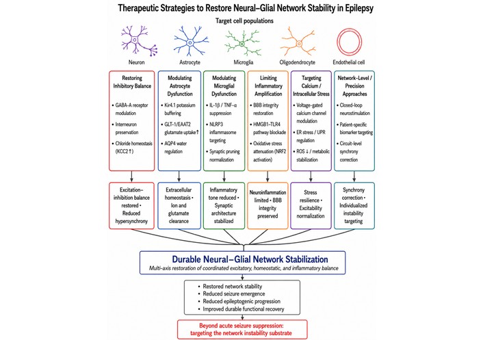
Therapeutic strategies to restore neural–glial network stability in epilepsy. Schematic overview of mechanism-informed therapeutic strategies that may promote durable stabilization of epileptic networks by targeting multiple interacting components of neural–glial instability. Major therapeutic domains include restoration of inhibitory balance, modulation of astrocyte dysfunction, modulation of microglial dysfunction, limitation of inflammatory amplification, targeting of calcium-linked intracellular stress, and network-level or precision approaches. These interventions act through intermediate effects on excitation–inhibition balance, extracellular homeostasis, inflammatory tone, neuroinflammatory restraint, stress resilience, and synchrony correction, which converge on durable neural–glial network stabilization. The figure emphasizes that effective therapy may require multi-axis restoration of coordinated excitatory, homeostatic, and inflammatory balance, with the broader goal of moving beyond acute seizure suppression toward reduced seizure emergence, reduced epileptogenic progression, and improved durable functional recovery.
The evidence reviewed here supports a clear conclusion: epilepsy is best understood not simply as a disorder of recurrent neuronal hyperexcitability, but as a condition driven by interacting instability mechanisms across neurons, glial cells, intracellular signaling systems, tissue homeostasis, and circuit organization [143, 253]. Across molecular, cellular, animal, translational, and human studies, epilepsy emerges not as a single-pathway disturbance, but as a multi-scale failure of coordinated regulation within neural–glial networks. This perspective does not replace classical concepts of neuronal hyperexcitability; rather, it places them within a broader and more biologically complete framework.
A central message of this review is that inhibitory dysfunction, calcium dysregulation, glial reactivity, inflammatory signaling, and circuit remodeling should not be treated as separate explanatory domains. Instead, they interact across time and scale to shape seizure emergence, epileptogenic transition, and chronic seizure susceptibility [143, 145]. Reduced inhibitory containment can lower the threshold for abnormal synchronization; calcium dysregulation can link acute activity to intracellular stress and long-term remodeling; astrocytes and microglia can regulate extracellular stability, inflammatory tone, and tissue permissiveness; and chronic circuit reorganization can stabilize pathological network behavior once instability is established. The epileptic state is therefore best understood as the product of converging and self-reinforcing processes rather than of any single defect in one cell type or signaling pathway.
This integrated view has important implications for both research and therapy. Experimentally, it argues for study designs that move beyond neuron-centered reductionism and instead examine how cell types, signaling systems, and circuit properties interact dynamically across disease stages [209, 253]. Methodologically, it supports the continued integration of animal models, in vitro systems, human tissue studies, organoids, electrophysiology, calcium imaging, omics, and computational approaches to resolve epilepsy as a systems-level disorder rather than a set of disconnected mechanisms. Translationally, it suggests that durable therapeutic progress may depend less on suppressing neuronal firing alone and more on restoring the broader architecture of network stability. In this sense, the future of epilepsy therapy may lie in multi-level, mechanism-informed, and increasingly patient-specific strategies that address the particular forms of instability sustaining disease in different biological contexts.
At the same time, this framework should be advanced with care. Epilepsy is heterogeneous, and not all mechanisms reviewed here are equally dominant across syndromes, developmental stages, etiologies, or anatomical regions [254, 255]. Neural–glial instability should therefore be understood as an organizing framework rather than a uniform explanatory template. Its value lies in helping integrate diverse findings into a coherent model while preserving biological complexity and disease-specific variation. Future work will need to define more precisely which combinations of inhibitory failure, glial dysfunction, inflammatory activation, calcium-related stress, and remodeling are most relevant in particular forms of epilepsy, and at which stages they are most actionable.
Several future directions appear especially important. One is the development of multimodal biomarkers that can identify dominant mechanisms of instability in individual patients or disease subtypes [255, 256]. Another is the refinement of patient-specific and human-relevant experimental systems, including stem cell-derived and organoid-based platforms that better capture neural–glial interactions in context. A third priority is the use of longitudinal and stage-aware study designs to define when particular instability mechanisms emerge, intensify, or become therapeutically actionable. Closely related to this is the need for interventions aimed not only at terminating seizures, but at preventing acute disturbances from being biologically consolidated into chronic epileptic states.
Taken together, the accumulated evidence supports a careful but firm conclusion: epilepsy should be understood as a disorder of interacting neuronal and glial instability mechanisms. Evidence across experimental systems supports a multi-scale model in which inhibitory dysfunction, calcium dysregulation, glial reactivity, inflammatory signaling, and circuit remodeling converge to shape seizure emergence and epileptogenesis [143, 145, 253]. Recognizing this convergence does not simplify epilepsy; it clarifies why the disorder is so persistent, heterogeneous, and difficult to treat. It also points toward a more durable therapeutic goal: not only suppressing seizures, but restoring stable function across the interacting neuronal and glial systems that determine whether epileptic networks recover or remain chronically destabilized.
BBB: blood–brain barrier
MMN: Conceptualization, Writing—original draft, Writing—review & editing. The author read and approved the submitted version.
The author declares that there are no conflicts of interest.
Not applicable.
Not applicable.
Not applicable.
Not applicable.
Not applicable.
© The Author(s) 2026.
Open Exploration maintains a neutral stance on jurisdictional claims in published institutional affiliations and maps. All opinions expressed in this article are the personal views of the author(s) and do not represent the stance of the editorial team or the publisher.
Copyright: © The Author(s) 2026. This is an Open Access article licensed under a Creative Commons Attribution 4.0 International License (https://creativecommons.org/licenses/by/4.0/), which permits unrestricted use, sharing, adaptation, distribution and reproduction in any medium or format, for any purpose, even commercially, as long as you give appropriate credit to the original author(s) and the source, provide a link to the Creative Commons license, and indicate if changes were made.



View: 657
Download: 15
Times Cited: 0
Hoong Wei Gan ... William P. Whitehouse
Jerónimo Auzmendi, Alberto Lazarowski
Toshimitsu Suzuki ... Kazuhiro Yamakawa
Rodolfo Cesar Callejas-Rojas ... Ildefonso Rodriguez-Leyva
Anwaar M. Bennour ... Heba A. El-Zawawi


