 Review


Affiliation:
Laboratory of NeuroOntogeny, Institute of Higher Nervous Activity and Neurophysiology, Russian Academy of Sciences, Moscow 117485, Russian Federation
Email: midzyanovskaya.is@ihna.ru
ORCID: https://orcid.org/0000-0003-1660-6632


Explor Neurosci. 2026;5:1006145 DOI: https://doi.org/10.37349/en.2026.1006145
Received: April 14, 2026 Accepted: July 14, 2026 Published: August 11, 2026
Academic Editor: Jinwei Zhang, Chinese Academy of Sciences, China
The article belongs to the special issue Advances in Epilepsy Research
Autism spectrum disorder (ASD) is a clinically heterogeneous neurodevelopmental condition characterized by core social communication deficits and restricted/repetitive behaviors. The majority of cases present with certain medical and psychiatric comorbidities. These include epilepsy, gastrointestinal disorders, attention-deficit/hyperactivity disorder, anxiety, and metabolic dysregulation. While animal models are indispensable in pre-clinical research and studying ASD’s pathobiology, there remains a need to address these comorbidities while modelling major ASD clinical domains. The paper describes a spectrum of animal models for ASD and its comorbid disorders, with a focus on ASD & epilepsy co-occurrence.
Autism spectrum disorder (ASD) is a neurodevelopmental condition characterized by deficits in verbal and nonverbal social communication, a limited initiation of social contacts, and inflexible behaviors. Autism research started with the pioneering work of G. Sukhareva, who first described autistic individuals in terms of “schizoid psychopathy” [1, 2]. Kanner [3], in a 1943 paper, described “early infantile autism” as a distinct condition, separating it from childhood schizophrenia. Mnukhin [4, 5] linked early infantile autism to organic brain deficiency, emphasizing the importance of neurological disorders (vegetative dystonia, disorders of muscle tone, insufficiency of subcortical structures). Twin studies by Folstein and Rutter [6, 7] demonstrated ASD as a heritable neurobiological condition. The first genetic linkages identifying specific chromosomal regions associated with autism were reported by the International Molecular Genetic Study of Autism Consortium (1998) and other groups. Now, large-scale genomic studies have revealed the genetic complexity of autism, identifying hundreds of associated gene variants [8, 9].
At the moment, the ASD prevalence is reported to be 2.1–2.3% [10], which represents a significant source of financial burden for healthcare systems and families.
ASD encompasses multiple domains, the delineation of which remains an active subject of scientific and clinical debate. Precisely defining these subtypes is essential for elucidating the distinct genetic and epigenetic mechanisms that operate across critical developmental stages [11]. It is widely recognized that ASD arises from a complex interplay of genetic and epigenetic influences, spanning a continuum from mild to severe phenotypes. Given that direct genetic interventions remain limited, epigenetic mechanisms represent a particularly promising avenue for investigation and therapeutic targeting.
A large-scale analysis of an ASD cohort (approximately 5,000 individuals from the SFARI [12–14] database identified four distinct clinical subtypes [15]. Notably, at least one-third of individuals with moderate symptom severity show little evidence of identifiable causative genetic variants, while de novo mutations are frequently associated with more severe forms of ASD [15].
The first subgroup, comprising roughly one-third of cases, is characterized by moderate social and behavioral impairments, typical attainment of early developmental milestones (e.g., walking and language acquisition), and a high prevalence of comorbid conditions, including anxiety, depression, attention-deficit/hyperactivity disorder (ADHD), and obsessive-compulsive disorder (OCD). The genetic architecture of this phenotype is often linked to mutations that exert postnatal effects. The next subgroup of comparable size also exhibits typical early developmental trajectories, accompanied by moderate deficits in social communication and restricted or repetitive behaviors. In contrast to the first group, these individuals show a low prevalence of psychiatric comorbidities, and no clear genetic etiology has been identified.
A third subtype, representing approximately one-fifth of the cohort, is defined by delayed developmental milestones and pronounced communicative impairments in the absence of significant anxiety or depressive comorbidity. This phenotype is associated with a genetic background enriched for de novo and rare inherited variants. Finally, a fourth subgroup, accounting for around 10% of cases, is characterized by severe developmental delay, profound social-communication deficits, a high burden of comorbid mood disorders, and a high frequency of de novo mutations [15].
Taken together, these findings underscore the pronounced heterogeneity of ASD and highlight the need to stratify potential intervention targets while disentangling causal mechanisms from secondary exacerbating effects.
The vast majority of cases of ASD are also diagnosed with concomitant somatic, neurological, or psychiatric diseases. At the same time, pure, uncomplicated forms of autism are rather the exception to the rule. Prevalent somatic comorbidities include gastrointestinal (GI) disorders [16, 17], immune system dysregulation [18, 19], and metabolic disturbances [20], such as mitochondrial dysfunction [21, 22], oxidative stress [23, 24], and impaired methylation [25–27]. Common neurological and psychiatric comorbidities include ADHD, epilepsy, and anxiety disorders [28–31]. Also, ASD is frequently associated with sensory processing difficulties [32–34], affecting visual [35], auditory [36, 37], and somatosensory domains [38, 39]. Intraspecific communication is critically dependent on the integration of multisensory information. Consequently, impairments in sensory perception or processing directly disrupt these communicative exchanges. Disrupted interoception—the sense of the body’s internal state—represents a critical facet of sensory processing impairment in ASD. Furthermore, this interoceptive deficit can be compounded by comorbidities, reinforcing a cycle that may exacerbate the core symptoms of the condition [40–42]. Addressing the interoceptive impacts of comorbid conditions is a primary target for improving the quality of life of patients with ASD. Some evidence suggests that comorbid conditions may exacerbate core ASD symptoms. A fundamental question is how comorbid conditions contribute to the overall ASD phenotype, including the critical stages and mechanistic pathways involved.
Epilepsy co-occurring with ASD has been associated with increased impairments in social awareness and expressive communication. A study in a South Korean cohort reported higher autism severity scores in children with both ASD and epilepsy compared to those with ASD alone, implying a potential aggravating effect independent of cognitive function [43]. Although meta-analyses have not demonstrated sufficient efficacy of antiepileptic drugs in improving ASD-related behavioral symptoms [44–46], effective control of epilepsy still plays a role in preventing secondary symptom worsening. Therefore, epilepsy management may represent an important component of comprehensive, long-term ASD care [47, 48].
The surgical treatment of epilepsy has long provided a unique window into human brain function, as highlighted by the pioneering work of Wilder Penfield and Herbert Jasper [49, 50]; in later accounts, epilepsy itself was regarded as a “great teacher of neurobiology”.
Epileptogenesis (and other neurological disorders) is increasingly understood not merely as a disorder of neuronal excitability but as a process shaped by complex bidirectional interactions between central nervous system function and peripheral metabolic state, in the frame of “Metabolic Neurophylosophy”, as proposed by Gulyaeva [51]. Emerging evidence implicates several metabolic signaling pathways—ranging from insulin and glucocorticoid (GC) signaling to neurotrophic support, dietary interventions, and gut–brain communication—in modulating seizure susceptibility, epilepsy progression, and associated comorbidities (Figure 1).
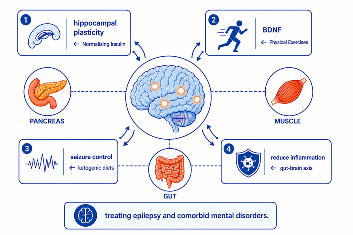
Schematic representation of body-to-brain metabolic interventions targeting epilepsy and comorbid mental disorders. The figure presents a framework for treating epilepsy and associated psychiatric comorbidities by intervening in body metabolism. Four possible intervention strategies are shown: (1) normalizing insulin signaling (pancreas) helps to support hippocampal plasticity; (2) physical exercise helps to sustain and elevate brain-derived neurotrophic factor (BDNF) levels; (3) ketogenic diets help to achieve seizure control; and (4) gut-targeted therapies help to modulate the gut–brain axis. These approaches reflect the emerging paradigm that brain disorders can be partly ameliorated via manipulation of peripheral metabolic pathways (conceptual framework after Gulyaeva [51]).
Insulin functions within the brain as a critical neuromodulator of synaptic plasticity, learning, and memory, beyond its classical role in peripheral glucose homeostasis. In particular, insulin signaling supports hippocampal-dependent cognitive processes. Conversely, insulin resistance—whether systemic or confined to the central nervous system—impairs hippocampal function, compromising memory formation and potentially lowering the seizure threshold in vulnerable individuals [52, 53]. These observations raise the possibility that restoring insulin sensitivity may exert protective effects against epilepsy-related cognitive decline and, partly, against epileptogenesis itself.
GCs, released upon activation of the hypothalamic–pituitary–adrenal (HPA) axis, act as metabolic orchestrators that prioritize energy supply to the brain, particularly to memory-encoding circuits. Moderate GC release enhances long-term potentiation (LTP)—the cellular correlate of memory—by directing metabolic resources toward the strengthening of synaptic connections that support adaptive behavior [54, 55]. However, chronic stress and sustained GC elevation disrupt neuronal energetics and may facilitate epileptogenesis by promoting excitotoxicity, impairing astrocytic glutamate clearance, and compromising mitochondrial function. Thus, the HPA axis represents a key bidirectional interface linking stress, metabolism, and seizure susceptibility.
Brain-derived neurotrophic factor (BDNF) operates as a “metabotrophin”—a molecular hub that integrates neuronal signaling with systemic metabolic state. BDNF not only supports synaptic plasticity and neuronal survival but also mediates the beneficial effects of metabolic interventions such as exercise and dietary modulation [56, 57]. Disruptions in BDNF signaling may represent a convergent pathway through which metabolic dysfunction exacerbates seizure susceptibility and cognitive comorbidity.
One of the most clinically validated metabolic interventions for seizure control is the ketogenic diet. For over a century, ketogenic and related metabolic regimens have consistently demonstrated anticonvulsant efficacy across various epilepsy syndromes [58]. A key mechanism underlying this effect is the metabolic shift induced by ketosis, which alters the central balance of excitatory and inhibitory neurotransmitters. Specifically, ketosis promotes the synthesis of GABA—the principal inhibitory neurotransmitter in the brain—thereby enhancing inhibitory tone and conferring an anticonvulsant effect [59]. This metabolic rerouting of neurotransmitter metabolism exemplifies how peripheral metabolic state directly modulates central neuronal excitability and seizure threshold [51].
The gut–brain axis constitutes a potential bidirectional metabolic link in epileptogenesis [51]. Metabolic disorders are frequently accompanied by intestinal dysbiosis, which disrupts the production of key microbial metabolites, including up to 90% of circulating serotonin and short-chain fatty acids, essential for maintaining the integrity of the blood–brain barrier. Dysbiosis leads to increased intestinal permeability (“leaky gut”), which is often associated with compromised blood–brain barrier integrity (“leaky blood–brain barrier”), thereby allowing neurotoxins and inflammatory mediators to enter the central nervous system [60, 61]. Such gut-derived inflammatory signals can activate microglia, promote neuroinflammation, and lower the seizure threshold. Indeed, impaired gut–brain axis function has now been documented across a wide spectrum of somatic and brain disorders, including psychiatric conditions [62, 63]. Extending these findings to epilepsy and ASD, dysbiosis and gut barrier dysfunction may contribute to a pro-inflammatory, hyperexcitable brain state, thereby facilitating epileptogenesis and its associated comorbidities.
In summary, these observations indicate that epileptogenesis is modulated by a network of peripheral–central metabolic interactions. Insulin resistance, chronic GC elevation, BDNF dysregulation, ketogenic shifts, and gut microbiome alterations each represent potential links between systemic metabolism and epileptogenesis [51]. Targeting these pathways—whether by normalizing insulin sensitivity, reducing chronic stress, enhancing BDNF signaling, implementing ketogenic diets, or restoring gut microbial homeostasis—may offer novel therapeutic strategies for the prevention of epileptogenesis and its associated neuropsychiatric comorbidities.
Epidemiological data consistently show that ADHD and ASD are highly prevalent neurodevelopmental conditions with substantial overlap. ADHD is one of the most frequent comorbid conditions to ASD [64], with systematic reviews reporting that approximately 30–50% of children with ASD also meet ADHD criteria [65, 66]. Conversely, at least half of children and adults with ADHD have another co-occurring condition, including ASD. This bidirectional association has been clarified by recent meta-analyses and large cohort studies. A meta-analysis of 63 studies found that about 38.5% of individuals with ASD have current ADHD and 40.2% meet lifetime criteria [66]. Registry‑ and population‑based studies indicate that ASD is markedly overrepresented among children with ADHD, and conversely, ADHD is highly prevalent among autistic children: meta‑analytic and cohort data typically report ASD in roughly 20–50% of ADHD cases and ADHD in approximately 30–40% of individuals with ASD, with higher rates in specialized clinical samples [67, 68]. These convergent findings support the view that ADHD–ASD comorbidity reflects a meaningful clinical intersection, not chance co-occurrence, and underscore the need to address comorbidity in neurobiological research [69].
GI issues, reported in approximately 30–50% of individuals with ASD, are increasingly recognized as a result of interplay between behavioral, microbial, and neurophysiological mechanisms. Dietary selectivity, a common feature of ASD, contributes to reduced dietary diversity and altered nutrient intake, which can secondarily affect gut motility and function. Recent studies demonstrate consistent alterations in gut microbiota composition [70] and metabolic activity in ASD, including reduced diversity and shifts in key microbial taxa, which are associated with both GI symptom severity and behavioral phenotypes [71, 72]. These microbiome changes influence immune responses, intestinal barrier integrity, and neuroactive metabolite production [73, 74]. In parallel, neuroendocrine and enteric nervous system dysfunctions—particularly involving serotonergic signaling, vagal pathways, and stress-axis regulation—have been implicated in altered gut motility and sensory processing in ASD [75]. Evidence for low-grade intestinal inflammation and barrier dysfunction further supports a role for immune-mediated mechanisms [76]. Importantly, these factors form a bidirectional network in which restrictive eating behaviors shape microbiota composition, which in turn modulates gut–brain signaling and contributes to both GI and behavioral symptoms [77].
Sleep disorders affect approximately 50–80% of children with ASD [78, 79]. These disturbances, including difficulties with sleep onset and maintenance, reduced sleep duration, and circadian dysregulation, are consistently associated with increased severity of ASD symptoms. In particular, poor sleep has been linked to heightened irritability, stereotyped behaviors, anxiety, and impairments in social communication and language development [80, 81]. A recent study suggests bidirectional interactions between sleep, sensory processing, and behavioral regulation [82]. Alterations in circadian rhythms, melatonin signaling, and autonomic function are thought to underlie these associations [83]. Therefore, systematic characterization of sleep disturbances is critical for accurate phenotyping and targeted intervention in ASD [84].
Animal models offer a crucial translational value for ASD research. While directly altering core ASD-like symptoms in model organisms remains impossible, experimentally manipulating comorbid states (in laboratory animals) and observing their convergent effects on behavioral phenotypic outcomes, as well as on translatable biomarkers, provides a feasible and powerful experimental paradigm. The most commonly used animal models for ASD can be categorized as genetic, environmentally induced, or idiopathic (Figure 2).
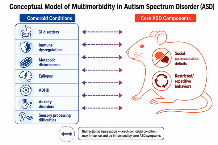
Conceptual model of multimorbidity in autism spectrum disorder (ASD). Core diagnostic components (social communication deficits and restricted/repetitive behaviors) are shown on a rodent figure. Comorbid conditions (gastrointestinal [GI] disorders, immune dysregulation, metabolic disturbances, epilepsy, attention-deficit/hyperactivity disorder [ADHD], anxiety disorders, and sensory processing difficulties) are listed on the left side. Bidirectional arrows indicate that each comorbidity may interact with and aggravate core ASD symptoms, and vice versa. This framework emphasizes the complex, reciprocal nature of ASD‑comorbidity interactions.
Genetic models, which primarily relate to syndromic forms of ASD, involve targeted mutations in high-risk ASD genes, enabling mechanistic studies of neuronal dysfunction. These are, for example, Fragile X Syndrome: the FMR1 KO mouse model recapitulates social deficits, repetitive grooming, and dendritic spine abnormalities [85–87]. Another example is Rett Syndrome: models with mutations in the MECP2 gene exhibit developmental regression, seizures, and motor stereotypies [88–93]. Genetic models enable the study not only of specific syndromes that include an autistic phenotype but also of mechanisms shared between syndromic and non-syndromic forms of autism. Among non-syndromic models, there are monogenic mutations such as NLGN3 R451C, SHANK3, and CNTNAP2. These three genes are among the most well-studied and influential genes in autism research, called “ASD risk genes” because mutations in them significantly increase the likelihood of developing autism and related conditions. These particular genes illustrate different genetic paths to ASD: a specific point mutation in a synaptic gene (NLGN3), the complete disruption of a critical synaptic scaffold leading to a defined syndrome (SHANK3), and common variations in a gene linking language and neuronal excitability (CNTNAP2). Mice with the NLGN3 R451C mutation show altered social interaction and enhanced inhibitory synaptic transmission [94–96]. The SHANK3 mutants display ASD-like behaviors, including reduced social novelty preference and stereotypic grooming, alongside marked striatal hypertrophy [97–102]. The CNTNAP2 knockout models exhibit hyperactivity, seizures, and a reduced population of cortical interneurons [103–105].
From a preclinical perspective, established models of focal and generalized seizures help determine how sustained epileptogenic activity alters neural circuits implicated in core ASD behaviors, such as social cognition and repetitive patterns. Clinically, epidemiological data are essential for correlating epilepsy syndromes, seizure types, and electroencephalographic (EEG) signatures with the prevalence and severity of autistic traits. The convergence of these complementary lines of inquiry—from animal models to human data—is indispensable for identifying causative links and shared biological substrates, including excitatory-inhibitory imbalance, chronic neuroinflammation, and synaptopathies.
Animal models of epilepsy are particularly valuable for deconstructing ASD pathophysiology. They provide a controlled system to study how epilepsy-induced network disruptions corrupt complex social behaviors—such as social recognition, dominance hierarchies, and group cohesion. This disruption is likely mediated by three key processes: neuronal hyperexcitability, glia-driven neuroinflammation, and structural alterations within limbic regions (hippocampus, amygdala, prefrontal cortex). These regions are not only fundamental for the expression of integrated social behavior but are also highly vulnerable to the disruptive process of epileptogenesis, creating a convergent pathway from seizure activity to autistic-like symptomatology [11, 106, 107]. Rats with increased susceptibility to kindled seizures and comorbid behavioral abnormalities showed multiple brain structural differences on magnetic resonance imaging (MRI) compared to their kindling-resistant controls: enlarged cerebrum, corpus callosum, third ventricle, and posterior inferior cerebellum, but a reduced anterior vermis. Histological examination of these seizure-prone rats further revealed cerebellar abnormalities, specifically expanded white matter, Purkinje cell loss, and a thinner molecular layer [108]. Also, the experimental induction of focal epileptiform activity in the prefrontal cortex of pre-pubertal rats leads to deficits in attention and social behavior [109, 110]. These studies may provide a mechanistic insight into a putative local pathway underlying ASD-epilepsy comorbidity. By employing the experimental model of epilepsy, it identifies the shared neural substrates that link epileptogenesis with the emergence of ASD-like symptoms in animals. This approach not only elucidates potential causal relationships but also establishes a translational framework for discovering therapeutic targets relevant to both conditions. Importantly, animal models of epilepsy often provide a possibility of a step-wise control of epileptogenesis by scheduling the experimental seizure provocations [111, 112].
Genetic animal models of generalized nonconvulsive epilepsies are often inbred rodent strains [113]. The two widely used rat models are the Genetic Absence Epilepsy Rats from Strasbourg (GAERS) and the Wistar Albino Glaxo/Rijswijk (WAG/Rij) strains. In these models, epilepsy manifests as spontaneous, recurrent generalized spike-wave discharges on the EEG, which are concomitant with behavioral arrest and a loss of contact with the surroundings. The evidence regarding social motivation in these models is equivocal. While some studies report no significant deficit [114, 115], others find only mild impairment [116, 117]. More specifically, zoosocial deficits seem to be confined to a subpopulation of the WAG/Rij strain that presents a mixed epilepsy phenotype [118]—i.e., a predisposition to both convulsive and non-convulsive epilepsies [119]—whereas in GAERS rats, social approach deficits have been reported exclusively in females [120]. Another animal model, the Krushinsky-Molodkina (KM) inbred rat strain, a genetic model of convulsive audiogenic epilepsy, exhibits consistent social deficits characterized by prominent freezing behavior [121, 122] and social aversion, but not anhedonia [123] or aggression [124]. These social deficits were seen in seizure-naïve rats and were resistant to rare seizure provocations [125] but were significantly aggravated by a 20-day regimen of audiogenic kindling [126]. This suggests common neurodevelopmental disturbances that contribute to both elevated seizure susceptibility and autistic-like phenotypes [127].
Environmentally induced models probe gene-environment interactions that impact neurodevelopment. The oldest type of epigenetic model is prenatal valproic acid (VPA) administration. The use of the anticonvulsant VPA during pregnancy is a well-established environmental risk factor for ASD. The link between prenatal VPA exposure and increased ASD risk in humans [128–130] led to the establishment of this model. Even transient prenatal exposure to valproate, particularly during critical periods of neural tube closure, is sufficient to induce long-term neurological effects. Rodents exposed to VPA in utero show decreased social motivation, increased stereotypies, and altered sensorimotor gating [131–134]. Interestingly, some of these deficits can be rescued by postnatal environmental enrichment [135–137]. This particular effect closely parallels the well-characterized impact of behavioral interventions on the phenotypic expression of autism, in clinical practice.
Maternal immune activation (MIA) is also a popular model for translational ASD research [138–141]. It is widely recognized that maternal infections during pregnancy, particularly those associated with prolonged fever, increase the risk of adverse neurodevelopmental outcomes in the offspring. In this model, immune activation in pregnant dams via Poly(I:C) (a component of bacterial walls) injection induces an interleukin surge (notably IL-6), leading to ASD-like traits in the offspring [140, 141]. In animals, these phenotypes can be prevented by anti-IL-6 antibodies [142, 143].
Idiopathic models are inbred strains that capture polygenic, spontaneous ASD phenotypes with no single known cause, making them resemble the majority of human ASD cases, which are also polygenic [15]. BTBR T+tf/J mouse strain is a good example: the animals display reduced social approach, low reciprocal interactions, and impaired juvenile play compared to controls. They also show impaired social transmission of food preference, indicating communication deficits, and high levels of novelty-induced hyperlocomotion and repetitive behavior (self-grooming) [144]. The consensus from the literature is that BTBR mice also display clear hyperactive and impulsive traits, but often do not show the classic attention deficits that are central to the ADHD diagnosis [145, 146]. Some studies show that stimulants like amphetamine can reduce hyperactivity in BTBR mice, which is a classic response seen in ADHD. However, other studies note that BTBR mice can have an atypical response, sometimes showing hypersensitivity or no response, potentially due to their complex underlying neurobiology, which includes ASD-related traits [145, 146]. The amelioration of social and cognitive deficits, along with repetitive behaviors, was achieved in these mice through the mitigation of pro-inflammatory cytokines and oxidative stress by a ketogenic diet [147]. This established mouse line exemplifies the utility of stable laboratory models for elucidating the ASD phenotype. By providing a consistent genetic background, such models enable researchers to isolate the effects of specific manipulations on disease mechanisms-from immune dysregulation to joint inflammation-establishing causal relationships that are often obscured in heterogeneous human populations.
While a number of animal models exist for the major comorbidities of ASD, a critical and often overlooked question is their translational value for ASD research. A priority would be to systematically characterize these models to determine which ones recapitulate both comorbidities and core ASD phenotypes. Elucidating the underlying mechanisms driving this convergence—or, conversely, the factors that protect certain models from developing core traits—is essential for refining our etiological models and identifying coherent biological subtypes.
Social behavior in animal models for GI is essential due to the gut–brain axis, which links gut health to neurological and behavioral outcomes. GI issues are found in 30–50% of ASD individuals, with higher prevalence in non-verbal populations [148, 149]. It was shown that GI distress itself could modify social aspects of behavior in laboratory rodents [150–152]. For gregarious species, a behavioral mechanism that induces social distancing in response to signs of GI distress or inflammation—a potential indicator of transmissible infection—would be adaptive [153]. This would enhance the survival prospects of the social group by limiting contagion. However, in clinical samples, microbial interventions ameliorated some behavioral deficits without rescuing the whole pathophysiology of ASD [154]. Therefore, interventions aimed at modulating gut health represent a strategic, accessible starting point for probing GI effects on social distancing and contact motivation.
Animal models of ADHD hold significant translational value for studying this major comorbid condition in ASD. Several animal models demonstrate face validity for the co-occurrence of ASD and ADHD, including genetic models (e.g., SHANK3, Fmr1) [93, 99], environmentally induced models (e.g., prenatal VPA) [131], and idiopathic strains (e.g., BTBR T+tf/J) [144]. The WKY/NCrl substrain presents a comorbidity of physiological and neurobehavioral traits, featuring hypertension alongside a spectrum of disorders that include ADHD, depression, high anxiety, and specific ASD-relevant deficits in social interest and communication (ultrasonic vocalizations) [155]. Importantly, these behavioral characteristics are not present in all WKY substrains: while the WKY/NCrl substrain displayed autistic-like traits, the WKY/NHsd substrain did not, establishing the latter as an ideal genetic control. Unfortunately, our search of the scientific literature did not yield new studies on ASD employing the aforementioned two WKY substrains.
There are significant links between aberrant sensory processing and core autistic-like traits. Sensory disruptions are not merely peripheral symptoms but are fundamentally connected to social behaviors and circuit-level brain abnormalities. In the BTBR mouse model of autism [144–146], auditory processing abnormalities were linked to reduced parvalbumin-positive neurons and an elevated glutamate/GABA ratio in the auditory cortex. The transcriptomic analysis identified Scn1a as a recurrent ASD-associated gene, expressed specifically in cortical inhibitory neurons, which offers mechanistic insight into the specific sensory dysfunction [146]. Impaired multisensory integration was consistently observed across three other monogenic models—GAD65, SHANK3, and Mecp2 knockout mice—all of which exhibited behavioral phenotypes and parvalbumin-circuit abnormalities within the insular cortex. Together, these findings offer developmental insight into putative cortical circuits implicated in neuropsychiatric conditions with social deficits, such as schizophrenia and autism [156].
Anxiety disorders are known to impair social functioning. Selectively bred Wistar rat substrains, diverging in high- and low-anxiety-related behavior on the elevated plus-maze, provide a reliable model for investigating the neurobiology underlying anxiety trait and its comorbidities, with depression-like behavior and social deficits. After decades of selective breeding, these lines exhibit consistent differences in anxiety, depressive-like, and social behaviors, with a confirmed causative mutation—a single nucleotide polymorphism in the vasopressin promoter—driving the neuropeptide overexpression in the high-anxiety substrain. Thus, this and similar models remain a valuable tool for exploring the multifaceted pathology of high anxiety and its associated comorbidities [157].
The remarkable heterogeneity characterizing ASD likely arises from the dynamic interplay between an individual's genetic makeup, environmental exposures, and acquired comorbid conditions. The scientific literature provides several compelling examples of how environmental stressors can exacerbate ASD-like symptoms in animal models, with studies often exploring the “two-hit” or gene-environment interaction paradigm. A 2020 study demonstrated that administering p-cresol—an environmental toxin and gut bacterial metabolite sometimes found at elevated levels in children with ASD—to BTBR mice led to exacerbated ASD-like symptoms [158]. Analysis of tissue levels of monoaminergic neurotransmitters and their metabolites revealed significantly increased dopamine turnover in the amygdala, as well as in the dorsal and ventral striatum, following p-cresol administration [158]. Research published in 2025 showed that subjecting Cntnap2+/– mice to early-life stress (using a limited bedding and nesting paradigm) resulted in unique behavioral changes not observed with either factor alone. The combination led to perseverative risk-taking behaviors and morphological abnormalities within the amygdala, highlighting how environmental factors can shape phenotypic outcomes based on genetic vulnerability [159]. A 2025 study investigated the combined impact of maternal deprivation (an early-life stressor) and postnatal exposure to VPA. The combination synergistically exacerbated anxiety-like behaviors and aggravated social impairment, particularly in female rats, underscoring how early-life stress and environmental toxins can interact to worsen neurodevelopmental outcomes [160].
Building on this line of inquiry, it is valuable to investigate which specific components of the ASD phenotype can be aggravated—and to what extent—by particular environmental (e.g., stress) or internal (e.g., seizure activity or sensory processing abnormalities) factors.
Several limitations of animal models warrant consideration. Strain-dependent variability in VPA and MIA models complicates cross-study replication. Sex differences are frequently overlooked, with most studies focusing on males. Modeling multi-system comorbidities remains technically challenging. Finally, single-mechanism rescue studies have limited translational validity, as ASD and its comorbidities arise from multifactorial etiologies unlikely to be reversed by targeting isolated pathways.
In summary, two fundamental questions emerge: whether (and to what extent) ASD-associated comorbidities can independently elicit social deficits in neurotypical animals, and whether core autistic traits may reciprocally exacerbate those comorbidities. Investigating these questions requires multi-trait phenotyping combined with longitudinal designs to infer causality. Pharmacological manipulation of specific comorbidities—including epilepsy, sleep dysfunction, and anxiety—can clarify their contribution to social impairments. Exploring brain–body interactions further necessitates parallel assessment of interoception, although this remains technically challenging in animal models. Environmental factors, particularly stress, can unmask latent functional links and determine whether the comorbidities are causal drivers or merely epiphenomena. This holds particular relevance for ASD subtypes associated primarily with developmental and environmental influences. Translational animal models are indispensable for elucidating these mechanisms and guiding the development of personalized therapies.
ADHD: attention-deficit/hyperactivity disorder
ASD: autism spectrum disorder
BDNF: brain-derived neurotrophic factor
EEG: electroencephalogram
GAERS: Genetic Absence Epilepsy Rats from Strasbourg
GC: glucocorticoid
GI: gastrointestinal
HPA: hypothalamic–pituitary–adrenal
MIA: maternal immune activation
VPA: valproic acid
WAG/Rij: Wistar Albino Glaxo/Rijswijk (rat strain)
The author used DeepSeek AI for assistance in translating content and refining the readability of the manuscript. The author thanks ChatGPT (OpenAI) for creating the figures according to the author’s prompts. After utilizing the tool, the author(s) reviewed and edited the content as necessary and take(s) full responsibility for the final content of the publication.
MIS: Conceptualization, Writing—original draft, Writing—review & editing, Funding acquisition. The author read and approved the submitted version.
The author declares no conflicts of interest.
Not applicable.
Not applicable.
Not applicable.
Not applicable.
The article was prepared in full within the state assignment of the Ministry of Education and Science of the Russian Federation for IHNA & NPh RAS. The funder(s) had no role in study design, data collection and analysis, decision to publish, or preparation of the manuscript.
© The Author(s) 2026.
Open Exploration maintains a neutral stance on jurisdictional claims in published institutional affiliations and maps. All opinions expressed in this article are the personal views of the author(s) and do not represent the stance of the editorial team or the publisher.
Copyright: © The Author(s) 2026. This is an Open Access article licensed under a Creative Commons Attribution 4.0 International License (https://creativecommons.org/licenses/by/4.0/), which permits unrestricted use, sharing, adaptation, distribution and reproduction in any medium or format, for any purpose, even commercially, as long as you give appropriate credit to the original author(s) and the source, provide a link to the Creative Commons license, and indicate if changes were made.



View: 1410
Download: 13
Times Cited: 0
Hoong Wei Gan ... William P. Whitehouse
Jerónimo Auzmendi, Alberto Lazarowski
Toshimitsu Suzuki ... Kazuhiro Yamakawa
Rodolfo Cesar Callejas-Rojas ... Ildefonso Rodriguez-Leyva
Anwaar M. Bennour ... Heba A. El-Zawawi
Moawiah M Naffaa


