 Review


Affiliation:
Department of Life Sciences, Manchester Metropolitan University, M15GD Manchester, UK

Affiliation:
Department of Life Sciences, Manchester Metropolitan University, M15GD Manchester, UK
Email: a.hasib@mmu.ac.uk
ORCID: https://orcid.org/0000-0002-5388-6461


Explor Endocr Metab Dis. 2026;3:101484 DOI: https://doi.org/10.37349/eemd.2026.101484
Received: May 12, 2026 Accepted: September 14, 2026 Published: October 22, 2026
Academic Editor: Amedeo Lonardo, Università di Modena (-2023), Italy
The article belongs to the special issue Innovative Strategies for Diabetes and Metabolic Disorders: Current and Future Directions
Type 2 diabetes mellitus (T2DM) is characterised by progressive insulin resistance and pancreatic β-cell dysfunction, ultimately resulting in impaired glucose homeostasis. While metabolic and genetic factors are central to disease development, increasing evidence indicates that disruption of the pancreatic islet extracellular matrix (ECM) may be an important contributor to β-cell failure. The islet ECM is not merely a structural scaffold but a dynamic signalling network that regulates β-cell adhesion, survival, proliferation and insulin secretion through integrin-mediated signalling and mechanotransduction pathways. In healthy islets, a tightly organised ECM composed of collagens, laminins, fibronectin and glycosaminoglycans maintains basement membrane integrity, vascular support and coordinated cell-matrix communication. In T2DM, chronic hyperglycaemia, inflammation and oxidative stress are associated with pathological ECM remodelling and are thought to contribute to alterations in islet ECM composition characterised by fibrosis, altered matrix composition, dysregulated proteolysis and excessive ECM protein deposition. These changes are potentially associated with impaired integrin signalling, altered mechanotransduction and compromised β-cell function and viability in experimental models. ECM remodelling may also promote islet stiffening, vascular dysfunction, amyloid deposition and inflammatory signalling, further amplifying β-cell decline. This review synthesises current understanding of islet ECM composition, cell-matrix interactions and the molecular mechanisms linking metabolic stress to ECM remodelling. It also highlights emerging preclinical evidence that incretin-based therapies, including semaglutide and tirzepatide, are associated with changes in ECM-related pathways, while emphasising that these effects may be secondary to improved metabolic control and that direct ECM-targeting effects have yet to be established. Finally, we discuss emerging therapeutic strategies targeting fibrosis, mechanotransduction and matrix signalling pathways, and identify key knowledge gaps regarding the causal contribution of ECM remodelling to β-cell dysfunction and loss in T2DM.
Diabetes mellitus encompasses a group of metabolic disorders characterised by chronic hyperglycaemia resulting from impaired insulin secretion, insulin resistance or dysregulated glucagon production [1, 2]. Among its major forms, type 2 diabetes mellitus (T2DM) accounts for the vast majority of cases worldwide and continues to rise in global prevalence [3]. The disease develops through a progressive combination of peripheral insulin resistance and pancreatic β-cell dysfunction, ultimately leading to impaired glucose homeostasis [2].
Pancreatic β-cells reside within the islets of Langerhans, specialised endocrine micro-organs composed of multiple hormone-secreting cell types including α-, β-, δ-, PP- and ε-cells [4]. Within this highly organised microenvironment, β-cells interact closely with neighbouring endocrine cells, vascular endothelial cells, neuronal inputs and immune cells, forming an integrated signalling network that regulates insulin secretion in response to metabolic demand [5–7]. These interactions are essential for maintaining β-cell function, proliferation and differentiation throughout development and adult life [8, 9]. β-cells are closely associated with the endothelial lining of intra-islet capillaries [10]. β-cells also contribute to the local microenvironment through production of factors including vascular endothelial growth factor A (VEGF-A), insulin-like growth factor 1 (IGF-1), platelet-derived growth factor (PDGF), and connective tissue growth factor (CTGF). These factors contribute to islet vascularisation, matrix organisation, and the regulation of β-cell maturation and functional competence [11–15].
In addition to these cellular interactions, β-cells are embedded within a specialised ECM that provides structural support and biochemical signals. Beyond providing structural support, the ECM actively regulates tissue organisation by influencing cell adhesion, migration, proliferation, and survival [16]. ECM proteins interact with cell-surface receptors such as integrins and cluster of differentiation 44 (CD44) to initiate intracellular signalling pathways that regulate cellular behaviour [17, 18]. Through these interactions, the ECM generates a dynamic network of biochemical and biomechanical signals that are essential for β-cell differentiation, maintenance and functional competence.
Increasing evidence from experimental pancreatic models suggests that pathological alterations in ECM composition and cell-matrix signalling may contribute to β-cell dysfunction during T2DM [19]. Chronic hyperglycaemia, oxidative stress and inflammatory signalling are associated with ECM remodelling in pancreatic islets, although much of the mechanistic evidence derives from experimental models [19–21]. These changes may alter the composition and mechanical properties of the islet microenvironment, potentially disrupting signalling pathways that support β-cell survival and insulin secretion.
This review summarises current knowledge of ECM composition within pancreatic islets, examines mechanisms by which ECM-dependent signalling regulates β-cell physiology, discusses how pathological ECM remodelling may contribute to β-cell dysfunction and loss in T2DM, and highlights emerging therapeutic approaches targeting the islet microenvironment.
The pancreatic islet is surrounded by a specialised ECM that provides structural support and regulates β-cell function through biochemical and biomechanical interactions. Major components include collagens, laminins, fibronectin, and glycosaminoglycans, which collectively establish the structural and signalling environment of the islet (Table 1).
Major extracellular matrix components and their roles in pancreatic β-cell function under physiological and diabetic conditions.
| ECM molecule | Integrin/Receptor | Healthy function (Homeostasis) | T2DM modification (Pathology) | Resulting β-cell outcome | Representative evidence/Key findings |
|---|---|---|---|---|---|
| Laminin-511 | α3β1, α6β1, α6β4, α7β1, αvβ3 | Supports β-cell adhesion, survival and functional competence through integrin-mediated basement-membrane signalling. | Altered/modified basement-membrane organisation | Potential impairment of β-cell function and survival | Mouse islets [26]: Evidence for laminin-associated vascular basement membrane/islet niche interactions and β-cell responses.Human islets [27, 28]: Evidence for laminin in the human islet vascular basement membrane and effects on β-cell phenotype.T2DM [87, 153]: Evidence of basement-membrane/ECM remodelling in diabetic pancreatic tissue. |
| Collagen IV | α1β1, other collagen-binding integrins reported in pancreatic/ islet models | Supports β-cell adhesion, survival, and insulin secretion. | Altered/Disorganised deposition and basement-membrane remodelling | Associated with islet fibrosis and β-cell dysfunction | Human β-cells/islets [23, 24]: Demonstrated collagen IV-associated β-cell adhesion, insulin secretion, and human islet function.3D/In vitro [25]: Demonstrated collagen-mediated cell–matrix interactions supporting β-cell survival and insulin secretion.T2DM [87, 153]: Reported basement-membrane and pancreatic ECM abnormalities. |
| Hyaluronan (HA) | Non-integrin ECM receptor (CD44-mediated signalling) | Maintains hydration and ECM spacing via CD44 signalling. | Increased HA accumulation/ turnover reported in diabetic metabolic tissues | CD44/TLR-mediated inflammation | Human T2DM [162]: Reported increased circulating HA.Mouse models [165, 168]: Demonstrated effects of CD44/HA modulation on metabolic outcomes and β-cell preservation.β-cell/Islet models [50, 161, 166]: Reported associations between HA/CD44 signalling, inflammatory responses and β-cell function. |
| Fibronectin | α5β1 and αv-containing integrins | Promotes β-cell adhesion via integrin α5β1 signalling. | Pathological overexpression & glycation | Associated with islet fibrosis and β-cell dysfunction | Pancreatic/T2DM [156]: Demonstrated association of fibronectin-related remodelling with islet fibrosis and β-cell dysfunction.Non-pancreatic diabetic/High-glucose model [157]: Demonstrated increased fibronectin expression under diabetic/high-glucose conditions.Human diabetes [154, 155]: Reported increased circulating fibronectin and associations with metabolic/inflammatory abnormalities |
CD44: cluster of differentiation 44; ECM: extracellular matrix; T2DM: type 2 diabetes mellitus.
Collagen, particularly collagen IV, is an important component of the islet basement membrane and supports β-cell adhesion, survival, and insulin secretion [22–25]. Their interaction with β-cells contributes to maintenance of a supportive extracellular niche and is important for preserving β-cell functional competence.
Laminins are major basement membrane glycoproteins enriched within the perivascular ECM of pancreatic islets [26, 27]. Multiple laminin isoforms have been identified in pancreatic islets, including laminin-211, -411, and -511 in experimental models, while laminin-411 and laminin-511/521 are important components of human islet basement membranes [27, 28]. These laminins contribute to β-cell adhesion, proliferation, and maintenance of endocrine phenotype [27, 28]. Laminin-integrin interactions can also regulate downstream signalling pathways, as demonstrated across different cellular contexts [29–31].
Fibronectin is a multifunctional ECM glycoprotein present within the pancreatic islet microenvironment and contributes to cell adhesion and matrix organisation [32, 33]. Fibronectin interacts with integrin receptors, particularly α5β1, as well as non-integrin receptors including dystroglycan and syndecans [34–38]. These interactions have been implicated in cell survival and function, including in pancreatic islet models [39, 40].
Hyaluronan (HA) is a glycosaminoglycan that contributes to extracellular matrix (ECM) organisation and cell signalling [41–45] and has been identified within pancreatic islet-associated tissues [46–48]. Through interactions with receptors such as CD44, HA can regulate cellular and inflammatory responses [45, 49], while experimental studies have also associated HA with modulation of insulin secretion [50].
Importantly, these ECM components are not intrinsically pathogenic; rather, changes in their abundance, organisation, biochemical state or mechanical properties during metabolic stress may alter β-cell-ECM communication.
Integrins are the principal transmembrane receptors that mediate β-cell interactions with the ECM. By physically linking ECM proteins to the cytoskeleton, integrins coordinate structural organisation with biochemical signalling and regulate cellular behaviour in both physiological and pathological contexts [51]. Integrins are heterodimeric receptors composed of non-covalently associated α- and β-subunits. In humans, 18 α- and 8 β-subunits combine to form 24 distinct receptors, enabling substantial ligand specificity and functional diversity [52]. The α-subunit primarily determines ECM binding specificity, whereas the β-subunit mediates intracellular signal transduction [53, 54]. Upon ligand engagement, integrins undergo conformational activation and initiate adhesion-dependent signalling cascades that regulate survival, proliferation, differentiation and cytoskeletal organisation [55].
Pancreatic β-cells express several integrin heterodimers, including α3β1, α5β1, α6β1 and αv-containing complexes [56, 57]. These receptors mediate adhesion to major islet ECM components such as collagen, laminin and fibronectin, thereby contributing to islet architecture and functional stability [58–60]. Disruption of cell-matrix interactions following islet isolation has been associated with reduced integrin expression, impaired insulin secretion, and increased β-cell apoptosis, whereas restoration of matrix proteins can preserve β-cell viability and function [61].
Specific ECM motifs further regulate integrin engagement. The arginine-glycine-aspartate (RGD) sequence, present in several ECM proteins including fibronectin and certain laminin isoforms, binds several integrins, including αv-containing integrins, thereby promoting receptor activation [62]. Studies in pancreatic β-cell models demonstrate that engagement of RGD-containing ligands has been associated with enhanced cell survival [63]. Integrin engagement therefore provides an important mechanism through which ECM composition and organisation can influence β-cell behaviour. However, disruption of integrin-ECM signalling caused by altered matrix composition or mechanical properties may affect downstream signalling and contribute to β-cell dysfunction, although the extent to which these changes are causal in human T2DM remains to be established [64].
The ECM regulates intracellular signalling in pancreatic β-cells through integrin and non-integrin receptors, translating extracellular biochemical and mechanical cues into cellular responses [58, 65]. Integrin engagement promotes formation of focal adhesion complexes involving focal adhesion kinase (FAK)/protein tyrosine kinase 2 (PTK2) and SRC family kinases, which regulate cytoskeletal organisation and mechanotransduction [66–69]. Downstream activation of mitogen-activated protein kinase (MAPK)/extracellular signal-regulated kinase (ERK) and phosphatidylinositol 3-kinase (PI3K)/Akt pathways influences β-cell survival, proliferation, glucose responsiveness and gene expression [65]. Other ECM-associated signalling networks include Wnt, JAK/STAT, TLR4/NF-κB, RhoA/ROCK and transforming growth factor beta (TGF-β), while crosstalk with growth factor receptors such as fibroblast growth factor receptor 1 (FGFR1) further integrates ECM and growth factor signalling (Table 2).
Key signalling pathways involved in islet β-cell function and survival.
| Signalling pathway | Functional outcome |
|---|---|
| FAK | β-cell survival [225] |
| ERK/MAPK | β-cell proliferation, survival and insulin secretion [226, 227] |
| PI3K/Akt/PKB | β-cell proliferation and survival [141, 228] |
| Wnt | Insulin secretion and β-cell proliferation [229] |
| JAK/STAT | β-cell apoptosis and islet inflammation [230] |
| TLR4/NF-kB | Inflammatory signalling, lipotoxicity and β-cell dysfunction [231] |
| RhoA/ROCK | Insulin secretion and cytoskeletal organisation [232] |
| TGF-β | β-cell proliferation, apoptosis, and function [233] |
ERK: extracellular signal-regulated kinase; FAK: focal adhesion kinase; MAPK: mitogen-activated protein kinase; PKB: protein kinase B; TGF-β: transforming growth factor beta.
ECM components can also influence β-cell function indirectly by modulating inflammatory and growth-factor signalling. General studies have established links between ECM remodelling and regulation of inflammatory pathways [70, 71], while pancreatic β-cell studies have shown that ECM interactions can influence inflammatory and cytokine responses [72]. Pancreas-specific ECM has also been shown to activate NF-κB downstream of integrin/FAK signalling and improve β-cell survival, proliferation and glucose-stimulated insulin secretion [72–74]. ECM-associated growth factors, including VEGF and CTGF, may additionally contribute to β-cell survival and functional regulation [15, 75]. Consistent with these findings, human islets cultured in decellularised pancreatic ECM exhibited improved glucose responsiveness, stress adaptation and survival ex vivo [21].
Under physiological conditions, these signalling pathways support β-cell survival, insulin secretion and adaptation. During T2DM, altered ECM composition and mechanical properties may disrupt ECM-dependent signalling and contribute to impaired β-cell function [19, 57, 58]. However, the extent to which these signalling alterations are causally responsible for β-cell failure in human T2DM remains to be established.
ECM remodelling is a tightly regulated process that maintains tissue homeostasis in response to physiological signals such as mechanical load, hypoxia and inflammation. Under normal conditions, a dynamic balance between matrix synthesis and degradation preserves structural organisation and signalling integrity [16, 76]. In metabolic disease, chronic hyperglycaemia, persistent low-grade inflammation and oxidative stress can disrupt this equilibrium, promoting changes in matrix composition, organisation and biomechanical properties [77].
Importantly, ECM remodelling should not be viewed solely as a pathogenic process. Dynamic remodelling is essential for maintaining tissue homeostasis, supporting β-cell adaptation and facilitating tissue repair [16, 76]. During the early stages of obesity and metabolic stress, alterations in ECM composition and organisation may represent adaptive responses that help maintain tissue homeostasis and islet integrity [76, 77]. Similar adaptive responses have been described in other metabolically active tissues, where transient matrix remodelling facilitates tissue repair and restoration of homeostasis [77, 78]. However, persistent metabolic stress may overwhelm these homeostatic mechanisms, resulting in maladaptive remodelling characterised by excessive matrix deposition, fibrosis, altered mechanotransductive signalling and impaired cell-matrix communication [77–80]. Thus, ECM remodelling may represent both a compensatory response to metabolic stress and, when persistent or dysregulated, a contributor to progressive β-cell dysfunction. This stage-dependent transition provides a framework for understanding the bidirectional relationship between metabolic stress and ECM dysfunction (Figure 1), although the mechanisms governing this transition remain incompletely defined.
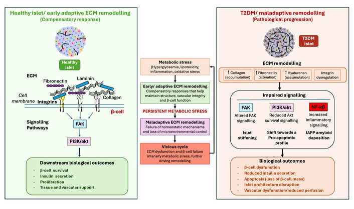
Conceptual framework of stage-dependent ECM remodelling in T2DM. Schematic representation of the proposed transition from physiological/early adaptive ECM remodelling to maladaptive remodelling during persistent metabolic stress. The left panel depicts a healthy islet microenvironment in which organised ECM-integrin interactions support β-cell survival, insulin secretion and tissue/vascular integrity. Persistent metabolic stress, including hyperglycaemia, lipotoxicity, inflammation and oxidative stress, may initially induce compensatory ECM remodelling. When homeostatic mechanisms are overwhelmed, maladaptive remodelling characterised by altered ECM composition, increased stiffness, integrin dysregulation, inflammatory signalling and amyloid deposition may impair β-cell function and islet architecture. Bidirectional interactions between ECM dysfunction and β-cell/metabolic stress may establish a vicious cycle that contributes to disease progression. ECM: extracellular matrix; FAK: focal adhesion kinase; T2DM: type 2 diabetes mellitus.
Within pancreatic islets, remodelling may alter the biochemical composition and mechanical properties of the microenvironment, affecting β-cell-matrix interactions and intracellular signalling [77, 79]. These changes may subsequently influence islet fibrosis, amyloid deposition and vascular function, potentially reinforcing β-cell impairment (Figure 1). The following sections examine how metabolic stress alters specific components and processes of the islet ECM and how these changes affect β-cell and islet function.
Chronic hyperglycaemia is widely recognised as a major driver of ECM remodelling across multiple tissues [78], including the pancreas, although many mechanistic insights derive from non-pancreatic models. Sustained glucose exposure promotes increased synthesis of fibrillar collagens, particularly types I and III [81, 82], enhances fibronectin production [83, 84], induces basement membrane thickening, and activates pro-fibrotic signalling pathways, including TGF-β signalling [85, 86]. Within the pancreas, these processes are associated with fibrosis affecting endocrine and exocrine compartments [87, 88].
In addition to increasing ECM abundance, chronic hyperglycaemia promotes post-translational modification of matrix proteins through non-enzymatic glycation and oxidation [89, 90]. These modifications may alter critical integrin-binding motifs, including the RGD sequence present in fibronectin and other ECM components [91, 92], potentially affecting integrin engagement and focal adhesion formation [93, 94]. Because integrin-mediated signalling is important for β-cell function [95, 96], such changes may influence cell-matrix communication under diabetic conditions. Hyperglycaemia also activates the receptor for advanced glycation end-products (RAGE), which amplifies oxidative stress, inflammatory responses and pro-fibrotic signalling [97, 98]. RAGE activation can further sustain matrix remodelling [99]. Together, these findings suggest that hyperglycaemia can initiate ECM remodelling through coordinated changes in matrix synthesis, biochemical modification and pro-fibrotic signalling, although the extent to which these alterations directly contribute to β-cell dysfunction remains incompletely defined.
In addition to increased matrix synthesis and biochemical modification, impaired ECM degradation may contribute to fibrotic remodelling in T2DM [100]. MMPs, particularly MMP-2 and MMP-9, regulate ECM turnover and are normally controlled by tissue inhibitors of metalloproteinases (TIMPs) to maintain matrix homeostasis [101–105]. In T2DM, inflammatory, oxidative, and metabolic stress may disrupt this balance, resulting in altered matrix turnover and accumulation [106, 107]. Altered MMP activity may also affect islet amyloid homeostasis, as suggested by reduced MMP-9 expression in human T2DM islets [108].
Pathological ECM remodelling is frequently accompanied by altered integrin expression and integrin-mediated signalling [109–113]. Changes in matrix composition and stiffness can modify integrin engagement and downstream signalling pathways involved in metabolic regulation [114, 115]. Evidence from skeletal muscle demonstrates associations between ECM accumulation, altered integrin signalling and insulin resistance [112, 116–118]. Similarly, adipose tissue ECM remodelling has been linked to metabolic dysfunction [119, 120], while increased MMP activity has been associated with metabolic and vascular abnormalities in obesity and T2DM [121]. Experimental studies in diabetic cardiomyopathy further suggest that limiting ECM expansion can improve tissue insulin sensitivity and prevent pathological remodelling [122].
Hyperglycaemia can also alter integrin expression in experimental systems [123, 124], while increased HA deposition has been associated with insulin resistance and its enzymatic degradation can improve insulin signalling [111]. However, much of this evidence derives from peripheral metabolic tissues rather than pancreatic islets. These findings therefore provide mechanistic context for a potential relationship between ECM dysfunction and systemic metabolic disturbance, but direct evidence linking islet ECM remodelling to insulin resistance remains limited.
Chronic low-grade inflammation is a hallmark of T2DM and contributes to both ECM remodelling and β-cell dysfunction. In pancreatic β-cell models, inflammatory cytokines can modulate adhesion-dependent signalling and β-cell function, with TNF-α-associated inflammatory stress linked to β-cell dysfunction, while the effects of IL-1β appear to depend on exposure level and duration [125, 126]. In particular, short-term exposure to low concentrations of IL-1β has been shown to enhance insulin secretion through focal adhesion and actin remodelling, highlighting the context-dependent effects of cytokine signalling on β-cell function [126]. Evidence from non-pancreatic experimental systems further demonstrates that inflammatory cytokines can promote ECM synthesis, matrix remodelling and fibrogenic responses [127–132]. For example, TNF-α and TGF-β regulate the production of fibronectin and collagens [128], while IL-1β and TNF-α can alter MMP/TIMP balance and collagen production [129]. Cytokine activation of stromal and stellate cells may further reinforce fibrogenic responses [130, 131], while TNF-α can promote MMP activation through NF-κB-dependent mechanisms [132].
Although much of the evidence for cytokine-driven ECM remodelling is derived from non-pancreatic systems, these findings provide mechanistic support for a potential link between chronic inflammation and islet ECM dysregulation. Further pancreatic-specific studies are required to determine the extent to which these mechanisms operate in human T2DM islets.
Pathological ECM remodelling can alter the biochemical, structural and mechanical environment in which pancreatic β-cells operate. The consequences extend beyond changes in matrix composition to affect β-cell adhesion and signalling, tissue mechanics, amyloid deposition, inflammatory activity and vascular support. However, the strength of evidence differs between human tissue studies, isolated islets and experimental models [133]. The following sections therefore consider how specific ECM alterations may influence β-cell and islet function, while distinguishing established observations from proposed mechanisms.
Fibrotic matrix accumulation is a prominent feature of islet remodelling in T2DM [87]. Excessive deposition of fibrillar collagens, particularly types I and III, together with increased fibronectin, shifts the ECM away from the basement membrane-associated environment and is associated with increased tissue rigidity and altered endocrine organisation [134–137].
Under physiological conditions, collagen IV supports β-cell adhesion and survival, whereas collagen I is associated with a less supportive matrix environment [138–140]. Islets cultured on collagen I exhibit greater apoptosis than those maintained on collagen IV, while collagen IV can enhance insulin secretion in human β-cells [139, 140]. However, prolonged exposure to collagen IV may also influence insulin gene transcription and intracellular insulin content, indicating that ECM effects are context-dependent [140].
Collagen-integrin interactions provide an additional mechanism through which matrix composition may influence β-cell behaviour. Integrin α3β1, for example, has been associated with enhanced adhesion and insulin secretion in human β-cells cultured on collagen matrices [141]. Conversely, fibrillar collagen I may promote islet amyloid polypeptide (IAPP) aggregation and the formation of cytotoxic intermediates [142]. Because ECM components, including heparan sulfate proteoglycans, can facilitate amyloid formation [143–145], fibrotic remodelling may create a microenvironment that favours amyloid-associated β-cell stress. However, evidence linking these ECM-amyloid interactions specifically to fibrotic remodelling of human diabetic islets remains limited [146].
Laminins contribute to the physiological basement membrane environment that supports β-cell adhesion, survival and functional competence. In particular, laminin-511 signalling through integrin receptors promotes β-cell survival and functional responses [26, 28]. Disruption of this organised basement membrane environment may therefore compromise cell-matrix communication.
In diabetic conditions, biochemical modification and structural remodelling of basement membrane components may alter their interaction with β-cell receptors. Non-enzymatic glycation and AGE formation can modify basement membrane proteins and increase matrix rigidity [147–149], potentially affecting laminin-integrin interactions and downstream signalling [150, 151]. In addition, inflammatory and proteolytic activity may further disrupt basement membrane organisation, although supporting evidence is largely derived from experimental systems [20, 152]. These changes may weaken the spatial and biochemical interactions between β-cells and their surrounding matrix and vasculature.
Importantly, basement membrane thickening and ultrastructural abnormalities have been reported in T2DM [79, 153], but increased ECM deposition does not necessarily indicate improved matrix support. Excessive or disorganised accumulation may produce a denser microenvironment with altered mechanical properties and potentially impair diffusion of oxygen and nutrients [79, 90]. Thus, the functional consequence of laminin remodelling is likely to depend on the composition, organisation and mechanical state of the matrix rather than on total ECM abundance alone.
Fibronectin is an important mediator of cell adhesion and integrin signalling, but its pathological accumulation or structural modification may alter β-cell-matrix interactions. Increased fibronectin deposition has been reported in diabetic tissues and is associated with pancreatic fibrosis and matrix remodelling [154–157]. In the diabetic microenvironment, non-enzymatic glycation and other post-translational modifications may alter fibronectin structure and its interaction with integrins [158–160].
Changes in ECM ligand structure may be particularly relevant because integrin binding depends on the presentation and accessibility of specific adhesion motifs. Modification of RGD-containing ECM proteins can alter integrin affinity, receptor clustering, and focal adhesion formation [93, 94, 160]. Such changes could influence downstream FAK-, PI3K/Akt-, and MAPK/ERK-dependent signalling pathways involved in β-cell survival and insulin secretion [95, 96]. However, much of the mechanistic evidence for these effects derives from experimental systems, and direct evidence demonstrating that fibronectin modification is a primary driver of β-cell failure in human T2DM remains limited.
Altered ECM degradation represents another important consequence of diabetic islet remodelling. MMPs and their endogenous inhibitors, TIMPs, regulate turnover of basement membrane and interstitial ECM components [101–105]. Disruption of this balance can alter matrix organisation and the availability of ECM ligands for cell-surface receptors.
Recent evidence further suggests that stressed β-cells may actively contribute to ECM remodelling rather than acting solely as passive targets of extracellular changes. In type 1 diabetes, cytokine exposure and hyperglycaemia increased β-cell expression of MMP-3, which correlated with degradation of peri-islet collagen IV in both mouse and human islets [20]. Although these findings were obtained in type 1 diabetes, they suggest that β-cell stress itself may promote basement membrane remodelling and provide a potential mechanism relevant to chronic metabolic stress in T2DM.
MMP activity may also influence islet amyloid homeostasis. MMP-9 can degrade human IAPP and thereby limit amyloid formation in experimental systems; notably, MMP-9 expression is reduced in human T2DM islets [108]. Thus, altered proteolytic activity may contribute to both ECM dysregulation and amyloid accumulation, although the direction and causal significance of this relationship remain unclear.
Together, these findings suggest that ECM degradation is not simply a passive process opposing matrix accumulation. Altered proteolytic activity can modify basement membrane integrity, cell-matrix signalling and amyloid handling. Nevertheless, because MMPs have diverse physiological functions, whether their dysregulation represents a primary pathogenic mechanism or a secondary response to diabetic stress remains unclear.
In T2DM, HA turnover becomes dysregulated within the inflammatory and metabolically stressed islet microenvironment [161]. Increased HA accumulation has been reported in skeletal muscle and other metabolic tissues, while circulating HA concentrations are elevated in individuals with T2DM [111, 162]. Emerging pancreatic studies suggest that similar mechanisms may contribute to β-cell dysfunction [161], although direct evidence from human diabetic islets remains limited.
Rather than serving solely as a structural component, excessive HA accumulation may contribute to pathological ECM remodelling. During disease progression, HA degradation by hyaluronidases and oxidative stress can generate low-molecular-weight (LMW) fragments [163]. These fragments can function as damage-associated molecular patterns (DAMPs) that activate toll-like receptors, particularly TLR2 and TLR4, on resident and infiltrating immune cells, promoting pro-inflammatory cytokine production and reinforcing inflammatory signalling [71, 163, 164]. HA-CD44 interactions may further amplify immune recruitment and cytokine release [164], strengthening ECM-immune crosstalk within the islet microenvironment (Figure 2). Consistent with this, studies in diet-induced obese mice have shown that anti-CD44 antibody treatment reduced hyperglycaemia, improved insulin resistance and attenuated adipose inflammation and hepatic steatosis [165]. These findings provide mechanistic support for a role of CD44-mediated inflammatory signalling in metabolic disease, although their relevance to pancreatic islets remains to be established.
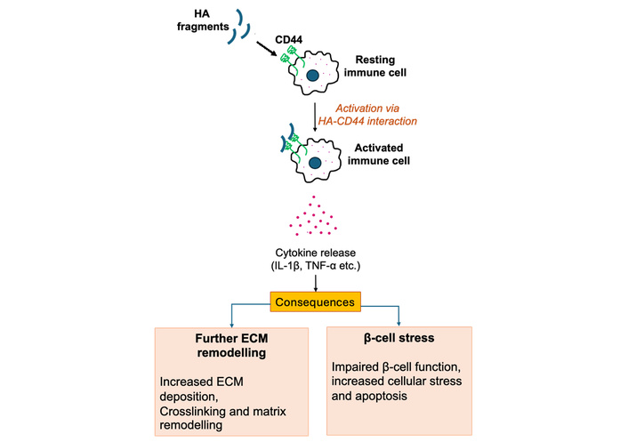
ECM-immune crosstalk model. Proposed HA-CD44-mediated mechanism based on experimental and non-pancreatic evidence. HA fragments, generated during metabolic stress, bind to the CD44 receptor on immune cells. This interaction triggers immune cell activation and the subsequent release of pro-inflammatory cytokines, specifically IL-1β and TNF-α. These inflammatory mediators drive a proposed pathological feedback loop resulting in further ECM remodelling and chronic β-cell stress, contributing to the progressive decline of islet function. CD44: cluster of differentiation 44; ECM: extracellular matrix; HA: hyaluronan.
Functional studies in β-cell models demonstrated that CD44 variant (CD44v) suppresses insulin biosynthesis by inhibiting L-type amino acid transporter 1 (LAT1)-mediated uptake of neutral amino acids required for proinsulin production, whereas CD44 depletion enhances insulin secretion and intracellular insulin stores [166]. These findings suggest that CD44 signalling may influence β-cell function in addition to its role in inflammatory pathways.
HA may also influence the physical properties of the islet microenvironment [161]. Alterations in HA abundance and molecular composition may influence vascular integrity and endothelial barrier function [167], potentially contributing to changes in the islet microenvironment. Changes in the physical properties of the extracellular environment may, in turn, affect calcium dynamics and insulin secretory responses [19]. Together, these findings suggest that HA accumulation may link ECM remodelling with inflammatory signalling and altered tissue mechanics, although the relative contribution of these mechanisms to human T2DM remains uncertain.
Importantly, experimental evidence suggests that HA-CD44 signalling can contribute to β-cell loss in diabetic mouse models. In obese diabetic mice, inhibition of HA synthesis with 4-methylumbelliferone (4-MU) or genetic deletion of CD44 preserved β-cell mass, maintained insulin production, and improved glycaemic control [168]. Mechanistically, HA-CD44 signalling was associated with suppression of the pro-survival factor survivin and increased susceptibility to caspase-3-mediated apoptosis [168]. These findings identify the HA-CD44 axis as a potential therapeutic target, but whether modulation of this pathway directly improves ECM homeostasis or β-cell function in human T2DM remains to be determined.
Fibrotic remodelling is associated with increased ECM stiffness, altering the mechanical properties of the islet microenvironment. Matrix stiffening is increasingly recognised as an important regulator of mechanotransduction through integrin-mediated focal adhesion signalling, although much of the mechanistic evidence has been derived from studies of pancreatic fibrosis, pancreatic cancer and other experimental systems rather than pancreatic islets [64]. Under physiological conditions, the pancreas is a relatively soft tissue [169, 170]; however, diabetes-associated ECM remodelling and increased matrix deposition may alter the mechanical properties of the islet microenvironment [19, 147, 171].
Emerging pancreatic studies indicate that β-cells sense mechanical signals primarily through integrin β1 [57], which links ECM ligands to the intracellular cytoskeleton. In experimental systems, increased matrix rigidity can promote integrin clustering and focal adhesion signalling, including activation of FAK and downstream PI3K/Akt and MAPK/ERK pathways [57, 66–68, 172, 173]. Recent studies using biomimetic three-dimensional scaffolds further demonstrated that increased matrix stiffness impairs glucose-stimulated insulin secretion in both mouse and human islets through activation of the mechanosensitive ion channel piezo-type mechanosensitive ion channel component 1 (Piezo1) and disruption of intracellular Ca2+ dynamics, providing experimental evidence that altered mechanical properties of the islet microenvironment can impair β-cell function [19]. These pathways regulate cytoskeletal organisation, gene expression and cell survival. Because glucose-stimulated insulin secretion depends on tightly regulated cytoskeletal remodelling and vesicle trafficking [174, 175], disruption of mechanical signalling can directly impair insulin granule mobilisation. Changes in actin dynamics, focal adhesion turnover and intracellular tension may interfere with vesicle docking and exocytosis [170, 174, 175]. In addition, mechanosensitive ion channels, including Piezo1, contribute to calcium influx and membrane excitability [19, 176], further linking ECM stiffness to β-cell dysfunction (Figure 3). Thus, excessive matrix stiffening in T2DM may impair insulin secretion through dysregulated mechanotransduction in addition to inflammatory and metabolic stress pathways.
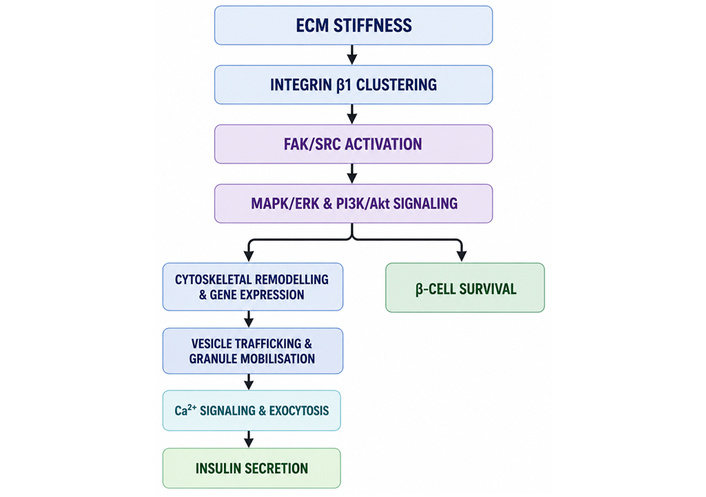
ECM stiffness-integrin signalling pathway and its impact on β-cell function. Schematic representation of proposed mechanotransductive signalling pathways activated in pancreatic β-cells in response to ECM stiffening. Increased ECM stiffness may promote integrin β1 clustering and focal adhesion formation, leading to activation of the FAK/SRC complex and downstream MAPK/ERK and PI3K/Akt signaling pathways. These pathways influence cytoskeletal remodelling, gene expression and β-cell survival, while coordinating vesicle trafficking, granule mobilisation, Ca2+-dependent exocytosis and insulin secretion. ECM: extracellular matrix; ERK: extracellular signal-regulated kinase; FAK: focal adhesion kinase; MAPK: mitogen-activated protein kinase; PI3K: phosphatidylinositol 3-kinase.
Importantly, the relationship between matrix stiffness and β-cell function is not necessarily linear. Cellular responses may depend on the magnitude and duration of mechanical stimulation, ECM composition, and cellular context. Evidence from human and mouse islet systems [19] provides mechanistic support for a role of integrin-dependent mechanotransduction in β-cell responses, but whether pathological stiffening is a primary driver of β-cell failure in human T2DM remains unresolved.
Pancreatic islets are highly vascularised [177], and the vascular basement membrane, composed mainly of laminin, collagen IV and heparan sulfate proteoglycans, contributes to endothelial integrity and β-cell function [10, 11, 27, 178]. In T2DM, alterations in vascular structure and function may disrupt this specialised niche. Human studies have reported differences in islet vascular organisation in T2DM [179], while experimental studies indicate that diabetes-associated vascular ECM changes can impair endothelial function and oxygen delivery [100, 180, 181]. Reduced perfusion may therefore increase metabolic stress and compromise β-cell function and survival [182].
Vascular and ECM remodelling are closely interconnected, as changes in basement membrane composition may alter endothelial-β-cell communication, while endothelial dysfunction can further influence the local matrix environment [10, 100, 181]. Experimental evidence also links endothelial activation and oxidative stress with islet dysfunction [183]. However, most mechanistic evidence remains derived from experimental models, and the extent to which vascular ECM remodelling contributes directly to β-cell failure in human T2DM remains uncertain. Further studies using well-characterised human islets are therefore needed to establish the causal relationships between vascular dysfunction, ECM remodelling and β-cell decline.
Although increasing evidence implicates ECM remodelling in pancreatic islet dysfunction, direct evidence from human T2DM remains comparatively limited. Much of our current understanding is derived from rodent models, isolated islets and ex vivo experimental systems, which have been instrumental in defining ECM composition, integrin signalling and mechanotransductive pathways [19, 53, 57–59, 133]. Representative studies investigating ECM components in human pancreatic islets are summarised in Table 1, providing an overview of the experimental models and supporting evidence available from human studies. In contrast, studies investigating ECM remodelling in human pancreatic tissue are relatively scarce and are often restricted to end-stage disease samples, limiting insight into the evolution of ECM changes during diabetes progression [133, 180, 182].
Existing human studies demonstrate several features consistent with pathological ECM remodelling, including peri-islet fibrosis, basement membrane thickening, altered collagen and fibronectin deposition, islet amyloid accumulation and microvascular abnormalities [87, 88, 133, 153–155, 180, 182]. These findings support the concept that disruption of the islet microenvironment is closely associated with β-cell dysfunction in T2DM. However, whether these alterations actively contribute to disease progression or primarily represent adaptive responses to chronic metabolic stress and β-cell injury remains unresolved. Furthermore, many mechanistic insights into ECM-dependent signalling pathways, including HA-CD44 signalling, integrin-mediated mechanotransduction, and matrix regulation of β-cell function, have largely been established in experimental models and require validation in human tissues [19, 57–59, 168, 171]. Thus, the available evidence ranges from associative observations in human tissues to mechanistic studies in isolated human islets and experimental systems, with stronger causal evidence predominantly derived from animal models.
Human studies are further complicated by several technical and biological challenges. Access to well-characterised pancreatic tissue representing different stages of T2DM is limited, and donor heterogeneity can introduce considerable variability, making it difficult to distinguish primary disease mechanisms from secondary tissue remodelling [180, 182]. In addition, the complex cellular architecture of the pancreas and the limited availability of longitudinal tissue samples restrict assessment of disease progression. Importantly, mechanistic insights into immune-mediated ECM remodelling have been more extensively investigated in type 1 diabetes, where inflammatory infiltration is more pronounced, whereas comparable studies in human T2DM remain relatively limited [20, 46, 47, 161].
Species differences further complicate translation of experimental findings to human T2DM. Although rodent models have been valuable for defining ECM-dependent mechanisms, differences in islet architecture, cellular organisation and ECM composition between rodent and human islets may influence β-cell responses to matrix cues [18, 19, 26–28]. Conversely, studies using isolated human islets provide greater translational relevance but may be affected by donor variability, isolation-associated disruption of cell-ECM interactions and loss of the native vascular and stromal context [61]. Experimental findings should therefore be interpreted as providing mechanistic insight rather than direct representations of human T2DM pathology.
Emerging technologies, including single-cell and spatial transcriptomics, spatial proteomics and advanced imaging approaches, now provide unprecedented opportunities to characterise the human islet ECM at cellular resolution [184–187]. Integration of these approaches with well-phenotyped human tissue cohorts, including resources such as the Human Pancreas Analysis Program (HPAP) [188], will be essential to define the molecular composition of the diabetic islet microenvironment, establish when ECM remodelling occurs during disease progression, and determine whether ECM alterations represent adaptive responses, downstream consequences of metabolic stress or active contributors to β-cell failure. Such studies will be critical for translating experimental findings into clinically relevant therapeutic strategies.
Given the potential contribution of ECM remodelling to islet fibrosis, altered mechanotransduction, inflammatory signalling, amyloid deposition and vascular dysfunction, targeting matrix-associated pathways represents a potential complementary strategy to conventional glucose-lowering treatments [189] (Figure 4). However, most ECM-directed therapeutic evidence is derived from non-pancreatic tissues or preclinical models, with relatively few studies directly investigating the diabetic islet ECM. Evidence for direct therapeutic modification of the human diabetic islet ECM is currently limited. Importantly, because ECM remodelling may represent both an adaptive response and a maladaptive process depending on disease stage, therapeutic strategies should aim to restore matrix homeostasis rather than broadly suppress ECM synthesis or signalling.
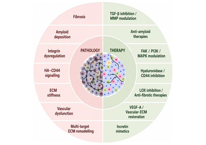
Potential therapeutic strategies targeting pathological ECM remodelling in pancreatic islets during T2DM. Schematic overview of major ECM alterations associated with T2DM, including fibrosis, amyloid deposition, dysregulated integrin signalling, HA-CD44-mediated inflammatory signalling, ECM stiffening and vascular dysfunction, together with potential therapeutic strategies targeting these processes. These include anti-fibrotic and MMP-modulating approaches, anti-amyloid strategies, modulation of integrin and mechanotransduction pathways, inhibition of HA-CD44 signalling, approaches targeting matrix stiffness and vascular-ECM interactions, and incretin-based therapies associated with changes in ECM-related pathways in preclinical models. Most approaches remain experimental or preclinical, and direct restoration of pancreatic islet ECM homeostasis in human T2DM has not yet been established. CD44: cluster of differentiation 44; ECM: extracellular matrix; HA: hyaluronan; T2DM: type 2 diabetes mellitus.
Given the involvement of fibrosis in diabetic islet remodelling, several therapeutic strategies have been proposed to limit excessive ECM deposition. Pancreatic stellate cells are an important source of ECM components during pancreatic fibrosis [190], and their activation can be promoted by oxidative and inflammatory stimuli [80, 132, 191, 192]. These observations provide a rationale for targeting fibrogenic pathways; however, the extent to which stellate-cell activation directly drives β-cell dysfunction during human T2DM remains incompletely defined.
TGF-β signalling represents one potential therapeutic target because of its established role in fibrogenesis and ECM deposition. Experimental studies in pulmonary fibrosis have shown that attenuation of TGF-β-associated fibrogenic responses can reduce fibroblast proliferation and ECM deposition, while fibroblast-specific inhibition of TGF-β1 signalling has been shown to attenuate fibrosis [193, 194]. TGF-β signalling has also been implicated in fibrosis and matrix remodelling in cardiovascular disease [195]. Similarly, studies of metabolic disease suggest that limiting pathological ECM expansion can improve tissue function and insulin sensitivity in other organs [111, 113]. However, these findings provide indirect support for pancreatic applications, and evidence that pharmacological inhibition of fibrogenic pathways directly preserves β-cell function through reduction of islet fibrosis remains limited.
In addition, the interaction between fibrillar collagen and IAPP provides a potential link between matrix remodelling and amyloid-associated β-cell toxicity [142]. Strategies aimed at modifying ECM-amyloid interactions or enhancing amyloid clearance may therefore have therapeutic potential, although evidence from pancreatic IAPP/T2DM models remains limited [142, 196, 197]. Furthermore, modulation of ECM turnover represents another potential therapeutic approach. MMPs and their endogenous tissue inhibitors regulate matrix degradation and remodelling [198–200], and altered MMP activity has been implicated in metabolic and fibrotic disease [108, 112, 199, 201]. In addition, MMPs and other matrix-regulating enzymes have important physiological functions, making selective modulation of ECM turnover challenging.
Overall, antifibrotic strategies provide a mechanistic rationale for restoring a more supportive islet microenvironment, but whether reducing established islet fibrosis improves β-cell function in human T2DM remains to be determined.
Integrin-mediated signalling provides an important interface between ECM composition, cell adhesion and intracellular signalling in β-cells [58]. Experimental modulation of downstream pathways, including FAK, PI3K/Akt and MAPK, can alter cellular responses to matrix interactions [202, 203]. Studies in other fibrotic tissues have also shown that modulation of integrin-associated pathways can influence fibrosis and inflammatory signalling [110, 113, 204]. These findings support further investigation of integrin signalling as a potential therapeutic target in diabetic islets. However, integrins are also essential for normal β-cell adhesion, survival and function; therefore, broad inhibition may disrupt physiological cell-matrix interactions. Therapeutic approaches will likely require selective modulation of pathological signalling while preserving normal integrin activity.
Changes in ECM stiffness can influence integrin-dependent mechanotransduction and cellular behaviour [205]. Experimental studies in fibrotic tissues have investigated approaches targeting collagen crosslinking, including inhibition of lysyl oxidase (LOX)-like enzymes [206], as well as antifibrotic agents such as pirfenidone [207]. Modulation of mechanosensitive signalling pathways, including FAK-associated pathways, has also been proposed as a means of altering pathological responses to mechanical stress [208, 209].
These approaches highlight matrix mechanics as a potential therapeutic interface between ECM remodelling and cellular function. However, matrix stiffness is not intrinsically pathogenic, and β-cell responses depend on the magnitude, duration, and biochemical context of mechanical cues. Consequently, strategies aimed at normalising tissue mechanics rather than simply reducing stiffness may be more appropriate. Whether pharmacological modification of islet stiffness can improve β-cell function in human T2DM remains unknown.
The HA-CD44 axis represents another potential therapeutic target because altered HA accumulation and CD44 signalling have been associated with inflammatory and metabolic dysfunction [210]. Experimental approaches targeting HA turnover or CD44 signalling have been investigated in metabolic disease models [165, 211, 212], while pancreatic studies suggest that inhibition of HA synthesis can influence β-cell survival [168]. Inhibition of HA synthesis with 4-MU preserved β-cell mass and improved glycaemic control in obese diabetic mice [168]. These findings support a potential role for excessive HA accumulation in β-cell injury, although they do not establish that HA modulation directly reverses ECM pathology in human T2DM. Other experimental approaches, including hyaluronidase inhibitors such as glycyrrhizin and hyaluromycin, have been investigated in relation to HA degradation [213, 214], while anti-CD44 treatment improved metabolic abnormalities in diet-induced obese mice [165]. However, these studies were largely performed in non-pancreatic or preclinical systems, and their relevance to human diabetic islet ECM remains uncertain.
Thus, targeting HA-CD44 signalling may represent a potential strategy for modifying ECM-associated inflammatory responses, but further studies are required to determine whether selective modulation of this pathway can safely restore pancreatic ECM homeostasis and improve β-cell function.
Because ECM organisation and vascular integrity are closely interconnected within the islet, strategies that improve vascular function may indirectly support the islet microenvironment. Controlled local VEGF delivery has improved revascularisation and reduced early islet hypoxia in experimental transplantation models [215]. Similarly, preservation of vascular basement membrane components may support β-cell function and proliferation [26]. These findings suggest that maintaining vascular-ECM interactions could complement approaches directed at matrix remodelling. However, whether restoration of vascular-matrix homeostasis directly reverses ECM pathology in human T2DM remains unclear.
Emerging preclinical evidence suggests that incretin-based therapies may be associated with changes in ECM-associated pathways in diabetic islets (Figure 5). However, whether these effects reflect direct actions on pancreatic ECM biology or occur secondarily to improvements in glycaemic control, body weight and systemic inflammation remains unresolved. Therefore, incretin-based therapies should currently be considered potential modulators of ECM-associated changes rather than established direct ECM-targeting therapies.
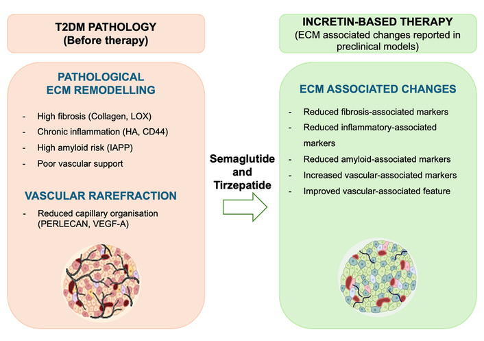
Impact of incretin-based therapies on islet ECM-associated pathology. The left panel depicts pathological ECM remodelling during T2DM, including fibrosis, collagen accumulation, HA-CD44-mediated inflammatory signalling and IAPP-associated amyloid deposition, together with vascular and basement membrane alterations that disrupt the islet microenvironment. The right panel illustrates ECM-associated changes reported following incretin-based therapy in preclinical models, including reductions in fibrosis-associated, inflammatory, and amyloid-associated markers and improvements in vascular-associated markers. CD44: cluster of differentiation 44; ECM: extracellular matrix; HA: hyaluronan; IAPP: islet amyloid polypeptide; LOX: lysyl oxidase; T2DM: type 2 diabetes mellitus.
Cardoso et al. [216] reported that semaglutide was associated with changes in several ECM-related features in diet-induced obese mice. Treatment reduced expression of collagen I, collagen VI, and LOX and altered MMP-2 and MMP-9 activity. Semaglutide also reduced expression of heparanase, syndecan-4, and HA-associated pathways, while increasing perlecan and VEGF-A expression and endothelial labelling [216]. In addition, IAPP and beta-site amyloid precursor protein-cleaving enzyme 2 (BACE2) expression were reduced [216]. Because heparan sulfate proteoglycans can influence IAPP aggregation [217], these findings raise the possibility that semaglutide may influence several interconnected features of the diabetic islet microenvironment.
Similarly, Reis-Barbosa et al. [218] reported that tirzepatide was associated with changes in ECM integrity and vascularisation in a mouse model of obesity, diabetes and oestrogen deficiency. Tirzepatide reduced collagen I, collagen VI, MMP-2, MMP-9, Has2, CD44, IAPP and BACE2 expression, while increasing VEGF-A and basement membrane-associated proteoglycans [218]. These findings are consistent with a shift towards a less fibrotic and better vascularised islet microenvironment, but the study does not establish whether the observed ECM changes resulted from a direct pancreatic action of tirzepatide or from improvements in the systemic metabolic environment.
This distinction is particularly important because chronic hyperglycaemia, obesity and inflammation are themselves associated with ECM remodelling [77, 79, 80, 85, 156]. Improvements in these metabolic stressors could therefore secondarily normalise ECM composition and turnover. Conversely, the changes observed in experimental studies raise the possibility that incretin signalling may also influence ECM-associated pathways, including matrix turnover and basement membrane organisation [216, 218]. At present, the relative contribution of these direct and indirect mechanisms cannot be determined.
Clinical studies demonstrating beneficial effects of semaglutide and tirzepatide on tissue remodelling in organs such as the liver, together with renoprotective effects of semaglutide in patients with T2DM, provide evidence that incretin therapies can influence tissue pathology beyond glycaemic control [219–221]. However, these findings cannot be directly extrapolated to the pancreatic islet ECM, and direct clinical evidence demonstrating restoration of human islet ECM composition, architecture or mechanical properties following incretin treatment is currently lacking.
Future studies combining well-characterised human pancreatic tissue with spatial transcriptomics, spatial proteomics, single-cell analyses and advanced imaging will be important for determining whether ECM remodelling changes following incretin therapy and whether such changes are associated with β-cell recovery. Establishing whether ECM restoration contributes independently to therapeutic response, rather than simply reflecting improved metabolic control, will be essential before ECM modulation can be considered a direct therapeutic mechanism.
Collectively, experimental studies identify several potentially targetable interfaces between ECM remodelling and β-cell dysfunction, including fibrogenic signalling, ECM turnover, integrin-mediated mechanotransduction, HA-CD44 signalling and vascular-matrix interactions. However, the majority of evidence remains preclinical, and the therapeutic window for modifying ECM pathways without disrupting their physiological functions is not yet established. Future therapeutic development should therefore focus on restoring ECM homeostasis and stage-appropriate matrix signalling rather than indiscriminate suppression of ECM components. Importantly, validation in human T2DM tissue and demonstration of a causal relationship between ECM restoration and improved β-cell function will be necessary before these approaches can be translated into clinical practice.
Recent advances in tissue engineering, biomaterials, pancreatic organoids, organoid-on-chip platforms, single-cell and spatial multi-omics, the HPAP, and artificial intelligence-assisted digital pathology are transforming our understanding of pancreatic islet biology and ECM organisation [184–188, 222–224]. Single-cell and spatial transcriptomic and proteomic approaches can resolve cell-specific changes in ECM production, turnover and signalling, while advanced imaging can provide spatial information on ECM organisation, vascular architecture and tissue mechanics within human pancreatic tissue. Organoid, organoid-on-chip and biomaterial-based models may further enable controlled investigation of cell-matrix interactions and mechanotransduction under defined metabolic conditions. Integration of these approaches with functional metabolic analyses and well-characterised clinical cohorts will be important for determining how dynamic changes in the islet microenvironment relate to β-cell dysfunction, disease progression and therapeutic responsiveness.
An important priority is to determine what molecular events govern the transition from potentially adaptive to maladaptive ECM remodelling under persistent metabolic stress. Future studies should determine which ECM changes are protective, which become detrimental, and whether these effects vary according to disease stage, duration of diabetes and individual metabolic characteristics. Particular attention should also be given to the relationship between ECM composition, matrix stiffness, integrin signalling, inflammation, vascular function and amyloid deposition, rather than considering these processes in isolation.
Translating mechanistic discoveries into human disease represents an equally important challenge. Much of the current understanding of pancreatic ECM remodelling is derived from rodent models, isolated human islets or ex vivo systems, and the extent to which these findings reflect human T2DM remains uncertain. Although recent clinical studies suggest that incretin-based therapies can influence tissue remodelling in organs such as the liver and kidney [219–221], direct evidence demonstrating modification of the human pancreatic ECM following treatment is currently lacking. The inability to longitudinally assess pancreatic tissue in vivo remains a major obstacle to determining whether ECM alterations are causes, consequences or adaptive responses to metabolic deterioration. Future studies integrating non-invasive imaging, circulating ECM-derived biomarkers and spatial multi-omics of human pancreatic tissue may help overcome these limitations and identify biomarkers of disease progression and therapeutic response.
Despite substantial progress, several fundamental questions remain unresolved. It is unclear whether ECM remodelling actively contributes to the initiation and progression of β-cell failure or predominantly reflects established metabolic and cellular injury. The extent to which ECM responses differ between human and animal islets, and among individuals with different stages and phenotypes of T2DM, also requires further investigation. Importantly, it remains unknown whether therapeutically restoring ECM composition or mechanical homeostasis is sufficient to improve β-cell function, or whether ECM changes must be targeted together with metabolic and inflammatory pathways. Addressing these questions will require longitudinal studies that combine spatial and single-cell measurements with functional analyses in clinically characterised human tissue and experimental models that permit controlled manipulation of the ECM.
Finally, increasing evidence suggests that recreating aspects of the native pancreatic microenvironment may itself represent a therapeutic strategy. Biomaterial-based approaches incorporating pancreatic ECM components into three-dimensional culture systems have already demonstrated improved human islet survival, stress resilience and glucose responsiveness ex vivo [21]. Together with emerging approaches targeting mechanotransduction, ECM crosslinking, matrix turnover and HA signalling [206, 208–214], these findings highlight the potential of manipulating the islet microenvironment. However, translation of such approaches will require demonstration of efficacy, safety and reproducibility in human systems before ECM-directed strategies can be considered viable disease-modifying therapies.
The ECM is increasingly recognised as an active regulator of pancreatic islet biology rather than a passive structural scaffold. Dynamic interactions between ECM components, integrin-mediated signalling, mechanotransduction and inflammatory pathways play important roles in maintaining β-cell survival, function and adaptive capacity. In T2DM, alterations in ECM composition, organisation and mechanical properties are associated with fibrosis, vascular dysfunction, altered cell-matrix signalling and progressive β-cell impairment.
Current evidence suggests that ECM remodelling is unlikely to represent a purely pathogenic process but may initially reflect an adaptive response to metabolic stress that becomes maladaptive when homeostatic capacity is exceeded. Thus, ECM remodelling may represent both a consequence of metabolic and cellular stress and, once maladaptive changes are established, a potential contributor to β-cell dysfunction. However, the causal relationships between these processes remain incompletely defined.
Although considerable advances have been made in understanding islet ECM biology, important questions remain regarding the stage-dependent regulation of ECM remodelling, differences between human and experimental islets, its causal contribution to β-cell failure and its influence on therapeutic responsiveness. Emerging preclinical evidence that incretin-based therapies are associated with changes in ECM-related pathways provides a rationale for further investigation, but whether these effects reflect direct ECM modulation or secondary improvements in metabolic control remains unresolved. Continued integration of spatial and single-cell technologies with mechanistic studies and well-characterised human tissue will be essential for determining whether restoration of ECM homeostasis can contribute to disease modification and preservation of β-cell function in T2DM.
4-MU: 4-methylumbelliferone
BACE2: beta-site amyloid precursor protein-cleaving enzyme 2
CD44: cluster of differentiation 44
CTGF: connective tissue growth factor
ECM: extracellular matrix
ERK: extracellular signal-regulated kinase
FAK: focal adhesion kinase
HA: hyaluronan
HPAP: Human Pancreas Analysis Program
IAPP: islet amyloid polypeptide
LOX: lysyl oxidase
MAPK: mitogen-activated protein kinase
MMP: matrix metalloproteinase
PI3K: phosphatidylinositol 3-kinase
Piezo1: piezo-type mechanosensitive ion channel component 1
RAGE: receptor for advanced glycation end-products
RGD: arginine-glycine-aspartate
T2DM: type 2 diabetes mellitus
TGF-β: transforming growth factor beta
TIMPs: tissue inhibitors of metalloproteinase
VEGF-A: vascular endothelial growth factor A
During the preparation of this work, the authors used Microsoft PowerPoint to create and finalise all figures and take full responsibility for their final content.
IA: Writing—original draft, Investigation, Visualization, Writing—review & editing. AH: Conceptualization, Investigation, Visualization, Writing—review & editing, Supervision. Both authors read and approved the submitted version.
The authors declare that they have no conflicts of interest.
Not applicable.
Not applicable.
Not applicable.
Not applicable.
Not applicable.
© The Author(s) 2026.
Open Exploration maintains a neutral stance on jurisdictional claims in published institutional affiliations and maps. All opinions expressed in this article are the personal views of the author(s) and do not represent the stance of the editorial team or the publisher.
Copyright: © The Author(s) 2026. This is an Open Access article licensed under a Creative Commons Attribution 4.0 International License (https://creativecommons.org/licenses/by/4.0/), which permits unrestricted use, sharing, adaptation, distribution and reproduction in any medium or format, for any purpose, even commercially, as long as you give appropriate credit to the original author(s) and the source, provide a link to the Creative Commons license, and indicate if changes were made.



View: 41
Download: 3
Times Cited: 0
Huiling Huang ... Dongsheng Li
Huiling Huang ... Dongsheng Li
Dorothy E. Vatner ... Stephen F. Vatner
Maria-Kalliopi Spanorriga ... Konstantinos Tsioufis
Ananyaa Sridhar ... Dawood Khan
Christina Dias ... Nirav Thosani


