 Case Report


Affiliation:
1Department of Cardiology, Kocaeli City Hospital, Kocaeli 41060, Türkiye
Email: dumanahmetberk@gmail.com
ORCID: https://orcid.org/0009-0008-1678-4272


 ,
Affiliation:
2Department of Internal Medicine, Altınova State Hospital, Yalova 77700, Türkiye
ORCID: https://orcid.org/0000-0001-7918-2334
,
Affiliation:
1Department of Cardiology, Kocaeli City Hospital, Kocaeli 41060, Türkiye
ORCID: https://orcid.org/0009-0000-2432-1106
,
Affiliation:
1Department of Cardiology, Kocaeli City Hospital, Kocaeli 41060, Türkiye
ORCID: https://orcid.org/0009-0008-2463-9622
,
Affiliation:
1Department of Cardiology, Kocaeli City Hospital, Kocaeli 41060, Türkiye
ORCID: https://orcid.org/0009-0003-0635-3915
Explor Endocr Metab Dis. 2026;3:101485 DOI: https://doi.org/10.37349/eemd.2026.101485
Received: July 27, 2026 Accepted: September 14, 2026 Published: November 03, 2026
Academic Editor: Per-Olof Hasselgren, Harvard Medical School, USA
Advanced atrioventricular (AV) block can accompany severe hypothyroidism, but thyroid dysfunction does not by itself establish reversibility. A 75-year-old man presented approximately 30 minutes after the onset of dizziness and general deterioration. At the initial documented assessment, temperature was 36.6°C, Glasgow Coma Scale score was 15, oxygen saturation was 98% on room air, and blood pressure was 100/60 mmHg while receiving norepinephrine at 0.05 µg/kg/min. The admission electrocardiogram (ECG) showed complete AV block with an atrial rate of approximately 41–42 beats/min and a markedly slow, wide-complex ventricular escape at approximately 13–14 beats/min. Temporary transvenous pacing increased ventricular rate to approximately 80 beats/min, and coronary angiography showed a dominant right coronary artery, non-obstructive plaques, no culprit lesion, and TIMI 3 flow in all major epicardial vessels. Despite effective rate correction, progressive haemodynamic, respiratory and renal deterioration could no longer be explained by the AV block alone, prompting evaluation for an alternative systemic cause. As respiratory failure progressed, SpO2 fell into the 30% range. At controlled endotracheal intubation, blood pressure was 90/60 mmHg during norepinephrine infusion at 0.2 µg/kg/min, while the temporary pacemaker continued to provide ventricular pacing. Cardiac arrest occurred shortly afterwards, with return of spontaneous circulation after approximately 40 minutes of cardiopulmonary resuscitation. After resuscitation, the previously requested thyroid-function results became available and demonstrated severe primary hypothyroidism (TSH 106 mIU/L, fT3 1.18 pg/mL, fT4 0.38 ng/dL); further history revealed approximately one month of levothyroxine non-adherence. The evolving multisystem syndrome yielded a score of 120 under the 2026 joint-consensus adaptation of the Popoveniuc system, highly suggestive of myxoedema coma. Intravenous hydrocortisone and levothyroxine were initiated. Refractory shock and multiorgan failure persisted, and the patient died on day 3. This case highlights that restoring ventricular rate may not correct a concurrent systemic endocrine crisis and that thyroid dysfunction alone does not prove AV-block reversibility.
Thyroid hormone influences cardiac automaticity, conduction, contractility, and vascular tone at several levels. Two primary rodent studies directly demonstrate thyroid-hormone regulation of the HCN2 pacemaker-channel transcript and the voltage-gated potassium channel Kv1.5 in the rat heart [1, 2], while recent mechanistic and cardiovascular reviews integrate these findings with effects on calcium handling, excitation-contraction coupling, adrenergic responsiveness, and the broader arrhythmic phenotype of hypothyroidism [3, 4]. Severe hormone deficiency can therefore produce bradycardia, low-voltage electrocardiographic complexes, delayed relaxation and impaired cardiovascular reserve; advanced atrioventricular (AV) block remains uncommon but is well described.
The reversibility of AV block in this setting is more difficult to define than the biochemical diagnosis of hypothyroidism. Thyroid-hormone deficiency produces potentially reversible functional electrophysiological changes, including altered pacemaker- and potassium-channel expression, reduced beta-adrenergic responsiveness and autonomic dysregulation; the specific sympathovagal pattern reported in hypothyroidism is heterogeneous [3, 4]. Severe hypothyroidism has also been associated with interstitial oedema and mucinous or mucopolysaccharide myocardial change, while more extensive fibrosis may limit recovery. By contrast, established fibrosis or calcific degeneration of the His-Purkinje system represents a fixed anatomical substrate [5]. Because histological examination of the conduction axis is rarely available during life and was not performed in this patient, these mechanisms provide an explanatory framework rather than patient-specific proof. Degenerative AV block becomes more common with age and with comorbid hypertension and diabetes, and the anatomical level of block cannot always be inferred reliably from the surface electrocardiogram (ECG) alone [5]. A narrow, stable junctional escape may favour a more proximal block, whereas a markedly wide escape complex raises concern for more distal conduction-system disease but is not by itself diagnostic.
Major pacing guidelines agree that potentially reversible causes should be identified and treated, but neither provides a thyroid-specific observation interval before permanent pacemaker implantation [5, 6]. The 2018 ACC/AHA/HRS guideline specifically notes that severe thyroid disease such as myxoedema can rarely be associated with reversible AV block, while also emphasizing that lesser thyroid-function abnormalities often do not eliminate the need for permanent pacing [5].
The clinical literature is similarly heterogeneous. Complete AV conduction recovered after thyroid replacement in individual case reports [7, 8], whereas a retrospective series of 50 patients with thyroid dysfunction and advanced bradyarrhythmia found that 44 received a permanent pacemaker and that thyroid dysfunction was considered unrelated to the AV block in 80% [9]. In that cohort, some conduction recovery occurred only after more than 21 days and after a pacemaker had already been implanted [9]. No evidence-based minimum period of observation on temporary pacing has therefore been defined for thyroid-associated AV block, and the reported time to recovery ranges from a few days to several weeks. No thyroid-specific clinical, biochemical, or electrocardiographic predictor of AV-conduction recovery has been validated. Broader AV-block cohorts have reported heterogeneous associations with recovery or permanent pacing, including younger age, acute coronary syndrome or beta-blocker exposure, absence of aortic-valve calcification, admission blood pressure (BP), sex, and left-ventricular ejection fraction, but these findings were not derived in thyroid-associated AV block and should not be used as a thyroid-specific decision rule [10, 11]. Neither thyroid-stimulating hormone (TSH) magnitude nor escape-QRS width has been prospectively validated as a predictor of recovery in this setting.
Temporary pacing and endocrine treatment address different time horizons rather than competing indications. The 2021 ESC pacing guideline recommends temporary transvenous pacing for haemodynamically compromising bradyarrhythmia refractory to intravenous chronotropic therapy and emphasises that its duration should be minimised because of procedure- and lead-related complications, including perforation, bleeding, malfunction, arrhythmia, accidental displacement, infection and thrombotic complications [6]. In parallel, the 2026 joint consensus statement on myxoedema coma recommends early empiric stress-dose glucocorticoids, intensive supportive care and cautious thyroid-hormone replacement without delaying treatment in a clinically suspected endocrine emergency [12]. Temporary pacing provides immediate rate support, whereas thyroid replacement treats the endocrine substrate but cannot be relied upon for immediate restoration of AV conduction. In an unstable older patient with coronary disease, the two strategies should proceed in parallel, with pacing used as a bridge while cautious endocrine correction is undertaken.
Case reports may over-represent recovery, whereas retrospective cohorts include older patients with competing intrinsic conduction disease and variable durations of observation. A practical assessment therefore requires temporal concordance, exclusion of ischaemic, drug-related and electrolyte causes, review of previous ECGs when available, consideration of the escape-QRS morphology and, when survival permits, direct observation of the rhythm response to thyroid replacement. We report a patient in whom complete AV block was initially treated as an intrinsic or ischaemic conduction emergency, but persistent systemic deterioration despite successful pacing led to recognition of severe hypothyroid decompensation.
The verified clinical sequence is summarised in Table 1. The timeline distinguishes findings present at admission from abnormalities that developed later during the evolving endocrine and multiorgan crisis.
Clinical timeline.
| Relative time | Clinical course |
|---|---|
| At presentation | Symptoms had begun approximately 30 minutes earlier. Complete AV block; bedside ventricular rate approximately 25–30 beats/min. Temperature 36.6°C, GCS 15, SpO2 98% on room air, blood pressure 100/60 mmHg while receiving norepinephrine 0.05 µg/kg/min. No peripheral oedema. |
| Within the first hour | Temporary transvenous pacemaker inserted; ventricular rate increased to approximately 80 beats/min. Coronary angiography performed; dominant RCA, non-obstructive plaques, no culprit lesion, TIMI 3 flow in all major epicardial vessels. |
| Approximately 2 hours | Transthoracic echocardiography: LVEF 40–45%, inferior akinesia, moderate mitral regurgitation, mild aortic and tricuspid regurgitation, PASP approximately 25 mmHg, and a newly documented small posterior pericardial effusion (~5 mm) without tamponade. |
| Following hours | Clinical recovery was incomplete despite pacing. Cold extremities, bilateral 2+ pretibial oedema, pulmonary oedema and progressive renal dysfunction developed. |
| Approximately 6 hours | Persistent systemic deterioration despite effective pacing and absence of an acute coronary culprit could no longer be explained by the presenting AV block alone; thyroid-function testing was requested as part of a search for an alternative systemic cause. |
| Later critical deterioration | SpO2 fell into the 30% range. At controlled endotracheal intubation, blood pressure was 90/60 mmHg during norepinephrine infusion at 0.2 µg/kg/min; the temporary pacemaker continued to provide ventricular pacing. Midazolam and rocuronium bromide were administered as part of the intubation regimen. Venous blood during intubation: pH 7.020, pCO2 77.4 mmHg. Cardiac arrest occurred shortly afterwards; return of spontaneous circulation was achieved after approximately 40 minutes of CPR. A second brief cardiac arrest was subsequently resuscitated. |
| After resuscitation | The previously requested thyroid-function results became available and showed severe primary hypothyroidism (TSH 106 mIU/L, fT3 1.18 pg/mL, fT4 0.38 ng/dL). Integrated with the evolving multisystem dysfunction, myxoedema coma was recognised. Further history disclosed approximately one month of levothyroxine non-adherence. Intravenous hydrocortisone and levothyroxine were initiated. |
| Final ~6 hours/Days 2–3 | Anuria, refractory shock, and progressive multiorgan failure. Temporary pacing continued. Death on hospital day 3. |
AV: atrioventricular; CPR: cardiopulmonary resuscitation; fT3: free triiodothyronine; fT4: free thyroxine; GCS: Glasgow Coma Scale; LVEF: left ventricular ejection fraction; PASP: pulmonary artery systolic pressure; pCO2: partial pressure of carbon dioxide; RCA: right coronary artery; SpO2: peripheral oxygen saturation; TIMI: thrombolysis in myocardial infarction; TSH: thyroid-stimulating hormone.
A 75-year-old man presented to the emergency department approximately 30 minutes after the onset of dizziness and general deterioration. His medical history included diabetes mellitus, hypertension, coronary artery disease, and primary hypothyroidism. He had been prescribed levothyroxine 100 µg/day; the fact that he had not taken it for approximately one month was not known at presentation and was established later during the admission. Medication reconciliation identified no beta-blocker, non-dihydropyridine calcium-channel blocker, digoxin, amiodarone, or ivabradine. No previous ECG was available for reliable comparison.
At the initial documented assessment, body temperature was 36.6°C, BP was 100/60 mmHg while receiving norepinephrine at 0.05 µg/kg/min, respiratory rate was 20–25 breaths/min, and peripheral oxygen saturation (SpO2) was 98% on room air. Glasgow Coma Scale score was 15. Peripheral oedema was absent at that time. Initial venous blood-gas analysis showed mild acidemia without hypercapnia (pH 7.308, partial pressure of carbon dioxide [pCO2] 43.7 mmHg, bicarbonate 21.4 mmol/L, lactate 0.9 mmol/L).
Bedside monitoring showed profound bradycardia at approximately 25–30 beats/min. The admission 12-lead ECG, recorded at 25 mm/s and 10 mm/mV, captured complete AV block with regular atrial activity at approximately 41–42 beats/min and a ventricular escape rhythm at approximately 13–14 beats/min (Figure 1). There was no fixed AV relationship. The escape QRS was markedly wide (> 200 ms) and limb-lead QRS voltages were low.
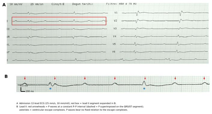
Admission electrocardiogram. (A) Twelve-lead tracing recorded at 25 mm/s and 10 mm/mV showing complete atrioventricular block. (B) Expanded lead II segment: red arrowheads indicate P waves (dashed where superimposed on the QRS/ST segment) and asterisks indicate ventricular escape complexes. Atrial rate was approximately 41–42 beats/min and the ventricular escape rate approximately 13–14 beats/min.
Because the presentation was dominated by haemodynamically significant complete AV block, intrinsic or ischaemic conduction-system disease was initially considered. A temporary transvenous pacemaker was inserted within the first hour and increased the ventricular rate to approximately 80 beats/min. Coronary angiography was performed during the same early period. Although rate correction was effective, the patient’s systemic and haemodynamic condition did not improve as expected.
During the subsequent hours, the extremities became cold, bilateral 2+ pretibial oedema and pulmonary oedema developed, and renal function deteriorated. Despite a paced ventricular rate of approximately 80 beats/min and the absence of an acute coronary culprit, the progressive haemodynamic and respiratory deterioration could no longer be explained by the presenting AV block alone. Thyroid-function testing was therefore requested approximately 6 hours after presentation as part of a search for an alternative systemic and potentially reversible cause.
As respiratory status subsequently worsened, continuous pulse-oximetry monitoring showed that SpO2 fell into the 30% range, consistent with profound hypoxaemia. At that time, BP was 90/60 mmHg while norepinephrine was continued at 0.2 µg/kg/min, and the temporary pacemaker continued to provide ventricular pacing. Controlled endotracheal intubation was therefore performed. Midazolam and rocuronium bromide were administered as part of the intubation regimen. Venous blood obtained during intubation demonstrated severe acidemia and marked hypercapnia (pH 7.020, pCO2 77.4 mmHg, bicarbonate 19.5 mmol/L, base excess –11.4 mmol/L, lactate 2.68 mmol/L). A post-intubation venous sample continued to show severe hypercapnia (pH 7.117, pCO2 78.9 mmHg, bicarbonate 24.9 mmol/L, lactate 3.03 mmol/L). Shortly after intubation, cardiac arrest occurred. Return of spontaneous circulation was achieved after approximately 40 minutes of cardiopulmonary resuscitation. A second brief cardiac arrest was subsequently resuscitated.
Following resuscitation, the previously requested thyroid-function results became available and demonstrated severe primary hypothyroidism (TSH 106 mIU/L, free triiodothyronine [fT3] 1.18 pg/mL, free thyroxine [fT4] 0.38 ng/dL). When integrated with the persistent cardiovascular, respiratory, renal and metabolic deterioration that was not explained by the corrected AV block, the overall syndrome was recognised as myxoedema coma. Further history established that the patient had not taken his prescribed levothyroxine for approximately one month. Intravenous hydrocortisone 100 mg every 8 hours was administered. Intravenous levothyroxine was given as a total first-day loading dose of 300 µg, divided into three 100-µg doses at 8-hour intervals as 30-minute infusions under continuous cardiac monitoring, followed by 50 µg intravenously once daily. Temporary pacing was continued.
Despite intensive supportive and endocrine-directed treatment, shock and multiorgan failure progressed. The patient was anuric during the final approximately 6 hours. He died on hospital day 3.
Coronary angiography showed a patent, dominant right coronary artery. The left anterior descending, left circumflex, and right coronary arteries contained atherosclerotic plaques without significant stenosis or an acute culprit lesion; thrombolysis in myocardial infarction (TIMI) 3 flow was present throughout all major epicardial vessels (Figure 2). In this context, acute coronary occlusion or AV-nodal ischaemia was considered an unlikely explanation for the complete AV block.
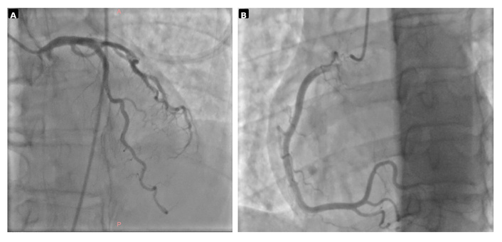
Coronary angiography. (A) Left coronary system and (B) dominant right coronary artery. All major epicardial vessels showed TIMI 3 flow without a significant stenosis or acute culprit lesion. TIMI: thrombolysis in myocardial infarction.
Transthoracic echocardiography approximately 2 hours after presentation showed a left ventricular ejection fraction of 40–45%, inferior wall akinesia, moderate mitral regurgitation, mild aortic and tricuspid regurgitation, an estimated pulmonary artery systolic pressure of approximately 25 mmHg, and a newly documented small posterior pericardial effusion with a maximum depth of approximately 5 mm, without tamponade physiology. The left-ventricular systolic function and inferior wall-motion abnormality were similar to previous echocardiographic findings; no new major structural explanation for the haemodynamic deterioration was identified.
Thyroid-function testing was requested approximately 6 hours after presentation during otherwise unexplained systemic deterioration. The results became available after resuscitation and showed TSH 106 mIU/L, fT3 1.18 pg/mL and fT4 0.38 ng/dL, establishing severe primary hypothyroidism. Serial measurements are shown in Table 2. During profound critical illness, the short-term fall in TSH while fT3 and fT4 remained markedly low could not distinguish inadequate hormone exposure from critical-illness-related, distributional or assay effects. Administration and sampling times, fT4/fT3 trajectories and the clinical response therefore require integrated interpretation.
Serial thyroid-function tests.
| Time point | TSH (mIU/L) | fT3 (pg/mL) | fT4 (ng/dL) |
|---|---|---|---|
| Day 1* | 106 | 1.18 | 0.38 |
| Day 2† | 78 | 0.79 | 0.29 |
| Day 3† | 42 | 0.51 | 0.28 |
*Day 1 sample obtained approximately 6 hours after presentation and before thyroid-hormone replacement. †Day 2 and Day 3 samples were obtained before the daily levothyroxine dose. Local laboratory reference intervals: TSH, 0.27–4.2 mIU/L; fT3, 2.00–4.40 pg/mL; fT4, 0.80–1.76 ng/dL. fT3: free triiodothyronine; fT4: free thyroxine; TSH: thyroid-stimulating hormone.
The admission laboratory profile did not support hyperkalaemia as the cause of the presenting complete AV block: venous-blood-gas potassium was 4.42 mmol/L, while the simultaneous serum potassium of 4.9 mmol/L was flagged as affected by haemolysis. Selected admission laboratory data are summarised in Table 3. A glucose nadir of 45 mg/dL was documented later after intubation, while hyperkalaemia developed only during severe acidosis and multiorgan deterioration.
Selected laboratory findings at presentation.
| Parameter | Admission value | Unit | Notes |
|---|---|---|---|
| Sodium | 140 | mmol/L | - |
| Potassium | 4.42‡ | mmol/L | Venous-blood-gas value |
| Chloride | 108 | mmol/L | - |
| Total calcium | 9.8 | mg/dL | - |
| Magnesium | 2.35 | mg/dL | - |
| Creatinine | 2.13 | mg/dL | - |
| eGFR (CKD-EPI) | 29 | mL/min/1.73 m2 | - |
| BUN | 28 | mg/dL | - |
| Glucose | 69.9 | mg/dL | - |
| Lactate | 0.9 | mmol/L | Venous blood gas |
| hs-Troponin T | 96.6 | ng/L | - |
| NT-proBNP | 8,899 | ng/L | - |
| WBC | 8.51 | × 109/L | - |
| Haemoglobin | 13.0 | g/dL | - |
| Platelets | 232 | × 109/L | - |
| CRP | 1.75 | mg/L | - |
‡ Potassium is the venous-blood-gas value. The simultaneous serum potassium was 4.9 mmol/L but was flagged as affected by haemolysis and was not used for causal assessment of the presenting AV block. BUN: blood urea nitrogen; CKD-EPI: Chronic Kidney Disease Epidemiology Collaboration; CRP: C-reactive protein; eGFR: estimated glomerular filtration rate; hs-Troponin T: high-sensitivity troponin T; NT-proBNP: N-terminal pro-B-type natriuretic peptide; WBC: white blood cell count.
The admission electrocardiographic measurements are summarised in Table 4. Because atrial and ventricular activity were dissociated, a PR interval could not be assigned. The escape complexes were broad and slurred, with an idioventricular configuration that did not conform to a typical right or left bundle-branch-block pattern. Limb-lead QRS voltages were low. No gross frontal-plane axis deviation was apparent; however, a precise numerical axis was not assigned because of the very low limb-lead voltage and the quality of the archived tracing. The QT interval could not be measured reliably because T-wave termination was indistinct and atrial deflections were superimposed; QTc was therefore not reported. No previous ECG was available for comparison.
Admission electrocardiographic features.
| Parameter | Finding |
|---|---|
| Atrial rate | Approximately 41–42 beats/min |
| Ventricular escape rate on admission 12-lead ECG | Approximately 13–14 beats/min |
| Bedside monitored ventricular rate | Approximately 25–30 beats/min |
| AV relationship | Complete AV dissociation/Complete AV block |
| PR interval | Not assignable |
| QRS duration | Markedly wide, > 200 ms |
| Bundle-branch-block morphology | Broad, slurred idioventricular escape complexes; no typical RBBB or LBBB pattern |
| Limb-lead QRS voltage | Low |
| Frontal QRS axis | No gross deviation apparent; precise numerical axis not assigned because of very low limb-lead voltage and archived-tracing quality |
| QT/QTc | QT not reliably measurable because T-wave termination was indistinct and atrial deflections were superimposed; QTc not reported |
| Previous ECG | Not available for reliable comparison |
AV: atrioventricular; ECG: electrocardiogram; LBBB: left bundle-branch block; RBBB: right bundle-branch block.
Myxoedema coma is a life-threatening decompensated hypothyroid state defined clinically by multisystem dysfunction rather than by a single biochemical threshold [12, 13]. The patient was not hypothermic and was fully conscious at presentation, and serum sodium remained normal; this represents an atypical early phenotype. Applying the 2026 joint-consensus adaptation of the Popoveniuc diagnostic system, the score was 120 when all documented features across the evolving clinical course were incorporated; a score of 60 or above is highly suggestive of myxoedema coma [12, 14]. When restricted to findings documented during the first hours after presentation, with the later-established precipitating event and later abnormalities excluded, the score was 70. Table 5 reports the two calculations separately. Hypotension was scored on the basis of vasopressor dependence rather than the absolute blood-pressure value. Because norepinephrine had already been started before the first documented BP, a sensitivity analysis was also performed: excluding the 20 hypotension points yielded an early-presentation score of 50, within the 45–59 range indicating risk, and an evolving-course score of 100, which remained highly suggestive. The score supports but does not independently establish the diagnosis and must be interpreted with the clinical and biochemical findings, particularly because other critical illnesses can produce overlapping features [12, 14].
Clinical findings assessed with the 2026 joint-consensus adaptation of the Popoveniuc diagnostic score for myxoedema coma.
| Domain | Documented finding | Timing | Early score | Evolving score |
|---|---|---|---|---|
| Thermoregulatory dysfunction | Temperature 36.6°C | Presentation | 0 | 0 |
| Central nervous system effects | GCS 15; no qualifying impairment | Presentation | 0 | 0 |
| Gastrointestinal findings | None documented | Clinical course | 0 | 0 |
| Precipitating event | Levothyroxine non-adherence (~1 month) | Established later | 0 | 10 |
| Bradycardia | Heart rate < 40 beats/min | Presentation | 30 | 30 |
| Pericardial/pleural effusion | Small posterior pericardial effusion | ~2 hours | 10 | 10 |
| Pulmonary oedema | Pulmonary oedema documented | Later deterioration | 0 | 10 |
| Cardiomegaly | Not documented | - | 0 | 0 |
| Hypotension | Vasopressor-dependent; BP 100/60 mmHg on norepinephrine | Presentation | 20 | 20 |
| Hyponatraemia | Absent; sodium 140 mmol/L | Presentation | 0 | 0 |
| Hypoglycaemia | Glucose nadir 45 mg/dL | Later | 0 | 10 |
| Hypoxaemia | SpO2 fell into the 30% range | Later | 0 | 10 |
| Hypercarbia | pCO2 77.4 mmHg during intubation | Later | 0 | 10 |
| Decrease in GFR | eGFR 29 mL/min/1.73 m2 | Presentation | 10 | 10 |
| Total | 2026 consensus-adapted score | Separate calculations | 70 | 120 |
The 2026 consensus adaptation of the Popoveniuc system integrates thermoregulatory, neurological, cardiovascular, metabolic and precipitating features [12, 14]. The early-presentation score was 70, and the evolving-course score was 120. Later hypoxaemia, hypercarbia, hypoglycaemia, pulmonary oedema and the precipitating event were included only in the evolving-course calculation. The 2026 adaptation does not include the older ‘other ECG changes’ item and assigns 10 points to pulmonary oedema. Because the first documented BP was obtained during norepinephrine treatment, a sensitivity analysis excluded the 20 hypotension points; the corresponding scores were 50 (risk range, 45–59) and 100 (highly suggestive), respectively. BP: blood pressure; ECG: electrocardiogram; GCS: Glasgow Coma Scale; GFR: glomerular filtration rate; pCO2: partial pressure of carbon dioxide; SpO2: peripheral oxygen saturation.
Because severe hypothyroidism did not by itself prove that the AV block was endocrine-mediated, competing causes were considered systematically (Table 6). Degenerative conduction-system disease remained the principal competing explanation for the block itself.
Differential diagnosis of complete atrioventricular block in this patient.
| Potential cause | Patient-specific evidence | Assessment |
|---|---|---|
| Acute coronary ischaemia | Dominant RCA; no culprit lesion or significant stenosis; TIMI 3 flow in all major epicardial vessels. | Unlikely |
| Rate-limiting drug effect | No beta-blocker, non-dihydropyridine calcium-channel blocker, digoxin, amiodarone or ivabradine. | Unlikely |
| Hyperkalaemia/Electrolyte disturbance | Admission VBG K 4.42 mmol/L; sodium, calcium and magnesium not substantially abnormal. Hyperkalaemia developed only later during severe acidosis. | Not the presenting cause |
| Severe hypothyroidism/Myxoedema coma | TSH 106 mIU/L, fT4 0.38 ng/dL; evolving multiorgan syndrome; 2026 consensus-adapted Popoveniuc score 120, highly suggestive of myxoedema coma. | Strongly supported as a systemic endocrine emergency |
| Degenerative conduction-system disease | Age 75 years, hypertension, diabetes, CAD; markedly wide escape QRS; no previous ECG or EPS. | Cannot be excluded; principal competing cause of the AV block |
| Hypothermia | Temperature 36.6°C at presentation. | Absent |
| Infiltrative disease | No specific diagnostic work-up was undertaken; no direct evidence established during the short course. | Not excluded, not supported |
| Infective/inflammatory AV block | No specific Lyme/sarcoid-directed work-up; initial WBC and CRP were not suggestive of marked systemic inflammation. | Not established |
| Iatrogenic AV block | No preceding cardiac procedure or intervention before presentation. | Unlikely |
| Increased vagal tone / acute neurological cause | No compatible trigger or acute neurological syndrome was documented. | Not supported |
AV: atrioventricular; CAD: coronary artery disease; CRP: C-reactive protein; ECG: electrocardiogram; EPS: electrophysiological study; RCA: right coronary artery; TSH: thyroid-stimulating hormone; TIMI: thrombolysis in myocardial infarction; VBG: venous blood gas.
A patient perspective could not be obtained because of the rapid clinical deterioration and subsequent death.
Severe primary hypothyroidism was unequivocal in this patient. The clinical question was whether the complete AV block could be attributed to thyroid-hormone deficiency and therefore regarded as reversible. This could not be resolved during the short clinical course.
Severe hypothyroidism was clearly present, but the conduction abnormality cannot be assigned to thyroid deficiency with certainty. Thyroid hormone can influence cardiac ion-channel expression and adrenergic responsiveness [1–4], and recovery of complete AV block after replacement has been reported [7, 8]. However, the dedicated thyroid-dysfunction cohort did not establish a validated predictor model: 80% of AV block was judged unrelated to the thyroid abnormality, and 88% of patients underwent permanent pacing [9]. Broader AV-block cohorts have identified clinical associations with recovery or permanent pacing, but these signals are heterogeneous and not thyroid-specific [10, 11]. Neither TSH magnitude nor a particular surface-ECG feature can currently be used as a validated thyroid-specific predictor of reversibility. In this patient, age 75 years, diabetes, hypertension, coronary disease, and the markedly wide escape QRS increased concern for coexisting intrinsic conduction-system disease, but the wide QRS itself is not a validated predictor of non-recovery in thyroid-associated AV block. No prior ECG was available, no electrophysiological study (EPS) was performed to localise the block, and the three-day fatal course did not permit observation of late AV-conduction recovery. Severe hypothyroidism was therefore a major systemic contributor, but its presence does not establish that the complete AV block was reversible.
Acute and long-term pacing decisions should be separated. This patient had haemodynamically significant complete AV block and required immediate temporary pacing. Both major pacing guidelines emphasise treatment of reversible causes, but neither defines a thyroid-specific observation period [5, 6]. The ACC/AHA/HRS guideline notes that severe thyroid disease such as myxoedema can rarely be associated with reversible AV block, while lesser thyroid abnormalities often do not eliminate the need for permanent pacing [5]. The ESC guideline simultaneously cautions that conventional temporary transvenous pacing has substantial procedure- and lead-related risks and should remain in place for as short a period as possible [6]. The available thyroid-specific cohort evidence argues against assuming rapid recovery, because some recovery occurred only after more than 21 days [9]. Temporary pacing therefore provides immediate haemodynamic rate support, while endocrine therapy treats a potentially reversible substrate in parallel. Recovery of AV conduction, when it occurs, has been reported over days to weeks and cannot be assumed to provide immediate rate support. Had the patient survived, the subsequent rhythm course, comorbidity burden, response to endocrine therapy and overall evidence for intrinsic disease would all have informed the permanent-pacemaker decision.
The published evidence is subject to selection bias. Case reports preferentially capture unusual reversibility and cannot estimate its frequency, whereas retrospective cohorts include older patients with competing degenerative disease and heterogeneous definitions of thyroid-related AV block. In addition, clinicians may implant permanent devices before a prolonged endocrine recovery can be observed. The three reports summarised in Table 7 illustrate this problem: two single cases recovered without permanent pacing [7, 8], while the larger cohort showed frequent permanent-pacemaker implantation and uncertainty about causality [9].
Focused comparison of selected reports of advanced AV block associated with thyroid dysfunction.
| Study | Population/Thyroid disorder | Conduction abnormality | Temporary pacing | Permanent pacemaker | Recovery/Interpretation |
|---|---|---|---|---|---|
| Schoenmakers et al., 2008 [7] | 90-year-old woman; severe hypothyroidism | Complete AV block | Yes | No | AV conduction restored; resolution documented at 6 weeks |
| Seol et al., 2012 [8] | 75-year-old man; severe hypothyroidism | Complete AV block | Yes | No | AV conduction recovered by day 4; complete block absent at 4 weeks |
| Ozcan et al., 2012 [9] | Retrospective cohort; 50 patients with thyroid dysfunction | 2nd/3rd-degree AV block or bradyarrhythmia ≤ 40/min | NR | 44/50 | Thyroid dysfunction judged unrelated in 80%; recovery after > 21 days in some patients |
AV: atrioventricular; NR: not reported.
Myxoedema coma is a life-threatening state of decompensated hypothyroidism with multisystem dysfunction, and its phenotype is heterogeneous. A recent systematic survey of 698 cases found that classic findings such as altered mental status and hypothermia were frequent but not universal [13]. This patient was normothermic, fully conscious and normonatraemic at presentation, constituting an atypical early phenotype. Nevertheless, profound bradycardia, vasopressor-dependent hypotension, pericardial effusion and renal dysfunction were present early, and the subsequent course added pulmonary oedema, documented profound hypoxaemia, severe hypercapnia and hypoglycaemia. Under the 2026 joint-consensus adaptation of the Popoveniuc system, the score was 70 from early findings and 120 across the evolving course [12, 14]. After exclusion of the 20 hypotension points because the first documented pressure was obtained during norepinephrine treatment, the corresponding scores were 50, indicating risk, and 100, remaining highly suggestive. These values support the diagnosis but are not interpreted in isolation, because sepsis and major cardiovascular events may produce overlapping high-scoring features [12]. The profound biochemical hypothyroidism, later-established non-adherence and otherwise unexplained multisystem deterioration were therefore considered together with the score. This interpretation is consistent with current consensus guidance, which emphasises early clinical recognition and treatment [12].
The patient was already vasopressor-dependent and had chronic left-ventricular dysfunction, pulmonary oedema, a pericardial effusion and severe hypothyroid decompensation when respiratory status worsened. SpO2 fell into the 30% range. Immediately before controlled endotracheal intubation, BP was 90/60 mmHg while norepinephrine continued at 0.2 µg/kg/min, and the temporary pacemaker continued to provide ventricular pacing. Midazolam and rocuronium bromide were administered as part of the intubation regimen. Shortly after intubation, cardiac arrest occurred. In the INTUBE international cohort, major peri-intubation adverse events were common in critically ill patients, with cardiovascular instability in 42.6% and cardiac arrest in 3.1% [15]. Contemporary reviews describe the transition from spontaneous to positive-pressure ventilation, induction-related sympatholysis/vasodilation, and limited preload reserve as interacting mechanisms of peri-intubation cardiovascular collapse in physiologically fragile patients [16]. The blood-gas sequence documents profound ventilatory failure: the initial venous pCO2 was 43.7 mmHg, whereas the sample obtained during intubation showed pCO2 77.4 mmHg and pH 7.020, with persistent severe hypercapnia after intubation. These data demonstrate marked physiological vulnerability and a temporal relationship but do not establish that intubation itself caused the arrest.
TSH fell from 106 to 42 mIU/L over three days, while fT3 and fT4 declined and remained markedly reduced. Short-interval thyroid measurements during shock, resuscitation and multiorgan failure are difficult to interpret because critical illness can alter thyroid-hormone metabolism and hypothalamic-pituitary-thyroid signalling [17]. Nevertheless, failure of fT4 and fT3 to rise after intravenous treatment warrants review of the administered exposure, dose timing, sampling time and possible distributional or assay effects; current consensus recommends interpreting serial hormone trajectories together with the clinical response and reconsidering treatment when biochemical improvement is absent [12]. The pattern therefore neither proves nor excludes inadequate treatment. The reported 300-µg first-day intravenous levothyroxine dose reflects the regimen actually administered. Current consensus recommends individualising the initial dose and considering 100–200 µg in older patients or those with coronary disease or arrhythmia [12]; this caution is particularly relevant to the present 75-year-old patient with coronary disease and complete AV block. The fatal three-day course precluded assessment of a longer-term endocrine or conduction response.
The principal limitation is that the patient died before the conduction system could be observed after sustained thyroid replacement. No previous ECG was available, no EPS was performed, and no post-mortem conduction-system examination was undertaken. The exact causal contribution of severe hypothyroidism to the complete AV block therefore remains uncertain. The focused literature comparison is also small and includes highly selected case reports. Future prospective data should record the anatomical level of block, escape-QRS width and morphology, baseline ECG status, timing and route of thyroid replacement, duration of temporary pacing and the time to AV-conduction recovery before permanent pacing decisions are made.
Complete AV block can be the dominant presenting abnormality in severe hypothyroid decompensation, but correction of the electrical disturbance does not correct the systemic hypometabolic crisis. In this patient, temporary pacing restored ventricular rate and angiography excluded an acute coronary culprit, yet circulatory, respiratory and renal failure continued to evolve until severe primary hypothyroidism and myxoedema coma were recognised.
The case also illustrates why thyroid status should not be used as a surrogate for conduction-system reversibility. Severe hypothyroidism can coexist with intrinsic degenerative AV block. Older age and a markedly wide escape QRS may raise concern for coexisting intrinsic disease, although neither establishes non-reversibility. Immediate pacing and endocrine treatment should proceed in parallel when the patient is unstable, while permanent pacing, when survival permits, should be based on the observed rhythm course rather than on the TSH value alone. The absence of a baseline ECG, electrophysiological localisation and prolonged rhythm follow-up precludes determination of AV-block reversibility in this patient. Prospective studies should evaluate escape-QRS characteristics, anatomical level of block and a standardised observation interval before permanent pacing.
Complete AV block may dominate the initial presentation of severe hypothyroid decompensation and can obscure the broader systemic diagnosis.
Successful temporary pacing restores ventricular rate but does not reverse the metabolic, respiratory or circulatory consequences of myxoedema coma.
Myxoedema coma may evolve without hypothermia, hyponatraemia or coma at presentation; diagnosis should integrate the full multisystem clinical course.
Thyroid dysfunction does not prove that advanced AV block is reversible; long-term pacing decisions should be based on the subsequent conduction course and competing evidence for intrinsic disease.
AV: atrioventricular
BP: blood pressure
ECG: electrocardiogram
EPS: electrophysiological study
fT3: free triiodothyronine
fT4: free thyroxine
pCO2: partial pressure of carbon dioxide
SpO2: peripheral oxygen saturation
TSH: thyroid-stimulating hormone
During the preparation of this work, the authors used ChatGPT (OpenAI) and Antigravity to assist with language refinement and editorial organisation. These tools were not used to make clinical decisions or determine the scientific conclusions. The authors independently reviewed and verified the clinical data, analyses, figure annotations, and references, edited the final text, and take full responsibility for the manuscript.
ABD: Conceptualization, Data curation, Writing—original draft, Writing—review & editing. HG: Data curation, Writing—review & editing. TE: Investigation, Writing—review & editing. SB: Investigation, Writing—review & editing. DK: Supervision, Writing—review & editing. All authors read and approved the submitted version.
The authors declare that they have no conflicts of interest.
This case report was prepared in accordance with the Declaration of Helsinki. All clinical information was obtained during routine clinical care and was de-identified. Under the institutional policy applicable at Kocaeli City Hospital, formal ethics committee approval is not required for publication of a single anonymised case report.
As the patient died during the admission, written informed consent to participate was obtained from the patient’s next of kin on his behalf.
As the patient died during the admission, written informed consent for publication of the anonymised case details and images was obtained from the patient’s next of kin.
The de-identified clinical, laboratory, electrocardiographic and imaging data supporting this case report are available from the corresponding author upon reasonable request, subject to institutional approval and patient-privacy restrictions.
This research received no external funding.
© The Author(s) 2026.
Open Exploration maintains a neutral stance on jurisdictional claims in published institutional affiliations and maps. All opinions expressed in this article are the personal views of the author(s) and do not represent the stance of the editorial team or the publisher.
Copyright: © The Author(s) 2026. This is an Open Access article licensed under a Creative Commons Attribution 4.0 International License (https://creativecommons.org/licenses/by/4.0/), which permits unrestricted use, sharing, adaptation, distribution and reproduction in any medium or format, for any purpose, even commercially, as long as you give appropriate credit to the original author(s) and the source, provide a link to the Creative Commons license, and indicate if changes were made.



View: 65
Download: 7
Times Cited: 0


