 Review


Affiliation:
1Department of Internal Medicine, The University of Texas Health Science Center at Houston, Houston, TX 77030, USA
ORCID: https://orcid.org/0000-0002-4553-1847

 ,
,
Affiliation:
1Department of Internal Medicine, The University of Texas Health Science Center at Houston, Houston, TX 77030, USA
Affiliation:
2Department of Gastroenterology, Hepatology and Nutrition, The University of Texas Health Science Center at Houston, Houston, TX 77030, USA
ORCID: https://orcid.org/0000-0003-1100-3904
,
Affiliation:
2Department of Gastroenterology, Hepatology and Nutrition, The University of Texas Health Science Center at Houston, Houston, TX 77030, USA
Email: nirav.thosani@uth.tmc.edu
ORCID: https://orcid.org/0000-0002-4607-6241

Explor Endocr Metab Dis. 2026;3:101482 DOI: https://doi.org/10.37349/eemd.2026.101482
Received: May 19, 2026 Accepted: August 25, 2026 Published: September 23, 2026
Academic Editor: Dawood Khan, Ulster University, United Kingdom
The article belongs to the special issue Innovative Strategies for Diabetes and Metabolic Disorders: Current and Future Directions
Metabolic diseases including obesity, type 2 diabetes mellitus (T2DM), and metabolic dysfunction-associated steatotic liver disease (MASLD) are increasing rapidly worldwide and contribute substantially to morbidity, mortality, and healthcare costs. Current therapies, including glucagon-like peptide-1 receptor agonists (GLP-1RAs), have transformed metabolic disease management but remain limited by high costs, adverse effects, treatment discontinuation, and the need for chronic administration. Duodenal mucosal resurfacing (DMR) and gene therapy represent potential long-term strategies for metabolic disease modulation. DMR is a minimally invasive endoscopic procedure that ablates and regenerates the duodenal mucosa, with clinical studies demonstrating improvements in glycemic control, insulin sensitivity, and liver fibrosis markers in patients with T2DM and MASLD. Although the precise mechanisms remain incompletely understood, recent evidence suggests that DMR may induce significant genetic, enteroendocrine, and gut microbiota changes involving pathways associated with glucose uptake, intestinal differentiation, and metabolic signaling. In parallel, advances in gene therapy using adeno-associated viral (AAV) vectors and lipid nanoparticles (LNPs) have shown potential in sustained modulation of insulin secretion, GLP-1 signaling, and β-cell regeneration. These approaches may overcome several limitations of conventional pharmacotherapy by providing longer-lasting therapeutic effects with fewer pharmacokinetic fluctuations. This review highlights the biological rationale underlying DMR and gene therapy, evaluates current preclinical and clinical evidence, and discusses limitations related to safety, efficacy, delivery specificity, manufacturing scalability, and ethical accessibility. Additionally, it explores the potential intersection between DMR and gene therapy, proposing that genes identified through DMR-mediated metabolic remodeling may serve as future therapeutic targets. This review also hypothesizes how DMR and gene therapy could be used together to have long-lasting complementary effects. Together, these technologies represent promising and potentially complementary strategies that could reshape the therapeutic landscape for obesity, diabetes, and MASLD.
Metabolic diseases like obesity, metabolic dysfunction-associated steatotic liver disease (MASLD), and diabetes are increasing in prevalence around the world. Nearly 1 billion people, an eighth of the world’s population, are obese [1]. It is estimated that 1.3 billion people will have diabetes by 2050 [1]. MASLD continues to rise in prevalence, as 75% of cases are associated with central obesity, diabetes, or visceral fat [2]. Each of these diseases is associated with serious downstream effects and contributes significantly to mortality [2–4]. It is projected that the economic burden of diabetes and obesity will exceed $2.5 trillion by 2030 [1]. All three diseases are increasing in parallel and are influenced by a multitude of factors, including genetics, environment, and behavior [3–5].
Given the prevalence of these diseases and their downstream effects, it is important to identify new potential therapeutic solutions [6, 7]. Duodenal mucosal resurfacing (DMR) is a novel therapy that has relevance in diabetes, MASLD, and likely obesity. It is proposed that DMR could transform the gut mucosa, resulting in differential gene expression in the duodenum, which has the potential to be therapeutic in metabolic diseases via the gut-pancreas axis [8]. Gene therapy is another rapidly growing field that has the potential to alter the natural history of metabolic disease. This review highlights recent advances in metabolic modulation through DMR and gene therapy, defines future directions to refine these technologies for widespread application, and explores how DMR and gene therapy could intersect and elucidate new treatment avenues.
The databases explored included PubMed, ClinicalKey, Embase, and Google Scholar. The search strategy covered literature from 2006 to the present, with intentional emphasis on primary research and clinical trials published in 2015 or later to identify timely clinical and technological developments.
To identify relevant literature, combinations of the following free-text keywords across all categories were utilized using Boolean operators (AND, OR):
Target conditions: (“Metabolic Diseases” OR “Type 2 Diabetes Mellitus” OR “T2DM” OR “Type 1 Diabetes” OR “Obesity” OR “MASLD” OR “Non-alcoholic Fatty Liver Disease” OR “NAFLD”)
DMR: (“Duodenal Mucosal Resurfacing” OR “DMR” OR “duodenal ablation” OR “hydrothermal ablation” OR “duodenal mucosal remodeling” OR “small intestine alterations”)
Gene therapy approaches: (“Gene Therapy” OR “Adeno-associated Virus” OR “AAV” OR “Lipid Nanoparticles” OR “LNP” OR “GLP-1 gene delivery” OR “in vivo gene editing”)
Mechanistic hypotheses/incretin targets/current therapies: (“Glucagon-like Peptide-1 Receptor Agonists” OR “GLP-1” OR “GIP” OR “Incretins” OR “PDZK1” OR “GATA6” OR “Mafa” OR “Pdx1” OR “PAX4” OR “gut microbiota” OR “bile acid signaling” OR “bariatric surgery”)
Reference lists from retrieved systematic reviews were used to guide further database searches and identify relevant directions for this review. Clinical trial registries were also explored to capture relevant early-stage clinical trial disclosures and unpublished preliminary data.
Study types: Peer-reviewed randomized controlled trials (RCTs), prospective and retrospective cohort studies, pilot studies, and preclinical in vivo (rodent) and in vitro models. It is important to note that a few of the sources included are websites, press releases, and conference abstracts displaying the only preliminary information available for some new gene therapy trials and the unpublished data from preliminary studies in these trials. To call attention to information derived from these sources as it is presented, there are clear warnings throughout the review.
Population/Target: Human participants with type 2 diabetes mellitus (T2DM), obesity, or MASLD/NAFLD, or corresponding animal models evaluating metabolic interventions.
Interventions: Studies evaluating DMR or gene delivery platforms targeting metabolic mechanisms.
Outcomes: Glycemic control metrics, body weight/adiposity changes, insulin sensitivity/resistance markers, liver fat content/fibrosis scores, and transcriptomic or gut microbiome alterations.
Non-peer-reviewed articles, commentary letters, editorials, and opinion pieces (except where preliminary trial data were only available via official developer disclosures/filings).
Studies published before 2005 (to maintain contemporary clinical relevance).
Surgical bariatric procedures (e.g., Roux-en-Y gastric bypass, sleeve gastrectomy) unless cited briefly for mechanistic comparison.
Studies lacking clear reporting of methodology, biological endpoints, or therapeutic mechanisms.
Sources were reviewed by the authors to ensure they met inclusion and exclusion criteria and were relevant to the scope of the article. A narrative synthesis approach was used for this review to include the following.
Pathophysiology of the relevant metabolic diseases and limitations of current therapies.
Topics in DMR, including procedural approach, endpoints and outcomes, safety, efficacy, and potential mechanistic pathways.
Current approaches in gene therapy, new avenues for metabolic disease therapy with gene therapy, efficacy and safety of gene therapy, and barriers to widespread application.
The pathogenesis of obesity is multifactorial and influenced by environmental, socioeconomic, genetic, and behavioral factors [3]. Energy intake consistently exceeds energy use, resulting in excess energy stored in adipose tissue [9]. As adipocytes experience hypertrophy, adjacent tissues experience hypoxia and fibrosis, resulting in a constant pro-inflammatory state [3]. The stress and dysfunction associated with chronic inflammation lead to obesity-associated diseases, including T2DM, cardiovascular disease, MASLD, hypertension, and cancer [3].
T2DM results from excess exposure to circulating free fatty acids and hyperglycemic states [4]. As tissues undergo chronic exposure to glucose and fatty acids, their insulin signaling mechanisms are disrupted [4]. The tissues become more resistant to insulin, requiring more and more secretion of insulin from pancreatic β-cells [4]. Over time, these β-cells become exhausted and eventually become dysfunctional [4]. T2DM is associated with several downstream effects including kidney dysfunction, cardiovascular events, neuropathy, retinopathy, and dyslipidemia [4]. Obesity is the strongest risk factor for T2DM [4].
In MASLD, excess lipids circulating in the body, especially in obesity, begin to accumulate in hepatocytes [2]. Obesity and diabetes can lead to insulin resistance, promoting the accumulation of free fatty acids in the liver [2]. The accumulation of these fatty acids results in mitochondrial dysfunction and inflammatory stress, leading to hepatocellular injury [2]. Specifically, they overwhelm mitochondrial functions, causing accumulation of reactive oxygen species [2]. The subsequent oxidative stress activates hepatic stellate cells to induce inflammation and fibrosis within the liver [2]. This leads to impaired liver function and eventually cirrhosis [2]. A large majority of MASLD cases occur in people with diabetes, obesity, or both [2].
The duodenum plays an important role in regulating metabolic function. Following food ingestion, the duodenal mucosa reacts by releasing incretin hormones, including glucagon-like peptide 1 (GLP-1) from L cells and glucose-dependent insulinotropic polypeptide (GIP) from K cells [8]. While K cells are mostly located further down in the intestine, they are also present in the duodenum [8]. In the pancreas, GLP-1 and GIP stimulate pancreatic β-cells to secrete insulin [8]. Approximately 65% of postprandial insulin secretion is modulated by GLP-1 and GIP [8]. GLP-1 also inhibits glucagon secretion from pancreatic α-cells and induces satiety by delaying gastric emptying [8]. Via these incretins, the L and K cells modulate pancreatic activity and biological signaling relevant to metabolic diseases.
GLP-1RAs are an increasingly important class of medications being used to treat diabetes and obesity [10]. While many other therapies remain relevant, GLP-1RAs are impressively efficacious and have diverse indications in metabolic disease. A meta-analysis showed that tirzepatide can reduce HbA1c by an average of 2.1 points and CagriSema can reduce body weight by an average of 14 kilograms [11]. GLP-1RAs reduce cholesterol, the risk of major adverse cardiovascular events, and the progression of chronic kidney disease [11, 12]. GLP-1RAs show promise in treating MASLD by reducing hepatic lipid content [13]. From 2010 to 2024, prescription rates rapidly increased, especially in patients who carry a diagnosis of diabetes [14]. These drugs have quickly changed the landscape for treating metabolic diseases.
However, GLP-1RAs carry significant limitations. They currently require weekly injections or daily oral dosing, which can be challenging for patients [12]. GLP-1RAs can be cost-prohibitive for many patients, given that they are not always covered by insurance companies for use in obesity [14]. A major limitation of GLP-1RA therapy is that these drugs show their best effect with long-term use [15]. After discontinuing GLP-1RA therapy for a short period of time, many trials show that weight regain rivals the weight lost during the treatment period [15]. HbA1c rises by an average of 0.65% after discontinuing GLP-1RAs [16]. Twenty-two percent of patients choose to discontinue GLP-1RA therapy due to cost [15]. Another 20% of patients cite that treatment scheduling difficulties were prohibitive to them continuing treatment [15]. These drugs also have significant adverse events. About 77% of people taking GLP-1RAs experience nausea [15]. The gastrointestinal side effects of GLP-1RAs are associated with pharmacokinetic spikes with weekly dosing [1]. GLP-1RAs combined with GIP receptor agonists have a greater impact on weight loss, but these formulations are still associated with rapid weight gain after discontinuation and gastrointestinal side effects during treatment [17]. It is estimated that nearly 70% of people discontinue GLP-1RA therapy by the end of the first year [1]. Given the importance of long-term GLP-1RA therapy and these significant barriers to long-term therapy for many patients, it is important to consider how treating T2DM, MASLD, and obesity could be done in a way that ensures long-term effects while mitigating these barriers.
DMR is a modality that can be used to modulate metabolic diseases like obesity, MASLD, and diabetes. In bariatric surgeries, a nutrient pathway is created in which food bypasses the duodenum [18]. Undergoing bariatric surgery is associated with improved insulin sensitivity. [18]. When bariatric surgeries are reversed and the bypassed duodenum is re-exposed to nutrients, insulin resistance recurs [18]. It is hypothesized that gut hormones may act centrally through the gut-brain-liver axis to modulate glucose metabolism [19]. Increased circulation of adipokines and hepatokines increases insulin sensitivity and reduces inflammation [19]. While the exact mechanism of increased insulin sensitivity is not fully understood, it exemplifies that food passing through the duodenum likely plays an important role in insulin resistance. Small intestinal hypertrophy and increased numbers of L and K cells have been observed in diabetes and obesity [20, 21]. Small intestinal hypertrophy and increased numbers of L and K cells likely contribute to insulin resistance. Therefore, DMR was proposed as a less invasive alternative to bariatric surgery in which this hypertrophy could be mitigated to modulate the progression of insulin resistance in diabetes and obesity.
DMR involves trans-oral introduction of an endoscope that is advanced to the duodenum (Figure 1) [18]. Saline is injected submucosally in the post-papillary duodenum, distal to the ampulla of Vater and proximal to the ligament of Treitz, to elevate any troughs in the superficial surface of the duodenum into a smooth, uniform surface for ablation [22]. Circumferential hydrothermal energy is applied to the duodenal mucosa, causing ablation of existing tissue [8, 22]. The previously injected saline acts as an insulator between the superficial and deep tissues to prevent the hydrothermal energy from damaging deeper structures [22]. It is thought that this ablative technique promotes regeneration of the duodenal mucosa and restores normal metabolic signaling pathways [8].
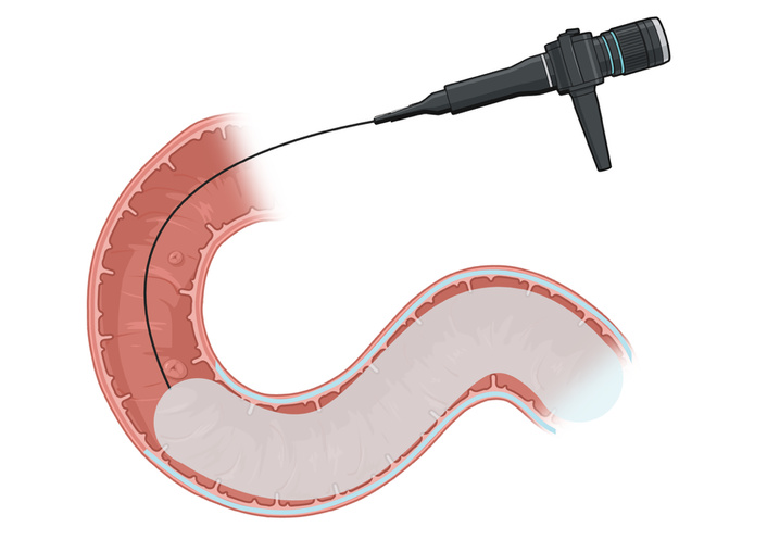
A depiction of duodenal mucosal resurfacing. Saline is injected into the duodenal walls to separate the superficial mucosa from deeper tissues. A balloon is introduced after the ampulla of Vater. Hydrothermal energy is applied to the duodenal mucosa via the balloon in a circumferential fashion. Created in BioRender. Dias, C. (2026) https://BioRender.com/dpfdcgk.
Multiple studies have shown improvements in metabolic syndrome markers after DMR. The first in-human trial demonstrated an overall cohort reduction in HbA1c by 1.2% after six months [22]. Even greater effects were seen in patients who received ablation over a larger area of their duodenum [22]. This study was limited to a six-month follow-up time and may fail to account for long-term effects [22]. Another prospective study called REVITA-1 consisted of patients (n = 67) who underwent a nine-centimeter or longer ablation [18]. These patients demonstrated a 1.1% overall reduction in HbA1c and a significant reduction in liver enzymes related to MASLD [18]. Fib-4 scores, which indicate the degree of MASLD liver fibrosis, also reduced after DMR [18]. That same study demonstrated that a sub-cohort (n = 14) experienced important modulations involved in improving cholesterol, insulin sensitivity, and antioxidative function [18]. This was assessed using a metabolomic analysis method that analyzed serum for lipids, glucose metabolism byproducts, inflammatory markers, and antioxidant compounds [18]. The results of this study are significantly limited by the small sample size of the original cohort and the sub-cohort. The small sample size significantly reduces the power of this study and increases the possibility that these results could be related to error. REVITA-2 was an RCT that again demonstrated significant reductions in HbA1c after DMR, now in multiple international groups [23]. This study was conducted mostly in Europe, with sites in Italy, the United Kingdom, Belgium, the Netherlands, and Brazil [23]. Therefore, these results lack generalizability to the world’s population. Additionally, the change in HbA1c in the Brazilian group was statistically insignificant, triggering post-hoc subgrouping which could introduce selective reporting bias and over-interpretation [23].
These clinical outcomes are promising, but the mechanisms of these changes remain incompletely understood [8]. One study showed that L and K cells do not change in concentration at three months after DMR [8]. GLP-1 and GIP increase after DMR in rats, but remain stable in humans [24, 25]. Significant macroscopic and microscopic histologic changes in duodenal tissue at three months after DMR are not seen, implying the duodenal mucosa regenerates completely with ablation [8]. All of these generally unexpected results suggest that a more nuanced enteroendocrine or genetic process could be involved in these changes [8].
Future directions in DMR research should focus on obtaining a better understanding of its mechanism. PDZK1 is a gene that likely encodes glucose uptake transporters in the intestinal epithelium, and loss of its activity may increase insulin resistance [8]. It may also have a critical role in cholesterol metabolism and insulin sensitivity, and its expression is increased when inflammasomes are inhibited [8, 26]. GATA6 is a transcription factor that mediates gastrointestinal epithelial growth and intestinal enteroendocrine cell differentiation [8]. Both have increased expression in patients whose diabetes responded to a combination of DMR and GLP-1RA therapy [8]. Therefore, PDZK1 and GATA6 could be key players in the mechanism by which DMR influences insulin resistance. However, these could simply be markers of the metabolic effects associated with DMR. Additionally, the use of GLP-1RAs in conjunction with DMR confounds this evidence. Accordingly, further study is needed to validate whether this effect exists after DMR, independently of GLP-1RA therapy.
As mentioned earlier, bypassing the duodenum via bariatric surgery is associated with changes in gut hormones that modulate glucose sensitivity via their central effect on the brain [19]. Polypeptide tyrosine-tyrosine (PYY) is a gut hormone that reverses pancreatic islet cell dysfunction and is increased after bariatric surgery [19]. Therefore, it could be reasonable to explore how gut hormones like PYY are impacted after DMR and whether gut hormones are the key to its mechanism [19]. Inflammation and oxidative stress are essential factors in the development of metabolic diseases [2, 3]. It would be worthwhile to explore if DMR has systemic anti-inflammatory or anti-oxidative effects that are significant enough to explain the metabolic impact of DMR. It would also be prudent to confirm that DMR does not affect the number of L and K cells. The study that asserted this finding had a sample size of only 16 patients [8]. The effects of DMR could be reasonably explained by a change in the number of L and K cells, and the data regarding DMR and L and K cell concentration seem counterintuitive to what would be expected.
Another reasonable mechanism to consider would be how DMR impacts the gut microbiota and bile acid signaling. A small clinical study investigated how glycated HbA1c, a hallmark of diabetes, and liver MRI proton density fat fraction (PDFF), an indicator of MASLD, were affected by changes in the gut microbiota after DMR and GLP-1RA combination therapy [27]. They found that HbA1c correlated negatively with alpha diversity, the richness of the microbiota [27]. PDFF correlated positively with beta diversity, the dissimilarity of community composition between two samples [27]. This study suggested that changes in gut microbiota could be responsible for the metabolic effects of DMR [27]. A spin-off of the same study also noticed alterations in bile acid signaling [28]. Bile acids activate receptors in the gut-liver axis called farsenoid X receptor (FXR) and fibroblast growth factor 19 (FGF19) [28]. FGF19 stimulates hepatic glycogen synthesis while inhibiting lipogenesis and gluconeogenesis [28]. Increased unconjugated bile acids after DMR and GLP-1RA therapy indicated a change in the gut microbiota, as gut bacteria are responsible for deconjugating bile [28]. After gastric bypass, similar changes are seen in bile acid signaling and gut microbiota [28]. Therefore, DMR alterations in gut microbiota could play a role in nutrient sensing, leading to altered bile acid signaling and alterations in hepatic contributions to metabolic disease [28]. A larger multi-center study without the concurrent use of a GLP-1RA is needed to validate these results and explore if changes in gut microbiota and bile acid signaling contribute to the mechanism of the metabolic changes after DMR.
While the mechanism of influence of DMR remains unclear, it has clear potential to significantly transform the management of metabolic diseases. It has potential to have long-lasting effects with one treatment. Further elucidation of its mechanisms will be key in fully defining its true therapeutic and preventive potential. The potential therapeutic impact of DMR on obesity should be explored further. Overall, DMR data remains limited to small cohort prospective studies with short follow-up periods and study populations that are incompletely representative of the global population [25]. While only minimal, mild, and reversible adverse effects have been reported thus far, expansion of the therapeutic trials is needed to confirm safety, efficacy, reproducibility among populations, durability and long-term effects, applicability to obesity, and provide robust data that can be reliably used to understand the nuanced mechanism in which DMR modulates metabolic diseases [8, 18, 22, 23].
Gene therapy is a young but rapidly growing field in the treatment of metabolic disease. It is a potential way in which GLP-1RA effects could be elicited more permanently. Gene therapy for metabolic diseases employs viral or non-viral carriers to deliver therapeutic genes to cells of interest to induce changes in expression of the mediators of metabolic diseases [29]. Adenovirus and adeno-associated viral (AAV) vectors are potential delivery methods [29]. AAVs are derived from nonpathogenic parvovirus and require the presence of a true adenovirus or herpes virus to replicate [30]. AAVs have low rates of immunogenicity compared to true adenoviruses and can exhibit transgenes for more than 4 years [31].
AAVs express markers that allow them to dock specifically on the type of target cells they are designed to affect (Figure 2) [31]. Once the AAV docks at the target cell, it is enveloped into an endosome within the target cell [30]. The AAV escapes the endosome and travels to the nucleus of the target cell [30]. Once the AAV enters the nucleus, the capsid is uncoated [30]. The now-exposed genetic contents persist in the nucleus of the target cell in the form of circular episomes in nonreplicating cells [30]. In replicating cells, the genetic material is incorporated randomly into the cell’s existing genetic material [30]. It is then transcribed and translated alongside the original genetic material of the cell [30]. It is important to note that many vector genomes remain as episomes and are quickly lost in the process of cellular replication before they have the chance to be randomly integrated into the cellular genome [30]. While AAV-based gene therapy in metabolic disease is a new field, several trials are proposed or in process to evaluate how metabolic disease could be modulated with AAV gene therapy. The information regarding these trials is widely limited to corporation-driven communication and will need extensive validation.
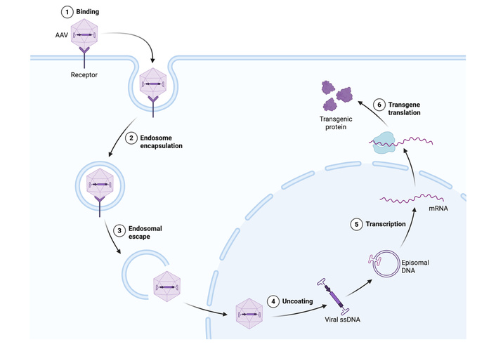
A depiction of adeno-associated virus (AAV)-based gene therapy. The AAV docks at the target cell and enters the cell via an endosome. Once inside the cell, the AAV escapes the endosome and travels into the nucleus. The AAV capsid is uncoated, and the genetic material is exposed. Through transcription and translation, the desired protein is created. Created in BioRender. Dias, C. (2026) https://BioRender.com/2dulsz7.
Genprex studied the use of adeno-associated virus 8 (AAV8) gene therapy for T2DM in mice [32]. They studied the use of AAV8 to deliver 1 × 10¹¹ viral genomes containing Pdx1 and Mafa to pancreatic cells via infusion into the pancreatic duct [32]. Mafa is a transcription factor that allows Pdx1 to produce β-cells from α-cells and endocrine precursor cells [33]. They also used cytomegalovirus and rat insulin promoters that better allow the AAV to directly target pancreatic islet cells [32]. Four weeks after the treatment, the mice underwent intraperitoneal glucose tolerance testing, insulin tolerance testing, glucose-stimulated insulin secretion tests, and a glucose secretion assessment [32]. At the American Society of Gene and Cell Therapy annual meeting, they stated that the mice showed better glucose tolerance, improved insulin secretion, and potentially increased insulin sensitivity [32]. Genprex proposed that these results could be translatable to human models using endoscopic retrograde cholangiopancreatography (ERCP) to deliver AAV8 to human pancreatic ducts [32]. These data and assertions have not been published formally and have only been presented via a poster presentation. Therefore, the findings should be viewed with caution.
Jaguar Gene Therapy has proposed an investigational study (JAG301) using adeno-associated virus 9 (AAV9) to treat type 1 diabetes mellitus (T1DM) [34]. In T1DM, an autoimmune response triggers immune cells called T-cells to destroy insulin-producing β-cells [35]. As a result, patients have a lifelong dependence on insulin injections or pumps [35]. PAX4 is a transcription factor that drives the development of β-cells and maintains mature β-cell identity to ensure intact insulin-producing function [36]. When PAX4 is lost, endocrine precursors preferentially differentiate into α-cells [36]. Jaguar will deliver PAX4 via AAV9 to glucagon-producing α-cells in the pancreas to induce them to transdifferentiate into β-cells that secrete insulin [34]. While the clinical stage of this trial remains incomplete, they produced preclinical proof-of-concept data that they claim is “encouraging” [34]. This study could be useful in the future development of a simple subcutaneous injection that is therapeutic for T1DM, given the function of PAX4 [36]. JAG301 could also be useful in late-stage T2DM. While early T2DM is driven by insulin resistance, late stages are marked by β-cell burnout with decreased insulin secretion [37]. JAG301 could potentially bolster insulin secretion by increasing the number of functional β-cells. While it wouldn’t be curative for insulin resistance, it could be a useful adjunct in insulin-dependent T2DM. However, JAG301 is an incomplete investigational study, and the aforementioned assertions regarding “encouraging” data and potential utility are derived from the Jaguar website. There is a high risk of bias in these assertions, and published, peer-reviewed, reliable data is needed to back up these claims.
Fractyl Health has proposed a method for local delivery of AAVs to the pancreas to treat obesity and T2DM [38]. As of May 11, 2026, it will be the first T2DM AAV-based gene therapy candidate to begin clinical testing [39]. They will use endoscopic ultrasound to guide the intrapancreatic injection of AAVs carrying RJVA-001 to pancreatic β-cells [38]. A poster presentation at the American Society of Gene and Cell Therapy annual meeting stated that RJVA-001 allowed pancreatic β-cells to produce GLP-1 locally in an in vitro model and is expected to have a similar effect in humans [38, 40]. Historically, AAV-based gene therapy at pancreatic targets has been challenging given AAVs do not selectively target the pancreas individually [41]. By using endoscopic ultrasound to locally deliver RJVA-001, it is thought that the virus will dock at pancreatic β-cells more efficiently and there will be a reduced risk of ectopic GLP-1 expression in untargeted cells [40]. The pancreatic β-cells could create a GLP-1-rich environment for themselves, allowing for stable exposure to GLP-1. According to the poster presentation, this therapy in mice showed significant body weight, adipose weight, liver weight, hepatic steatosis, and glycemic control improvements [40]. As substantiated by their in vitro and murine models, Fractyl hypothesizes that RJVA-001 could produce long-term positive effects on obesity and diabetes [38, 40]. These assertions are all collected from a poster presentation at the American Society of Gene and Cell Therapy annual meeting, a United States Securities and Exchange Commission filing, and the Fractyl website. The data are unpublished, and these assertions will need to be backed by reliable data from clinical testing.
These innovative AAV trials show promise in developing treatments for obesity and diabetes, but it is important to consider the limitations of AAV-based gene therapy. Ex vivo trials for virus-delivered gene therapies have historically advanced well, but there are a number of barriers to clinical translation [31]. The vectors are quite difficult and expensive to manufacture [1]. It is estimated that AAV manufacturing capacity could currently serve less than three percent of people who would benefit from these therapies [31]. Given the systemic nature of traditional AAV-based gene therapy, it can be associated with ectopic expression at non-target tissues and is also limited by low overall transduction efficiency [29, 31]. In a study of AAV2, transduction in humans was six to eight times lower than in pre-clinical rodent models [31]. If this same effect is seen in AAV8 and AAV9, the utility of these proposed therapies will be severely limited. However, mouse models that employ pancreas-directed injection and use cytomegalovirus and rat insulin promoters showed success in cell specificity, efficient transduction, and minimal ectopic expression [42]. This shows promise that Fractyl and Genprex’s proposed methods of directed injection could overcome AAV limitations and actually be successful in human models. It will be important for these AAV-based gene therapy trials to critically evaluate for any off-target effects and to determine if their proposed methods would be efficient enough to overcome manufacturing and transduction inefficiencies that are intrinsically associated with these therapies.
Lipid nanoparticles (LNPs) are a low-cost alternative delivery shuttle for therapeutic genes [1]. They have lower immunogenicity than viral carriers, can carry a boundless amount of genetic material, and are easier to manufacture than virus carries [31]. They are easily able to target adipocytes and hepatocytes given their lipophilicity [1, 43]. Like AAVs, they dock at specific receptors on target cells (Figure 3) [44]. Next, they enter the cell via an endosome that they eventually escape [44]. They travel to the nucleus, where they release their DNA payload for transcription to mRNA and translation to proteins [45]. LNPs can also be used to deliver mRNA directly into the cytoplasm [46].
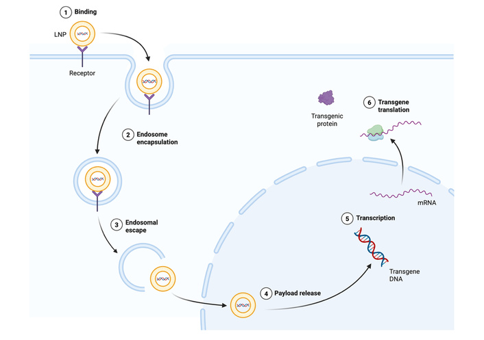
A schematic representation of Lipid nanoparticle (LNP) gene therapy. The LNP docks at a receptor and enters the target cell via an endosome. It escapes the endosome to travel to the nucleus, where it delivers DNA. The DNA is transcribed into mRNA that is translated into proteins. Created in BioRender. Dias, C. (2026) https://BioRender.com/4t5jqkq.
Exendin-4 is a synthetic peptide that is thought to act as a GLP-1 receptor agonist [43]. In a study that packaged a gene encoding exendin-4 in an LNP, mouse liver cells sustainably secreted exendin-4 up to 28 weeks later [43]. They found that these mice had a knock-in rate of one percent [43]. Food intake decreased by an average of 29%, body weight decreased by an average of 34%, and the mice had increased insulin sensitivity, evidenced by decreased blood glucose levels after intraperitoneal insulin injection [43]. Another study packaged DNA encoding exendin-4 and a modified version of GLP-1 in an adipocyte-targeting LNP they developed called Prometheus [1]. The mice that received the injection showed 14% weight loss after exendin-4 and 20% weight loss after GLP-1 DNA [1]. The mice had improved insulin sensitivity, with a 26% reduction after exendin-4 and a 28% reduction after GLP-1 treatment in the area under the curve after insulin challenge [1]. The treated mice also showed reduced appetite and better glycemic control [1]. The transgene remained within the subcutaneous tissue where it was injected for 6 months and exhibited disease-modifying effects that show superiority to daily exenatide injections [1]. The study demonstrated that LNP-mediated gene therapy could be delivered almost identically to traditional GLP-1RA therapy, while having more sustained biological activity [1]. However, this study employed a murine cohort that was small (n = 34) and was subsequently divided further into diet groups and treatment groups [1]. The power of this study is significantly limited by the short follow-up period and small cohort size. LNP-mediated gene therapy could also be successful in MASLD treatment, given LNP ease in targeting hepatocytes [47]. One trial used LNPs to deliver genetic material to encode proteins that regulate heat shock protein 47, a protein involved in liver fibrosis [47, 48]. LNP-mediated gene therapy was shown to be safe in patients with moderate to severe liver fibrosis, but therapeutic effects have not yet been reported [47]. It is important to consider that LNP-mediated gene therapy is limited by poor transduction efficiency when compared to viral carriers, preference for a limited subset of cells, and a lack of infrastructure for widespread manufacturing [31, 46].
Both AAV- and LNP-mediated gene therapy have extensive limitations that will be barriers to advancement. Individually, they lack the ability to have nuanced control over the amount of the transduced gene that is expressed [1, 31]. However, Clustered Regularly Interspaced Short Palindromic Repeat interference (CRISPRi) and Clustered Regularly Interspaced Short Palindromic Repeat activation (CRISPRa) allow for titratable control over gene expression [31]. Gene therapy developers will need to consider how therapeutic gene expression will approach overexpression and how a dose-response could be created by combining CRISPRi and CRISPRa with AAV- and LNP-mediated gene therapy. Many of the effects that these studies propose could lead to dangerous effects if left unchecked. More data are needed to confirm the long-term efficacy and safety of gene therapy for metabolic diseases in humans. Almost all of the studies are in the preclinical phase or are just approaching clinical translation to humans. They lack long-term data on the durability and safety of these therapies. Increased research and clinical success in these trials may help overcome manufacturing capacity limitations by forcing increased production that would drive costs down.
As with any gene-editing topic, it will be essential to consider the ethical implications of gene therapy for metabolic diseases. Given the goal is permanent genetic alteration, extensive trials will have to be conducted in pre-clinical models and controlled human trials to confirm long-term safety and efficacy before gene therapy can be released for clinical use. Even then, strict drug regulations will have to ensure that the therapies released are efficacious and provide benefits that outweigh any potential harm from off-target effects. Additionally, metabolic diseases disproportionately affect people of lower socioeconomic status [49]. Two-thirds of gene therapy trials are currently concentrated in Western countries, but the majority of patients with metabolic disease reside outside of these areas [31]. The effects of these therapies must be studied in populations that carry the majority of metabolic disease before they can be adopted globally. The issue of accessibility is likely the largest barrier to ethical acceptance of gene therapy. The aforementioned manufacturing costs and lack of application outside of Western populations exclude people in lower socioeconomic groups [31]. Affluent populations may overcome metabolic disease, while the majority of metabolic disease will continue to accumulate in groups that have fewer healthcare resources to begin with [49]. In accordance with ethical justice, it will be of utmost importance that gene therapy for metabolic disorders be made accessible to people of all socioeconomic statuses.
As discussed above, DMR is suggested to likely have a more intricate and sophisticated mechanism of action than originally thought [8]. While changes in endocrine signaling and histology at the site of the resurfacing may not be present, increased expression of PDZK1 and GATA6 was noted after DMR procedures in human models [8]. These may just be markers of DMR efficacy in improving metabolic disease, but further exploration into the impetus for this increased expression could provide important insights in understanding the mechanism of DMR. We speculate that there may be other gene expression-related changes that occur with DMR that could explain the mechanism of its function. Extensive further research is needed to identify genes or transcription factors that are reliably present in patients after DMR and to confirm their roles in metabolic disease signaling [8]. For example, it is hypothesized that apical sodium bile acid transporters (ASBT) have reduced expression after DMR, leading to less reuptake of the bile acids and more deconjugation [28]. As discussed earlier, the change in bile acid signaling could have relevance in metabolic disease therapy.
If PDZK1, GATA6, and the genes encoding ASBT are scientifically confirmed as crucial players in how DMR modulates metabolic diseases, they could be important future avenues for gene therapy. We suggest that inducing increased expression of PDZK1 and GATA6 or repressing the genes encoding ASBT via Cas-mediated transcription repression at the duodenum could potentially bolster the effects of DMR or mimic it. Studies of gene expression and biomarkers after DMR will likely expose more relevant targets for gene therapy in metabolic diseases. This will need extensive experimental validation. Subsequently, we propose that gene therapy and DMR may be used concurrently in a therapeutic approach to metabolic disease in the future. For example, DMR could be used to induce initial changes in the duodenal mucosa while gene therapy might be used afterwards to have systemic effects or maintain its effects. This proposed intersection is highly speculative and requires extensive research before it can really be considered. A murine model could be given both DMR and gene therapy and compared to groups treated with either therapy. This would be a helpful step in understanding how the therapies might interact negatively or positively bolster the effects of each other. Ultimately, understanding the mechanism of DMR will be paramount in understanding the future directions for DMR and gene therapy intersecting to affect long-term changes in obesity, T2DM, and MASLD. Once this is understood, further experimentation to explore the viability of this proposed intersection of DMR and gene therapy is needed.
Metabolic diseases like diabetes, obesity, and MASLD are increasing rapidly in prevalence, are associated with extensive comorbidities, and significantly contribute to mortality globally. Highly efficacious and long-lasting therapies are needed to address these diseases. DMR could provide a safe and effective therapeutic pathway for mediating diabetes, MASLD, and likely obesity. DMR is allowing for the elucidation of genes potentially involved in the gut-pancreas signaling axis and broadening our understanding of their function. It is possible that these genes have roles in modulating metabolic diseases and could be important targets for gene therapy at the pancreas in the future. Gene therapies for metabolic diseases via AAV and LNP carriers have shown promise in preclinical models and are expected to have success in clinical translation. They have the potential to modulate diabetes, obesity, and MASLD over long periods of time with minimized invasiveness and side effects.
Both DMR and gene therapy require further mechanistic research, sophistication, and improved accessibility to all populations. Safety, durability of effects, clinical validity, and applicability to all populations of both therapies remain unclear. Large cohort studies with long-term follow-up are needed to better understand how these therapies might compare to existing therapies like GLP1-RAs and bariatric surgery. It is unclear if DMR and gene therapy will be superior to existing therapies when considering cost, efficacy, long-term effects, and acceptability to patients. Continued investigation of these therapies is necessary and will likely be fruitful in further understanding how they could intersect. The data available for DMR is very limited and requires further study in larger, diverse cohorts with longer follow-up time. For gene therapy specifically, manufacturing capacity will have to increase substantially before gene therapy can reach all populations. Regulation of gene therapy and the need for a dose-response currently pose large hurdles that must be overcome before gene therapy could be realistically applied widely. More human trials will be an essential step in understanding the potential utility of gene therapy.
In a landscape of rapidly increasing rates of metabolic diseases around the world and corresponding skyrocketing healthcare expenses, new therapeutic options for metabolic diseases are vital. Further study of DMR, gene therapy, and how they could work together could make a meaningful shift in how we treat or even cure metabolic diseases.
AAV: adeno-associated virus
ASBT: apical sodium bile acid transporters
CRISPRa: Clustered Regularly Interspaced Short Palindromic Repeat activation
CRISPRi: Clustered Regularly Interspaced Short Palindromic Repeat interference
DMR: duodenal mucosal resurfacing
FGF19: fibroblast growth factor 19
GIP: glucose-dependent insulinotropic polypeptide
GLP-1: glucagon-like peptide 1
GLP-1RA: glucagon-like peptide-1 receptor agonist
LNPs: lipid nanoparticles
MASLD: metabolic dysfunction-associated steatotic liver disease
PDFF: proton density fat fraction
PYY: polypeptide tyrosine-tyrosine
RCTs: randomized controlled trials
T1DM: type 1 diabetes mellitus
T2DM: type 2 diabetes mellitus
During the preparation of this work, the authors used Google Gemini to brainstorm topic ideas, outline the initial structure of the paper, and to refine language. After using this tool, the authors reviewed and edited the content as needed and take full responsibility for the final publication.
CD: Conceptualization, Investigation, Methodology, Validation, Visualization, Writing—original draft, Writing—review & editing. NT: Conceptualization, Project Administration, Supervision, Writing—review & editing. SP: Writing—review & editing. TG: Conceptualization, Project administration, Supervision, Validation, Writing—review & editing. All authors read and approved the submitted version.
The authors declare that they have no conflicts of interest.
Not applicable.
Not applicable.
Not applicable.
Not applicable.
Not applicable.
© The Author(s) 2026.
Open Exploration maintains a neutral stance on jurisdictional claims in published institutional affiliations and maps. All opinions expressed in this article are the personal views of the author(s) and do not represent the stance of the editorial team or the publisher.
Copyright: © The Author(s) 2026. This is an Open Access article licensed under a Creative Commons Attribution 4.0 International License (https://creativecommons.org/licenses/by/4.0/), which permits unrestricted use, sharing, adaptation, distribution and reproduction in any medium or format, for any purpose, even commercially, as long as you give appropriate credit to the original author(s) and the source, provide a link to the Creative Commons license, and indicate if changes were made.



View: 298
Download: 10
Times Cited: 0
Huiling Huang ... Dongsheng Li
Huiling Huang ... Dongsheng Li
Dorothy E. Vatner ... Stephen F. Vatner
Maria-Kalliopi Spanorriga ... Konstantinos Tsioufis
Ananyaa Sridhar ... Dawood Khan


