Review

Affiliation:
1Department of Pharmaceutical Sciences, Faculty of Science & Engineering, Dibrugarh University, Dibrugarh 786004, Assam, India
Email: kalyakster@gmail.com
ORCID: https://orcid.org/0000-0002-1421-8962
Affiliation:
1Department of Pharmaceutical Sciences, Faculty of Science & Engineering, Dibrugarh University, Dibrugarh 786004, Assam, India
ORCID: https://orcid.org/0000-0003-0803-5470
Affiliation:
1Department of Pharmaceutical Sciences, Faculty of Science & Engineering, Dibrugarh University, Dibrugarh 786004, Assam, India
ORCID: https://orcid.org/0000-0003-0147-6556
Affiliation:
1Department of Pharmaceutical Sciences, Faculty of Science & Engineering, Dibrugarh University, Dibrugarh 786004, Assam, India
2Pratiksha Institute of Pharmaceutical Science, Guwahati 781026, Assam, India
ORCID: https://orcid.org/0000-0003-0035-0329
Affiliation:
1Department of Pharmaceutical Sciences, Faculty of Science & Engineering, Dibrugarh University, Dibrugarh 786004, Assam, India
ORCID: https://orcid.org/0009-0001-6834-2117
Affiliation:
1Department of Pharmaceutical Sciences, Faculty of Science & Engineering, Dibrugarh University, Dibrugarh 786004, Assam, India
ORCID: https://orcid.org/0009-0003-3734-5360
Affiliation:
2Pratiksha Institute of Pharmaceutical Science, Guwahati 781026, Assam, India
ORCID: https://orcid.org/0009-0006-2540-0432
Affiliation:
3Dibrugarh University Institute of Engineering and Technology, Faculty of Science & Engineering, Dibrugarh University, Dibrugarh 786004, Assam, India
ORCID: https://orcid.org/0000-0002-0884-8693
Affiliation:
3Dibrugarh University Institute of Engineering and Technology, Faculty of Science & Engineering, Dibrugarh University, Dibrugarh 786004, Assam, India
ORCID: https://orcid.org/0000-0003-2367-7535
Affiliation:
3Dibrugarh University Institute of Engineering and Technology, Faculty of Science & Engineering, Dibrugarh University, Dibrugarh 786004, Assam, India
ORCID: https://orcid.org/0000-0001-8884-8556
Affiliation:
4Department of Mechanical Engineering, Indian Institute of Technology Ropar, Ropar 140001, Punjab, India
ORCID: https://orcid.org/0009-0000-2179-5322
Explor Med. 2023;4:1135–1167 DOI: https://doi.org/10.37349/emed.2023.00200
Received: August 30, 2023 Accepted: October 26, 2023 Published: December 29, 2023
Academic Editor: Nermeen A. Elkasabgy, Cairo University Faculty of Pharmacy, Egypt
The article belongs to the special issue Exploration of 3D and 4D Printing in the Biomedical and Personalized Medicine Fields: Merits and Challenges
The integration of three-dimensional (3D) printing techniques into the domains of biomedical research and personalized medicine highlights the evolving paradigm shifts within contemporary healthcare. This technological advancement signifies potential breakthroughs in patient-specific therapeutic interventions and innovations. This systematic review offers a critical assessment of the existing literature, elucidating the present status, inherent challenges, and prospective avenues of 3D printing in augmenting biomedical applications and formulating tailored medical strategies. Based on an exhaustive literature analysis comprising empirical studies, case studies, and extensive reviews from the past decade, pivotal sectors including tissue engineering, prosthetic development, drug delivery systems, and customized medical apparatuses are delineated. The advent of 3D printing provides precision in the fabrication of patient-centric implants, bio-structures, and devices, thereby mitigating associated risks. Concurrently, it facilitates the ideation of individualized drug delivery paradigms to optimize therapeutic outcomes. Notwithstanding these advancements, issues concerning material biocompatibility, regulatory compliance, and the economic implications of avant-garde printing techniques persist. To fully harness the transformative potential of 3D printing in healthcare, collaborative endeavors amongst academicians, clinicians, industrial entities, and regulatory bodies are paramount. With continued research and innovation, 3D printing is poised to redefine the trajectories of biomedical science and patient-centric care. The paper aims to justify the research objective of whether to what extent the integration of 3D printing technology in biomedicine enhances patient-specific treatment and contributes to improved healthcare outcomes.
In the realm of manufacturing, an emerging technology known as rapid prototyping or additive manufacturing has demonstrated substantial promise. This technology has undergone significant advancements, evolving into a valuable tool for diverse fields including research, manufacturing, design, engineering, and science. This convergence of disciplines gave rise to the three-dimensional (3D) printer, encapsulating design, manufacturing, electronics, materials, and business elements [1]. 3D printing involves the layer-by-layer creation of 3D objects using material. Unlike traditional manufacturing, which often employs subtractive methods involving grinding, bending, forging, molding, cutting, gluing, welding, and assembly, 3D printing utilizes an additive approach. Initially perceived primarily as a means to give shape to artistic and unique designs, 3D printing has progressed to a stage where it can fabricate mechanical components and necessary parts, fundamentally transforming not just industrial and manufacturing sectors but also the future fabric of our daily lives [2].
Two primary techniques dominate 3D printing for consumer-level additive manufacturing: fused deposition modeling (FDM) and stereolithography (SLA). These processes incrementally add material, layer-by-layer, to construct objects. SLA employs ultraviolet light to selectively cure resin, while FDM extrudes semi-liquid plastic according to the desired layout. The rapid growth of this technology has enabled significant breakthroughs. Particularly, 3D printing, notably the FDM technique, has markedly reduced manufacturing costs, build times, and object weight while minimizing waste compared to certain traditional manufacturing methods. Consequently, 3D printing has become accessible to the average consumer [3–4], illustrating the graph depicting the reduction in manufacturing costs [3–4] and time consumption in 3D printing compared to conventional manufacturing techniques. The evolution of 3D printing, also known as additive manufacturing, has been a remarkable journey that has transformed numerous industries by redefining traditional manufacturing processes, fostering innovation, and enabling unprecedented levels of customization, efficiency, and design flexibility. This technology has grown from a niche concept to a powerful tool with broad applications across diverse sectors [5]. The aerospace and defense industry were an early adopter of 3D printing for rapid prototyping and design validation. The capability of the technology to produce intricate designs while minimizing weight has paved the way for its use in crafting lightweight parts such as brackets, hinges, and air ducts. As materials and processes advanced, critical components like turbine blades and engine parts began to be 3D printed, contributing to enhanced performance and fuel efficiency [6]. This evolution has reduced lead times in production, lower costs, and improved sustainability due to reduced material waste.
In healthcare, 3D printing has revolutionized patient care by enabling the production of personalized medical devices, implants, and prosthetics. Surgeons can create patient-specific anatomical models for preoperative planning, enhancing surgical precision [7]. Bioprinting has opened doors to creating functional tissues and organs, addressing organ transplant shortages. Dental applications have also benefited from 3D printing, producing accurate dental crowns, braces, and aligners. 3D printing has significantly impacted the automotive sector by expediting the prototyping and development of new vehicle designs. Its ability to manufacture lightweight yet strong components has led to improved fuel efficiency and vehicle performance [8–10]. Automakers use 3D printing to produce intricate parts, tooling, and even vehicle prototypes. Customization and on-demand production have become feasible, reducing inventory costs and enabling the creation of specialized parts for specific vehicle models [11]. The architecture and construction industries have been exploring 3D printing for creating complex building components and structures [12]. Large-scale 3D printers can use various materials, such as concrete, to fabricate walls, columns, and even entire buildings. This approach reduces construction time, labor requirements, and material waste. Architects can experiment with innovative designs that were previously challenging to execute using traditional methods. 3D printing has democratized manufacturing, allowing individuals and small businesses to create custom consumer goods with intricate designs and personalized features [13–15]. Jewelry, fashion accessories, and home decor items can be produced with unique characteristics tailored to individual preferences. This has prompted a transition from bulk manufacturing to an eco-friendlier and customer-focused production methodology [16, 17].
Educational institutions and researchers have embraced 3D printing as a hands-on learning and experimentation tool. Students can bring abstract concepts to life by creating physical models, enhancing their understanding of complex subjects [18]. Researchers use 3D printing to produce prototypes, equipment, and scientific models to advance their fields. The food industry has ventured into 3D printing to create intricate food designs and personalized nutrition solutions. Culinary professionals experiment with creating visually appealing dishes, while researchers explore the possibility of printing nutritious and customized meals for individuals with specific dietary needs. In the energy sector, 3D printing has improved efficiency and cost savings by enabling the production of complex parts for turbines, pumps, and industrial machinery [19, 20]. Customization of components for specific applications enhances performance and reduces downtime for maintenance and repairs. The transition of 3D printing into the biomedical sector has been a transformative journey, revolutionizing how medical professionals approach patient care, research, and the development of medical devices [11]. This convergence of cutting-edge technology and healthcare needs has opened up new horizons, enabling remarkable advancements in personalized medicine, surgical procedures, and even the creation of human tissues and organs.
One of the most significant impacts of 3D printing in the biomedical sector is the creation of personalized medical devices. With the ability to produce intricate and patient-specific implants, prosthetics, and orthotics, 3D printing has improved patient outcomes and quality of life. Custom-fit implants can be designed based on individual anatomical scans, ensuring a precise fit and reducing the risk of complications 3D printing has transformed surgical planning and training by providing surgeons with physical, tangible models of patients’ anatomies [12]. This technology is particularly favored for numerous applications in the healthcare sector, offering significant advantages, especially in medical and dental imaging. It excels in managing tasks related to patient-specific anatomical structures and plays a crucial role in medical device design and production. Biocompatible materials enable diverse applications, including creating tissue without harming living cells, blood vessel production, dental implants, and specialized medical prostheses [13].
Researchers are actively exploring its potential in repairing or replacing defective organs like kidneys and hearts, even paving the way for creating organs replicating their originals’ biological functions. This holds great promise for numerous patients [13, 21], leading to an increasing research focus on its diverse biomedical applications [22]. While 3D printer technology has gained traction across various sectors, its use in biomedical applications has recently gained prominence. This study introduces 3D technology, discusses various 3D methods, highlights their exceptional properties, and delves into their utilization across surgical procedures, medical imaging, the pharmaceutical industry, the production of patient-specific medical prostheses and implants, veterinary medicine, skin engineering, stem cell research, and organ printing. Furthermore, the study explores the anticipated widespread benefits of this technology in biomedical applications, outlines current developmental challenges, identifies trends, and envisions future opportunities [21, 22]. These models help surgeons visualize complex structures, plan intricate procedures, and simulate surgeries before entering the operating room. This reduces surgical risks, minimizes operating time, and enhances overall surgical outcomes.
Medical education has greatly benefited from 3D printed anatomical models. Medical students can interact with accurate and detailed physical models, improving their understanding of human anatomy [21]. These models also enhance patient communication, allowing healthcare professionals to explain conditions and procedures effectively. One of the most revolutionary aspects of 3D printing in the biomedical sector is bioprinting. This process involves the layer-by-layer deposition of living cells and biomaterials to create functional tissues and even organs. While still in its early stages, bioprinting holds immense potential for addressing organ transplantation shortages, drug testing, and disease modeling. Further, 3D printed models of patient anatomy enable enhanced preoperative planning. Surgeons can evaluate various approaches and potential challenges, leading to more successful surgeries. These models also serve as valuable communication tools, helping patients understand their conditions and treatment options more comprehensively [22].
Dentistry has embraced 3D printing to fabricate dental crowns, bridges, braces, and aligners. This technology allows for accurate and rapid production of dental devices, reducing patients’ time in the dentist’s chair and improving overall oral health. 3D printing has also paved the way for the production of personalized drug delivery systems, tailoring medications to individual patients’ needs. Additionally, pharmaceutical researchers use 3D printing to create intricate drug formulations and dosage forms with precise release profiles. 3D printing has accelerated biomedical research by enabling the creation of complex research models, lab equipment, and microfluidic devices. This technology promotes innovation in drug development, disease modeling, and understanding biological processes [12, 13]. The transition of 3D printing into the biomedical sector has been marked by groundbreaking advancements that have reshaped healthcare practices and medical research. As the technology continues evolving, it promises further personalized medicine, regenerative therapies, and the fabrication of functional human tissues and organs.
The emergence of 3D printing with biomedical applications has illuminated a path toward a healthier and more innovative future in healthcare [22]. The evolution of 3D printing has been accompanied by challenges, including developing suitable materials, quality control standards, and scalability. As materials science progresses, more materials are becoming available for 3D printing, expanding its capabilities. As research and development continue to push the boundaries of what is possible, 3D printing is poised to drive further advancements and disrupt traditional manufacturing in unprecedented ways [20, 22].
The protocol applied systematic methods, which had not been previously registered, to conduct a thorough search for pertinent studies, meticulously screen them for eligibility, and rigorously assess their quality. The review follows the Preferred Reporting Items for Systematic Reviews and Meta-Analyses guidelines. A literature search was performed using PubMed/MEDLINE, IEEE Xplore, Web of Science, Scopus, Embase, Google scholar, and Research Gate. The search strategy utilized a combination of keywords and MeSH terms, including but not limited to “3D printing”, “additive manufacturing”, “biomedicine”, “personalized care”, “medical applications”, “tissue engineering”, “drug delivery”, “implants”, “patient-specific”, “biomedical devices”, and “surgery planning”.
The inclusion criteria for the literature analysis encompassed articles published from 1984 to the present (2023), written in English, and comprising research articles, reviews, and conference papers that focused on the application of 3D printing in biomedicine, including personalized medical devices, tissue engineering, drug delivery systems, surgical planning, and patient-specific implants. Additionally, studies reporting positive, negative, or non-significant findings and encompassing both preclinical and clinical research were considered for inclusion in the review. The exclusion criteria comprised articles not written in English, studies not directly related to 3D printing applications in biomedicine, articles lacking original research content (e.g., opinion pieces or editorials), and duplicate publications or studies with incomplete data. The literature search was conducted from 18th August 2023 to ensure the inclusion of the most recent publications up to that date. A total of 214 articles were initially identified through database searches. After applying the inclusion and exclusion criteria, 110 articles were selected for the review. The review team conducted a thorough assessment of these 100 articles to provide a comprehensive overview of the role of 3D printing in advancing personalized care in biomedicine.
In this review, potential publication bias was addressed and mitigated through a comprehensive search strategy encompassing multiple databases, grey literature sources, and contact with study authors to include unpublished data. The inclusion of grey literature and the use of reporting bias assessment tools were instrumental in identifying and accounting for potential publication bias. Transparent reporting and discussion of this bias, along with sensitivity analyses, contributed to a more robust evaluation of the available evidence. These measures collectively aimed to minimize the impact of publication bias and enhance the reliability of the review’s conclusions regarding the diverse applications of 3D printing in biomedicine.
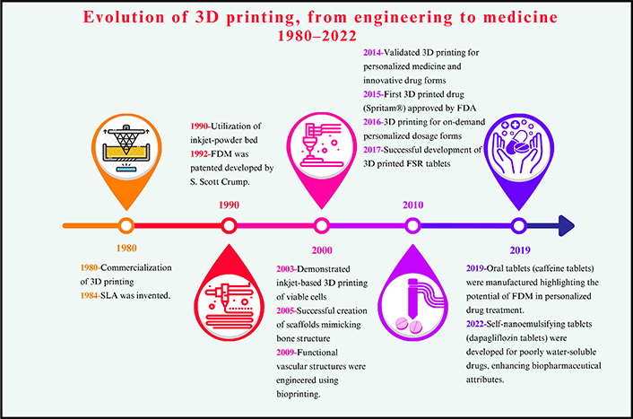
Digital fabrication technology, or 3D printing or additive manufacturing, constructs tangible objects from a geometrical blueprint through successive material additions. This innovation has rapidly emerged, finding widespread utility across various sectors. The versatility of 3D printing is particularly evident in mass customization, generating open-source designs, and catering to sectors like agriculture, healthcare, automotive, locomotive, and aviation. Using a geometric design, 3D printing constructs objects by progressively adding material [1]. This method has undergone remarkable expansion in recent years, marking a significant milestone. Charles Hull pioneered the commercialization of 3D printing processes in 1980 [5]. At present, the applications of 3D printing are diverse, encompassing the creation of artificial heart pumps [23], jewelry assortments [24], 3D printed corneas [25], PGA rocket engines [26], a steel bridge in Amsterdam [27], and various aviation and food industry-related products. Rooted in the layer-by-layer fabrication of 3D structures from computer-aided design (CAD) blueprints, 3D printing technology is an innovative and versatile platform [8].
Integrating 3D printing in the medical realm offers numerous advantages, including customizing and individualizing medical products, medications, and equipment; cost-efficiency; heightened productivity; democratized design and manufacturing processes; and strengthened collaborative efforts [28]. The first 3D printing used in medicine was to manufacture anatomically accurate patient-specific surgical and orthopedic components, including surgical planning [29] and custom implants [30].
In 2003, Dr. Thomas Boland had applied for his patent for a technique that involved the printing of viable cells at Clemson University. It was an approach for generating an array of viable cells, detailed in the disclosed invention, whether in two or three dimensions. The method involved inkjet printing to deposit a cellular composition containing diverse cell types, such as eukaryotic, prokaryotic, or cell aggregates, onto a substrate. This innovation presented a promising method for arranging and preserving viable cells for diverse applications [31].
Using a 3D printing research setup, high-resolution channel structures ideal for bone tissue engineering (BTE) were crafted. Scaffolds with interlinked channels were produced, and the granular structure of the scaffolds was preserved after sintering, resulting in significant microporosity. The researchers designed a test structure with inclined layers to enhance cell attachment and proliferation without clogging. By modifying the cell seeding protocol, seeding efficiency improved. Histological analysis revealed that isolated cells were present on scaffold surfaces after one day of culture, while cell quantity increased significantly after a week. In statically cultured scaffolds, cells formed multiple layers on granule surfaces, while dynamically cultured scaffolds exhibited more profound cell growth within granule cavities, possibly due to improved nutrient supply. Both methods demonstrated cell attachment, proliferation, and characteristic morphology, crucial for successful BTE with mesenchymal stem cells. The microporosity resulting from the 3D printing process enhanced scaffold surface area and porosity, promoting the remodeling process necessary for bone regeneration. The study demonstrated the ability to create 3D printed hydroxyapatite scaffolds tailored to a specific patient’s anatomical data obtained from radiographic images. Simultaneously, mimicking the internal bone structure was possible, creating a conducive environment for cultivating live cells in anticipation of the implantation process [32].
A surgical planning study reported by Robiony [29] demonstrated the collaborative utilization of virtual reality (VR), SLA (a form of 3D printing), and reverse engineering (RE). A maxillofacial surgeon treated a 9-year-old girl with right-sided hemifacial microsomia. Sophisticated imaging methods such as orthopantomograms, cephalograms, and 3D computed tomography (CT) scans were used to determine the extent of facial asymmetry for treatment planning. These CT images were transformed into 3D models through RE processes and image segmentation software, enabling detailed reconstruction of anatomical structures. A unilateral vertical ramus distraction procedure was planned based on these models. Virtual surgical simulations guided the creation of physical models using rapid prototyping techniques. Surgeons replicated these simulations on the physical model to ensure the procedure’s feasibility. The same approach was then executed in the operating room, where the virtual simulation and the physical model guided bone cuts and distractor placement, leading to successful mandibular ramus lengthening. A surgical template facilitated the transfer of information from the standard triangulation language (STL) model to the patient. Intraoral distraction devices were utilized, achieving effective mandibular osteodistraction. Postoperative assessments confirmed positive outcomes, including esthetic facial balance, vertical maxillary growth, and occlusal stability. This case showcases the synergy between 3D printing, virtual simulations, and surgical practice, ultimately yielding excellent surgical results in terms of function and aesthetics [29].
In 2009, a significant milestone was reached when Organovo secured the first National Institutes of Health (NIH) grant dedicated to bioprinting aimed explicitly at creating blood vessels [24]. This grant not only underscored the innovative potential of bioprinting but also marked a pivotal step forward in the quest to engineer functional vascular structures. This achievement highlighted the growing recognition of the importance of bioprinting technology in medical research and regenerative medicine, as efforts were directed toward developing techniques for constructing intricate biological structures like blood vessels [33]. An innovative approach reported the successful creation of complex bilayer tablet formulations using an affordable desktop 3D printer, achieving release profiles comparable to commercially manufactured tablets. The researchers manufactured guaifenesin bilayer tablets using semi-solid extrusion and compared them to commercially available dosage forms. Similar release profiles of 3D printed and branded tablets demonstrated the versatility of semi-solid extrusion 3D printing and offered a more straightforward approach to drug manufacturing. This highlights the potential for 3D printing to enable novel formulation types like unique geometries, intricate multi-layer, and multi-reservoir tablets. The technology’s promise extends to innovating treatments for chronic conditions such as asthma, arthritis, and diabetes [34].
A similar study conducted by Khaled [35] utilized 3D extrusion-based printing as an innovative approach to manufacturing multi-active tablets characterized by distinct and accurately regulated release patterns for three different medications. A 3D additive process produces a “polypill”, consolidating complex medication regimens into a single, customizable tablet. The tablet includes an osmotic pump for captopril and sustained-release compartments for nifedipine and glipizide. This formulation could hold promise in treating hypertensive diabetics. This study uses room temperature extrusion with common pharmaceutical excipients for analysis. Drug-excipient interactions are assessed using techniques like attenuated total reflection-Fourier transform infrared (ATR-FTIR) and X-ray powder diffraction (XRPD), and United States Pharmacopeia (USP) dissolution testing measures drug release. Results show that the captopril compartment follows zero-order drug release, resembling an osmotic pump.
In contrast, the nifedipine and glipizide compartments exhibit first-order release or Korsmeyer-Peppas kinetics based on the active/excipient ratio. The study successfully demonstrates 3D extrusion printing of a multi-compartment tablet, delivering three active agents through diffusion and osmotic release. There are no significant drug-excipient interactions, except for glipizide, which turns amorphous. This multi-compartment design prevents compatibility issues and offers flexibility for each drug’s environment. The research is seen as a significant stride towards validating 3D printing for tailored medicine production, potentially influencing future developments in personalized care and treatment [35].
The concept of “polypill” refers to a single tablet that combines several drugs. This concept is highly beneficial for the geriatric population, as patients of this age category are prone to multiple disorders and, hence, multiple therapies. The use of 3D extrusion printing has paved the way for a groundbreaking multi-active solid dosage form, often termed a polypill. This novel method facilitated the integration of five distinct drugs into individual compartments, each having specific and determined release patterns. This progress simplifies intricate medication schedules by merging them into one tailored tablet, presenting a hopeful answer for patients juggling various medications. The adaptable nature of this technology enables the customization of drug combinations and release patterns to suit individual patient needs. The polypill design focused on cardiovascular treatment, with immediate and sustained release compartments for aspirin, hydrochlorothiazide, pravastatin, atenolol, and ramipril. Using techniques like XRPD and ATR-FTIR confirmed the absence of drug-excipient interactions and changes in drug properties due to 3D printing. This achievement validates the successful creation of a complex polypill with distinct geometries through 3D extrusion printing, highlighting its potential to deliver multiple active ingredients via various release mechanisms effectively. This milestone holds substantial promise in improving patient adherence and personalizing dosages within the convenient format of a single tablet, particularly within cardiovascular disease prevention and treatment [36].
In 2015, the pharmaceutical industry achieved a milestone with Spritam®, the first 3D printed prescription drug. ZipDose, the technology used, employs 3D printing to layer a watery fluid over powdered medication, which rapidly dissolves upon contact with liquid. This technique combines an active pharmaceutical ingredient (API) with a carrier material in the powder. Within the ZipDose manufacturing process, the powder is dispensed onto a forming area and passed beneath a printing device that applies a specific liquid pattern. The dosage form’s configuration is predetermined using a digital print image that differs for each product. The process concludes after the powder dispensing and liquid deposition steps are repeated several times specific to the product. Essentially, the ZipDose approach arranges and binds the powder layers with a binding fluid, resulting in a highly porous dosage form that disintegrates upon contact with a small quantity of liquid [37]. Each year, the pharmaceutical industry embraces innovation to enhance drug manufacturing, therapy, and patient care precision by addressing patient needs through unique and efficient drug production methods, resulting in improved productivity and patient experiences.
3D printing methods build objects layer-by-layer, enabling intricate structures and integrating multiple materials in one process. The categorization of 3D printing techniques is based on the physical mechanisms used to consolidate these layers. Noteworthy methods encompass photochemical and thermal transformation and binding and adhesion processes. Among the prominent 3D printing technologies are SLA [38], inkjet-powder bed [39], and material extrusion, which includes FDM [40]. These techniques enable the realization of diverse designs and material combinations, proving invaluable in creating intricate and multifunctional objects.
3D printing explored the feasibility of using FDM to create extended-release tablets with customizable dosages, addressing a key challenge in personalized medicine. Prednisolone-loaded polyvinyl alcohol (PVA) filaments were used for tablet fabrication, achieving successful control over tablet mass by manipulating design volume. Prednisolone was loaded into PVA filaments, and the drug’s physical form was analyzed using differential scanning calorimetry (DSC) and XRPD. High-performance liquid chromatographic (HPLC) and pH change flow-through dissolution tests were conducted to evaluate dose accuracy and drug release patterns. The FDM-based 3D printer effectively transformed prednisolone-loaded PVA filaments into solid, ellipse-shaped tablets, and tablet mass correlated well with design volume (R2 = 0.9983). Across different target drug contents (2, 3, 4, 5, 7.5, and 10 mg), a strong correlation between targeted and achieved doses was established (R2 = 0.9904), with dose accuracy spanning 88.7–107%. Thermal analysis and XRPD indicated the predominance of amorphous prednisolone within the tablets. In vitro release studies demonstrated extended drug release for up to 24 h. The study showcased FDM-based 3D printing as a promising avenue for producing and controlling doses of extended-release tablets, offering a digitally controlled, adaptable, cost-effective platform for crafting patient-specific medications. The precision and feasibility of dose control highlight its potential significance in advancing personalized medicine [41].
This 3D printing research addresses the demand for precise tablet production in personalized medicine. It introduces a flexible dose tablet system for immediate and extended-release tablets, combining hot melt extrusion (HME) and low-cost FDM (3D printing). The method achieves precise dose control (91–95% accuracy) with various polymers. Higher-resolution printing minimally impacts release patterns and weight accuracy, while theophylline in the tablet primarily exists in a crystal form. Integrating FDM 3D printing with HME required a temperature adjustment during printing. This study is the first to apply this 3D printing method to commonly used methacrylic and cellulose-based polymers. Leveraging the cost-effectiveness, compact size, and compatibility with healthcare networks, FDM 3D printing shows potential for clinical applications, providing an innovative solution for tailored treatment in personalized medicine [42].
Research focusing on utilizing polyvinylpyrrolidone (PVP) as a pharmaceutical-grade polymer for the immediate on-demand production of tablets through 3D printing was conducted, allowing personalized dosage forms. Dipyridamole and theophylline-loaded filaments were produced using a combination of API and PVP via HME. Computer software created the tablet’s design, and scanning electron microscopy (SEM) evaluated its surface morphology. XRPD, thermal analysis, and HPLC were used to assess drug integrity and form following FDM 3D printing. In vitro, drug release studies were conducted using a USP II dissolution apparatus. By combining 3D printing with HME and talc as a filler, the study achieved successful tablet fabrication at low temperatures (110°C). The integrity of model drugs was maintained, while XRPD revealed some crystalline theophylline in the tablet. The tablets demonstrated favorable mechanical properties, batch consistency, and immediate in vitro release. Ultimately, the approach combining PVP and FDM 3D printing has the potential to expand the range of drugs suitable for on-demand manufacturing of personalized dosage forms [43].
The technique of FDM, a form of 3D printing, was utilized in research to produce tablets for intragastric use with floating and sustained release (FSR) characteristics. The research focused on domperidone (DOM), an insoluble weak base, to explore FSR’s potential to enhance its oral bioavailability and reduce administration frequency. DOM was successfully integrated into hydroxypropyl cellulose (HPC) filaments via HME. Subsequently, hollow tablets were 3D printed by altering the shell counts and infill ratios. During the fabrication process, most of the DOM underwent an amorphous transformation. An optimized formulation (containing 10% DOM, two shells, and 0% infill) displayed sustained release characteristics and floated for approximately 10 h in vitro. Radiographic imaging revealed that BaSO4-labeled tablets remained in rabbit stomachs for over 8 h. Pharmacokinetic studies showed the FSR tablet’s relative bioavailability was 222.49% ± 62.85% compared to reference commercial tablets. The study confirmed the feasibility of FDM-based 3D printing for intragastric drug delivery devices, with hollow DOM-FSR tablets exhibiting buoyancy linked to tablet densities. The tablets provided prolonged floating and release in vitro and in vivo, potentially reducing administration frequency and enhancing patient compliance. The study’s implications encompass the creation of a versatile and cost-effective platform for drug screening and personalized medical care through FDM 3D printing technology [43, 44].
Okwuosa et al. [43] highlighted the transformative capabilities of digital fabrication technology, commonly known as 3D printing or additive manufacturing, which constructs physical objects from a geometric representation by sequentially adding materials. This rapidly emerging technology has widespread applications across various industries, including agriculture, healthcare, automotive, locomotive, and aviation. It enables mass customization and the production of open-source designs with remarkable precision. 3D printing operates by layering materials based on CAD models, reshaping manufacturing, and designing landscapes [43].
A recent study highlighted potential safety concerns with using economical 3D printing, especially 3D pens, in educational environments [44]. The research analyzes filament washing and particle emissions during 3D pen printing, focusing on toxicity and cellular effects. Findings suggest that while most filaments are benign, those with copper additives exhibit higher stress levels, cell death, and metabolic changes. The study advises caution when using 3D pen printers with filaments containing redox-active metals as additives in home environments [45]. A graphical presentation illustrates the surge in additive manufacturing within 3D printing, offering readers a clear understanding of this revolutionary progress by referencing seminal works (Figure 1).
3D printing has emerged as a prominent tool in pharmaceutical manufacturing and drug delivery. 3D printing can be used to create self-nanoemulsifying tablets designed to enhance the water solubility of the drug dapagliflozin propanediol monohydrate [46]. This research employed a semisolid pressure-assisted microsyringe (PAM) extrusion technique for 3D printing. These tablets demonstrated rapid in vitro drug release, highlighting the method’s efficacy for immediate-release self-nanoemulsifying tablets. By merging a self-nanoemulsifying drug delivery system (SNEDDS) with 3D printing and using poloxamer 188 as both a surfactant and solidifying agent, the study achieved stable nanoemulsions within the tablet, enhancing drug characteristics and dosage precision. This pioneering strategy offers a blueprint for crafting effective solid dosages for drugs with poor water solubility [46].
This comprehensive review delves deeply into the evolution and promising future of 3D printing in the field of biomedicine. This review explores its diverse applications, including personalized implants and organ printing, while also addressing the challenges and emerging trends that shape its potential in healthcare. This analysis provides a detailed overview of the development and bright prospects of 3D printing within the biomedicine arena.
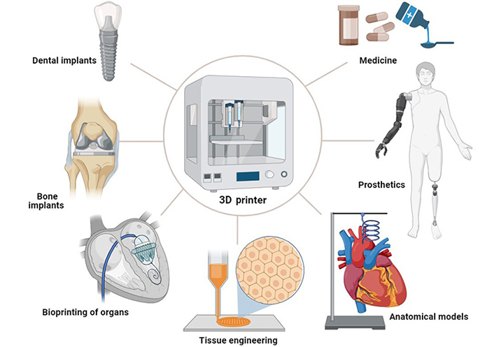
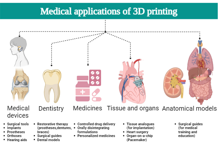
The realm of biomedical applications has been profoundly influenced by additive manufacturing, commonly known as 3D printing. This technology’s capability to construct intricate structures with exactitude and personalization has opened novel avenues in the realm of healthcare, spanning from crafting medical devices to the domain of tissue engineering. This piece delves into a selection of ongoing biomedical applications of 3D printing, accentuating pertinent instances. Foremost among these applications is the production of medical devices, a significant domain in biomedicine. Personalization stands pivotal in elevating patient outcomes, and 3D printing facilitates the creation of implants, prosthetics, and orthotics curated to each patient’s unique anatomy. For instance, cranial implants can be fine-tuned to correspond to individual anatomical attributes, ameliorating their aesthetic and functional aspects [47].
Furthermore, 3D printed prosthetics furnish economical alternatives, particularly suited for burgeoning children necessitating recurrent replacements. Additionally, 3D printing has unfurled advances in tissue engineering and regenerative medicine. One can progressively print structures that mimic tissues by utilizing bioinks containing live cells. This approach has proven its potential in crafting skin, bone, and cartilage constructs for transplantation. The ongoing pursuit in this sector envisions the eventual realization of fully operational transplantable organs. The arena of personalized drug delivery has been revolutionized by 3D printing, rendering bespoke drug delivery systems calibrated to individual patient requisites. Bestowing enhanced therapeutic efficacy [48], printers can fabricate intricate drug-laden configurations that dispense medications with precision. This platform also emboldens the creation of intricate dosage forms, including amalgamations of multiple drugs within a solitary tablet [49]. Translating medical imaging data into 3D models is a pivotal aid in surgical planning and medical education. Surgeons are empowered to rehearse intricate procedures on faithful replicas of anatomical structures, augmenting their skill sets and heightening patient safety [50]. These models concurrently nurture experiential learning among medical students, augmenting their comprehension and retention of anatomical intricacies and procedural nuances.
Bioprinting, a nascent yet promising realm, entails the methodical deposition of bioinks to fashion living structures (Figure 2). This domain exhibits immense potential in conjuring functional organs within laboratory settings. Researchers have successfully fabricated heart valves, blood vessels, and miniature organ simulations for drug testing. While challenges persist, encompassing aspects like ensuring vascularization and sustained viability, bioprinting emerges as a conceivable solution to alleviate the dearth of organ transplants. In summation, the advent of 3D printing has wrought transformative changes across the biomedical landscape, endowing innovative resolutions for medical device creation, tissue engineering, pharmaceuticals, surgical planning, and bioprinting. Its versatility coupled with its aptitude to craft intricate structures has fostered medical strides benefiting both patients and healthcare practitioners [49]. Zhou et al. [50] reported the substantial challenges posed by bone defects for patients, orthopedic surgeons, and healthcare resources. These defects can result from various conditions, including trauma, tumors, inflammation, and osteoporosis. While auto- and allograft transplantation have emerged as common clinical treatments, with autologous bone grafts as the gold standard, addressing bone defects—particularly large-volume defects in elderly patients or those complicated by systemic diseases—remains a clinical challenge in regenerative medicine. Fortunately, the rapid advancement of biomaterials and nanomedicine has opened doors to more efficient bone regeneration therapies. This review provides a concise overview of novel biomaterial and nanomedical approaches to bone regeneration and explores the ongoing clinical challenges that impede their widespread application in treating bone defects [50]. As technological strides continue, the horizon augurs potential for more sophisticated 3D printing applications within the realm of biomedicine [51]. Some of the significant biomedical applications of 3D printing are described below.
Integrating 3D printing technology into prosthetics and orthopedics has ushered in a new era of customization and innovation. This cutting-edge approach allows for the creation of personalized prosthetics and bone replacements specifically tailored to individual patients’ needs. Following the most recent software and 3D printing developments, the use of personalized orthopedic implants to treat complex surgical cases has gained more popularity. In orthopedics, 3D printing has revolutionized the approach to bone replacements [51, 52]. Traditional implants may not always align perfectly with a patient’s anatomy, leading to potential complications and discomfort. Through 3D printing, orthopedic surgeons can generate implants that precisely match the patient’s bone structure. This personalized approach contributes to better implant integration, reduced post-operative complications, and improved patient outcomes. The advent of modern 3D printing technology has made significant contributions to the field of orthopedics, particularly in enhancing our comprehension of intricate bone fracture patterns, streamlining surgical procedures, and ensuring precise implant placement in preclinical investigations [52, 53]. In the realm of orthopedic practice, a diverse array of implants is readily available for standardized surgeries related to bone substitution across various anatomical regions of the human body. Nevertheless, these standardized implants often fall short of providing effective solutions when confronted with non-traditional scenarios characterized by patients whose bony geometries deviate from the typical range of standard implant applications, whether in terms of implant size or specific disease-related requirements [54]. In such instances, salvage surgeries and arthrodesis are typically indicated, albeit with a notably low success rate and diminished patient satisfaction, particularly within the domain of hip surgery [48, 54]. The 3D printed materials for personal dentistry, their methods of production, and a comparison with conventional materials are summarized in Table 1.
Comparison of 3D printed dental materials, their production methods, and properties relative to conventional dental materials [48, 55, 56]
| Material | Production method | Properties (3D printed) | Properties (conventional) |
|---|---|---|---|
| Polymeric resins | SLA or DLP | Biocompatible, customizable, smooth finish | May lack customization, potentially less biocompatible depending on source |
| Ceramics | Binder jetting or SLA with ceramic-filled resins | High strength, esthetic, biocompatible | Highly esthetic, durable, but less customizable |
| Metal alloys (e.g., cobalt-chrome) | SLM or EBM | Durable, high tensile strength, customizable | Extremely durable, used for long-term restorations, not easily customizable |
| Thermoplastic materials (e.g., PEEK, PEKK) | FDM | Durable, biocompatible, flexible, good for temporary prosthetics | Durable, but more suited for mass production |
| Wax | FDM or SLA/DLP with wax-like resins | Useful for casting applications, easy to melt and shape | Traditional in lost-wax casting, manually shaped |
| Bioinks (for tissue engineering) | Bioprinting | Can incorporate live cells, for potential tissue regeneration | Traditional tissue grafts, less customizable |
| Composite resins | SLA/DLP with composite resins | Combination of ceramic and polymer; esthetic, durable | Often hand-layered, with good esthetics but potentially less precision |
PEEK: polyetheretherketone; PEKK: polyetherketoneketone; DLP: digital light processing; SLM: selective laser melting; EBM: electron beam melting
Traditional prosthetics often come with limitations in terms of fit, comfort, and functionality. However, with 3D printing, the design and production of prosthetic limbs can be meticulously tailored to each patient’s unique anatomy. This level of customization not only ensures a better fit but also enhances the overall comfort and usability of the prosthetic. Patients can experience improved mobility and quality of life due to prosthetics that closely mimic the function and appearance of natural limbs. Custom-fabricated 3D printed prostheses offer valuable utility in the surgical management of bone defects manifesting in diverse anatomical locales or instances characterized by anatomical irregularities, rendering conventional implant deployment impractical [53, 54]. In the context of patient-specific conditions, it is noteworthy that the imposition of silver ion coatings upon the surface of these bespoke implants holds the potential to mitigate infection rates. Furthermore, the acceleration of the osseointegration process can be realized through the strategic application of hydroxyapatite surface coatings within the bone-implant interface zones. In the domain of joint replacement implants, a metal-polyethylene interface exhibits utility [48, 54]. In light of their personalized nature, custom-fabricated implants harmoniously conform to the unique anatomical contours of the patient, facilitating seamless intraoperative implantation and concomitantly fostering substantial enhancement in functional outcomes [48, 55, 56].
Peng et al. [57] addressed the challenges of repairing articular osteochondral defects, which are hindered by the complex tissue structure and limited chondrocyte proliferation. Traditional clinical treatments, including microfracture, osteochondral transplantation, and cell-based approaches, have shown limited efficacy. Consequently, the study explores tissue engineering as a solution, harnessing biomaterials’ regenerative potential to control cell behavior. Osteochondral tissue’s gradient structure, involving changes in various factors, necessitates bioinspired gradient scaffolds that mimic these characteristics. Such scaffolds enhance osteochondrogenesis and promote the formation of osteochondral interfaces, outperforming homogeneous scaffolds. They investigated these strategies and assessed their potential for clinical application [57]. The contemporary design of 3D printed personalized implants encompasses both precise structural geometries tailored to individual patient needs and the capacity for biomechanical assessment under patient-specific loading conditions, enabling pre-fabrication design modifications to potentially reduce pain, expedite recovery, enhance osseointegration, and optimize overall functional outcomes [57, 58]. The utilization of 3D printing technology presents a range of significant benefits within the prosthetics and orthopedics realm. Firstly, its precision facilitates the crafting of intricate and highly accurate designs, ensuring a seamless integration of prosthetics and implants with the patient’s individual anatomy. This process is further enhanced by the capacity for customization, wherein the unique anatomical features of each patient can be meticulously incorporated into the design and fabrication, consequently enhancing both comfort and functionality. Speed also emerges as a key advantage, as 3D printing enables swift prototyping and production, effectively minimizing the temporal gap between diagnosis and implantation. Notably, the technology contributes to reduced waste generation compared to conventional manufacturing methods, given its ability to optimize material usage and align with more sustainable practices. Beyond these practical benefits, 3D printing fosters an environment of innovation, permitting researchers and designers to explore novel materials and structures that have the potential to catalyze advancements in the realm of prosthetic and orthopedic technologies.
Dental disorders have emerged as the most significant barrier to overall human health and wellness. The evolution of 3D printing technologies has led to their widespread application in orthodontics, restoration of edentulous arches, and root canal treatments [59].
The advancements in precision medicine suggest that comparable headway has been achieved in the realm of oral orthodontics. These advancements are expected to be progressively implemented to achieve personalized and tailored treatment approaches, leading to enhanced treatment efficiency [60]. Prior research aimed to establish a foundation for the evolution of computer technology and biomedical science. They are also concerned with their present and potential applications to precise dental orthodontics. In the future decades, the potential for individualized care and biomechanical planning provided by 3D imaging technology and advancements in computer hardware and software will be more completely realized. When coupled with 3D printing, these tools have already been harnessed to tailor the production of devices like aligners and retainers [61]. The desirable characteristics of smart devices and mechanical properties in suitable materials serve as compelling indications for the foreseeable potential of tailoring the production of orthodontic brackets. In the realm of biomedicine, researchers are actively pursuing a foundational understanding of cartilage growth and bone biology within animal organisms. This exploration pertains to the alteration of mandible growth and the regulation of tooth movement. This area of study holds the potential for significant advancements, such as changes in growth patterns, the acceleration of orthodontic tooth movement, and the improvement of tooth stabilization and preservation. These advancements could eventually yield important applications within oral orthodontics. The field of orthodontics stands to gain from these emerging discoveries. As additional genomic and proteomic information becomes available, orthodontic diagnoses and treatments can become even more tailored and precise. In the upcoming decades, the concept of precision orthodontics is poised to continue benefiting from progress across diverse disciplines. This entails incorporating technological breakthroughs, amalgamating insights from biomedical and clinical research, and ultimately fostering a framework for personalized orthodontic treatments that are characterized by excellence, efficiency, safety, reliability, and reproducibility [62]. In addition, 3D force plays a crucial role in predicting tooth movement during orthodontic therapy.
With dental implants, the edentulous mandibular arch may now be restored in a predictable and satisfying manner. The focus of early implant research has been on bone integration. Advancements have been made in the realm of digital technology, specifically in the enhancement of digital 3D imaging and CAD, along with technologies such as cone beam CT (CBCT) and 3D printing. These technological improvements have been integrated with the concept of digital guided surgery [63]. This innovative approach holds the promise of achieving accurate and streamlined surgical implant procedures. Furthermore, these techniques have the potential to reduce the necessity for extensive surgical intervention, making them less invasive compared to conventional implant methods. This also leads to decreased post-operative discomfort and shorter healing periods.
Root canal therapy is an approach employed to manage the microbial ecosystem residing within a tooth by disinfecting, shaping, and effectively sealing the root canal system to promote healing around the apex of the tooth’s root. The presence of calcification within the pulp cavity and root canal system can impact this process, potentially complicating the root canal treatment. This calcification, also known as occlusion or calcification, emerges due to extensive damage to the tooth’s pulp, often caused by factors like dental diseases (caries, loss of tooth structure), trauma to the alveolar ridge, or certain surgical interventions like pulp covering, pulp removal, or the absence of orthodontic treatment [64]. It’s worth noting that although canal obliteration doesn’t inevitably lead to the death of the pulp or the development of periapical diseases, its presence significantly amplifies the intricacy of locating and navigating the root canal. The level of complexity is contingent on the inherent morphology of the tooth, the nature and extent of the calcification overlay, and even the stiffness of the tooth. Additionally, the challenges posed by the entry of teeth into the oral cavity cannot be overlooked.
Past research has indicated that while the precision of the printing system has its limitations, the provisional materials suitable for 3D printing can still be employed within the mouth due to their mechanical properties that align well with oral requirements.
Temporary restorations play a critical role in several aspects, such as safeguarding pulpal and periodontal tissues, upholding oral functionality, and maintaining aesthetics. To accomplish this objective, significant emphasis needs to be placed on guaranteeing the form and appropriateness of these interim restorations. The adequacy of the restoration is predominantly reliant on the fabrication process [65]. Based on the manufacturing approach, the methods employed to create temporary crowns can be categorized into direct and indirect techniques. Using the direct approach, the provisional crown is promptly fabricated directly onto the prepared teeth. Conversely, the alternative technique involves casting the temporary crown on a stone model before placing it in the mouth. Nonetheless, the heat generated during the polymerization of resin in the direct method could potentially cause heat-related injury to the dental pulp. Furthermore, any remaining resin monomers might harm the oral mucosa, potentially resulting in moss-like reactions or allergic stomatitis.
The technique of 3D digital mapping is utilized to prevent inadvertent harm to the root during root canal therapy by guiding around vanishing ductwork. This method ensures avoidance of iatrogenic damage to the root. Through a combination of CBCT scans and intraoral scans of the dental structure, computer software aids in creating a digital treatment plan for complex root canal systems in severely occluded anterior teeth. This plan serves as the foundation for digital design and 3D printing, which produces an endodontic guide for the intended treatment [66]. The 3D printed template assists in precisely directing a customized drill hole to the root canal orifice. This stands in stark contrast to the traditional approach, where root canal treatment is carried out after the root canals have been negotiated.
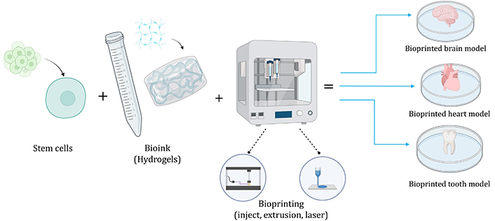
The concept of bioprinting essentially extends from additive manufacturing techniques utilized for constructing intricate tissue structures in a layer-by-layer manner [67]. Broadly, this procedure can be delineated into three key stages [68, 69]: A) preprocessing, involving the preparation of bioink and the creation of a CAD blueprint; B) processing step, which typically encompasses the 3D printing of the structure; and C) postprocessing, where the printed construct is cultivated within a bioreactor. The inclusion of the postprocessing phase primarily aims to promote the maturation of the printed structure, enabling its evolution into a functional tissue. Various groups have compiled the requirements for creating biological tissues and organs (Figure 3) [70]. The organization and resolution needed on a microscale, however, are in dispute. Categorized according to their operational principles, 3D bioprinting technologies can be predominantly divided into three groups: inkjet-based, extrusion-based bioprinting, and laser-assisted.
In 1988, the inception of bioprinting occurred through the utilization of a bioink solution containing collagen and fibronectin, ingeniously adapted for use in a commonly available Hewlett-Packard inkjet printer [71]. This groundbreaking technique involves the precise layer-by-layer deposition of organized cell assemblies onto a scaffold or substrate, typically a hydrogel, utilizing principles akin to traditional inkjet printing. This process involves loading cellular material into a specialized cartridge. These scaffolds play a pivotal role by providing nourishment and anchoring sites, thereby facilitating cellular proliferation and the development of targeted tissue structures. However, it’s worth noting that the inkjet-based approach, despite its ability to achieve resolutions as fine as 50 μm, is constrained by its reliance on pressure-based mechanisms, resulting in suboptimal support for extremely low cell densities, specifically less than 106 cells per mL [72]. Despite its cost-effectiveness and promising viability of enclosed cells, the application spectrum of inkjet bioprinting remains limited, particularly when it comes to the fabrication of intricate, cell-rich architectures [73]. The demand for the production of meticulously detailed structures that faithfully replicate native tissue poses a formidable challenge within the realm of clinical applications. This intricate requirement tends to be elusive when employing inkjet technology as the primary tool for bioprinting.
Enabling the precise placement of living cells onto designated positions while enveloping them within a hydrogel matrix, extrusion-based bioprinting technologies have emerged as a noteworthy advancement. Presently, these technologies, which involve a syringe, nozzle, and pressure system, exhibit considerable promise as the favored approach for fabricating 3D tissue or organ constructs that match clinically relevant dimensions and forms [67]. Before initiating the printing process using this method, cells or proteins are ensconced within a hydrogel and loaded into sterilized syringes equipped with micronozzles. Subsequently, the hydrogel-incorporated cells or cell spheroids are dispensed onto the substrate in accordance with a customized design, achieved through either air pressure or the controlled movement of a motorized plunger.
Laser-assisted bioprinting (LaB) employs a pulsed laser source, an absorption layer, and a substrate to position multiple cells and biological components with precision directly onto a chosen surface. This process employs laser beams to create intricate patterns of living tissues or organs [74]. Before the laser comes into play, the transparent absorption layer, designed to be compatible with the laser’s wavelength, is coated with biological materials, often referred to as bioink, which encloses the living cells and/or proteins. Subsequently, a focused laser beam is directed onto the absorption layer, generating heat that propels the cell suspension towards the substrate [67]. The absorption layer functions crucially to prevent direct interaction between the laser and the biological materials. LaB showcases the capacity to produce small volumes of cell suspension with exceptional precision [75]. The volume of the printed droplets, ranging from 10 pL to 7,000 pL, can be finely controlled by manipulating the viscosity and thickness of the bioink layer. Additionally, LaB enables the printing of high cell densities and viscous hydrogels, which poses a challenge for other techniques like inkjet printing [76]. Recently, LaB has found widespread application in the creation of diverse tissue constructs [77]. Notable instances include attempts at bone regeneration through the printing of human osteoprogenitors alongside nanohydroxyapatite, an essential inorganic bone component [74]. In the context of skin regeneration, keratinocytes and fibroblasts embedded in collagen were printed to replicate the natural cellular arrangement of skin. Furthermore, the potential of LaB in adipogenesis was showcased by printing undifferentiated stem cells sourced from human adipose tissue, which exhibited the ability to develop into adipogenic lineages [78]. However, concerns persist around the limited availability of suitable photocurable materials and the cytotoxic effects stemming from ultra violet (UV) exposure, warranting attention [79]. Some of the commonly used bioinks (Table 2), bioprinting techniques (Table 3), and bioprinted organs and their current stage (Table 4) are listed below.
| Bioink material | Properties | Common use |
|---|---|---|
| Alginate | Biocompatible, gels rapidly | Cartilage, vascular tissues |
| Gelatin | Thermoresponsive, biodegradable | Skin, liver |
| Collagen | Native extracellular matrix component, supports cell growth | Various tissues |
| Hyaluronic acid | Hydrophilic, biodegradable | Cartilage, skin |
| Technique | Principle | Best for |
|---|---|---|
| Inkjet bioprinting | Uses droplets of bioink | Thin tissues, high-resolution structures |
| Extrusion bioprinting | Continuous stream of bioink | Vascular structures, thicker tissues |
| LaB | Laser pulses to deposit bioink | High precision structures |
| Organ | Current stage |
|---|---|
| Skin | Clinical application |
| Heart | Experimental |
| Kidney | Pre-clinical trials |
3D bioprinting harnesses the technique of layer-by-layer deposition to fabricate tissue-like structures from biological materials, with the ultimate goal of producing functional organs for transplantation. This intricate process begins with organ design using CAD software, followed by the selection of cell-infused bioinks, precise layering through advanced printers, and final maturation in bioreactors. As of now, the technology is employed for drug testing tissue models, and while simpler tissues like skin and cartilage have been successfully bioprinted, more complex organs like the heart and kidneys remain in the research phase. The path forward faces challenges in ensuring sufficient vascularization, post-transplant organ functionality, and achieving the right organ size while preserving cell viability. Nevertheless, as this technology evolves, the medical field anticipates a transformative shift towards the routine use of bioprinted organs, potentially revolutionizing organ donation and transplantation [67, 73].
Combining 3D printing technology with individualized drug delivery has resulted in an innovative method that offers customized medications and smart release systems. This convergence is poised to revolutionize drug delivery, offering enhanced treatment precision and patient-centric therapeutic outcomes [80, 81]. In addition to tailored medications, 3D printing brings forward innovative release techniques, adding a fresh perspective to drug delivery. Traditional formulations might need to align with individual needs, leading to consistent dosing or suboptimal administration. Personalized delivery mechanisms encompass controlled release, targeted delivery, and adaptable dosing. These mechanisms enable medications to be released in response to specific triggers or physiological cues, ensuring the highest therapeutic impact while mitigating potential side effects [81]. The recent FDA approval of the 3D printed drug product Spritam® has ignited a surge of interest in 3D printing technology. This innovative approach is poised to reshape pharmaceutical product development, particularly in personalized therapy [82]. With the ability to adapt from early development to end-product manufacturing, 3D printing is becoming a game-changer, offering simplified design and production cycles. This method has garnered popularity for its automated process and cost-effectiveness, surpassing traditional manufacturing methods, especially in on-demand production [81, 82]. The applications of 3D printing in personalized medicine are summarized in Table 5.
| Application area | Description | Benefits |
|---|---|---|
| Drug dosage forms | Customized drug release profiles and dosages | Tailored drug delivery, potential for improved patient adherence |
| Medical implants | Patient-specific implants (e.g., hip, dental, cranial plates) | Perfect fit, reduced surgery time, improved outcomes |
| Prosthetics | Custom-made prosthetic limbs or parts | Better fit, improved mobility, enhanced patient comfort |
| Hearing aids | Tailored hearing devices | Improved comfort and acoustic performance |
| Bioprinted tissues/organs | Printing cells and biomaterials for tissue regeneration | Potential solutions for organ donor shortages, tailored tissue grafts |
| Disease models | 3D printed tissues for drug testing and research | Accurate disease representation, improved drug testing accuracy |
| Personalized medical devices | Custom-fit devices (e.g., orthotics, braces) | Enhanced comfort, optimized functionality |
| Anatomical models | Patient-specific models for surgical planning | Improved surgical preparation, reduced operation time |
At its core, 3D printing entails the layer-by-layer deposition of materials based on digital designs, allowing for the creation of intricate 3D objects. The concept originated in the 1980s, and it was initially used for prototyping purposes in fields like automotive and aerospace. However, the pivotal moment came with the FDA approval of Spritam® in 2015, marking a new era for 3D printing in pharmaceuticals. Among various 3D printing techniques, FDM stands out [81, 82]. This technology’s potential lies in its capacity to enable precision medicine, tailoring therapeutic approaches to individual patients’ unique physiological and lifestyle requirements. Conventional pharmaceutical tablet manufacturing often employs a one-size-fits-all approach based on phase 3 clinical studies. However, this method can lead to suboptimal dosing, potentially causing toxicities, adverse events, or reduced therapeutic efficacy. 3D printing offers a solution by allowing selective material deposition and precise control of factors, including API compartmentalization for drug combinations [83, 84]. Advantages of 3D printing, over traditional methods include personalization, enhanced complexity, and on-demand manufacturing. It empowers dose tailoring based on factors like body mass index, metabolism, and genetic variations, leading to improved treatment adherence. Complex designs, like multi-drug doses, can also be effortlessly manufactured, enhancing treatment efficacy.
Additionally, 3D printing’s adaptability to on-demand manufacturing holds promise for resource-constrained settings, such as emergencies or disaster zones, minimizing wastage and maximizing efficiency. 3D printing is revolutionizing pharmaceutical production, particularly in the fields of prosthetics and orthopedics. It is a new era of personalized solutions, intricate designs, and efficient on-demand manufacturing. As technology evolves, this approach is set to reshape the pharmaceutical landscape, driving improved patient outcomes and resource utilization [81, 85]. The integration of 3D printing into personalized drug delivery offers a range of distinct advantages. Firstly, 3D printing enables the precise crafting of medications tailored to the unique profiles of individual patients, thereby optimizing treatment outcomes through personalized approaches. Secondly, the design of customized medications promotes seamless integration into patients’ daily routines, boosting adherence and overall treatment effectiveness. Thirdly, by refining dosages based on individual attributes, personalized drug delivery via 3D printing mitigates adverse effects and enhances treatment safety. Fourthly, the alignment between medications and patients’ specific parameters guarantees superior therapeutic outcomes, ultimately contributing to improved disease management and heightened patient satisfaction. Lastly, the pursuit of personalized drug delivery using 3D printing has spurred a wave of innovation in drug formulations, fostering the development of advanced delivery systems catered to the precise needs of individual patients [86].
The process of utilizing FDM 3D printing to produce personalized drug products from digital designs involves a sequential workflow encompassing five critical stages [80–82, 86]. First, the design phase involves creating a 3D digital model of the dosage form tailored to the specific patient’s needs. Following this, the designed 3D models are converted into STL format files, which are compatible with the 3D printer. The third stage involves setting precise printing parameters and segmenting the STL file into a layer-by-layer design, essentially “slicing” it for the printing process. Subsequently, the feedstock material, used to craft the drug product, is meticulously formulated and prepared for fabrication. Finally, in the fifth and last stage, the actual printing of the dosage form takes place, followed by a rigorous evaluation process to ensure its quality and functionality [86].
3D printing’s profound impact on personalized medicine arises from its unique capability to craft individualized solutions, often informed by precise patient data sourced from imaging techniques like magnetic resonance imaging (MRI) and CT scans. However, this promising landscape isn’t without its challenges: regulatory barriers, concerns about biocompatibility, and existing technological constraints present hurdles. Nevertheless, ongoing research and innovation in this domain hold the promise of cementing 3D printing’s pivotal role in advancing personalized medical care.
The utilization of personalized 3D models tailored to individual patients’ needs has been documented across various medical and surgical fields. Cardiology has benefited from 3D models to comprehend the complicated and diverse aspects of congenital heart conditions. These models have also aided in sizing implantable devices for procedures like closing the left atrial appendage. In the domain of neurosurgery, 3D models have proven crucial in planning surgical approaches and providing real-time guidance during operations involving complex skull-base tumors and cerebrovascular aneurysms. Orthopedic cases, often involving reconstructions and hardware, have harnessed 3D models to visualize anatomical structures, select appropriate implant sizes, and chart drilling paths. These models have significantly contributed to surgeries addressing challenges such as acetabular defects and scoliosis [87]. Similarly, otolaryngology and craniomaxillofacial surgery have utilized 3D models to simulate surgeries and customize reconstruction plates for procedures involving the mandible, orbital, and other craniofacial reconstructions. In 2017, Marconi and his team [88] revealed that 3D printed models offer a swifter and clearer grasp of surgical anatomy. This aids medical students, surgeons, and radiologists in spending less time assessing these models compared to interpreting traditional two-dimensional CT scans and 3D virtual reconstructions.
In the ever-evolving landscape of modern medicine, the integration of 3D technology with personalized patient care has given rise to a new era of innovation in the development of custom tools and patient-specific surgical models. This shift in approach is poised to redefine medical procedures by tailoring interventions to individual patients, thus optimizing precision and ultimately elevating the overall outcomes of treatments. Custom instruments offer remarkable benefits across various medical fields. In orthopedics, for instance, surgical tools are now designed with meticulous attention to a patient’s unique anatomy, ensuring precise alignment and improving joint function in procedures like knee replacements [89]. Similarly, in dentistry, 3D printed surgical guides, tailored to a patient’s oral structure, are revolutionizing processes like dental implant placement [90].
Neurosurgery also benefits from specialized tools that navigate individual cranial contours, minimizing risks in complex procedures [91]. Also, patient-specific surgical models have proven very useful, particularly in complex surgeries. In cardiovascular surgery, these models provide comprehensive insights into a patient’s cardiac anatomy, enabling meticulous preoperative planning and guiding precise interventions. From addressing cardiac anomalies to enhancing complex valve repairs, patient-specific heart models have the potential to significantly reduce surgical risks and streamline procedures [92]. Neurosurgeons now have the advantage of patient-specific 3D printed brain models, allowing them to simulate tumor removals and protect vital brain functions during surgery. Such models also aid in proactive preparations for surgeries like facial reconstruction, where practicing on tailored models ensures surgical finesse and minimizes complications.
The benefits of these advancements are manifold. The fusion of custom tools and patient-specific models results in more precision, reducing the likelihood of errors during procedures. This approach not only enhances accuracy but also leads to more time-efficient surgeries. Surgeons armed with comprehensive insights from patient-specific models can perform procedures more efficiently, thus minimizing operation times. Beyond the immediate operating room applications, these technologies serve as invaluable educational tools for aspiring surgeons, enabling them to refine their skills on lifelike models before encountering real-life scenarios. However, these advancements are not without challenges. Developing and implementing custom tools and models comes with financial considerations, necessitating innovative solutions to ensure accessibility across diverse medical settings. Additionally, the introduction of personalized tools and models requires rigorous validation and regulatory approvals to guarantee patient safety and procedural effectiveness. Looking ahead, the potential for custom tools and patient-specific models is vast. As advancements continue, their applications are likely to extend to a broader range of medical specialties, transforming surgical approaches across various disciplines [92].
Incorporating 3D printed anatomical models into medical practice empowers healthcare practitioners with immersive and tangible resources for comprehending detailed anatomical structures. Surgeons are bestowed with the capacity to meticulously plan and simulate complex procedures on patient-specific models, thus augmenting their proficiency in dealing with individualized anatomical variations. Moreover, medical pedagogy is significantly enriched as these models offer hands-on learning opportunities, fostering a deeper grasp of complex physiological nuances among aspiring medical professionals.
3D anatomical models have become essential tools in medical education and practice, offering a dynamic bridge between theory and real-world application. These models transform learning by providing hands-on insights into complex anatomy. For instance, cardiac education benefits from 3D heart models, aiding understanding of intricate structures and diseases. Surgical precision is enhanced through customized bone models for procedures like joint replacements. These models extend to neurosurgery, where they aid visualization during significant interventions. Patient education is elevated too, as these models offer tangible explanations of health conditions. Ultimately, 3D anatomical models exemplify technology’s role in reshaping medical education, surgical planning, and patient engagement [93]. The utilization of 3D planning and printing technologies has seen a growing trend in the field of revision total hip arthroplasty (THA). Employing 3D simulations and models has emerged as a valuable strategy for aiding implant positioning, especially in complex cases of revision THA. These advanced technologies offer a precise platform for comprehensive preoperative planning and real-time intraoperative guidance, proving particularly beneficial in addressing complex acetabular defects. Empirical investigations have consistently demonstrated that the incorporation of 3D models contribute to improved surgical outcomes and a marked reduction in complications associated with revision hip arthroplasty [94]. Simulation practices offer several advantages, including a secure practice environment where medical professionals can enhance their clinical skills. Through virtual patient interactions, augmented reality, and high-fidelity mannequins, these practices replicate real-world medical scenarios. This enables medical practitioners to refine their skills and decision-making without jeopardizing patient safety, while also fostering critical thinking abilities by emulating diverse medical situations and emergencies. The integration of models and simulations brings forth comprehensive learning, bridging the gap between theory and practice to enrich medical education. Medical students delve deeper into anatomy insights, while practitioners hone their skills and techniques [91]. These tools support continuous learning, extending beyond initial education to facilitate ongoing professional development and aiding the adoption of emerging medical technologies and techniques. However, challenges remain. The accuracy and authenticity of anatomical models necessitate meticulous attention to detail, and simulations must closely mimic clinical settings for realistic decision-making. Incorporating feedback mechanisms is crucial to identify areas for improvement and enhance the learning experience. Way forward, anatomical models and simulations hold promise for transformative learning experiences, reshaping medical education through immersive approaches. These tools contribute to nurturing competent, confident, and compassionate medical professionals, while the synergy between technology and medical knowledge continues to evolve, further enhancing learning endeavors.
Personalized medicine stands as a transformative force in healthcare, aiming to tailor pharmaceutical treatments to individual patients by factoring in their unique physiology, drug responses, and genetic profiles, shifting away from the traditional “one size fits all” approach. Among the emerging technologies driving this shift, 3D printing holds a prominent position [95]. 3D printing involves the progressive layering of materials, guided by sophisticated computer software, to fabricate intricate 3D objects. Its versatile applications in pharmaceuticals encompass the creation of drug dosage forms with diverse shapes, release profiles, and drug combinations. 3D printing in the pharmaceutical sector holds significant promise in the realm of personalized medicine. It possesses the capability to customize dosage forms to suit individual patient requirements. This can be achieved through the creation of tailored dosage forms, dosage adjustments, combining multiple medications, or altering the release profiles of these dosage forms, all in accordance with the specific needs of patients [96].
3D printing offers considerable potential in achieving dose flexibility tailored to individual patient needs, with a particular focus on population groups like pediatrics, where therapeutic doses vary based on factors such as age and body weight. Within this context, various dosage forms mentioned earlier can be effectively adapted using 3D printing technology to deliver the precise dose required for patients. For instance, in the case of orally disintegrating film (ODF) formulations, this adaptability is easily realized by adjusting the quantity of liquid API dispensed onto the film. Furthermore, ODFs can undergo alterations in shape and dimensions to individualize treatments. Similarly, dose strength can be modified in other dosage forms such as tablets or patches to meet specific patient requirements. An illustrative example includes the work of Awad et al. [97], who utilized FDM and HME to 3D print theophylline tablets with doses ranging from 60 mg to 300 mg by manipulating the printing scale. In the past years, achieving dose flexibility involved manual tablet splitting or the use of tablet splitters, which has proven ineffective due to variations in the characterization parameters of the divided tablets, often falling short of pharmacopoeial standards. Zheng et al. [98] reported that split tablets with 3D printed subdivided tablets, ultimately concluding that 3D printed subdivided tablets were more accurate, safer, and held significant potential for customization [96]. Additionally, 3D printed pellets or mini-printlets, akin to mini-tablets, have been developed, offering a personalized approach [96]. These mini-printlets can also serve the purpose of combining two different drugs, further enhancing personalization possibilities. Moreover, mini-printlets can be combined and encapsulated according to the specific dosage requirements to achieve a high degree of personalization [96].
3D printing offers a versatile approach for tailoring drug release profiles to meet specific individual requirements. One effective method involves the manipulation of tablet shapes and geometries. For instance, the creation of immediate-release tablets for low-dose drugs has shown that reducing tablet thickness or introducing voids within the tablets can significantly enhance drug release rates, even achieving complete release within as little as 5 min. In another study, paracetamol tablets were crafted into various shapes, including cube, disc, sphere, pyramid, and torus, revealing that drug release could be finely tuned by altering the surface area-to-volume ratio. Complex geometric structures, such as honeycomb-shaped tablets, produced via 3D printing, with variations in honeycomb cell sizes, allowed for the attainment of diverse release profiles, showcasing the potential of dosage form geometries in influencing drug release [96].
Additionally, 3D printing has facilitated the creation of tablets with intricate release profiles, combining multiple release mechanisms within a single dosage form. These innovations include immediate-extended release tablets with dual pH-based release mechanisms, breakaway tablets comprising three sections, two of which erode within 45 min in a gastrointestinal environment, and enteric dual pulsatory tablets with two release pulses at 1 h and 8 h. Moreover, dual pulsatory tablets with sections exhibiting opposing pH-based solubility patterns have been developed, enabling one section to erode during the acidic dissolution stage within 30 min, while the other begins eroding 5 h later in the higher pH stage. Beyond solid tablets, 3D-designed patches have also explored shape variations, resulting in hydrogel patches of cylinder, torus, and gridline shapes, each exhibiting distinct drug release patterns. Optimized capsular devices have been engineered to regulate drug release from immediate-release tablets within them while being suspended in gastric fluid. Furthermore, 3D printing has been employed to fabricate osmotic dosage forms, where the alteration of the shape of the cellulose acetate shell surrounding the osmotic core was used to modify drug release. Coated tablets have demonstrated that factors such as the extent of tablet coating, the number of coats, and the sides of the tablets coated significantly influence drug release profiles [99]. Excipients used in 3D printing formulations have also been found to modulate drug release. Studies have shown that the percentage of cross-linkable polymers in tablets can impact drug release rates, with higher ratios of poly(ethylene glycol) diacrylate leading to decreased dissolution rates, while increased concentrations of polyethylene glycol 300 (PEG 300) enhance drug release. Similarly, the quantity of hydroxypropyl methylcellulose (HPMC) in the formulation of 3D printed tablets has been found to retard drug release, further highlighting the versatility of 3D printing in tailoring drug release profiles through variations in structure, coating, and excipient composition [95, 96].
3D printing in personalized medicine offers a groundbreaking application known as “polypills”. These innovative tablets combine multiple drugs into a single dose, allowing tailoring to individuals with complex medication needs, especially benefiting geriatric populations by simplifying medication regimens [10]. Khaled et al. [36] successfully 3D printed polypills, one with three drugs for potential use in diabetes and hypertension, featuring an osmotic compartment for captopril and sustained release compartments for nifedipine and glipizide. Another polypill from the same team included five compartments designed for cardiovascular therapy, incorporating aspirin, hydrochlorothiazide, pravastatin, atenolol, and ramipril. Another research group explored PVA-based polypills containing four drugs (lisinopril, amlodipine, rosuvastatin, and indapamide) with variations in multilayer and unimatrix structures. Unimatrix tablets exhibited slower drug release compared to individual tablets, while multilayered polypills had varying drug release influenced by their location within the structure [35].
Robles-Martinez et al. [100] utilized SLA 3D printing to create multilayered polypills containing six drugs with different geometries, such as cylindrical and ring-shaped configurations. The 3D printer was adapted to change resin solutions during printing [96]. Advancing this concept, 3D printing was used to develop polypill capsules with customizable release profiles by combining FDM with hot-filling syringes. Two capsule designs featured four separate compartments each, with one having a concentric configuration for early and delayed release and the other adopting a parallel configuration with non-dissolving capsule shells, free passageways, and dissolution rate-limiting pores for varied release profiles. These capsules were composed of PVA and polylactic acid, and their release profiles were tailored by adjusting shell thickness or orifice size [96, 97].
3D printing in biomedicine presents a myriad of advantages that are reshaping the healthcare sector. A primary benefit is its capacity for customization, allowing for patient-specific solutions that lead to enhanced treatment effectiveness and patient outcomes. Additionally, while the initial setup may be costly, the technology promises long-term cost-effectiveness by enabling on-site production of intricate medical components [91]. The rapid prototyping capabilities of 3D printing not only facilitate the swift development and production of medical tools and devices, especially vital in emergencies but also democratizes healthcare by making high-quality solutions accessible in underserved areas. Beyond direct patient care, the innovative potential of 3D printing propels advancements in medical research and drug discovery, with researchers utilizing it to produce accurate models of biological structures, thus broadening our comprehension of diseases and paving the way for new treatments. In essence, 3D printing’s myriad benefits range from tailored patient care and cost reductions to accelerating production and pioneering medical innovations, underscoring its transformative role in the future of medicine [35, 96]. Nanomaterials in medicine necessitate thorough nanotoxicological assessments for safe application in living organisms. Artificial intelligence (AI) and machine learning (ML) offer robust tools to analyze vast datasets in toxicology, including toxicological databases and high-content image-based screening data. Predictive models like physiologically based pharmacokinetic (PBPK) and nano-quantitative structure-activity relationship (QSAR) models aid in forecasting nanomaterial behavior and toxic effects, respectively. Nano-QSAR modeling encounters numerous hurdles, stemming from the intricate characteristics of nanomaterials, the limited availability of high-quality experimental data, a lack of industry-standard protocols, the potential for overfitting, and the dynamic nature of nanoparticle interactions within evolving environmental conditions and biological systems. These intricacies present substantial challenges in achieving precise nanotoxicity predictions. To tackle these obstacles effectively, it is imperative to meticulously curate training data and select appropriate structural descriptors, while also embracing novel computational methodologies tailored for diverse datasets. Additionally, harnessing cutting-edge ML algorithms and implementing stringent validation techniques holds the potential to significantly improve the accuracy and reliability of nano-QSAR models [100].
Recent advancements in orthopedic prosthesis design have notably enhanced the quality of life for individuals with orthopedic disabilities. Nevertheless, several substantial challenges remain that necessitate attention to further augment prosthesis functionality. Key areas of concern encompass the improvement of biocompatibility to facilitate seamless integration with natural tissues, enhancing durability to withstand the rigors of daily use, and refining sensory feedback mechanisms for better movement control. Addressing these issues has given rise to promising emerging solutions, including smart prosthetics, 3D printing, regenerative medicine, and AI. These innovative technologies hold significant potential for elevating the capabilities of orthopedic prostheses. Realizing the full potential of these next-generation orthopedic prostheses entails addressing critical factors such as fostering interdisciplinary collaboration among experts in orthopedics, materials science, biology, and engineering, increasing investments in research and development, standardizing components to ensure quality and reliability, and improving accessibility to prosthetic solutions [101].
Material biocompatibility is an overarching concern in the realm of 3D printing for biomedical applications, wielding a significant influence on the safety and efficacy of medical devices and implants. These challenges span a spectrum of factors that intricately shape the healthcare landscape. The selection of materials for 3D printing poses multifaceted challenges. The inherent variability in patient responses necessitates a quest for universally biocompatible materials. Furthermore, the limited availability of suitable materials constrains design possibilities and can elevate costs. Materials can undergo degradation over time, raising the specter of adverse reactions. Ensuring sterility throughout the 3D printing process, especially for complex structures, is another formidable challenge [35, 102]. Postprocessing steps like sterilization can impact material properties, further complicating biocompatibility considerations. Material toxicity concerns stem from residual chemicals and degradation byproducts. Mismatched mechanical properties and surface finish issues can lead to complications like wear and infections. Long-term biocompatibility challenges include potential inflammatory responses and rare allergic reactions to certain materials. Regulatory compliance demands stringent testing, adding complexity and cost. While ongoing research explores 3D printed material biocompatibility, numerous uncertainties, especially regarding long-term effects, remain. The impact of these challenges on 3D printed medical devices and implants is profound, potentially leading to adverse outcomes, regulatory complexities, and ongoing material safety research efforts. Manufacturers must prioritize robust research on material biocompatibility, adherence to stringent testing protocols, and vigilant monitoring of emerging concerns related to materials to ensure the safety and efficacy of these transformative healthcare innovations [101].
Despite the transformative potential of 3D printing in biomedicine, it grapples with multifaceted challenges. Technical constraints, such as printer resolution limitations and material incompatibility, can compromise the precision and efficacy of medical products. Regulatory intricacies further compound these challenges, with the evolving nature of 3D printing demanding robust frameworks to ensure the balance between innovation and patient safety. Moreover, the groundbreaking realm of bioprinting, which hints at organ replacements, ignites profound ethical debates, particularly around the manipulation of living tissues and the fundamental value of life. Beyond these immediate challenges, there’s an imperative to understand the long-term implications and safety of 3D printed medical solutions, ensuring their durability and biocompatibility over extended periods. Therefore, while 3D printing promises a healthcare revolution, it mandates a judicious blend of technological advancement, regulatory diligence, and ethical contemplation to truly benefit patients and uphold societal values [103].
The rapid evolution of 3D printing is poised to redefine biomedicine, marking an epoch of unprecedented healthcare transformation. At the helm is bioprinting, which is inching closer to fabricating full organs, potentially obviating transplantation waiting lists by using a patient’s own cells. This marvel is further amplified by the fusion of AI and robotics, enhancing precision in crafting bespoke medical solutions through AI-driven design optimization and robot-assisted fabrication. Moreover, the future of drug discovery looks brighter, with 3D printed tissue models offering a more human-like environment for drug testing, potentially speeding up approvals while reducing reliance on animal models. Crucially, 3D printing stands to democratize healthcare by introducing advanced medical tools and treatments to remote and underserved locales globally. As 3D printing and biomedicine converge, we stand on the precipice of a healthcare revolution, envisioning a world of personalized, efficient, and universally accessible medical care, shaping a promising future for all [104].
Ensuring regulatory compliance is a critical challenge in 3D printing for medical applications. The production of medical devices via 3D printing necessitates strict adherence to regulatory standards to guarantee their safety and efficacy. These standards can vary by country or region, demanding thorough evaluation and documentation of any design or production process modifications to meet regulatory prerequisites. Addressing these challenges entails the establishment of robust quality control measures encompassing material testing, postprocessing validation, orientation optimization, and compliance testing. Furthermore, collaboration with regulatory authorities is imperative for defining appropriate standards and guidelines for additive manufacturing technology in the medical field. The development and implementation of quality control procedures are essential to ensure that 3D printed components meet precise specifications and uphold safety standards for human use [104, 105]. Over the past decade, the regulatory landscape for 3D printed medical devices has experienced substantial evolution. Initially marked by uncertainty in the early 2010s, regulatory agencies grappled with categorization and assessment due to the innovative nature of 3D printing in healthcare. Subsequently, the FDA took a pivotal step by publishing guidance documents in 2014, providing much-needed clarity on regulatory expectations [105].
As 3D printing technology gained broader acceptance in medical applications, regulatory agencies worldwide began revising their evaluation frameworks, often looking to the FDA’s guidance as a reference point. International harmonization efforts aimed to streamline approval processes across countries and regions, facilitated by organizations like the International Medical Device Regulators Forum (IMDRF). Regulatory emphasis shifted towards material science, especially concerning bioprinting and implantable devices, with the development of standards for material safety and quality. The advent of patient-specific 3D printed implants posed regulatory challenges, necessitating the creation of frameworks accommodating customization while ensuring safety. The regulatory landscape remains dynamic, adapting to emerging technologies like bioprinting and nanoscale 3D printing, reflecting the continuous evolution of this field [106].
Collaborative efforts have played a pivotal role in advancing 3D printing in biomedicine, fostering innovation, and pushing the boundaries of what is possible in healthcare. Here are some noteworthy interdisciplinary projects and partnerships that have made significant contributions.
Organovo, a biotechnology company, collaborated with Autodesk, a software giant, to create 3D bioprinted human tissues. This partnership combined Organovo’s expertise in bioprinting with Autodesk’s design and modeling software to accelerate tissue engineering research [107].
The Wyss Institute has been at the forefront of interdisciplinary collaboration in biomedicine. They have worked closely with various institutions and industry partners to develop 3D printed organs-on-a-chip, which mimic the structure and function of human organs. These chips are valuable for drug testing and disease modeling [108].
BioBots, a 3D bioprinting startup, collaborated with researchers at the University of Pennsylvania’s Perelman School of Medicine to create a handheld 3D bioprinter. This portable device allows on-the-spot bioprinting of tissues and has potential applications in surgeries and trauma care [109].
The 3DHEALS community is a global platform that brings together healthcare professionals, researchers, engineers, and entrepreneurs to foster collaboration in the field of 3D printing for healthcare. They organize conferences, webinars, and networking events to facilitate interdisciplinary partnerships [110].
Materialise, a leading 3D printing software and service provider, collaborated with McLaren Applied Technologies, known for Formula 1 racing innovation, to develop personalized, lightweight, and breathable 3D printed facemasks for patients with facial injuries. This partnership showcased how expertise from motorsports could be applied to healthcare [111].
InkSpace Imaging, a medical technology company, partnered with Doctors Without Borders to provide 3D printed medical models and anatomical replicas for training purposes in resource-limited settings. This collaboration illustrates the value of 3D printing in medical education and disaster response [112].
Stratasys, a leading 3D printing solutions provider, collaborated with The Jacobs Institute for Design Innovation to create patient-specific, 3D printed medical models for pre-surgical planning. These models enable surgeons to practice procedures and enhance surgical outcomes [113].
Russian company 3D Bioprinting Solutions partnered with the International Space Station (ISS) to 3D print functional organ tissue in microgravity. This project explores the potential of 3D bioprinting for long-duration space missions and has implications for regenerative medicine on Earth [114].
These collaborations showcase how the convergence of expertise from diverse fields, including biomedicine, engineering, software development, and aerospace, has driven the rapid advancement of 3D printing in biomedicine. They demonstrate the power of interdisciplinary partnerships in addressing complex healthcare challenges and opening up new possibilities for personalized medicine, surgical planning, and medical education.
The economic implications of integrating 3D printing into the realm of biomedicine are profound, touching on several crucial aspects of cost-effectiveness, scalability, and the potential for significant cost savings in the context of personalized healthcare. Firstly, 3D printing technology excels in cost-effectiveness by enabling the creation of highly tailored and patient-specific medical devices and implants, a process that would be prohibitively expensive or even impossible through traditional manufacturing methods reliant on mass production. Moreover, additive manufacturing approach for 3D printing minimizes material waste, reducing costs, especially for pricey materials like biocompatible metals. Additionally, the technology can automate repetitive tasks, thereby trimming labor costs over time. Secondly, 3D printing lends itself to scalability in ways that conventional manufacturing methods often cannot match. Its adaptability and flexibility empower rapid prototyping and the seamless adjustment of production levels in response to varying market demands. This adaptability can significantly streamline inventory management, ultimately resulting in cost savings [34, 115].
The global distribution potential of 3D printing, coupled with telemedicine and the digital dissemination of 3D printing files, can level the playing field, granting access to custom medical solutions for patients in remote or underserved regions and potentially reducing transportation and distribution expenditures. Lastly, the economic benefits of 3D printing in biomedicine extend to personalized healthcare. Patient-specific 3D printed implants and surgical guides enhance surgical precision, thereby potentially reducing complications, shortening hospital stays, and subsequently curbing healthcare costs while promoting better patient outcomes. Furthermore, the precise fit of personalized implants minimizes the need for revision surgeries, translating into fewer healthcare expenses in the long run. While there are initial hurdles to overcome, such as navigating regulatory requirements and making substantial capital investments in infrastructure, the promise of long-term cost savings and improved patient care makes the integration of 3D printing into biomedicine a compelling economic proposition [115].
Over the last ten years, additive manufacturing, often associated with 3D printing, has deeply embedded itself within the sphere of tissue engineering, heralding a new era of medical innovation. One of the standout techniques from this integration has been bioprinting. This method goes beyond merely creating structures; it paves the way for designing intricate scaffolds that house cells in an evenly distributed manner. The brilliance of bioprinting is its ability to methodically position an array of cell types within its support framework, a feature that simulates the nuanced compositions of tissues comprised of multiple cell types or those regions where two different tissues converge.
However, the efficacy and success of bioprinting are inextricably linked to the materials employed and the intricacy of the scaffold’s design. These elements are crucial as they govern the post-fabrication vitality, growth, and overall health of the embedded cells. An interesting observation is that, depending on the specific bioprinting technique adopted, there are noticeable variations in cell behavior and activity once the printing process concludes. Despite the leaps made in bioprinting, it’s essential to recognize that it’s still an evolving field, with a plethora of challenges looming on its path to widespread clinical adoption. This is particularly true when considering its potential for direct in situ applications, which remains largely uncharted. A comprehensive analysis indicates that there isn’t a universal bioprinting technique suitable for all needs. Instead, the choice hinges on numerous considerations, ranging from the requisite resolution and fabrication speed to economic factors and the unique ability to print in specific orientations, like vertically.
In light of these complexities, the future trajectory of bioprinting seems to be steering towards amalgamation. The current research ethos is inclined towards synergizing diverse techniques, allowing them to collaboratively enhance each other. This interdisciplinary approach aims to fine-tune the creation of structures that not only resemble but also function like native tissues.
3D: three-dimensional
AI: artificial intelligence
API: active pharmaceutical ingredient
CAD: computer-aided design
CT: computed tomography
DOM: domperidone
FDA: U.S. Food and Drug Administration
FDM: fused deposition modeling
FSR: floating and sustained release
HME: hot melt extrusion
LaB: laser-assisted bioprinting
PVA: polyvinyl alcohol
PVP: polyvinylpyrrolidone
QSAR: quantitative structure-activity relationship
SLA: stereolithography
STL: standard triangulation language
XRPD: X-ray powder diffraction
KP: Conceptualization, Methodology, Resources, Writing—review & editing, Supervision. RS: Conceptualization, Visualization, Writing—review & editing. AD: Conceptualization, Methodology, Supervision. DD, MAI, PP, and AP: Conceptualization, Data curation, Formal analysis, Funding acquisition, Investigation, Methodology. PPB, MS, PS, and BB: Conceptualization, Writing—review & editing.
The authors declare that they have no conflicts of interest.
Not applicable.
Not applicable.
Not applicable.
Not applicable.
Not applicable.
© The Author(s) 2023.
Copyright: © The Author(s) 2023. This is an Open Access article licensed under a Creative Commons Attribution 4.0 International License (https://creativecommons.org/licenses/by/4.0/), which permits unrestricted use, sharing, adaptation, distribution and reproduction in any medium or format, for any purpose, even commercially, as long as you give appropriate credit to the original author(s) and the source, provide a link to the Creative Commons license, and indicate if changes were made.

Pankaj Sharma, Vinay Jain
Anushikha Ghosh ... Abhik Mallick
Dasharath Ramavath ... Sudheer Reddy Devana
Gayan A. Appuhamillage ... Achintha Wijenayake