Review

Affiliation:
1NIMS Institute of Pharmacy, NIMS University, Jaipur 303121, Rajasthan, India
Affiliation:
2Department of Pharmaceutics, NIMS Institute of Pharmacy, NIMS University, Jaipur 303121, Rajasthan, India
ORCID: https://orcid.org/0000-0001-7673-6127
Affiliation:
3Department of Biotechnology, University School of Biotechnology, Guru Gobind Singh Indraprastha University, Delhi 110078, India
ORCID: https://orcid.org/0000-0001-5993-7576
Affiliation:
4Department of Pharmaceutics, SGT College of Pharmacy, SGT University, Gurgaon 122505, India
Email: mdsabiralam86@gmail.com
ORCID: https://orcid.org/0000-0002-2845-0156
Explor Med. 2022;3:516–539 DOI: https://doi.org/10.37349/emed.2022.00111
Received: June 22, 2022 Accepted: September 10, 2022 Published: November 14, 2022
Academic Editor: Lindsay A. Farrer, Boston University School of Medicine, USA
The article belongs to the special issue Techniques in Repurposing and Targeted Delivery: Bringing a New Life to Shelved Drugs
Fernandoa adenophylla (FA, Heterophragma adenophyllum) is a plant, cultivated throughout Africa and Southeast Asia. It contains potent phytochemicals such as novel naphthoquinones, their derivatives (peshwaraquinone, dilapachone, adenophyllone, indadone, and lapachol), and triterpenoids [ursolic acid (UA), β-sitosterol (BS), α-amyrin, and oleanolic acid (OA)] that have been assessed and reported to show potential pharmacological activities. The crude extract obtained from the plant has been investigated for certain pharmacological activities such as antibacterial, antifungal, anti-tubercular (TB), antihypertensive, and leishmanicidal activity. A novel drug delivery systems (NDDS) is the latest technique that combines innovative development, formulations, new technology, and methodologies for the safe delivery of pharmaceutical substances in the body. The present study reports the possible treatment opportunities of FA and recent possible novel drug delivery approaches for the natural medicinal phytochemicals.
Plants are the primary sources of certain active phytochemicals that can be used as a source of medicine in modern practice [1]. Herbal drugs are considered cost-effective due to their easy availability, further, these also have fewer or nil side effects and toxicity as compared to synthetic drugs. Synthetic drugs are more prone to bacterial resistance and are associated with harmful side effects. Due to all these reasons, the medications obtained as phytochemicals from herbs are gaining popularity these days. The recognizable proof of unrefined medications from restorative plants is dynamic, ethnomedical research is critical to its progress [2]. Modernistic research was directed in the search for akin species, and Fernandoa adenophylla (FA) was studied scientifically. FA belongs to the Bignoniaceae family, which is found throughout Africa and Southeast Asia [3]. Fernandoa is a genus of 15 species that are predominantly found in the tropics in the past. Only one species of the genus, FA, is found in India [4]. It has been found in Maharashtra, Gujarat, Rajasthan, and Assam forests in India.
FA or Heterophragma adenophyllum also called Ziron, Lotum-poh, Mostan-phul, Dhopa-phali, and Karen wood is generally utilized for snakebite (in the Morigaon district, Assam, India), skin disorders (in the Thai traditional medicine), hemorrhoids and constipation (in the Chakma tribe of Bangladesh) [5].
The aerial component of Heterophragma adenophyllum is important for the precaution and therapy of a variety of disorders. The leaves were used as a topical therapy for skin disorders in traditional medicine. Its fruits are used after cooking, and flowers are eaten fresh. Thus, traditional medicine makes considerable use of this plant. It’s used as a massage oil to relieve muscle stress. It is also cultivated as a tree, the wood is flexible, and is utilized in Burma to make bows and furniture. Its roots are used in folk medicine for piles and constipation, and as a drink for snake bites [6–8].
FA has been found to contain intense phytochemicals, for example, novel naphthoquinones, their subordinates (peshwaraquinone, dilapachone, adenophyllone, indadone, and lapachol), and triterpenoids [ursolic corrosive, sitosterol, amyrin, and oleanolic acid (OA)] that have been assessed and reported to have potential pharmacological activities. Pharmacological characteristics of FA crude extract have been investigated, including antibacterial, antifungal, anti-TB, antihypertensive, and leishmanicidal activity [5].
FA is a deciduous tree of about 10–20 m tall deciduous trees with ash-grey bark and terete, Ferrugineous tomentose, and lenticellate branchlets. Leaflets subsessile, obovate or broadly ovate, 4–22 cm × 3.5–13.5 cm, obtuse or shortly acute at apex, entire along margins, narrowly rounded at base, coriaceous, tomentose beneath, glabrous above except on veins; lateral veins 6–9 pairs, prominent beneath; leaves opposite, 30–45 cm long, imparipinnate; leaves opposite, 30–45 cm long, impair peduncles 10–12 cm long; cymes 3–7-flowered; involucral bracts narrowly lanceolate or triangular, 1 cm long, deciduous; inflorescences in terminal thyrses, erect, stout, lax-flowered, Ferrugineous tomentose; involucral bracts narrowly lanceolate or triangular, 1 cm long deciduous.
Calyx ventricose-campanulate, rusty-tomentose, 2–2.5 cm × 1–1.5 cm; lobes ovate, sub equal 6 mm long. Corolla brownish-yellow, dark inside, densely woolly-tomentose, when expanded, 5 cm across at mouth; lobes hardly crispate or crenate. Stamens 4, didynamous; filaments, 5 cm long; anther cells nearly separate, pendulous. Capsules large, sub-terete, cylindric, 50–80 cm × 2–3 cm, cork-screw-like; fruiting pedicels strongly curved, lenticellate, glabrous; septum flat, shining seeds ovoid, 8–10 mm × 10 mm.
INDIA (Andaman Islands, Arunachal Pradesh, Assam, Delhi, Gujarat, Kerala, Maharashtra, Rajasthan, Tamil Nadu, Uttar Pradesh, Uttarakhand, and West Bengal). Bangladesh, Laos, Malaysia, Myanmar, Thailand, and Vietnam [4, 9, 10].
A total literature search has been directed for the FA species by the method of books, journals, magazines, and scientific web-based search engines. Google scholar, Research gate, Medley, PubMed, Science Direct, and Scopus are being utilized as web indexes to gather the articles distributed and identified with the plant species. The pharmacological data of FA species has been re-seen and gathered as original research articles, extensive review articles, book chapters, web information, and unpublished proposal works.
FA contains novel naphthoquinones such as β-lapachone, adenophyllone [11, 12] peshawaraquinone [13] lapachol (2–5) and indadone [14]. Dilapachone, adenophyllone, dehydro-α-lapachone, lapachol, dehydroiso- α-lapachone, dehydrotectol, β-sitosterol (BS), and tectol [15] may be found in the shavings and roots of the heartwood. Rarely isolated components of the heartwood include 3-ydroxydehydroiso-α-lapachone, 3,8-dihydroxydehydroiso-lapachone,5-hydroxydehydroiso-α-lapachone,5-methoxydehydroiso-α-lapachone, and 8-methoxy-dehydroiso-α-lapachone [16]. The chemical analysis of the pods led to the discovery of fixed oils and fatty acids such as palmitic acid, lauric acid, myristic acid, stearic acid, oleic acid, linolenic acid, linoleic acid, and arachidic acid [17]. Myricyl alcohol, α-amyrin, BS, octacosane, friedelin, and triterpenoid are the primary components of unsaponifiable matter [17]. It has been discovered that beta-sitosterol and beta-amyrin are the two primary components of leaves [15]. Fernandoside, 2” O-β-apiosyl-verbascoside, salidroside, decaffeoylverbacoside, isoverbascoside, verbascoside, martynoside, plantaginoside C, leucosceptoside A, oxo-α-ionol glucoside, hispidulin-7-O-β glucoside, (+)-lyoniresinol 3a-O-β-glucoside, apigenin-7-O-β- glucuronide and hispidulin-7-O-β-glucuronide were isolated from the leaves and branches of the plant [18]. Ursolic acid (UA), OA, and lapachol are the three primary components that make up the ethanolic extract of the plant’s aerial section [14]. In addition to the chemical elements that have already been highlighted, other potentially bioactive phytoconstituents have been found. These include lupeol, quercitrin, and phytosterols. Previous work on this plant led to the isolation of lapachol, dehydro-α-lapachone, tecomaquinone-I, dehydro-iso-α-lapachone, BS, tectol, α-lapachone, α-lapachone, and β-amyrin [15, 19]. The phytochemical screening of FA showed the presence of various essential constituents i.e., alkaloids, flavonoids, tannins, saponins, terpenoids, steroids, glycosides, reducing sugars, and anthracene (Table 1) [20, 21].
FA plant parts pharmacological activity
| Serial number | Plant part | Plant extract used | Experimental model | Outcome | Reference number |
|---|---|---|---|---|---|
| 1 | Leaves | Aqueous extract | Earthworm | Analgesic, antipyretic and anti helminthic | [22–24] |
| 2 | Bark | Hexane, ethyl acetate extract/methane extract (by percolation method) | Culture media of Bacillus cereus, S. aureus, Pseudomonas aeroginosa, Salmonella typhi, Shigella Boydii and E. coli | Antibacterial and anti-oxidant | [25–31] |
| 3 | Root | Dichloro methane extract (using Soxhlet apparatus) | Culture media of Trichophyton mentagophyte, Microsporum gypsum, Epidermophyton floccosum, Penicillum expansum, and Aspergillus niger | Anti fungal | [31–35] |
| 4 | Leaves and seeds | Methanolic extract (by cold maceration process) | Culture media of S. aureus, E. coli, Pseudomonas aeroginosa, Bacillus subtilis and Staphylococcus epidermidis (S. epidermidis) | Anti microbial | [36–39] |
| 5 | Root (heartwood) | Methanolic extract (by cold extraction process) | BALB/c mice of both sexes | Antinociceptive, muscle relaxant, and sedative | [1, 40–42] |
| 6 | Root (heartwood) | Methanolic extract (using Soxhlet apparatus) | In silico docking study | Potent urease inhibitor | [43–46] |
| 7 | Leaves | Maceration process | Culture media of Pseudomonas aeroginosa, Klebsiella pneumonia, Citrobacter, E. coli, Enterobacter, Acinetobacter, Providencia, and methicillin-resistant S. aureus | Antibacterial (UTI) | [25–31] |
| 8 | Leaves | Methanolic extract (by using hot percolation method) | By formulation of culture media of Klebsiella pneumonia, Bacillus anthracis, Salmonella enterica, S. aureus, and E. coli | Anti microbial | [30, 36–39] |
| 9 | Stem | Cold extraction | BALB/c mice of both sexes | Anti-inflammatory effects | [47–51] |
E. coli: Escherichia coli; S. aureus: Staphylococcus aureus; UTI: urinary tract infections
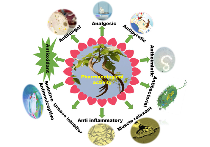
Pheretima posthuma (worm) species was researched for anthelmintic movement in this review. Pain relieving movement was tried in three gatherings of mice (n = 6) utilizing the hot plate technique, with each gathering getting 100, 200, and 300 mg/kg and measurements, everything being equal. Tramadol (0.1 mg/kg) and typical saline (0.9 g/L) were utilized as certain and negative controls, separately. Five gatherings of mice (n = 6) abstained for five hours before movement testing for antipyretic impact (Brewer’s yeast incited pyrexia) just water was permitted. The dosages of each of the three concentrates were 100, 200, and 300 mg/kg, separately. Paracetamol and sodium chloride were utilized as a norm and negative control, individually. The rectal temperature was required consistently for 5 h after the dose to notice temperature varieties. Toxicological testing was performed on all concentrates in mice gauging up to 4 g/kg. The watery concentrate (60 mg/mL) deadened P. posthuma in 15.48 min ± 1.9 min with regards to anthelmintic movement. At a measurement of 300 mg/kg for 1 h, the watery concentrate had a bigger pain-relieving impact than different concentrates, going on until 1 h and a half (P < 0.001). At 200 mg/kg after treatment, the antipyretic activity of the fluid concentrate was fundamentally expanded (P < 0.01), with the greatest levels found in the fourth and fifth hours. Therefore, the plant showed no harmfulness during toxicological examination in mice, the most noteworthy portion of concentrates was 4 g/kg [22–24] (Figure 1).
To begin, the antibacterial activity of (Petthan) FA bark extracts was investigated using the agar well diffusion method with six human pathogens: Shigella boydii, E. coli, Bacillus cereus, Salmonella typhi, Pseudomonas aeroginosa, and S. aureus. The ethyl acetate extract demonstrated considerable inhibition, while the methanol extract showed good results. Using two pathogens, Bacillus cereus and Shigella Boydii, antibacterial screening for ethyl acetate extract was performed using the immersion bio autography method. As a result, the fraction of ethyl acetate extract with a broad spectrum of activity (Rf values range 0.16–0.84) was discovered. Second, the antioxidant activity of ethyl acetate and methanol extract was determined using the 2,2-diphenylpicrylhydrazyl (DPPH) test method and compared to ascorbic acid. Both extracts are high in antioxidants. The antioxidant activity of the ethyl acetate extract was then tested using thin layer chromatography (TLC) by spraying a 20% solution of molybdo phosphoric acid in ethanol. Antioxidant chemicals provided Rf values that were within the range of Rf values that have a broad spectrum of antibacterial action. This discovery suggested that antibacterial and antioxidant chemicals could be present in the ethyl acetate of Petthan bark [25–27].
Phytochemical and antibacterial potentials of several solvent extracts from FA leaves were investigated in this study. In the preliminary phytochemical screening of leaf extracts, reducing sugars, alkaloids, flavonoids, glycosides, terpenoids, tannins, anthracenes, and steroids, were discovered. Fourier transform infrared spectroscopy research revealed the existence of various functional groups in compounds including amines, amides, alcohol, saturated and unsaturated hydrocarbons, aldehydes and ketones, carboxylic acids, esters, and ethers. Each of the concentrates showed significant antibacterial activity when tried against eight multidrug-safe bacterial strains gathered from urinary lot disease patients. FA leaf extricates were displayed to have a few key phytochemical parts, as well as critical anti-microbial activity against multidrug-safe bacterial strains that cause urinary tract infections [28, 29].
The analysts needed to check whether an ethyl acetic acid derivation part extricated from FA leaves had antibacterial action against an assortment of microscopic organisms, including Salmonella enterica, E. coli, Bacillus anthracis, Klebsiella pneumonia, and S. aureus. The analysis involved standard microbial sustenance stock as a positive control and streptomycin as a negative control. Ethyl acetic acid derivation division was utilized as the test, while dimethyl sulfoxide (DMSO) was utilized as the negative control. Given the consequences of the zone of hindrance, the ethyl acetic acid derivation division was displayed to have in vitro antibacterial movement, while the aftereffects of the most minimal inhibitory focus showed that all bacterial strains were vulnerable to the portion picked for the review [30, 31].
FA dichloromethane removes have antifungal viability against dermatophytes and wood decay growths. The movement is doubtlessly inferable from lapachol and β-lapachone, the two of which have been found in these plant separates. The arrangement of mycelium and conidia in filamentous parasites (fungi) is repressed by these concentrates, as indicated by a microscopical examination. The dichloromethane extracts of FA have antifungal effects, particularly against dermatophytes, according to the current study. The activity is most likely attributable to lapachol and β-lapachone, both of which have been found in these plant extracts [31–35].
This study investigated the antimicrobial viability of watery and methanolic concentrates of FA leaves and seeds, which have been utilized in customary medication for centuries to treat skin and urinary tract infections, as well as antidiabetic and anti-diarrhoeal prescriptions. In the early phytochemical screening of FA leaves, alkaloids, glycosides, and saponins were found, while tannins, alkaloids, flavonoids, glycosides, and saponins were found in seeds. The antimicrobial activity of the plant’s fluid and methanolic extricates was tried against three gram-positive strains, including S. aureus, S. epidermidis, and Bacillus subtilis, as well as two gram-negative strains, Pseudomonas aeroginosa, and E. coli, utilizing the disc diffusion strategy. FA leaves had a zone of restraint of 22.37 mm ± 0.35 mm against S. aureus; however, the fluid concentrate had no activity against S. epidermidis. The methanolic extract of the seeds was best effective against E. coli, with a zone of restraint of 16.33 mm ± 0.31 mm yet affected S. epidermidis. With hindrance zones of 22.13 mm ± 0.09 mm and 23.43 mm ± 0.35 mm, individually, the fluid concentrates of the seeds showed the most adequacy against S. aureus and S. epidermidis [36–39].
The motivation behind this examination was to evaluate the muscle relaxant, antinociceptive, and atomic docking properties of an original medication named (5aR,5a1R,6R,7aS,14bR,15R) 15-hydroxy- 7a-methyl-6-(2-methylprop-1-en-1-yl)-7,7a,14b,15-tetrahydro-5H-t-5a,15methanobenzo[g]benzo[5,6] azuleno[1,8-bc]-chromene-5,9,14,16(5a1H,6H)-tetraone (peshawaraquinone) was disconnected from a methanolic concentrate of FA in a creature model. The synthetic design of the detached substance was resolved utilizing progressed spectroscopic methods, which were thoroughly approved utilizing X-rays differaction (XRD) examination. Compound 1 was tried against hot plate-incited harmful improvements at various portions [2.5, 5, 10, and 15 mg/kg, intraperitoneal (i.p.)]. The open-field test was utilized to look at compound 1’s soothing potential, while the tendency and foothold tests were utilized to survey its muscle loosening-up adequacy. The secluded compound was likewise exposed to intense harmfulness testing. To sort out how the substance capacities were set through sub-atomic mooring research. Compound 1 had a huge (P < 0.05) pain-relieving influence in a portion subordinate way. Both exploratory creatures fostered an extensive muscle relaxant impact over the long haul. Synthetic 1 made a measurably huge narcotic difference (P < 0.05) and no unfriendly impacts in an intense poisonousness examine. Docking tests uncovered that narcotic and gamma-aminobutyric acid (GABA) receptors are liked. Therefore, the antinociceptive, muscle relaxant, and narcotic exercises are in all probability intervened [1, 40–42].
The efficacy of the methanolic extract of FA to inhibit urease, a key enzyme connected to gastrointestinal ailment, was explored. To dive deeper into the system of restraint, specialists involved in the molecular operating environment (MOE) direct in silico docking examinations. Peshawaraquinone and indanone subsidiaries hindered urease extensively [half-limiting dose (IC50), IC50 = 19.8 mmol/L + 0.56 mmol/L and 21.9 mmol/L + 1.12 mmol/L, individually], yet lapachone hindrance was unobtrusive (IC50 = 87.3 mmol/L + 2.17 mmol/L). Lapachol showed powerful impact (IC50 = 23.1 mmol/L + 0.14 mmol/L), rather than the positive control thiourea (IC50 = 21 mmol/L + 0.23 mmol/L). In silico docking studies were directed to inspect the component of restraint of jack-beans urease (JBU) utilizing the MOE. As indicated by docking tests, synthetic substances that tight spot Ni molecules and the passage fold deposits, to be specific CME592 (a modified cysteine residue), inactivate the JBU. FA phytochemicals have shown potential as a urease inhibitor in the treatment of Helicobacter pylori diseases connected to gastrointestinal issues [43–46].
The customary therapeutic plant FA has been utilized to ease muscle strain and as a mitigating. Four secluded compounds, in particular (1) lapacho, (2) peshawaraquinone, (3) indanone subsidiaries, and (4) β-lapachone of FA, were read up for calming impact utilizing the carrageenan-and receptor actuated paw edema worldview. The calming impacts of the medications (1–4) tried were assessed in both the early and late phases of edema acceptance. In the beginning stage, every one of the medications tried (0.5–2.5 mg/kg, i.p.) had under a half impact, while intensifies 2 and 3 showed 85.66 and 89.87 percent constriction in the later stage, individually. Intensifies 1–4, additionally presented to receptor prompted aggravation, with intensifies 2 and 3 exhibiting superb impacts of 86.87 and 89.98 percent, separately, at 5 mg/kg after the second hour of organization, while intensifies 1 and 4 showed no tremendous impact when contrasted with the negative control. Sub-atomic docking results uncovered an exceptionally high intensity of compound given the protein-ligand interaction (PLI) profile, which was affirmed by a sub-atomic unique reproduction investigation. Thus, FA mitigating activity, which is connected to the presence of these bioactive mixtures (1–4), unequivocally upholds the plant’s conventional use for irritation treatment [47–51] (Table 2).
Pharmacological activity of phytoconstituents of FA
| Phytoconstituent | Pharmacological activity/dosing | Mechanism | References |
|---|---|---|---|
| β-Lapachone, lapachol | Antitumor activity (compared to 75% of the animals that received β-lapachone 50 mg/kg IT and 100% of the animals that received β-lapachone 75 mg/kg IT) | The potent antipancreatic cancer drug-lapachone is more bioavailable, effective, and toxic when it is complexed with HPβ-CD. | [52] |
| Lapachol | Lapachol exhibits relatively substantial action when administered orally, intraperitoneally, subcutaneously, intramuscularly, or in combination against Walker 256 carcinosarcoma. | [53] | |
| Naphthoquinone derivatives | A promising foundation for their potential use in the treatment of prostate cancer comes from the antitumoral activity of certain of the produced naphthoquinone derivatives. | [54] | |
| Lapachol (2-hydroxy-3-(3-methyl-2-butenyl)- 1,4-naphthoquinone derivative | When lapachol is acetylglucosylated, a substance that increases lapachol activity starts to work against mouse lymphocytic leukemia P-388. | [55] | |
| β-Lapachone, lapachol | β-Lapachone, an O-naphthoquinone derived from lapachol, is now being tested for use against Trypanosome cruzi, cancer, viruses, and bacteria. | [56] | |
| β-Lapachone | Changes in morphology in PC3 cells after single and combined-treatment with β-lapachone and genistein isoflavone, including cell shrinkage and chromatin condensation. | [57] | |
| β-Lapachone | Regardless of the tumor’s sensitivity to hormones, the chemotherapy for prostate cancer provided by the combination of β-lapachone and genistein will be effective. | [58] | |
| β-Lapachone | β-Lapachone’s ability to induce a seemingly unique, calpain-like-mediated apoptotic cell death could be beneficial in the treatment of breast and prostate cancer. | [59] | |
| β-Lapachone | Antibacterial and antifungal activity [inhibitory activity was found against Microsporum gypsum (50 μg/mL) as compared to standard salicylic acid (200 μg/mL)] | Even when the results were converted into mg/mL, β-lapachone outperformed ketoconazole in terms of effectiveness. If the toxicity is confirmed, many medical applications may be avoided. However, the impact they have on bacteria and fungi may be useful for protecting plants. | [52, 60] |
| β-Lapachone, lapachol | Anti-plasmodial activity (IC50< 1.25 μmol/L to 10 μmol/L) | Natural lapachol and β-lapachone were evaluated on the cultivation of the F32 strain of Plasmodium falciparum, and certain derivatives showed promising in vitro activity (IC50 < 10 μmol/L). | [61] |
| Naphthoquinone derivatives | Mollusicidal activity (LC50 values is 3.1 μg/mL) | The lipophilic feature of the amino alkyl side chain improved the molluscicidal action of amino derivatives of lapachol. These compounds have low to medium LC50 values, with the most potent derivative in the series having a 3.1 μg/mL value. | [62] |
| Hydroxynaphthoquinones derivatives | However, other aspects must also be taken into account. The trend in hydroxynaphthoquinones derivatives shows that bioreduction may play a significant role in the molluscicidal activity of these compounds. | [63] | |
| Lapachol, isolapachol, and dihydrolapachol | Antileishmanial activity [minimum lethal concentration (MLC) 100 μg/mL and minimum inhibitory concentration (MIC), 50 μg/mL] | Hydroxyquinone derivative has shown activity against Leishmania amazonensis and Leishmania braziliensis in vitro and in vivo, as well as being a very promising antileishmanial medication. | [64, 65] |
| 1,2-Naphthoquinones, lapachol, β-lapachone | Antimalarial activity (KD50 57.51 and KD90 98.32 was determined by probit analysis, while mortality value after 24 h was 10.6% ± 0.2%) | Utilizing isolates of parasites with varying susceptibilities to chloroquine and/or mefloquine, benzo[a]phenazines produced from 1,2-naphthoquinone, lapachol, β-lapachone, and other derivatives have been investigated for their antimalarial efficacy against Plasmodium falciparum in vitro. | [66, 67] |
| 1,4-Naphthoquinones and 1,2-naphthoquinones derivatives | Antiviral activity | Epstein-Barr virus early antigen (EBV-EA) activation caused by 12-O-tetradecanoylphorbol-13-acetate (TPA) was examined for the inhibitory effects of many derivatives made from the naphthoquinone lapachol and TPA. | [68] |
| Friedelin | Antiulcerogenic activity (doses of friedelin 20, 25, 30, 35, 40 and 45 mg/kg) | These results suggested that the anti-ulcer potential of friedelin, which may involve antioxidants, endogenous prostaglandins, nitric oxide, K+ATP channel opening, maintaining the balance between pro and anti-inflammatory cytokines, antiapoptotic function, could be a new useful natural gastroprotective tool against a gastric ulcer in the rat model. | [69] |
| Friedelin | Anti-diarrhoeal activity [friedelin (20 mg/kg) showed significant (P < 0.0001)] | The overall findings revealed that friedelin’s anti-diarrheal activity may be related to its anti-secretory and anti-motility properties, providing support for the traditional assertion. | [70] |
| Friedelin | Anti-inflammatory, analgesic, and antipyretic activity (40 mg/kg dose of friedelin with a significance value of P < 0.05) | Friedelin had substantial anti-inflammatory, analgesic, and antipyretic properties in vivo. Friedelin’s major mode of action may be to inhibit the synthesis or release of inflammatory mediators. | [71] |
| Friedelin | Hypolipidemic activity [friedelin (50 mg/kg and 70 mg/kg) caused a lowering of lipid levels in plasma and the liver showed a significant (P < 0.01)] | The current investigation found that friedelin had the greatest efficacy in decreasing increased cholesterol levels in Triton WR-1339 and high-fat diet-induced hyperlipidemic rats. | [72] |
| Friedelin | Hepatoprotective activity [friedelin at 40 mg/kg (IC50 21.1 mmol/L), hydroxyl (IC50 19.8 mmol/L), nitric oxide (IC50 22.1 mmol/L) and superoxide (IC50 21.9 mmol/L) radicals] | Friedelin demonstrated effective antioxidant, free radical scavenging, and liver protecting properties in both in vitro and in vivo investigations. It may be useful in avoiding or reducing the progression of several oxidative stress-related illnesses. | [73] |
| OA and UA | Osteoprotective effect [medium Ca diet (MCD, 0.6% Ca), high Ca diet (HCD, 1.2% Ca), MCD + FLL (700 mg/kg per day), MCD + OA (23.6 mg/kg per day) + UA (8.6 mg/kg per day)] | This study offers proof that OA + UA may be employed as a cutting-edge, orally administered treatment medication for osteoporosis management and prevention. | [74] |
| OA | In mature C57BL/6 ovariectomized (OVX) mice, OA dramatically enhanced bone mineral density, improved micro-architectural characteristics, decreased urine Ca excretion, and elevated 1,25(OH)2D3 and renal CYP27B1 messenger RNA (mRNA) expression. OA also enhanced bone characteristics. | [75] | |
| Oleanolic and UAs | Antibacterial activity [gram-positive bacteria: 4 mg/L against Enterococcus faecalis, and 8 mg/L against both strains of S. aureus, for UA; 8 mg/L against Enterococcus faecalis, 32 mg/L S. aureus American type culture collection (ATCC) 29213 and 64 mg/L against S. aureus ATCC 25923, for OA. Both compounds, UA and OA, were devoid of activity against gram-negative bacteria (≥ 256 mg/L, for E. coli and Pseudomonas aeruginosa)] | Numerous bacterial species, particularly gram-positive ones like mycobacteria, are susceptible to the effects of OA and UA. They prevent bacterial survival and growth, and a wide range of MICs values are available. | [76] |
| Oleanolic, ursolic and BA | When it comes to the treatment of bacterial infections, UA, OA, and BA establish the genuine value of such ethnopharmacology approaches by determining antibacterial activity as well as cellular impact (i.e., viability and cytotoxicity). | [77] | |
| Oleanolic and UA | Antimutagenic effect [UA (80 mg/kg b.w.); OA (80 mg/kg b.w.) and a mixture of UA and OA (80 mg/kg b.w.)] | Under the experimental conditions utilized in this study, UA and OA reduced doxorubicin (DXR) clastogenicity in mouse peripheral blood and bone marrow cells. It has promising candidates for the prevention of cancer and other disorders. | [78] |
| UA | UA has demonstrated that its in vitro chemopreventive properties can shield DNA from both endogenous and induced DNA damage in both types of cells. | [79] | |
| UA | Anti-neoplastic activity (20–120 μmol/L of UA for HT-29 cells) | As a strong anti-cancer drug for colorectal cancer therapy, usolic acid causes apoptosis in colorectal cancer cells by upregulating micro (miR)-4500 and inhibiting signal transducer and activator of transcription 3 (STAT3) activation. | [80] |
| UA | Due to its capacity to influence various intracellular signal STAT3, extracellular signal-regulated kinase (ERK), c-Jun N-terminal kinase (JNK), and p38 pathways targets, UA has a wide variety of actions. Both in vivo and in vitro, UA dramatically reduces the activation of several CRC-related signaling pathways. | [81] | |
| Glycyrrhetic, oleanolic, and UAs | Anti-ulcerogenic activity (HCT15 cells treated with UA 30 μmol/L and OA 60 μmol/L) | Pentacyclic triterpene derivatives (Gl, OA, UA) with anti-ulcer action have advanced to the point where a number of novel substances are available that are potenter than carbenoxolone, essentially devoid of mineralocorticoid activity, and possibly non-toxic. | [82] |
| OA and UA | Hepatoprotective activity [OA and UA with IC50 (Ki) values of 143.5 (74.2) μmol/L and 78.9 (41.0) μmol/L)] | The current investigation shows that UA inhibits CYP2C19 in human liver microsomes and that OA has an inhibitory impact on CYP1A2 and CYP3A4. | [83] |
| UA and asiatic acid | The processes by which UA and asiatic acid have demonstrated protective effects may be linked to direct protection of mitochondria and hepatocytes as well as the potent scavenging of reactive oxygen species (ROS), which may indirectly benefit mitochondria. | [84] | |
| BS | Anti-inflammatory activity [BS (0.1–200 μmol/L)] | BS strongly reduces vascular adhesion molecule 1 and intracellular adhesion molecule 1 expression in TNF-α-stimulated HAEC, as well as U937 cell binding to TNF-α-stimulated HAEC and attenuates nuclear factor-kB p65 phosphorylation. | [85] |
| BS | Anti-diabetic activity [BS treatment (10, 15 and 20 mg/kg, p.o.)] | In STZ-induced diabetes, BS protects serum and pancreatic tissue, as well as antioxidant status. | [86] |
| BS | The anti-hyperglycemic action of the BS extract may be attributed to both protection against oxidative damage in alloxanized diabetes and an increase in peripheral glucose consumption. | [87] |
p.o.: oral; TNF-α: tumor necrosis factor α; Ki: inhibition constant; CRC: colorectal carcinoma; b.w.: body weight; BA: betulinic acid; Ca: calcium; FLL: fructus ligustri lucidi; IT: intratumoral; HPβ-CD: hydroxypropyl-β-cyclodextrine; LC50: lethal concentration
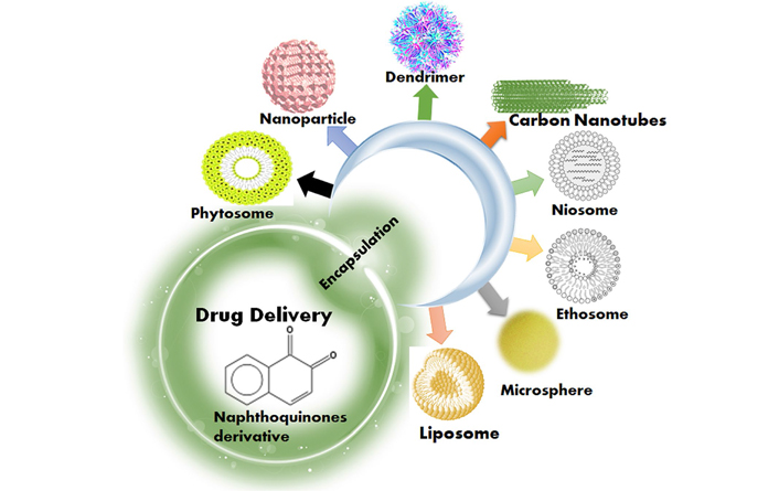
The transdermal drug delivery system (TDDS) is a practical option in contrast to oral medicine since it takes into account better persistence, consistency, and more predictable plasma drug fixations. Therefore, a transdermal patch containing Sigesbeckia Herba (SiH) separate was created in this review. SiH removal was detached from Sigesbeckia pubescence Makino, and afterward, transdermal patches containing SiH separate were made using the dissolvable vanishing technique. The transdermal patch formulation was changed given the in vitro skin permeability study. At long last, the best patch’s anti-inflammatory and pain-relieving qualities were researched [88].
Quercetin is an antihypertensive, vasodilator, anti-inflammatory, antiatherosclerosis, antihypercholesterolemic, and anti-obesity bioflavonoid found in over twenty plants. Free radicals are a major factor in the development of diseases such as vascular disorders, hypertension, and metabolic syndrome. The motivation behind this study was to foster a transdermal delivery system for quercetin as a once-daily anti-inflammatory dosage form [89].
Curcuma longa and Kaempferia parviflora have both been shown to have anti-inflammatory effects. When administered orally, both extracts have been displayed to have low bioavailability and extensive measures of first-pass metabolism. The goal of this work was to create transdermal delivery of both extracts in matrix-patch compositions with five distinct volatile oils that have analgesic and anti-inflammatory characteristics [90].
Phytosome is herbal products in the form of improved microspheres or cells that are easier to absorb and have a better pharmacodynamic and pharmacokinetic profile than typical herbal extracts. These are also known as herbosome. Phytosome technology makes herbal extracts more bioavailable. In phytosome technology, single components of a plant extract are linked to phosphatidylcholine (PC). Phytopharmaceuticals are being used to increase the bioavailability of botanical extracts for medical purposes using new drug delivery technology. According to a thorough review of the literature, some plant extracts, whether crude, partially purified, or fractionated, have been documented to have various major pharmacological or health-promoting characteristics. Plant extracts can be standardized and produced as phytosome for further exploration into their possible applications [91].
Bombax ceiba plants have long been used as a home cure for jaundice, spleen enlargement, and hepatoprotective action, according to certain sources. Natural medicines are now being used to treat the majority of common ailments and nutritional issues. The administration of a productive level of the medicinal active component is critical to the efficacy of any herbal treatment. However, when given orally or by topical treatments, their bioavailability is severely limited. Phytosome is herbal formulations that are more easily absorbed and have higher bioavailability and effects than traditional Phyto molecules or botanical extracts. An ethanolic extract of phytosome was produced in soya lecithin. Finally, phytosome were successfully synthesized and encapsulated. It has a longer release time and higher free radical scavenging action [92, 93].
On account of their upgraded bioavailability in the body and the way that the encapsulated molecules are safeguarded while staying utilitarian, liposomes keep on astonishing revenue. This study means to make “goliath” liposomes by hydrating lipid films with two unique phospholipids, phosphatidylserine (Ptd-L-Ser) and PC. The full scale and infinitesimal properties, complete phenol content, and cancer prevention agent limit of Stellaria media (L.) Vill. were first surveyed. The Stellaria media (L.) Vill. extract was then encapsulated in the two formulations phosphatidylcholine-based liposomes (PCE) and phosphatidylserine-based liposomes (PSE), and the liposomes were inspected for morphology, size distribution, and Zeta potential (ZP) utilizing optical microscopy and dynamic light scattering [94].
Synthetic and herbal medications have been integrated into lipid and liposomal formulations for the treatment of dermatological illnesses, with herbal extracts reducing the negative effects associated with incorporated synthetic drugs. Reverse phase evaporation was used to extract antioxidants from green tea leaves and roselle calyces into cholesterol-carrying liposomes. To work on the security of polyphenols and safeguard their helpful qualities, the roselle and green tea removes were embodied into liposomes by scattering soy lecithin in refined water utilizing the reverse phase evaporation strategy [95–100].
Curcumin is a well-known anticancer herbal treatment, but its bioavailability is low, necessitating research into more efficient and targeted delivery mechanisms. Due to its over-expression in numerous types of malignancies, human epidermal growth factor receptor 2 (HER2) is a good option for anticancer therapy. The cytotoxicity of curcumin encapsulated in ZHER2:342 Affibody-enhanced liposomes against the malignant cell lines SKBR3 and MCF-7 were researched in this work [101].
Nanoparticles are solid particles or dispersion of particles that act as drug carriers [nanoparticles (NPs)], often containing polymeric nano spheres, liposomes hydrogel, solid nanoparticles, and other nanoparticles. They’ve been frequently employed as an alternative to standard drug delivery and proven to be potential medication carriers. The goal of this project is to develop a unique strategy using herbal chitosan nanoparticles to improve therapeutic efficacy while reducing side effects. The current study uses the ionic gelation method to produce and evaluate chitosan-loaded nanoparticles of Allivum sativum. In this investigation, five distinct formulations were created by varying the polymer and Tri-polyphosphate (TPP) ratios [102–110].
The analysts needed to make Annonam uricata fruit extract stacked solid lipid nanoparticles (SLNs) and explore their cytotoxicity in an in vitro breast malignant growth model. High-pressure homogenizations followed by ultrasonication were used to successfully prepare extract-loaded SLNs, which were then optimized using a full factorial design. Percent cumulative drug release, ZP, percent entrapment efficiency (EE), and particle size (PS) were all used to describe the extract-loaded SLNs controlled drug release (CDR) [111].
Nanotechnology is a relatively recent area of pharmacological research with numerous applications in the engineering, biomedical, food, chemical, and pharmaceutical industries. The utilization of microorganisms and natural extracts to blend nanoparticles has piqued worldwide interest, and it is viewed as perhaps the most encouraging nanotechnology. This study will investigate the cytotoxicity and antioxidant activities of silver nanoparticles mediated by natural extracts of Justicia adhatoda and Ocimum sanctum utilizing ultraviolet (UV) visible spectroscopy [112, 113].
Microspheres are characterized as a “monolithic sphere or therapeutic agent distributed throughout the matrix either as a molecular scattering of particles” (or) a construction made out of a consistent period of at least one miscible polymer in which drug particles are scattered at the sub-atomic or plainly visible level. Microspheres are minuscule circular particles having aspects in the micrometer range (normally 1 mm to 1,000 mm). Micro-particles are another name for microspheres. However, because of its high polarity, it has poor absorption properties and is sensitive to heat, light, and pH. The point of this study was to create and manufacture a phytosome-loaded green tea leaf extract microsphere with good physicochemical properties to improve phytosome stability and delivery [114].
Bletilla striata (B. striata) polysaccharide (BSP) have already been tested in the development of a muco-adhesive carrier microsphere capable of long-term stomach retention. Stomach ulcers are treated with a traditional Chinese herbal composition containing B. striata and Panax notoginseng (Burk.). In this scientific study, panax notoginseng saponin (PNS) was changed into a hydrophobic dispersion with slow-release qualities. PNS was packed inside BSP/alginate microspheres to enhance the encapsulation rate. Diffusion and erosion released the active components from the PNS-loaded microspheres, which were spherical and had release qualities that matched the Weibull equation. In a synergistic approach, the produced microspheres enhanced the benefits of PNS while also exerting the medicinal influences of BSP on acute stomach ulcers [115].
In recent years, controlled medication delivery technology has advanced at a faster rate. Gelatine and collagen-gelatine microspheres were made using an emulsion technique with distilled water and paraffin oil containing 1% Span 80 after that, a combination of ethyl-3(3-dimethylaminopropyl) carbodiimide (EDC) and N-hydroxysuccinimide (NHS) was used to cross-link the microspheres. Dehydrothermal treatment (DHT) was also used to cross-link microspheres to compare chemical and physical cross-linking. As a pharmacological model, gathered microspheres were filled with Calendula officinalis flower extract [116].
Phospholipids, ethanol, and water make up “ethosomes” which are soft vesicles. They’re a “new carrier system” for delivering medications that don’t penetrate biological membranes well. Ethanolic extracts of Zingiber officianale (Z. officianale) rhizomes, Phyllanthus niruri (P. niruri) leaves, and Croton tiglium (C. tiglium) Linne seeds were made and put into ethosomal dosage forms to test hair development-boosting activity and security [117].
C. tiglium, Zingiber officinale, and P. niruri, combined plant extracts have been reported to have potential therapeutic benefits. Ethosome is remarkable in that they can encapsulate medications or plant extracts in a phospholipid’s bilayer with various hydrophobicity’s. The objective of this study is to investigate the cytotoxicity of joined plant extracts and ethosome-stacked consolidated plant extracts for topical use, as well as their effect on HaCa T cells stimulated with testosterone [118].
This research aims to create an ethosome from the seed extract of Sesamum indicum L., combine it into gel formulations, and use various parameters to define the ethosome and gel formulations. The current study found that an ethosomal vesicular delivery system for S. indicum L. seeds extract is a promising new technique for herbal extract [119].
Niosome is a novel drug delivery systems (NDDS) that encapsulates medication in a vesicle. These are vesicles that are made up of a bilayer of non-ionic surface-active substances and are extremely small and microscopic. Their aspects are on the nanometres scale. In malignant growth treatment, the low solvency of plant-separated agents like D-limonene is a significant issue. In this review, in vitro cytotoxicity tests of HepG2, MCF-7, and A549 cell lines to produce D-limonene-stacked niosome (D-limonene/Nio) for malignant growth treatment. The niosomal dosage form was made utilizing a film hydration approach utilizing Span® 40, Tween® 40, and cholesterol (35:35:30 molar proportion) and assessed for vesicle circulation size, shape, entrapment proficiency (EE percent), and in vitro release property. At a convergence of 20 mol/L, the stacked plan exhibited impressively expanded cytotoxic action against every one of the three malignant growth cell lines (A549, Macf-7, and HepG2). These findings suggest that a phytochemical-loaded niosome could be a potential nano-carrier for cancer therapy [120]. In this study, niosome carrying oleander (Nerium oleander) extract as the natural ingredient was prepared and characterized. For this situation, niosomal formulations of two separate oleander root extracts were made utilizing a thin film hydration process with Tween 60 (non-ionic surfactant) and a cholesterol combination at a 1:1 mol/L ratio [121]. The extremely lipoidal cell of Tuberculosis bacilli makes drug penetration difficult, resulting in treatment resistance, which can be addressed by adopting a nanosized niosomal delivery technique. The purpose of this study was to investigate the anti-tuberculosis properties of Spermacoce hispida extracts and to develop a new nanonoisome-based drug delivery system for Spermacoce hispida extracts to boost anti-tuberculosis activity [122].
Hexosomes are reverse hexagonal phases made up of infinite hexagonally close-packed water layers coated by a monolayer of surfactants. They have the potential to be used as an alternate delivery mechanism for drugs due to their unique structural features. There has been a flood sought after for regular fungicides to supplant manufactured ones on the grounds that hurtful build-ups endure in soils, sullying the climate and representing a genuine danger to overall general wellbeing. In this review, medicinal balms from the strips of four citrus organic products (Citrus lemon, Citrus aurantifolia, Citrus maxima, and Citrus sinensis) were separated and dissected utilizing gas chromatography-mass spectrometric investigation. Monoterpene hydrocarbons were found to overflow all four oils, with limonene being the most incessant. The antifungal properties of the oils were considered, and the most dynamic oil (Citrus lemon) was set into hexosomal scattering and tried once again against similar organisms [123].
Because of their fantastic inborn physical and synthetic properties of carbon nanomaterials and carbon nanotubes (CNTs) have been effectively investigated for a scope of utilization [124, 125]. CNTs can be utilized in gene transfer, malignant growth treatment, transdermal, and DNA applications. Functionalized multi-walled CNTs (MWCNTs) were utilized to make helpful nanoparticles for the vehicle of specific bioactive mixtures. Accordingly, the objective of this study is to examine the poisonousness of N-vinyl caprolactam-joined MWCNTs within the sight of pomegranate strip extract [Punica granatum (P. granatum)], titanium dioxide (TiO2), or potentially silver nanoparticles on male mice utilizing in vivo organic assessment (liver and kidney brokenness biomarkers), as well as histopathological examination [126].
The researchers used an extract from the herb Hempedu bumi (H. bumi) to investigate the antibacterial activity of functionalized single-walled CNTs (SWNTs). H. bumi concentrate and complexed SWNT were tried for their organic exercises against Aspergillus niger (microbe), E. coli (deft microorganism), and Bacillus sp. (microorganism). As indicated by the discoveries, SWNTs can possibly be utilized as a carrier of parts from plant extracts [127].
Prostate cancer is the second highest risk in men and the 6th driving reason for death from malignant growth. Naturally grown prescriptions are the most widely recognized kind of wellbeing treatment accessible to humankind. Regular synthetics are significant wellsprings against disease and novel cancer prevention agent atoms, bringing about a ton of variety and inventiveness. The field of anticancer drugs has been changed by nanotechnology, which has reformed imaging, diagnostics, and medicine conveyance using NPs. The cytotoxic and apoptotic impacts of Tamarixm annifera watery extract, functionalized SWNTs, and their blend on the PC3 cell line were explored in this scientific study [128].
Dendrimers are a drug delivery technology that works by sending a drug-loaded nano particle (10–9) into the body. The medicine could be placed onto the dendrimer’s terminal surface or enclosed within the dendrimer’s branches. Despite its low bioavailability and in vivo stability, curcumin has been linked to a variety of pharmacological effects. Current research focuses on the development and characterization of generation 4 (G4) Poly(amidoamine) (PAMAM) dendrimer-palmitic acid core-shell nanoparticles incorporating curcumin as antistress treatment, with the goal of enhancing curcumin bioavailability. Various formulations were created using different amounts of palmitic acid and curcumin ratio [129].
Anti-inflammatory, anticancer, antioxidant, and anti-microbial properties demonstrate this plant extract’s value in human health and therapy. Curcumin’s anticancer properties are owing to its ability to target a variety of cell and sub-atomic pathways engaged with cancer progression. Curcumin’s restricted dissolvability, low bioavailability, and fast metabolism on the other hand, severely limit its medicinal potential. Curcumin was conjugated to the surface of a polyamidoamine dendrimer at G4 to form a nano-carrier with sufficient delivery properties (PAMAM). According to the findings, the dendrimeric curcumin generated has a hydrodynamic diameter of around 100 nm. The pace of curcumin stacking on this nanostructure framework was likewise found to associate with 100 nm, as per the discoveries. The discoveries additionally uncovered that the pace of curcumin stacking on this nanostructure framework was around four curcumin atoms for every dendrimer [130].
Nanofibers are fibers with a diameter of less than a nanometre and can be made from a variety of polymers and so have a variety of physical properties and applications. Microbial invasion of burn sites by both local and external bacteria has become a major general wellbeing concern and a significant financial load. Novel antimicrobial injury dressings that confine bacterial colonization while accelerating the recuperating process are believed to be important, bringing about a more elevated level of care for patients. Since old times, naturally grown extracts from restoratively significant plants have been utilized to treat consumed wounds. This exploration depicts the utilization of electrospun nanofibers containing plant extracts [131].
Nanofibers film containing various polymers is a transdermal medicine delivery system made by mixing gels or creams. Flexible films can be affixed to the sensitive skin and controlled for medication release. Many researchers have been interested in the fabrication of biopolymer nanofiber as a matrix for medication and herbal extract release control until now. The focus of this study was on nanofiber manufacturing using biological polymers of polyhydroxyalkanoates (PHAs) and poly (butylene-adipate-co-terephthalate) (PBAT) mixing systems with chili herbal extract or capsicum extract [132].
The goal of this study was to create Aloe vera extract-loaded chitosan/polyethylene oxide (CS/PEO) electrospun nanofibers for biomedical applications. To optimize the nanofiber construction process, the polymer-to-extract ratio and electro-spinning parameters were studied [133].
The goal of this study was to make a nanogel with Cymbopogon citratus; Stapf’s volatile oil encapsulated in poly (D, L-lactide-co-glycolide) nanoparticles and test its anti-herpetic action in vitro. Solvent emulsification-diffusion was used to make polymeric nanoparticles, which were then integrated into carbomer hydrogels. The findings show that nanogel can prevent oil from control release, and improve anti-herpetic action and volatilization [134].
Nanotechnology’s production and use strategy for biologically produced nonmaterial has become ingrained. The aqueous extraction of root (Pongamia pinnata) and Fourier-transform infrared (FTIR) analysis of phytochemical substances and functional groups found in plant roots are the subjects of this work. Silver nanoparticles were generated using root extract (Pongamia pinnata); scanning electron microscopy (SEM), UV visible spectroscopy (UV-VIS) spectroscopy, and Energy dispersive analysis X-ray (EDAX) were used to confirm and characterize the created silver nanoparticles. The concentrations of silver nanoparticles and paraffin wax used to make nanogel were varied [135, 136].
For wound dressings, Mikania micrantha has been utilized and to support the mending of injuries for a really long time. This is because alkaloids and terpenoids/steroids molecules are present. Hyperglycaemia is an ideal environment for bacterial growth, which slows the healing process. The goal of this study was to see how nanogels carrying MMLE healed wounds in hyperglycaemic rodents as a model for diabetic injuries [137].
Microencapsulation is the technique of encapsulating one material within another on a very small scale, resulting in capsules ranging in size from less than one micron to several hundred microns. The goal of this study was to use a solvent evaporation method to create microcapsules from Macaranga gigantea (M. gigantea) leaves extract utilizing ethocel 10 centi poise (cP) and Eudragit E100 as a matrix. It can be concluded that employing Eudragit E100 and ethocel 10 cP as a polymer matrix, microcapsules of M. gigantea leaves extract may be made by solvent evaporation [138].
The antioxidant chemicals in mangosteen peel extract have reactive, temperamental and without problem oxidized properties. To protect bioactive molecules such as xanthone from destruction, a coating approach using ingredients with established effectiveness and efficiency, such as maltodextrin, is required. The point of this study is to take a gander at the best formulations and qualities that can be gotten by microencapsulating mangosteen peel extract with maltodextrin got from arenga starch [139].
Herbs have been used as galactagogues for a long time, and numerous commercial formulations have been made with them. In this work, ionotropic gelation was utilized to create microcapsules containing galactagogue herbs extract (Figure 2) [140].
β-Lapachone could be shown to be a potent inhibitor of transcriptase activity from myeloblastosis virus and Rauscher murine leukaemia virus. In addition, it affects eukaryotic DNA-dependent DNA-polymerase activity [20].
The potent anti-pancreatic cancer drug β-lapachone is more bioavailable, effective, and toxic when it is complexed with HPβ-CD [52].
A promising foundation for their potential use in the treatment of prostate cancer comes from the antitumoral activity of certain of the produced naphthoquinone derivatives [54].
Even when the results were converted into μg/mL, β-lapachone outperformed ketoconazole in terms of effectiveness. If the toxicity is confirmed, many medical applications may be avoided. However, the impact they have on bacteria and fungi may be useful for protecting plants [60].
Natural lapachol and β-lapachone were evaluated on the cultivation of the F32 strain of Plasmodium falciparum, and certain derivatives showed promising in vitro activity (IC50 < 10 μmol/L) [61].
Hydroxyquinone derivative has shown activity against Leishmania amazonensis and Leishmania braziliensis in vitro and in vivo, as well as being a very promising antileishmanial medication [64].
For a long time, there has been a tremendous disagreement between Ayurvedic treatment and Western medicine. While biomedicine and modern medications are primarily concerned with lowering pathology, Ayurveda is known for taking a holistic approach to ailments and general wellness. This review tried to report the pharmacological potential, extraction, and models used in the study and their significant results of the novel nano phytopharmaceutical approaches applied for drug delivery systems. Almost all parts (leaves, bark, stems, seeds, fruits, and roots) have pharmacological potential as the scientific reports said. Most of the studies are done with alcoholic extracts. FA (Heterophragma adenophyllum) is one of the most well-known Rasyana medicinal plants are having the role of analgesic, antipyretic, anti helminthic, anti-bacterial, antioxidant, anti-fungal, anti-microbial, antinociceptive, muscle relaxant, sedative, potent urease inhibitor and anti-inflammatory effects that has been explained in this article. This review will also provide researchers with a full overview of the tremendous pharmacological potential of FA extracts and compounds, with a focus on ethno-medical applications and various pharmacological activities, particularly neuroprotective and psychedelic effects. The discovery and characterization of metabolic products of bioactive components absorbed by the body, as well as elucidating of the pharmacological activity of FA, should be the focus of future research programs.
BS: β-sitosterol
BSP: Bletilla striata polysaccharide
CNTs: carbon nanotubes
E. coli: Escherichia coli
FA: Fernandoa adenophylla
HPβ-CD: hydroxypropyl-β-cyclodextrine
IC50: half-limiting dose
MCD: medium calcium diet
OA: oleanolic acid
PNS: panax notoginseng saponin
S. aureus: Staphylococcus aureus
SiH: Sigesbeckia Herba
SLNs: solid lipid nanoparticles
SWNTs: single-walled carbon nanotubes
UA: ursolic acid
TY and SKM: Writing-original draft. TY: Writing-review & editing. MSA and AW contributed to various areas of the manuscript and critically evaluated it. Each author reviewed and approved the version that was submitted after helping to revise the document.
The authors declare that they have no conflicts of interest.
Not applicable.
Not applicable.
Not applicable.
Not applicable.
Not applicable.
© The Author(s) 2022.
Copyright: © The Author(s) 2022. This is an Open Access article licensed under a Creative Commons Attribution 4.0 International License (https://creativecommons.org/licenses/by/4.0/), which permits unrestricted use, sharing, adaptation, distribution and reproduction in any medium or format, for any purpose, even commercially, as long as you give appropriate credit to the original author(s) and the source, provide a link to the Creative Commons license, and indicate if changes were made.

Neha Minocha ... Parijat Pandey
Monu Yadav ... Anil Kumar
Rouchan Ali ... Pooja A. Chawla
Rosy Yesela Mancilla Santa Cruz ... María Segunda Aurora Prado
Tausif Alam
Abhishek Chaurasiya ... Pooja A. Chawla
Dilpreet Singh ... Pooja A. Chawla
Shikha Choudhary ... Md Sabir Alam