• Open Access
    Review

    N-type calcium channel blockers: a new approach towards the treatment of chronic neuropathic pain

    Shikha Choudhary 1
    Raminderjit Kaur 1
    Aafrin Waziri 2
    Arun Garg 3
    Renu Kadian 4
    Md Sabir Alam 5*

    Explor Med. 2023;4:85–106 DOI: https://doi.org/10.37349/emed.2023.00126

    Received: June 28, 2022 Accepted: October 12, 2022 Published: February 28, 2023

    Academic Editor: Lindsay A. Farrer, Boston University School of Medicine, USA

    This article belongs to the special issue Techniques in Repurposing and Targeted Delivery: Bringing a New Life to Shelved Drugs

    Abstract

    Neuropathic pain (NP) remains maltreated for a wide number of patients by the currently available treatments and little research has been done in finding new drugs for treating NP. Ziconotide (PrialtTM) had been developed as the new drug, which belongs to the class of ω-conotoxin MVIIA. It inhibits N-type calcium channels. Ziconotide is under the last phase of the clinical trial, a new non-narcotic drug for the management of NP. Synthetically it has shown the similarities with ω-conotoxin MVIIA, a constituent of poison found in fish hunting snails (Conus magus). Ziconotide acts by selectively blocking neural N-type voltage-sensitized Ca2+ channels (NVSCCs). Certain herbal drugs also have been studied but no clinical result is there and the study is only limited to preclinical data. This review emphasizes the N-type calcium channel inhibitors, and their mechanisms for blocking calcium channels with their remedial prospects for treating chronic NP.

    Keywords

    N-type calcium channel blockers, neuropathic pain, conotoxin, ω-conotoxin, ziconotide, peptide inhibitor

    Introduction

    Neuropathic pain (NP) is the pain triggered by primary laceration or somatosensory disfunction as defined by International Association for the Study of Pain [1]. Throughout the world, approximate 9% population is affected by this pain [2]. This pain is mostly divided into two categories: one is peripheral which is the most common, while the other is central. The peripheral pain is the result of nerve injury, neuralgia, or radiculopathy. The reason for the central pain may be the brain injury, spinal injury, brain stroke, or multiple sclerosis [3]. NP is recognized by several symptoms such as spontaneous shooting, burning pain, and allodynia. It affects the life quality of a patient [4, 5], leading to depression, certain other mental and physical health issues [6, 7]. The explanation of NP is not much clear but rather quite confusing resulting in lack of fixed treatment strategy. So, certain invasive therapies are used for mitigation of pain due to a lack of treatment. Treatment is totally a literature-based outcome. Certain antidepressant and antiepileptic drugs are used to cure the symptoms although these methods are associated with certain side effects such as addiction. Beyond these, surgical methods are also used. So, there is much space for the development of new medication in this field, which should be specific with less side effects. The International Association for the Study of Pain (IASP) defines NP as pain initiated or caused by a primary lesion or due to dysfunction within the nervous system [8]. This condition arises as the outcome of sequence of several pathological mechanisms which are frequently described on the basis of anatomic localization [9]. Neuropathic syndromes are typically characterized with several intricate neuronal episodes that incorporate all types of allodynia, hyperalgesia, paraesthesia, and dysesthesias [10]. These signs are typically followed by depression, anxiety, and sleep disturbances [11]. Large numbers of neuropathic patients do not attain satisfactory relief from popular treatment methods [1214]. The failure of present treatment methods highlights the crucial requirement for brand new categories of drugs and improved use of available treatments. Medicines presently in use for treating NP are mentioned in Table 1 [1517]. Certain herbal drugs also have been preclinically studied for the treatment of NP such as Aconiti tuber, Curcuma longa, Ocimum sanctum, but with no clinical application yet.

    Pharmacotherapy for nerve pain

    TherapyDrug classDrugsMechanism of actionAdverse effects
    First-line therapyCalcium channel blockersGabapentinIt inhibits α2δ subunit of VGCCs.Peripheral swelling
    PregabalinIt inhibits α2δ subunit of VGCCs.Peripheral swelling and increased bodyweight
    TCAsAmitriptylineIt inhibits the reuptake of serotonin and norepinephrine.Arrhythmia and suicide risk
    SNRIDuloxetine, venlafaxineThese act by inhibiting the reuptake of serotonin and norepinephrine.Ataxia, hyperhidrosis and hypertension
    Second-line therapyOpioidsTramadolIt acts by binding to opioid receptors.Seizures and ataxia
    TapentadolIt inhibits the reuptake of serotonin and norepinephrine.Seizures and ataxia
    Topical treatmentsLidocaineIt acts by binding to sodium channels.Local erythema, itching and pain
    CapsaicinIt interacts chemically with sensory neurons for its action.Pain and itching
    Third-line therapyStrong opioidsMorphineIt acts by binding to opioid receptors.Dizziness and lethargy
    NeurotoxinsBotulinum toxinIt acts by decreasing the release of acetylcholine.Pain at injection site
    Display full size

    SNRI: serotonin-norepinephrine reuptake inhibitors; TCAs: tricyclic antidepressants; VGCCs: voltage-gated calcium channels

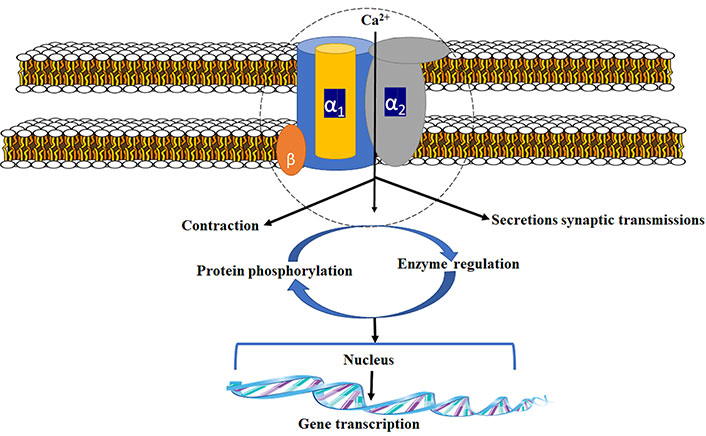
    Because of the crucial side effect offered by current treatments, novel approach towards management of nerve pain is desperately required [18]. Ziconotide (PrialtTM), which was studied as N-type calcium channel blocker, was developed on account of the new drug in the class of ω-conotoxins. Synthetically it was similar to conotoxin MVIIA, an N-type calcium channel inhibitor permitted for NP [19]. The calcium channel is made up of three different subunits (Figure 1). For channel activity α-1 subunit is important. Gabapentin/pregabalin binds at a site in the extracellular region of α2 subunit [20]. N-type voltage-dependent calcium channel (VDCC) controls the discharge of neurotransmitters towards synaptic cleft [21, 22]. These neurochemicals carry nociceptive signals across the afferent nerve, and the pain signals get suppressed due to antagonism at the channel. Structure of voltage-gated Ca2+ channel is shown in these articles [2325].

    Signal transduction of voltage gated

    Pathophysiology of NP

    Peripheral NP

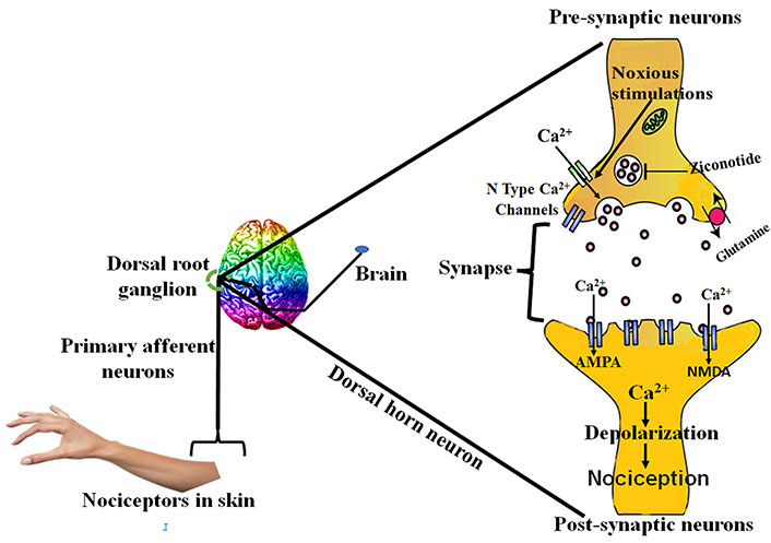
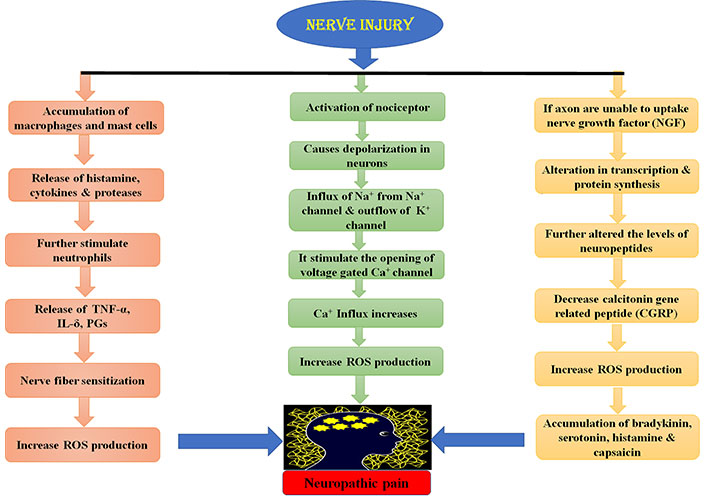
    Peripheral nerve lesion causes aberrant regeneration which unusually sensitizes the neurons that cause abnormal excitability [26]. This process is recognized as superficial stimulation, which results in NP. Peripheral nervous system injury causes the release of certain mediators of inflammation like histamine, cytokines, potassium, and neuropeptides [27]. These mediators cause the change in the quantity and location of sodium ion channels in the damaged nerve fibres [27, 28]. This results in the depression of depolarization threshold and ectopic discharge resulting in the higher nociceptor response to any type of external stimuli known as peripheral sensitization [28, 29]. Ectopic discharge results from the demyelinated nerve fibres due to shrivelled blood supply [30, 31]. Due to injury, the chemical mediated electrical connections are developed between adjoining neurons called ephaptic conductions resulting in pain in a normal calm nociceptor [27, 31, 32]. Hyperalgesia and burning sensation mediated through uninterrupted excretion in C-fibres [33]. Dysesthesias and paraesthesias are the results of sporadic spontaneous discharges in A-δ or A-β fibres, all due to peripheral sensitization [34, 35]. The pathophysiology of peripheral NP is stated below in Figure 2.

    Physiology of peripheral NP. TNF-α: tumor necrosis factor α; IL-δ: interleukin δ; PGs: prostaglandins; ROS: reactive oxygen species

    Central nervous system NP

    The tachykinins and neurotransmitters released as a result of peripheral injury causes the hype in the excitability of central nociceptor receptors present in the spinal cord [36]. Overexcitation of these receptors causes the hyperexcitation of N-methyl-D-aspartate (NMDA) receptor, rising intracellular calcium levels [37] through N-type calcium channels [38], considered imperative for the maintenance of central sensitization [38, 39]. The hyperactivity results in the biochemical abnormalities in the dorsal horn neurons of the spinal cord, which lowers the threshold for activation and thus response to stimuli is enhanced with enlarged receptive field [36]. Another mechanism responsible for central NP is suppression of central inhibitory control, resulting in overexpression of excitatory mechanism [39, 40]. All these mechanisms collectively lead to allodynia, which is a clinical state in NP in which the condition is induced by stimulus not usually evoking any pain [36, 41]. The drugs which are used to treat central NP act by affecting the levels of calcium, 5-hydroxytryptamine (5HT) and noradrenaline etc. Ziconotide, gabapentin and anticonvulsants drugs act by antagonizing N-type calcium channel, and oxcarbazepine, lamotrigine and lidocaine act by modulating the sodium channels [4244]. Tricyclic antidepressants act both peripherally and centrally. Peripherally by modulating sodium channels and centrally by modulating neurotransmitter levels (e.g., 5HT, NE). Opioids act by closure of calcium channels, and thus further reducing the release of key neurotransmitters involved in the pain transmission [36].

    Brain and central nervous system NP

    The major ascending nociceptive pathway constituted by the spinothalamic tract (STT) forms the neurons of the spinal cord [45]. Due to the rise in the spontaneous activity in the periphery, the background activity of STT neurons increases, the receptive fields became enlarged and the response to afferent impulse increases [46]. The stated event is called central sensitization [47]. The pathophysiology of central NP is stated below in Figure 3.

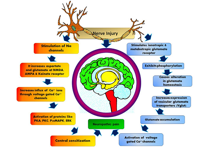
    Pathophysiology of central nervous system NP. AMPA: α-amino-3-hydroxy-5-methyl-4-isoxazolepropionic acid; PKA: protein kinase A; p38MAPK: p38 mitogen-activated protein kinase; ERK: extracellular signal-regulated protein kinase

    Cellular

    The detail peripheral and central pathophysiology depends on changes that occur at cellular and molecular levels [48]. Functional changes occur due to change in sodium and calcium channel subunit expression related to NP. During a nerve injury, the sodium and calcium channel subunits reshuffle, which results in the sudden firing of neurotransmitters [49].

    N-type VGCC (new target for NP)

    Pain, a complicated medical condition that has considerable unsatisfied clinical requirements. The opioid treatment remains standard despite of their side effects like tolerance and respiratory depression bounded to their continuous use. The foundation for pain comprises different pathways from dorsal root ganglion to brainstem [5052]. The downward pathways vigorously regulate the upward pain pathways [53]. G protein-coupled opioid receptor is the main target of opioid analgesics, and the neuronal excitability decreases when these receptors are activated [5457].

    VGCC channel α subunit inhibitors

    These channels are strongly blocked by numerous peptides obtained from the toxin of fish-trapping cone snails from Conus species, along with GVIA, MVIIA and CVID [5860]. At elevated concentrations the peptides even can block 0 channel subtypes [61, 62]. Along with MVIIA, a specific conotoxin Cav2.2 blocker is approved for managing neuronal pain. Medicinal prospective of ω-conotoxins specific for Cav2.2 for pain management has been recognized as the Cav2.2 channel, which plays a vital role in the transmission of pain [6365].

    Peptide isolated from Conus geographus

    The GVIA conotoxin obtained from Conus geographus permanently blocks Cav2.2 channels in the nanomolar range [61, 66]. GVIA has more potency in vivo, compared to other structurally similar peptides [67, 68]. GVIA is three to four folds more effective than other peptides, and almost 40 times more effective compared to morphine, when given to rats intrathecally. Because of the permanent blockage of the Cav2.2 channel, it is possibly difficult to decide the safe dose in an experimental setting [69].

    Peptide isolated from Conus magus

    MVIIA obtained from Conus magus, inhibits the Cav2.2 channels. The synthetic form (ziconotide) has been developed, which is injected intrathecally for treatment of NP clinically [7072]. The USA and Europe permitted MVIIA for managing NP [73, 74]. This peptide shows appreciable relief from pain. Like morphine, there is no adverse effect of tolerance and addiction in the case of MVIIA [75, 76].

    Peptide isolated from Conus catus

    CVID is obtained from Conus catus, the major selective antagonist of Cav2.2 channels among all peptides [77, 78]. It is presently under clinical trials by the name of AM336 [79]. In the previous studies, it has been proved that CVID is a highly selective antagonist for Cav2.2 rather than Cav2.1. Different Conus species [80] with their actions and the biological sources are shown in Table 2.

    Different Conus species with their action and biological source

    Name of peptide inhibitorBiological sourceMechanism of action
    GVIAConus geographusIrreversible inhibitor of Cav2.2
    MVIIAConus magusPotent inhibitor of Cav2.2
    CVIDConus catusSelective inhibitor of Cav2.2
    Display full size

    Ziconotide

    Ziconotide has a neuroprotective activity and is derived from the venom of cone snail. However, it is the synthetic peptide that has similarities with ω-conopeptide (MVIIA) obtained from Conus magus. The pharmacodynamics and pharmacokinetics affect living cells, and their dosage regimens have been reviewed and studied [81]. This drug is in the final stage of human testing to confirm its efficacy against NP and is one of the non-opioid neuroprotective agents [82]. Moreover, it acts by inhibiting Cav2.2 channels [83, 84]. The neuroactive peptides obtained from the venom of Conus snail have the unique property of high potency and specificity as this ziconotide is epitomizing as an excellent option for the management of nerve pain. Clinical studies have been done as proofs for the therapeutic potency of ziconotide over approximate 2,000 human subjects [85].

    (1) Protection of people against cerebral ischemia.

    (2) Study the surgical cases with bypass procedures used to treat coronary heart failure.

    (3) Pain relief in the patient having chronic pain disorder.

    Preclinical record of ziconotide

    The biological activity of ziconotide was proved by administering intracranial injections in mice that further produced shaking behaviour; this demonstrates its neuroactive property in mammals, which differentiates it from the other peptides obtained from the venom of Conus magus [86]. The Conus peptide belongs to the “Omega” class as it shares properties similar to the omega family [87]. One of the preclinical studies performed on isolated embryonic chick sympathetic ganglia shows that ziconotide blocks the electrical transmission of calcium along the synaptic end giving the evidence that it inhibits the voltage sensitive calcium channels present on presynaptic nerve ending [88, 89]. It has been observed that the pharmacodynamics and pharmacokinetics of ziconotide resemble ω-GVIA, one of the Conus peptides [90, 91].

    Biochemical interaction of ziconotide

    Ziconotide is the selective antagonist of voltage-gated selective Ca2+ channels. The predominant position of its activity is the nociceptive receptor within the neurons of the spinal cord [9295]. The analgesic potential of ziconotide is comparable to opiate and is successful against the NP in patients developed tolerance for opiate [96]. Analgesic effects of opiate are due to its binding to μ-opiate receptor resulting in its inactivation through the G protein-coupled receptor (GPCR) pathway that further blocks inflow of calcium in the cell [97]. Moreover, ziconotide directly antagonizes voltage-gated Ca2+ channels [98] as shown in Figure 4. However, ziconotide achieves complete blockade of channels as compared to opiates which only lowers the potential opening of voltage-gated Ca2+ channels. On the other hand, ziconotide also modulates other voltage-gated ion channels like potassium channels and glutamate receptors [99]. Ziconotide is considered as a preferred treatment against NP because of its following properties:

    (1) It’s effectiveness in opiate tolerant patients.

    (2) Patients do not develop tolerance against ziconotide.

    Therefore, it is considered as a novel treatment against the NP mechanism of ziconotide [100] as shown in Figure 4.

    Mechanism of ziconotide in relieving NP

    Contribution of GPCR in inhibiting the N-type VGCCs

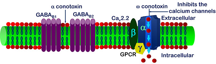
    It has been reviewed that inside cell the signal transduction modulates N-type VGCCs regulated by GPCR [101]. Inhibition of VGCCs decreases the span of signal transmission in spinal root ganglia nerve cells [102]. The observation shows the stimulation of γ-aminobutyric acid type B (GABAB) channels leads up the inhibition of VGCCs current [103107] (Figure 5). GABA can act either through ligand ion channels (GABAA) or by membrane receptor (GABAB) [108, 109]. GABAB receptor is made up of 2 subunits, GABAB1 and GABAB2 [110, 111], out of which GABAB1 is inotropic and GABAB2 is a metabotropic receptor [112117]. Stimulation of GABAB blocks the inflow of Ca2+ through VGCCs; this is only possible by activation of GPCR [118120] as shown in Figure 5.

    Inhibition of N-type calcium channels via GPCR

    Effective treatment of NP with μ-conotoxins

    U.S. Food and Drug Administration (FDA) and European Medicines Agency (EMA) authorized ziconotide (Prialt) ω-conotoxin MVIIA is administered in the management of extreme persistent pain related to malignancy, AIDS, and neuritis as the alternative medication for current pain therapy. The major problem is only with its administration through the intrathecal route to the patients [121]. However, it acts by the blockade of N-type VGCCs; therefore, it represents the new target for pain therapy with its evidence in knockout mice [122, 123]. The ability of binding Prialt with N-type VGCCs presents at A-d and C fibres of dorsal root ganglia (DRG) of the spinal cord only after its intrathecal administration [124]. It is the potent antagonist of sensitive signalling and thus has the potential to treat the pain. MVIIA and CVID during its in vitro testing show that these drugs have the fastest onset and offset of action. Intrathecal delivery is the only novel route for the administration of conotoxins [125127].

    Topical administration of drugs used for the therapy of NP

    The concept of topical therapies is consistent with current theory of mechanism-oriented pain treatment; thus, they may increase pain management quality and patient satisfaction with treatment. There are numerous preclinical trials in the literature demonstrating the antinociceptive impact of drugs applied topically in inflammatory and NP animal models. They are not currently used in clinical practise and so are not included in this evaluation. The topical agents act by either excitatory or inhibitory mechanism but through different receptor ion channel enzyme, many are currently being utilized in clinical practice (Table 3).

    Mechanisms of action of topical drugs for the therapy of NP for excitatory and inhibitory effects

    Mechanism of action of topical agentsReceptor ion channel enzymeTopical agent utilized in clinical practicePossible site of actionReference
    Mechanism for excitatory effect of drugsVoltage-gated sodium channels (NaVs)Lidocaine, amitriptyline, doxepin, phenytoin, ambroxol, TV-45070, opioids, NSAIDs, clonidineNeurons, keratinocytes[128132]
    TRPV1Capsaicin, NSAIDsNeurons, keratinocytes, immune cells[133135]
    VGCCGabapentin, lidocaineNeurons, keratinocytes[136138]
    NMDARKetamine, amitriptyline, diclofenacNeurons, keratinocytes, immune cells[139141]
    COX-2NSAIDsNeurons, immune cells, Schwann cells[142144]
    Mechanism for inhibitory effect of drugsGABARAmitriptylineNeurons, keratinocytes, immune cells[145148]
    GABAARKetamine, phenytoinNeurons, keratinocytes, immune cells[149, 150]
    GABABRBaclofenNeurons, keratinocytes, immune cells[151154]
    α2-ARClonidineNeurons[155157]
    OROpioidsNeurons, keratinocytes, immune cells[158160]
    CRCannabinoidsNeurons, keratinocytes, immune cells[161163]
    Display full size

    α2-AR: α2 adreno receptors; COX-2: cyclooxygenase-2; CR: cannabinoid receptors; GABAR: γ-aminobutyric acid receptors; GABAAR: γ-aminobutyric acid type A receptors; TV-45070: topical sodium channel inhibitor; NMDAR: NMDA receptors; NSAIDs: Non-steroidal anti-inflammatory drugs; OR: opioid receptors; TRPV1: transient receptor potential vanilloid 1

    Herbal drugs used for the therapy of NP

    Despite the abundance of medications available, there are no curative conventional treatments for NP. In the field of drug research, more emphasis is now being placed on herbal formulation. As a result, we conducted a thorough evaluation of herbal medications and plants that show anti-NP properties [164]. Certain herbal drugs are also used for the therapy of NP but only preclinical studies have been done. The following plants are some of the most popular ones that are used to treat NP: Acorus calamus [165, 166], Artemisia dracunculus [167, 168], Butea monosperma [169, 170], Citrullus colocynthis [171, 172], Curcuma longa [173, 174], Crocus sativus [175, 176], Elaeagnus angustifolia [177, 178], Ginkgo biloba [179, 180], Mitragyna speciosa [181, 182], Momordica charantia [183], and Nigella Sativa [184, 185] (Table 4).

    Herbal drugs undergone preclinical studies for NP

    Plant usedFamilyPlant partReference
    Aconiti tuberRanunculaceaeTuber[186]
    Acmella oleraceaAsteraceaeLeaves and flowers[187]
    Acorus calamusAraceaeRhizomes[188]
    Alstonia scholarisApocyanaceaeMilky juice of plant[189]
    Butea monospermaFabaceaeLeaves[190]
    Crocus sativusIridaceaeFlower[191]
    Curcuma longaZingiberaceaeRoots[192]
    Emblica officinalisPhyllanthaceaeFruit[193]
    Euphorbia tirucalliEuphor-biaceaeLatex, root, bark[194]
    Gelsemium elegansLoganiaceaeSeeds, stems[195, 196]
    Ginkgo bilobaGinkgoaceaeHerbal extract[197, 198]
    Momordica charantiaCucurbitaceaeFruit[199]
    Nauclea latifoliaRubiaceaeRoots[200, 201]
    Ocimum sanctumLamiaceaeLeaves[202]
    Olea europaeaOleaceaeOil and leaves[203]
    Paederia scandensRubiaceaeRoots, leaves, bark, and fruits[204]
    Punica granatumPunicaceaeThe juice, whole fruit, flowers, and roots[205]
    Senna spectabilisFabaceaeFlowers, fruits and leaves[206]
    Thymus caramanicus JalasLamiaceaeAerial parts[207]
    Tribulus terristrisZygophylla-CeaeComplete herb[208]
    Trigonella foenum-graecumFabaceaeSeeds[209]
    Vernonia cinereaAsteraceaeWhole plant[210]
    Display full size

    Future prospective of N-type VGCC blockers

    Certain limitations associated with the existing N-type VGCCs inhibitors provide a great opportunity for the development of better and new drugs with targeted therapeutic outcomes and lesser side effects. The peptide currently used has a drawback of narrow therapeutic window [211, 212]. Further, ziconotide shows peripheral interaction, so oral and intravenous administration are not possible, thus leaving with only choice of intrathecal route [213, 214]. The future research can be focused on development of novel ω-conotoxins with improved selectivity as inhibitor for N-type VGCCs, without any peripheral interactions, thus reducing the associated adverse effects. Although ω-conotoxin therapeutics currently is under clinical and preclinical development, there is still vast opportunity for the new research drug candidate with respect to NP. There are also several non-peptide small molecules under preclinical and clinical trials but till no successful outcome has been noticed. But of course, because of the small size and non-peptide nature they can lead to the development of molecule that can follow an alternate route with fewer side effects and more specific to the receptor [215221]. In recent years, lots of review and research have been published on recent advancements of nanotechnology and their application on neurological disease as well as NP [222233].

    Conclusions

    The existing pharmacotherapy for the NP is very challenging being non-specific in action. The therapies are non-invasive associated with several side effects and drug-drug interactions. There is a scope for the development of therapy which is target specific with lesser side effects. Ziconotide acts as N-type VGCC antagonist the only approved drug administered through intrathecal route. Certain herbal drugs also have been studied but no clinical result is there and the study is only limited to preclinical data. New research is being carried out on ω-conotoxins and small peptides have been obtained from Conus species. The vast number of Conus species and wide variety of conotoxin availability open a path for further research on more target specific and least side effect molecules.

    Abbreviations

    GABA:

    γ-aminobutyric acid

    GABAB:

    γ-aminobutyric acid type B

    GPCR:

    G protein-coupled receptor

    NP:

    neuropathic pain

    VGCCs:

    voltage-gated calcium channels

    Declarations

    Acknowledgments

    Our sincere thanks to Paramveer Singh, Shadab Husain and Jamilur Rahman Ansari for editing the language of this manuscript. The authors gratefully acknowledge the contributions of the collaborators and co-workers mentioned in the cited reference. SC, R Kaur, and AG acknowledges K.R. Mangalam University, AW acknowledges Guru Gobind Singh Indraprastha University (GGSIPU). R Kadian gratefully acknowledges the support and cooperation from the Director of Ram Gopal College, Gurugram. MSA gratefully acknowledges the Vice Chancellor and Dean of Pharmacy, Shree Guru Gobind Singh Tricentenary University (SGTU) for their kind cooperation and support.

    Author contributions

    SC, AW, R Kaur, and AG: Conceptualization, Investigation, Writing—original draft, Writing—review & editing. R Kadian and MSA: Validation, Writing—review & editing, Supervision.

    Conflicts of interest

    The authors declare that they have no conflicts of interest.

    Ethical approval

    Not applicable.

    Consent to participate

    Not applicable.

    Consent to publication

    Not applicable.

    Availability of data and materials

    Not applicable.

    Funding

    Not applicable.

    Copyright

    © The Author(s) 2023.

    References

    Merskey HE. Classification of chronic pain: descriptions of chronic pain syndromes and definitions of pain terms. Pain. 1986;Suppl 3:226.
    Bernetti A, Agostini F, de Sire A, Mangone M, Tognolo L, Di Cesare A, et al. Neuropathic pain and rehabilitation: a systematic review of international guidelines. Diagnostics. 2021;11:74. [DOI] [PubMed] [PMC]
    Lo J, Chan L, Flynn S. A systematic review of the incidence, prevalence, costs, and activity and work limitations of amputation, osteoarthritis, rheumatoid arthritis, back pain, multiple sclerosis, spinal cord injury, stroke, and traumatic brain injury in the United States: a 2019 update. Arch Phys Med Rehabil. 2021;102:11531. [DOI] [PubMed] [PMC]
    Seddighi AS, Seddighi A, Ghadirian M, Zali A, Far SMT. Neuropathic pain: mechanism, representation, management and treatment. J Int Clin Neurosci. 2022;9:e18. [DOI]
    Marchettini P, Formaglio F, Lacerenza M. Neuropathic pain. In: Eduardo Bruera IJ, Higginson CF, von Gunten TM, editors. Textbook of palliative medicine and supportive care. Boca Raton: CRC Press; 2021. pp. 30112. [DOI]
    Batmaz SB, Birinci G, Aslan EA. Quality of life of children with allergic disease: the effect of depression and anxiety of children and their mothers. J Asthma. 2022;59:177686. [DOI] [PubMed]
    Samadi Z, Jannati Y, Hamidia A, Mohammadpour RA, Hesamzadeh A. The effect of aromatherapy with lavender essential oil on sleep quality in patients with major depression. J Nurs Midwif Sci. 2021;8:6773. [DOI]
    Merskey H, Bogduk N. Classification of chronic pain. 2nd ed. Seattle: IASP Press; 1994.
    Colloca L, Ludman T, Bouhassira D, Baron R, Dickenson AH, Yarnitsky D, et al. Neuropathic pain. Nat Rev Dis Primers. 2017;3:17002. [DOI] [PubMed] [PMC]
    Pottoo FH, Sharma S, Javed MN, Barkat MA, Harshita, Alam MS, et al. Lipid-based nanoformulations in the treatment of neurological disorders. Drug Metab Rev. 2020;52:185204. [DOI] [PubMed]
    van Hecke O, Austin SK, Khan RA, Smith BH, Torrance N. Neuropathic pain in the general population: a systematic review of epidemiological studies. Pain. 2014;155:65462. [DOI] [PubMed]
    Pandey M, Saleem S, Nautiyal H, Pottoo FH, Javed MN. PINK1/Parkin in neurodegenerative disorders: crosstalk between mitochondrial stress and neurodegeneration. In: Uddin MS, Ashraf GM, editors. Quality control of cellular protein in neurodegenerative disorders. Hershey: IGI Global; 2020. [DOI]
    Finnerup NB, Sindrup SH, Jensen TS. The evidence for pharmacological treatment of neuropathic pain. Pain. 2010;150:57381. [DOI] [PubMed]
    Finnerup NB, Attal N, Haroutounian S, McNicol E, Baron R, Dworkin RH, et al. Pharmacotherapy for neuropathic pain in adults: a systematic review and meta-analysis. Lancet Neurol. 2015;14:16273. [DOI] [PubMed] [PMC]
    Fornasari D. Pharmacotherapy for neuropathic pain: a review. Pain Ther. 2017;6:2533. [DOI] [PubMed] [PMC]
    Vadivelu N, Kai A, Maslin B, Kodumudi G, Legler A, Berger JM. Tapentadol extended release in the management of peripheral diabetic neuropathic pain. Ther Clin Risk Manag. 2015;11:95105. [DOI] [PubMed] [PMC]
    Sanford M. Intrathecal ziconotide: a review of its use in patients with chronic pain refractory to other systemic or intrathecal analgesics. CNS Drugs. 2013;27:9891002. [DOI] [PubMed]
    Pottoo FH, Tabassum N, Javed MN, Nigar S, Sharma S, Barkat MA, et al. Raloxifene potentiates the effect of fluoxetine against maximal electroshock induced seizures in mice. Eur J Pharm Sci. 2020;146:105261. [DOI] [PubMed]
    Field MJ, Li Z, Schwarz JB. Ca2+ channel α2-δ ligands for the treatment of neuropathic pain. J Med Chem. 2007;50:256975. [DOI] [PubMed]
    Heinke B, Balzer E, Sandkühler J. Pre- and postsynaptic contributions of voltage-dependent Ca2+ channels to nociceptive transmission in rat spinal lamina I neurons. Eur J Neurosci. 2004;19:10311. [DOI] [PubMed]
    Dolphin AC, Menon-Johansson A, Campbell V, Berrow N, Sweeney MI. GABAB receptors and G proteins modulate voltage-dependent calcium channels in cultured rat dorsal root ganglion neurons: relevance to transmitter release and its modulation. Neurophysiology. 1994;26:2935. [DOI]
    Westenbroek RE, Hoskins L, Catterall WA. Localization of Ca2+ channel subtypes on rat spinal motor neurons, interneurons, and nerve terminals. J Neurosci. 1998;18:631930. [DOI] [PubMed] [PMC]
    King T, Ossipov MH, Vanderah TW, Porreca F, Lai J. Is paradoxical pain induced by sustained opioid exposure an underlying mechanism of opioid antinociceptive tolerance? Neurosignals. 2005;14:194205. [DOI] [PubMed]
    Yamamoto T, Nair P, Davis P, Ma SW, Navratilova E, Moye S, et al. Design, synthesis, and biological evaluation of novel bifunctional C-terminal-modified peptides for δ/μ opioid receptor agonists and neurokinin-1 receptor antagonists. J Med Chem. 2007;50:277986. [DOI] [PubMed] [PMC]
    Yamamoto T, Nair P, Jacobsen NE, Davis P, Ma SW, Navratilova E, et al. The importance of micelle-bound states for the bioactivities of bifunctional peptide derivatives for δ/μ opioid receptor agonists and neurokinin 1 receptor antagonists. J Med Chem. 2008;51:633447. [DOI] [PubMed] [PMC]
    Pottoo FH, Tabassum N, Javed MN, Nigar S, Rasheed R, Khan A, et al. The synergistic effect of raloxifene, fluoxetine, and bromocriptine protects against pilocarpine-induced status epilepticus and temporal lobe epilepsy. Mol Neurobiol. 2019;56:123347. [DOI] [PubMed]
    Siddall PJ, Cousins MJ. Spinal pain mechanisms. Spine. 1997;22:98104. [DOI] [PubMed]
    Sorkin LS, Xiao WH, Wagner R, Myers RR. Tumour necrosis factor-α induces ectopic activity in nociceptive primary afferent fibres. Neuroscience. 1997;81:25562. [DOI] [PubMed]
    Nicholson B. Gabapentin use in neuropathic pain syndromes. Acta Neurol Scand. 2000;101:35971. [DOI] [PubMed]
    Amir R, Devor M. Chemically mediated cross-excitation in rat dorsal root ganglia. J Neurosci. 1996;16:473341. [DOI] [PubMed] [PMC]
    Patil PG, Campbell JN. Lesions of primary afferent and sympathetic efferents as treatments for pain. In: Bonica’s management of pain. 3rd ed. Baltimore: Lippincott Williams & Wilkins; 2001. pp. 201122.
    Pottoo FH, Bhowmik M, Vohora D. Raloxifene protects against seizures and neurodegeneration in a mouse model mimicking epilepsy in postmenopausal woman. Eur J Pharm Sci. 2014;65:16773. [DOI] [PubMed]
    Schott GD. Visceral afferents: their contribution to ‘sympathetic dependent’ pain. Brain. 1994;117:397413. [DOI] [PubMed]
    Rowbotham MC, Petersen KL. Anticonvulsants and local anesthetic drugs. In: Loeser JD, Butler S, Chapman CR, Turk DC, editors. Bonica’s management of pain. 3rd ed. Philadelphia: Williams & Wilkins; 2001. pp. 172735.
    Pasero C. Pathophysiology of neuropathic pain. Pain Manage Nurs. 2004;5:38. [DOI] [PubMed]
    Pottoo FH, Javed MN, Barkat MA, Alam MS, Nowshehri JA, Alshayban DM, et al. Estrogen and serotonin: complexity of interactions and implications for epileptic seizures and epileptogenesis. Curr Neuropharmacol. 2019;17:21431. [DOI] [PubMed] [PMC]
    Kuner R, Flor H. Structural plasticity and reorganisation in chronic pain. Nat Rev Neurosci. 2017;18:2030. Erratum in: Nat Rev Neurosci. 2017;18:158. [DOI] [PubMed]
    Rowbotham MC, Yosipovitch G, Connolly MK, Finlay D, Forde G, Fields HL. Cutaneous innervation density in the allodynic form of postherpetic neuralgia. Neurobiol Dis. 1996;3:20514. [DOI] [PubMed]
    Ochoa JL, Campero M, Serra J, Bostock H. Hyperexcitable polymodal and insensitive nociceptors in painful human neuropathy. Muscle Nerve. 2005;32:45972. [DOI] [PubMed]
    Reichling DB, Levine JD. Critical role of nociceptor plasticity in chronic pain. Trends Neurosci. 2009;32:6118. [DOI] [PubMed] [PMC]
    Ratté S, Prescott SA. Afferent hyperexcitability in neuropathic pain and the inconvenient truth about its degeneracy. Curr Opin Neurobiol. 2016;36:317. [DOI] [PubMed]
    Beydoun A, Backonja MM. Mechanistic stratification of antineuralgic agents. J Pain Symptom Manage. 2003;25:S1830. [DOI] [PubMed]
    Yaksh TL. Calcium channels as therapeutic targets in neuropathic pain. J Pain. 2006;7:S1330. [DOI] [PubMed]
    Xiao WH, Bennett GJ. Synthetic omega-conopeptides applied to the site of nerve injury suppress neuropathic pains in rats. J Pharmacol Exp Ther. 1995;274:66672. [PubMed]
    Bridges D, Thompson SW, Rice AS. Mechanisms of neuropathic pain. Br J Anaesth. 2001;87:1226. [DOI] [PubMed]
    Attal N. Chronic neuropathic pain: mechanisms and treatment. Clin J Pain. 2000;16:S11830. [DOI] [PubMed]
    Woolf CJ. Evidence for a central component of post-injury pain hypersensitivity. Nature. 1983;306:6868. [DOI] [PubMed]
    Laird MA, Gidal BE. Use of gabapentin in the treatment of neuropathic pain. Ann Pharmacother. 2000;34:8027. [DOI] [PubMed]
    Simms BA, Zamponi GW. Neuronal voltage-gated calcium channels: structure, function, and dysfunction. Neuron. 2014;82:2445. [DOI] [PubMed]
    Sindrup SH, Jensen TS. Efficacy of pharmacological treatments of neuropathic pain: an update and effect related to mechanism of drug action. Pain. 1999;83:389400. [DOI] [PubMed]
    Latremoliere A, Woolf CJ. Central sensitization: a generator of pain hypersensitivity by central neural plasticity. J Pain. 2009;10:895926. [DOI] [PubMed] [PMC]
    Gracely RH, Lynch SA, Bennett GJ. Painful neuropathy: altered central processing maintained dynamically by peripheral input. Pain. 1992;51:17594. [DOI] [PubMed]
    Loeser JD, Treede RD. The Kyoto protocol of IASP basic pain terminology. Pain. 2008;137:4737. [DOI] [PubMed]
    Ibrahim AM, Pottoo FH, Dahiya ES, Khan FA, Kumar JBS. Neuron-glia interactions: molecular basis of alzheimer’s disease and applications of neuroproteomics. Eur J Neurosci. 2020;52:293143. [DOI] [PubMed]
    Truini A, Cruccu G. Pathophysiological mechanisms of neuropathic pain. Neurol Sci. 2006;27:S17982. [DOI] [PubMed]
    Vaillancourt PD, Langevin HM. Painful peripheral neuropathies. Med Clin North Am. 1999;83:62742. [DOI] [PubMed]
    Salter MW. Cellular signalling pathways of spinal pain neuroplasticity as targets for analgesic development. Curr Top Med Chem. 2005;5:55767. [DOI] [PubMed]
    Costigan M, Scholz J, Woolf CJ. Neuropathic pain: a maladaptive response of the nervous system to damage. Annu Rev Neurosci. 2009;32:132. [DOI] [PubMed] [PMC]
    Snutch TP. Targeting chronic and neuropathic pain: the N-type calcium channel comes of age. NeuroRx. 2005;2:66270. [DOI] [PubMed] [PMC]
    Heinricher MM, Tavares I, Leith JL, Lumb BM. Descending control of nociception: specificity, recruitment and plasticity. Brain Res Rev. 2009;60:21425. [DOI] [PubMed] [PMC]
    Hille B, Beech DJ, Bernheim L, Mathie A, Shapiro MS, Wollmuth LP. Multiple G-protein-coupled pathways inhibit N-type Ca channels of neurons. Life Sci. 1995;56:98992. [DOI] [PubMed]
    Bovill JG. Mechanisms of actions of opioids and non-steroidal anti-inflammatory drugs. Eur J Anaesthesiol. 1997;14:915. [DOI] [PubMed]
    Zamponi GW, Snutch TP. Modulation of voltage-dependent calcium channels by G proteins. Curr Opin Neurobiol. 1998;8:3516. [DOI] [PubMed]
    Christie MJ, Connor M, Vaughan CW, Ingram SL, Bagley EE. Cellular actions of opioids and other analgesics: implications for synergism in pain relief. Clin Exp Pharmacol Physiol. 2000;27:5203. [DOI] [PubMed]
    Schroeder CI, Lewis RJ. ω-conotoxins GVIA, MVIIA and CVID: SAR and clinical potential. Mar Drugs. 2006;4:193214. [DOI] [PMC]
    Olivera BM, Gray WR, Zeikus R, McIntosh JM, Varga J, Rivier J, et al. Peptide neurotoxins from fish-hunting cone snails. Science. 1985;230:133843. [DOI] [PubMed]
    Olivera BM, Miljanich GP, Ramachandran J, Adams ME. Calcium channel diversity and neurotransmitter release: the ω-conotoxins and omega-agatoxins. Annu Rev Biochem. 1994;63:82367. [DOI] [PubMed]
    Feng ZP, Doering CJ, Winkfein RJ, Beedle AM, Spafford JD, Zamponi GW. Determinants of inhibition of transiently expressed voltage-gated calcium channels by ω-conotoxins GVIA and MVIIA. J Biol Chem. 2003;278:201718. [DOI] [PubMed]
    Williams ME, Feldman DH, McCue AF, Brenner R, Velicelebi G, Ellis SB, et al. Structure and functional expression of α1, α2, and β subunits of a novel human neuronal calcium channel subtype. Neuron. 1992;8:7184. [DOI] [PubMed]
    Vanegas H, Schaible H. Effects of antagonists to high-threshold calcium channels upon spinal mechanisms of pain, hyperalgesia and allodynia. Pain. 2000;85:918. [DOI] [PubMed]
    de Souza AH, Castro CJ Jr, Rigo FK, de Oliveira SM, Gomez RS, Diniz DM, et al. An evaluation of the antinociceptive effects of Phα1β, a neurotoxin from the spider Phoneutria nigriventer, and ω-conotoxin MVIIA, a cone snail Conus magus toxin, in rat model of inflammatory and neuropathic pain. Cell Mol Neurobiol. 2013;33:5967. [DOI] [PubMed]
    Prado WA. Involvement of calcium in pain and antinociception. Braz J Med Biol Res. 2001;34:44961. [DOI] [PubMed]
    Ellinor PT, Zhang JF, Horne WA, Tsien RW. Structural determinants of the blockade of N-type calcium channels by a peptide neurotoxin. Nature. 1994;372:2725. [DOI] [PubMed]
    Vega T, De Pascual R, Bulbena O, García AG. Effects of omega-toxins on noradrenergic neurotransmission in beating guinea pig atria. Eur J Pharmacol. 1995;276:2318. [DOI] [PubMed]
    Scott DA, Wright CE, Angus JA. Actions of intrathecal ω-conotoxins CVID, GVIA, MVIIA, and morphine in acute and neuropathic pain in the rat. Eur J Pharmacol. 2002;451:27986. [DOI] [PubMed]
    Pin JP, Bockaert J. ω-conotoxin GVIA and dihydropyridines discriminate two types of Ca2+ channels involved in GABA release from striatal neurons in culture. Eur J Pharmacol. 1990;188:814. [DOI] [PubMed]
    Wang YX, Pettus M, Gao D, Phillips C, Scott Bowersox S. Effects of intrathecal administration of ziconotide, a selective neuronal N-type calcium channel blocker, on mechanical allodynia and heat hyperalgesia in a rat model of postoperative pain. Pain. 2000;84:1518. [DOI] [PubMed]
    Jain KK. An evaluation of intrathecal ziconotide for the treatment of chronic pain. Expert Opin Investig Drugs. 2000;9:240310. [DOI] [PubMed]
    Wermeling DP, Berger JR. Ziconotide infusion for severe chronic pain: case series of patients with neuropathic pain. Pharmacotherapy. 2006;26:395402. [DOI] [PubMed]
    Miljanich GP. Ziconotide: neuronal calcium channel blocker for treating severe chronic pain. Curr Med Chem. 2004;11:302940. [DOI] [PubMed]
    Wermeling DP. Ziconotide, an intrathecally administered N-type calcium channel antagonist for the treatment of chronic pain. Pharmacotherapy. 2005;25:108494. [DOI] [PubMed]
    Micheli L, Rajagopalan R, Lucarini E, Toti A, Parisio C, Carrino D, et al. Pain relieving and neuroprotective effects of non-opioid compound, DDD-028, in the rat model of paclitaxel-induced neuropathy. Neurotherapeutics. 2021;18:200820. [DOI] [PubMed] [PMC]
    Wang YX, Gao D, Pettus M, Phillips C, Bowersox SS. Interactions of intrathecally administered ziconotide, a selective blocker of neuronal N-type voltage-sensitive calcium channels, with morphine on nociception in rats. Pain. 2000;84:27181. [DOI] [PubMed]
    Adams DJ, Smith AB, Schroeder CI, Yasuda T, Lewis RJ. Omega-conotoxin CVID inhibits a pharmacologically distinct voltage-sensitive calcium channel associated with transmitter release from preganglionic nerve terminals. J Biol Chem. 2003;278:405762. [DOI] [PubMed]
    Lewis RJ, Nielsen KJ, Craik DJ, Loughnan ML, Adams DA, Sharpe IA, et al. Novel ω-conotoxins from Conus catus discriminate among neuronal calcium channel subtypes. J Biol Chem. 2000;275:3533544. [DOI] [PubMed]
    Li Q, Lu J, Zhou X, Chen X, Su D, Gu X, et al. High-voltage-activated calcium channel in the afferent pain pathway: an important target of pain therapies. Neurosci Bull. 2019;35:107384. [DOI] [PubMed] [PMC]
    Smith MT, Cabot PJ, Ross FB, Robertson AD, Lewis RJ. The novel N-type calcium channel blocker, AM336, produces potent dose-dependent antinociception after intrathecal dosing in rats and inhibits substance P release in rat spinal cord slices. Pain. 2002;96:11927. [DOI] [PubMed]
    Miljanich GP, Ramachandran J. Antagonists of neuronal calcium channels: structure, function, and therapeutic implications. Annu Rev Pharmacol Toxicol. 1995;35:70734. [DOI] [PubMed]
    Dolphin AC. Functions of presynaptic voltage-gated calcium channels. Function (Oxf). 2021;2:zqaa027. [DOI] [PubMed] [PMC]
    Bowersox SS, Luther R. Pharmacotherapeutic potential of ω-conotoxin MVIIA (SNX-111), an N-type neuronal calcium channel blocker found in the venom of Conus magus. Toxicon. 1998;36:16518. [DOI] [PubMed]
    Staats PS, Yearwood T, Charapata SG, Presley RW, Wallace MS, Byas-Smith M, et al. Intrathecal ziconotide in the treatment of refractory pain in patients with cancer or AIDS: a randomized controlled trial. JAMA. 2004;291:6370. [DOI] [PubMed]
    Thompson SW, Bennett DL, Kerr BJ, Bradbury EJ, McMahon SB. Brain-derived neurotrophic factor is an endogenous modulator of nociceptive responses in the spinal cord. Proc Natl Acad Sci U S A. 1999;96:77148. [DOI] [PubMed] [PMC]
    Terlau H, Olivera BM. Conus venoms: a rich source of novel ion channel-targeted peptides. Physiol Rev. 2004;84:4168. [DOI] [PubMed]
    Yeager RE, Yoshikami D, Rivier J, Cruz LJ, Miljanich GP. Transmitter release from presynaptic terminals of electric organ: inhibition by the calcium channel antagonist omega Conus toxin. J Neurosci. 1987;7:23906. [PubMed] [PMC]
    Doroshenko PA, Woppmann A, Miljanich G, Augustine GJ. Pharmacologically distinct presynaptic calcium channels in cerebellar excitatory and inhibitory synapses. Neuropharmacology. 1997;36:86572. [DOI] [PubMed]
    Chu LF, Clark DJ, Angst MS. Opioid tolerance and hyperalgesia in chronic pain patients after one month of oral morphine therapy: a preliminary prospective study. J Pain. 2006;7:438. [DOI] [PubMed]
    Kristipati R, Nádasdi L, Tarczy-Hornoch K, Lau K, Miljanich GP, Ramachandran J, et al. Characterization of the binding of omega-conopeptides to different classes of non-L-type neuronal calcium channels. Mol Cell Neurosci. 1994;5:21928. [DOI] [PubMed]
    Deer TR, Pope JE, Hanes MC, McDowell GC. Intrathecal therapy for chronic pain: a review of morphine and ziconotide as firstline options. Pain Med. 2019;20:78498. [DOI] [PubMed] [PMC]
    Dray A. Neuropathic pain: emerging treatments. Br J Anaesth. 2008;101:4858. [DOI] [PubMed]
    Malmberg AB, Yaksh TL. Effect of continuous intrathecal infusion of omega-conopeptides, N-type calcium-channel blockers, on behavior and antinociception in the formalin and hot-plate tests in rats. Pain. 1995;60:8390. [DOI] [PubMed]
    Saegusa H, Matsuda Y, Tanabe T. Effects of ablation of N- and R-type Ca2+ channels on pain transmission. Neurosci Res. 2002;43:17. [DOI] [PubMed]
    Nestler EJ, Alreja M, Aghajanian GK. Molecular and cellular mechanisms of opiate action: studies in the rat locus coeruleus. Brain Res Bull. 1994;35:5218. [DOI] [PubMed]
    Attali B, Saya D, Nah SY, Vogel Z. Kappa opiate agonists inhibit Ca2+ influx in rat spinal cord-dorsal root ganglion cocultures. Involvement of a GTP-binding protein. J Biol Chem. 1989;264:34753. [DOI] [PubMed]
    Adams DJ, Callaghan B, Berecki G. Analgesic conotoxins: block and G protein-coupled receptor modulation of N-type (CaV2.2) calcium channels. Br J Pharmacol. 2012;166:486500. [DOI] [PubMed] [PMC]
    Pirec V, Laurito CE, Lu Y, Yeomans DC. The combined effects of N-type calcium channel blockers and morphine on Aδ versus C fiber mediated nociception. Anesth Analg. 2001;92:23943. [DOI] [PubMed]
    Zamponi GW, McCleskey EW. Splicing it up: a variant of the N-type calcium channel specific for pain. Neuron. 2004;41:34. [DOI] [PubMed]
    Dunlap K, Fischbach G. Neurotransmitters decrease the calcium component of sensory neurone action potentials. Nature. 1978;276:8379. [DOI] [PubMed]
    Dunlap K, Fischbach GD. Neurotransmitters decrease the calcium conductance activated by depolarization of embryonic chick sensory neurones. J Physiol. 1981;317:51935. [DOI] [PubMed] [PMC]
    Zamponi GW, Lewis RJ, Todorovic SM, Arneric SP, Snutch TP. Role of voltage-gated calcium channels in ascending pain pathways. Brain Res Rev. 2009;60:849. [DOI] [PubMed] [PMC]
    Zamponi GW, Currie KP. Regulation of CaV2 calcium channels by G protein coupled receptors. Biochim Biophys Acta. 2013;1828:162943. [DOI] [PubMed] [PMC]
    Currie KP. G protein modulation of CaV2 voltage-gated calcium channels. Channels. 2010;4:497509. [DOI] [PubMed] [PMC]
    Altier C, Zamponi GW. Signaling complexes of voltage-gated calcium channels and G protein-coupled receptors. J Recept Signal Transduct Res. 2008;28:7181. [DOI] [PubMed]
    Tedford HW, Zamponi GW. Direct G protein modulation of Cav2 calcium channels. Pharmacol Rev. 2006;58:83762. [DOI] [PubMed]
    Enna SJ, McCarson KE. The role of GABA in the mediation and perception of pain. Adv Pharmacol. 2006;54:127. [DOI] [PubMed]
    Page AJ, O’Donnell TA, Blackshaw LA. Inhibition of mechanosensitivity in visceral primary afferents by GABAB receptors involves calcium and potassium channels. Neuroscience. 2006;137:62736. [DOI] [PubMed]
    Schwenk J, Metz M, Zolles G, Turecek R, Fritzius T, Bildl W, et al. Native GABAB receptors are heteromultimers with a family of auxiliary subunits. Nature. 2010;465:2315. [DOI] [PubMed]
    Bettler B, Kaupmann K, Mosbacher J, Gassmann M. Molecular structure and physiological functions of GABAB receptors. Physiol Rev. 2004;84:83567. [DOI] [PubMed]
    Page AJ, Blackshaw LA. GABAB receptors inhibit mechanosensitivity of primary afferent endings. J Neurosci. 1999;19:8597602. [DOI] [PubMed] [PMC]
    Patel S, Naeem S, Kesingland A, Froestl W, Capogna M, Urban L, et al. The effects of GABAB agonists and gabapentin on mechanical hyperalgesia in models of neuropathic and inflammatory pain in the rat. Pain. 2001;90:21726. [DOI] [PubMed]
    Smith GD, Harrison SM, Birch PJ, Elliott PJ, Malcangio M, Bowery NG. Increased sensitivity to the antinociceptive activity of (±)-baclofen in an animal model of chronic neuropathic, but not chronic inflammatory hyperalgesia. Neuropharmacology. 1994;33:11038. [DOI] [PubMed]
    Campbell V, Berrow N, Dolphin AC. GABAB receptor modulation of Ca2+ currents in rat sensory neurones by the G protein G0: antisense oligonucleotide studies. J Physiol. 1993;470:111. [DOI] [PubMed] [PMC]
    Menon-Johansson AS, Berrow N, Dolphin AC. Go transduces GABAB-receptor modulation of N-type calcium channels in cultured dorsal root ganglion neurons. Pflügers Arch. 1993;425:33543. [DOI] [PubMed]
    Morishita R, Kato K, Asano T. GABAB receptors couple to G proteins Go, G*o and G•i1 but not to Gi2. FEBS Lett. 1990;271:2315. [DOI] [PubMed]
    Herlitze S, Garcia D, Mackie K, Hille B, Scheuer T, Catterall WA. Modulation of Ca2+ channels βγ G-protein py subunits. Nature. 1996:380;25862. [DOI] [PubMed]
    Ikeda S. Voltage-dependent modulation of N-type calcium channels by G-protein βγ subunits. Nature. 1996;380:2558. [DOI] [PubMed]
    Bean BP. Neurotransmitter inhibition of neuronal calcium currents by changes in channel voltage dependence. Nature. 1989;340:1536. [DOI] [PubMed]
    Park J, Luo ZD. Calcium channel functions in pain processing. Channels. 2010;4:5107. [DOI] [PubMed] [PMC]
    Price N, Namdari R, Neville J, Proctor KJ, Kaber S, Vest J, et al. Safety and efficacy of a topical sodium channel inhibitor (TV-45070) in patients with postherpetic neuralgia (PHN): a randomized, controlled, proof-of-concept, crossover study, with a subgroup analysis of the Nav1.7 R1150W genotype. Clin J Pain. 2017;33:3108. [DOI] [PubMed] [PMC]
    McGivern JG. Targeting N-type and T-type calcium channels for the treatment of pain. Drug Discov Today. 2006;11:24553. [DOI] [PubMed]
    Li ZM, Chen LX, Li H. Voltage-gated sodium channels and blockers: an overview and where will they go? Curr Med Sci. 2019;39:86373. [DOI] [PubMed]
    Mould J, Yasuda T, Schroeder CI, Beedle AM, Doering CJ, Zamponi GW, et al. The α2δ auxiliary subunit reduces affinity of ω-conotoxins for recombinant N-type (Cav2.2) calcium channels. J Biol Chem. 2004;279:3470514. [DOI] [PubMed]
    Wright CE, Robertson AD, Whorlow SL, Angus JA. Cardiovascular and autonomic effects of ω-conotoxins MVIIA and CVID in conscious rabbits and isolated tissue assays. Br J Pharmacol. 2000;131:132536. [DOI] [PubMed] [PMC]
    Hwang SM, Jo YY, Cohen CF, Kim YH, Berta T, Park CK. Venom peptide toxins targeting the outer pore region of transient receptor potential vanilloid 1 in pain: implications for analgesic drug development. Int J Mol Sci. 2022;23:5772. [DOI] [PubMed] [PMC]
    Dib-Hajj SD, Rush AM, Cummins TR, Hisama FM, Novella S, Tyrrell L, et al. Gain-of-function mutation in Nav1.7 in familial erythromelalgia induces bursting of sensory neurons. Brain. 2005;128:184754. [DOI] [PubMed]
    Novakovic SD, Tzoumaka E, McGivern JG, Haraguchi M, Sangameswaran L, Gogas KR, et al. Distribution of the tetrodotoxin-resistant sodium channel PN3 in rat sensory neurons in normal and neuropathic conditions. J Neurosci. 1998;18:217487. [DOI] [PubMed] [PMC]
    Dib-Hajj SD, Fjell J, Cummins TR, Zheng Z, Fried K, LaMotte R, et al. Plasticity of sodium channel expression in DRG neurons in the chronic constriction injury model of neuropathic pain. Pain. 1999;83:591600. [DOI] [PubMed]
    Zhao P, Barr TP, Hou Q, Dib-Hajj SD, Black JA, Albrecht PJ, et al. Voltage-gated sodium channel expression in rat and human epidermal keratinocytes: evidence for a role in pain. Pain. 2008;139:90105. [DOI] [PubMed]
    Bennett DL, Woods CG. Painful and painless channelopathies. Lancet Neurol. 2014;13:58799. [DOI] [PubMed]
    Zhang N, Inan S, Cowan A, Sun R, Wang JM, Rogers TJ, et al. A proinflammatory chemokine, CCL3, sensitizes the heat- and capsaicin-gated ion channel TRPV1. Proc Natl Acad Sci U S A. 2005;102:453641. [DOI] [PubMed] [PMC]
    Obreja O, Rathee PK, Lips KS, Distler C, Kress M. IL-1 beta potentiates heat-activated currents in rat sensory neurons: involvement of IL-1RI, tyrosine kinase, and protein kinase C. FASEB J. 2002;16:1497503. [DOI] [PubMed]
    Jang Y, Kim M, Hwang SW. Molecular mechanisms underlying the actions of arachidonic acid-derived prostaglandins on peripheral nociception. J Neuroinflammation. 2020;17:30. [DOI] [PubMed] [PMC]
    Słoniecka M, Le Roux S, Boman P, Byström B, Zhou Q, Danielson P. Expression profiles of neuropeptides, neurotransmitters, and their receptors in human keratocytes in vitro and in situ. PLoS One. 2015;10:e0134157. [DOI] [PubMed] [PMC]
    Denda M, Fujiwara S, Hibino T. Expression of voltage-gated calcium channel subunit α1C in epidermal keratinocytes and effects of agonist and antagonists of the channel on skin barrier homeostasis. Exp Dermatol. 2006;15:45560. [DOI] [PubMed]
    Kumamoto J, Goto M, Denda S, Nakatani M, Takasugi Y, Tsuchiya K, et al. External negative electric potential accelerates exocytosis of lamellar bodies in human skin ex vivo. Exp Dermatol. 2013;22:4213. [DOI] [PubMed]
    Jang JH, Nam TS, Jun J, Jung SJ, Kim DW, Leem JW. Peripheral NMDA receptors mediate antidromic nerve stimulation-induced tactile hypersensitivity in the rat. Mediators Inflamm. 2015;2015:793624. [DOI] [PubMed] [PMC]
    Warncke T, Jørum E, Stubhaug A. Local treatment with the N-methyl-D-aspartate receptor antagonist ketamine, inhibit development of secondary hyperalgesia in man by a peripheral action. Neurosci Lett. 1997;227:14. [DOI] [PubMed]
    Morhenn VB, Murakami M, O’Grady T, Nordberg J, Gallo RL. Characterization of the expression and function of N-methyl-D-aspartate receptor in keratinocytes. Exp Dermatol. 2004;13:50511. [DOI] [PubMed]
    Ma W, Chabot JG, Vercauteren F, Quirion R. Injured nerve-derived COX2/PGE2 contributes to the maintenance of neuropathic pain in aged rats. Neurobiol Aging. 2010;31:122737. [DOI] [PubMed]
    Ahmed SU, Zhang Y, Chen L, Cohen A, St Hillary K, Vo T, et al. Effect of 1.5% topical diclofenac on clinical neuropathic pain. Anesthesiology. 2015;123:1918. [DOI] [PubMed]
    Derry S, Wiffen PJ, Kalso EA, Bell RF, Aldington D, Phillips T, et al. Topical analgesics for acute and chronic pain in adults - an overview of Cochrane reviews. Cochrane Database Syst Rev. 2017;5:CD008609. [DOI] [PubMed] [PMC]
    Nigam R, El-Nour H, Amatya B, Nordlind K. GABA and GABAA receptor expression on immune cells in psoriasis: a pathophysiological role. Arch Dermatol Res. 2010;302:50715. [DOI] [PubMed]
    Cevikbas F, Braz JM, Wang X, Solorzano C, Sulk M, Buhl T, et al. Synergistic antipruritic effects of gamma aminobutyric acid A and B agonists in a mouse model of atopic dermatitis. J Allergy Clin Immunol. 2017;140:45464.e2. [DOI] [PubMed] [PMC]
    Ngo DH, Vo TS. An updated review on pharmaceutical properties of gamma-aminobutyric acid. Molecules. 2019;24:2678. [DOI] [PubMed] [PMC]
    Wu C, Qin X, Du H, Li N, Ren W, Peng Y. The immunological function of GABAergic system. Front Biosci (Landmark Ed). 2017;22:116272. [DOI] [PubMed]
    Irifune M, Sato T, Kamata Y, Nishikawa T, Dohi T, Kawahara M. Evidence for GABAA receptor agonistic properties of ketamine: convulsive and anesthetic behavioral models in mice. Anesth Analg. 2000;91:2306. [DOI] [PubMed]
    Granger P, Biton B, Faure C, Vige X, Depoortere H, Graham D, et al. Modulation of the gamma-aminobutyric acid type A receptor by the antiepileptic drugs carbamazepine and phenytoin. Mol Pharmacol. 1995;47:118996. [PubMed]
    Whitehead RA, Puil E, Ries CR, Schwarz SK, Wall RA, Cooke JE, et al. GABAB receptor-mediated selective peripheral analgesia by the non-proteinogenic amino acid, isovaline. Neuroscience. 2012;213:15460. [DOI] [PubMed]
    Kopsky DJ, Keppel Hesselink JM. Neuropathic pain as a result of acromegaly, treated with topical baclofen cream. J Pain Symptom Manage. 2013;46:e45. [DOI] [PubMed]
    Barton DL, Wos EJ, Qin R, Mattar BI, Green NB, Lanier KS, et al. A double-blind, placebo-controlled trial of a topical treatment for chemotherapy-induced peripheral neuropathy: NCCTG trial N06CA. Support Care Cancer. 2011;19:83341. [DOI] [PubMed] [PMC]
    Buerkle H. Peripheral anti-nociceptive action of α2-adrenoceptor agonists. Best Pract Res Clin Anaesthesiol. 2000;14:4118. [DOI]
    Riedl MS, Schnell SA, Overland AC, Chabot-Doré AJ, Taylor AM, Ribeiro-da-Silva A, et al. Coexpression of α2A-adrenergic and δ-opioid receptors in substance P-containing terminals in rat dorsal horn. J Comp Neurol. 2009;513:38598. [DOI] [PubMed] [PMC]
    Shi TJ, Winzer-Serhan U, Leslie F, Hökfelt T. Distribution and regulation of α2-adrenoceptors in rat dorsal root ganglia. PAIN®. 2000;84:31930. [DOI] [PubMed]
    Kawasaki T, Kawasaki C, Ueki M, Hamada K, Habe K, Sata T. Dexmedetomidine suppresses proinflammatory mediator production in human whole blood in vitro. J Trauma Acute Care Surg. 2013;74:13705. [DOI] [PubMed]
    Eisenstein TK. The role of opioid receptors in immune system function. Front Immunol. 2019;10:2904. [DOI] [PubMed] [PMC]
    Smith MT, Wyse BD, Edwards SR, El-Tamimy M, Gaetano G, Gavin P. Topical application of a novel oxycodone gel formulation (tocopheryl phosphate mixture) in a rat model of peripheral inflammatory pain produces localized pain relief without significant systemic exposure. J Pharm Sci. 2015;104:238896. [DOI] [PubMed]
    Sehgal N, Smith HS, Manchikanti L. Peripherally acting opioids and clinical implications for pain control. Pain Physician. 2011;14:24958. [DOI] [PubMed]
    Maldonado R, Baños JE, Cabañero D. The endocannabinoid system and neuropathic pain. Pain. 2016;157:S2332. [DOI] [PubMed]
    Lötsch J, Weyer-Menkhoff I, Tegeder I. Current evidence of cannabinoid-based analgesia obtained in preclinical and human experimental settings. Eur J Pain. 2018;22:47184. [DOI] [PubMed]
    Bruni N, Della Pepa C, Oliaro-Bosso S, Pessione E, Gastaldi D, Dosio F. Cannabinoid delivery systems for pain and inflammation treatment. Molecules. 2018;23:2478. [DOI] [PubMed] [PMC]
    Forouzanfar F, Hosseinzadeh H. Medicinal herbs in the treatment of neuropathic pain: a review. Iran J Basic Med Sci. 2018;21:34758. [DOI] [PubMed] [PMC]
    Muthuraman A, Singh N, Jaggi AS. Effect of hydroalcoholic extract of Acorus calamus on tibial and sural nerve transection-induced painful neuropathy in rats. J Nat Med. 2011;65:28292. [DOI] [PubMed]
    Muthuraman A, Singh N. Attenuating effect of Acorus calamus extract in chronic constriction injury induced neuropathic pain in rats: an evidence of anti-oxidative, anti-inflammatory, neuroprotective and calcium inhibitory effects. BMC Complement Altern Med. 2011;11:24. [DOI] [PubMed] [PMC]
    Mohammad Reza S, Hamideh M, Zahra S. The nociceptive and anti-inflammatory effects of Artemisia dracunculus L. aqueous extract on fructose fed male rats. Evid Based Complement Alternat Med. 2015;2015:895417. [DOI] [PubMed] [PMC]
    Watcho P, Stavniichuk R, Ribnicky DM, Raskin I, Obrosova IG. High-fat diet-induced neuropathy of prediabetes and obesity: effect of PMI-5011, an ethanolic extract of Artemisia dracunculus L. Mediators Inflamm. 2010;2010:268547. [DOI] [PubMed] [PMC]
    Thiagarajan VR, Shanmugam P, Krishnan UM, Muthuraman A, Singh N. Antinociceptive effect of Butea monosperma on vincristine-induced neuropathic pain model in rats. Toxicol Ind Health. 2013;29:313. [DOI] [PubMed]
    Thiagarajan VR, Shanmugam P, Krishnan UM, Muthuraman A, Singh N. Ameliorative potential of Butea monosperma on chronic constriction injury of sciatic nerve induced neuropathic pain in rats. An Acad Bras Cienc. 2012;84:1091104. [DOI] [PubMed]
    Marzouk B, Marzouk Z, Haloui E, Fenina N, Bouraoui A, Aouni M. Screening of analgesic and anti-inflammatory activities of Citrullus colocynthis from southern Tunisia. J Ethnopharmacol. 2010;128:159. [DOI] [PubMed]
    Heydari M, Homayouni K, Hashempur MH, Shams M. Topical Citrullus colocynthis (bitter apple) extract oil in painful diabetic neuropathy: a double-blind randomized placebo-controlled clinical trial. J Diabetes. 2016;8:24652. [DOI] [PubMed]
    Zhao X, Xu Y, Zhao Q, Chen CR, Liu AM, Huang ZL. Curcumin exerts antinociceptive effects in a mouse model of neuropathic pain: descending monoamine system and opioid receptors are differentially involved. Neuropharmacology. 2012;62:84354. [DOI] [PubMed]
    Moini Zanjani T, Ameli H, Labibi F, Sedaghat K, Sabetkasaei M. The attenuation of pain behavior and serum COX-2 concentration by curcumin in a rat model of neuropathic pain. Korean J Pain. 2014;27:24652. [DOI] [PubMed] [PMC]
    Amin B, Hosseinzadeh H. Evaluation of aqueous and ethanolic extracts of saffron, Crocus sativus L., and its constituents, safranal and crocin in allodynia and hyperalgesia induced by chronic constriction injury model of neuropathic pain in rats. Fitoterapia. 2012;83:88895. [DOI] [PubMed]
    Amin B, Abnous K, Motamedshariaty V, Hosseinzadeh H. Attenuation of oxidative stress, inflammation and apoptosis by ethanolic and aqueous extracts of Crocus sativus L. stigma after chronic constriction injury of rats. An Acad Bras Cienc. 2014;86:182132. [DOI] [PubMed]
    Karimi G, Hosseinzadeh H, Rassoulzadeh M, Razavi BM, Taghiabadi E. Antinociceptive effect of Elaeagnus angustifolia fruits on sciatic nerve ligated mice. Iran J Basic Med Sci. 2010;13:97101.
    Ramezani M, Hosseinzadeh H, Daneshmand N. Antinociceptive effect of Elaeagnus angustifolia fruit seeds in mice. Fitoterapia. 2001;72:25562. [DOI] [PubMed]
    Kim YS, Park HJ, Kim TK, Moon DE, Lee HJ. The effects of Ginkgo biloba extract EGb 761 on mechanical and cold allodynia in a rat model of neuropathic pain. Anesth Analg. 2009;108:195863. [DOI] [PubMed]
    Taliyan R, Sharma PL. Protective effect and potential mechanism of Ginkgo biloba extract EGb 761 on STZ-induced neuropathic pain in rats. Phytother Res. 2012;26:18239. [DOI] [PubMed]
    Matsumoto K, Narita M, Muramatsu N, Nakayama T, Misawa K, Kitajima M, et al. Orally active opioid μ/δ dual agonist MGM-16, a derivative of the indole alkaloid mitragynine, exhibits potent antiallodynic effect on neuropathic pain in mice. J Pharmacol Exp Ther. 2014;348:38392. [DOI] [PubMed] [PMC]
    Carpenter JM, Criddle CA, Craig HK, Ali Z, Zhang Z, Khan IA, et al. Comparative effects of Mitragyna speciosa extract, mitragynine, and opioid agonists on thermal nociception in rats. Fitoterapia. 2016;109:8790. [DOI] [PubMed]
    Jain V, Pareek A, Paliwal N, Ratan Y, Jaggi AS, Singh N. Antinociceptive and antiallodynic effects of Momordica charantia L. in tibial and sural nerve transection-induced neuropathic pain in rats. Nutr Neurosci. 2014;17:8896. [DOI] [PubMed]
    Amin B, Taheri MM, Hosseinzadeh H. Effects of intraperitoneal thymoquinone on chronic neuropathic pain in rats. Planta Med. 2014;80:126977. [DOI] [PubMed]
    Tewari S, Salman M, Thadani S, Singh S, Ahmad A. A study of pregabalin, tramadol, their combination and Nigella sativa in neuropathic pain in rats. Int J Pharm Sci Res. 2015;6:4406. [DOI]
    Xu H, Arita H, Hayashida M, Zhang L, Sekiyama H, Hanaoka K. Pain-relieving effects of processed Aconiti tuber in CCI-neuropathic rats. J Ethnopharmacol. 2006;103:3927. [DOI] [PubMed]
    Dallazen JL, Maria-Ferreira D, da Luz BB, Nascimento AM, Cipriani TR, de Souza LM, et al. Distinct mechanisms underlying local antinociceptive and pronociceptive effects of natural alkylamides from Acmella oleracea compared to synthetic isobutylalkyl amide. Fitoterapia. 2018;131:22535. [DOI] [PubMed]
    Garg G, Adams JD. Treatment of neuropathic pain with plant medicines. Chin J Integr Med. 2012;18:56570. [DOI] [PubMed]
    Singh H, Arora R, Arora S, Singh B. Ameliorative potential of Alstonia scholaris (Linn.) R. Br. against chronic constriction injury-induced neuropathic pain in rats. BMC Complement Altern Med. 2017;17:63. [DOI] [PubMed] [PMC]
    Que W, Wu Z, Chen M, Zhang B, You C, Lin H, et al. Molecular mechanism of Gelsemium elegans (Gardner and Champ.) Benth. against neuropathic pain based on network pharmacology and experimental evidence. Front Pharmacol. 2022;12:792932. [DOI] [PubMed] [PMC]
    Samandar F, Tehranizadeh ZA, Saberi MR, Chamani J. CB1 as a novel target for Ginkgo biloba’s terpene trilactone for controlling chemotherapy-induced peripheral neuropathy (CIPN). J Mol Model. 2022;28:283. [DOI] [PubMed]
    Chen Y, Feng Z, Shen M, Lin W, Wang Y, Wang S, et al. Insight into Ginkgo biloba L. extract on the improved spatial learning and memory by chemogenomics knowledgebase, molecular docking, molecular dynamics simulation, and bioassay validations. ACS Omega. 2020;5:242839. [DOI] [PubMed] [PMC]
    Lim DW, Kim JG, Kim YT. Analgesic effect of Indian gooseberry (Emblica officinalis fruit) extracts on postoperative and neuropathic pain in rats. Nutrients. 2016;8:760. [DOI] [PubMed] [PMC]
    Palit P, Mukherjee D, Mahanta P, Shadab M, Ali N, Roychoudhury S, et al. Attenuation of nociceptive pain and inflammatory disorders by total steroid and terpenoid fraction of Euphorbia tirucalli Linn root in experimental in vitro and in vivo model. Inflammopharmacology. 2018;26:23550. [DOI] [PubMed]
    Xu Y, Qiu HQ, Liu H, Liu M, Huang ZY, Yang J, et al. Effects of koumine, an alkaloid of Gelsemium elegans Benth., on inflammatory and neuropathic pain models and possible mechanism with allopregnanolone. Pharmacol Biochem Behav. 2012;101:50414. [DOI] [PubMed]
    Liu M, Shen J, Liu H, Xu Y, Su YP, Yang J, et al. Gelsenicine from Gelsemium elegans attenuates neuropathic and inflammatory pain in mice. Biol Pharm Bull. 2011;34:187780. [DOI] [PubMed]
    Chen H, Ma D, Zhang H, Tang Y, Wang J, Li R, et al. Antinociceptive effects of oleuropein in experimental models of neuropathic pain in male rats. Korean J Pain. 2021;34:3546. [DOI] [PubMed] [PMC]
    Zhu C, Li W, Xu F, Li M, Yang L, Hu XY, et al. Effects of Ginkgo Biloba extract EGb-761 on neuropathic pain in mice: involvement of opioid system. Phytother Res. 2016;30:180916. [DOI] [PubMed]
    Jain V, Pareek A, Ratan Y, Singh N. Standardized fruit extract of Momordica charantia L protect against vincristine induced neuropathic pain in rats by modulating GABAergic action, antimitotoxic, NOS inhibition, anti-inflammatory and antioxidative activity. S Afr J Bot. 2015;97:12332. [DOI]
    Taïwe GS, Bum EN, Talla E, Dimo T, Dawe A, Sinniger V, et al. Nauclea latifolia Smith (Rubiaceae) exerts antinociceptive effects in neuropathic pain induced by chronic constriction injury of the sciatic nerve. J Ethnopharmacol. 2014;151:44551. [DOI] [PubMed]
    Taïwe GS, Bum EN, Talla E, Dimo T, Weiss N, Sidiki N, et al. Antipyretic and antinociceptive effects of Nauclea latifolia root decoction and possible mechanisms of action. Pharm Biol. 2011;49:1525. [DOI] [PubMed] [PMC]
    Kaur G, Jaggi AS, Singh N. Exploring the potential effect of Ocimum sanctum in vincristine-induced neuropathic pain in rats. J Brachial Plex Peripher Nerve Inj. 2010;5:e311. [DOI] [PubMed] [PMC]
    Kaeidi A, Esmaeili-Mahani S, Sheibani V, Abbasnejad M, Rasoulian B, Hajializadeh Z, et al. Olive (Olea europaea L.) leaf extract attenuates early diabetic neuropathic pain through prevention of high glucose-induced apoptosis: in vitro and in vivo studies. J Ethnopharmacol. 2011;136:18896. [DOI] [PubMed]
    Wang L, Jiang Y, Han T, Zheng C, Qin L. A phytochemical, pharmacological and clinical profile of Paederia foetida and P. scandens. Nat Prod Commun. 2014;9:87986. [DOI] [PubMed]
    Guerrero-Solano JA, Jaramillo-Morales OA, Velázquez-González C, De la O-Arciniega M, Castañeda-Ovando A, Betanzos-Cabrera G, et al. Pomegranate as a potential alternative of pain management: a review. Plants. 2020;9:419. [DOI] [PubMed] [PMC]
    Jothy SL, Torey A, Darah I, Choong YS, Saravanan D, Chen Y, et al. Cassia spectabilis (DC) Irwin et Barn: a promising traditional herb in health improvement. Molecules. 2012;17:10292305. [DOI] [PubMed] [PMC]
    Hajializadeh Z, Nasri S, Kaeidi A, Sheibani V, Rasoulian B, Esmaeili-Mahani S. Inhibitory effect of Thymus caramanicus Jalas on hyperglycemia-induced apoptosis in in vitro and in vivo models of diabetic neuropathic pain. J Ethnopharmacol. 2014;153:596603. [DOI] [PubMed]
    Gautam M, Ramanathan M. Saponins of Tribulus terrestris attenuated neuropathic pain induced with vincristine through central and peripheral mechanism. Inflammopharmacology. 2019;27:76172. [DOI] [PubMed]
    Moghadam FH, Vakili-Zarch B, Shafiee M, Mirjalili A. Fenugreek seed extract treats peripheral neuropathy in pyridoxine induced neuropathic mice. EXCLI J. 2013;12:28290. [PubMed] [PMC]
    Thiagarajan VR, Shanmugam P, Krishnan UM, Muthuraman A. Ameliorative potential of Vernonia cinerea on chronic constriction injury of sciatic nerve induced neuropathic pain in rats. An Acad Bras Cienc. 2014;86:143550. [DOI] [PubMed]
    Vink S, Alewood PF. Targeting voltage-gated calcium channels: developments in peptide and small-molecule inhibitors for the treatment of neuropathic pain. Br J Pharmacol. 2012;167:97089. [DOI] [PubMed] [PMC]
    Raj S, Manchanda R, Bhandari M, Alam MS. Review on natural bioactive products as radioprotective therapeutics: present and past perspective. Curr Pharm Biotechnol. 2022;23:172138. [DOI] [PubMed]
    Atanassoff PG, Hartmannsgruber MW, Thrasher J, Wermeling D, Longton W, Gaeta R, et al. Ziconotide, a new N-type calcium channel blocker, administered intrathecally for acute postoperative pain. Reg Anesth Pain Med. 2000;25:2748. [DOI] [PubMed]
    Waziri A, Bharti C, Aslam M, Jamil P, Mirza MA, Javed MN, et al. Probiotics for the chemoprotective role against the toxic effect of cancer chemotherapy. Anticancer Agents Med Chem. 2022;22:65467. [DOI] [PubMed]
    Knutsen LJ, Hobbs CJ, Earnshaw CG, Fiumana A, Gilbert J, Mellor SL, et al. Synthesis and SAR of novel 2-arylthiazolidinones as selective analgesic N-type calcium channel blockers. Bioorg Med Chem Lett. 2007;17:6627. [DOI] [PubMed]
    Mishra S, Sharma S, Javed MN, Pottoo FH, Barkat MA, Harshita, et al. Bioinspired nanocomposites: applications in disease diagnosis and treatment. Pharm Nanotechnol. 2019;7:20619. [DOI] [PubMed]
    Javed MN, Dahiya ES, Ibrahim AM, Alam MS, Khan FA, Pottoo FH. Recent advancement in clinical application of nanotechnological approached targeted delivery of herbal drugs. In: Beg S, Barkat M, Ahmad F, editors. Nanophytomedicine. Singapore: Springer; 2020. pp. 15172. [DOI]
    Javed MN, Alam MS, Pottoo FH, inventors; Javed MN, Alam MS, Pottoo FH, assignees. Metallic nanoparticle alone and/or in combination as novel agent for the treatment of uncontrolled electric conductance related disorders and/or seizure, epilepsy & convulsions. India patent WO2017060916. 2017 Apr 13.
    Javed MN, Pottoo FH, Shamim A, Hasnain MS, Alam MS. Design of experiments for the development of nanoparticles, nanomaterials, and nanocomposites. In: Beg S, editor. Design of experiments for pharmaceutical product development. Singapore: Springer; 2021. pp. 15169. [DOI]
    Aslam M, Javed MN, Deeb HH, Nicola MK, Mirza MA, Alam MS, et al. Lipid nanocarriers for neurotherapeutics: introduction, challenges, blood-brain barrier, and promises of delivery approaches. CNS Neurol Disord Drug Targets. 2022;21:95265. [DOI] [PubMed]
    Singhal S, Gupta M, Alam MS, Javed MN, Ansari JR. Carbon allotropes-based nanodevices: graphene in biomedical applications. In: Birla S, Singh N, Shukla NK, editors. Nanotechnology. Boca Raton: CRC Press; 2022. pp. 24169. [DOI]
    Javed MN, Akhter MH, Taleuzzaman M, Faiyazudin M, Alam MS. Chapter 10—Cationic nanoparticles for treatment of neurological diseases. In: Barhoum A, Jeevanandam J, Danquah MK, editors. Fundamentals of bionanomaterials. Amsterdam: Elsevier; 2022. pp. 27392. [DOI]
    Pandit J, Alam MS, Ansari JR, Singhal M, Gupta N, Waziri A, et al. Multifaced applications of nanoparticles in biological science. In: Pal K, Zaheer T, editors. Nanomaterials in the battle against pathogens and disease vectors. Boca Raton: CRC Press; 2022. pp. 1750. [DOI]
    Naseh MF, Ansari JR, Alam MS, Javed MN. Sustainable nanotorus for biosensing and therapeutical applications. In: Shanker U, Hussain CM, Rani M, editors. Handbook of green and sustainable nanotechnology. Cham: Springer; 2022. pp. 121. [DOI]
    Ibrahim AM, Chauhan L, Bhardwaj A, Sharma A, Fayaz F, Kumar B, et al. Brain-derived neurotropic factor in neurodegenerative disorders. Biomedicines. 2022;10:1143. [DOI] [PubMed] [PMC]
    Kumari N, Daram N, Alam MS, Verma AK. Rationalizing the use of polyphenol nano-formulations in the therapy of neurodegenerative diseases. CNS Neurol Disord Drug Targets. 2022;21:96676. [DOI] [PubMed]
    Bharti C, Alam MS, Javed MN, Saifullah MK, Almalki FA, Manchanda R. Silica based nanomaterial for drug delivery. In: Nimesh S, Gupta N, Chandra R, editors. Nanomaterials: evolution and advancement towards therapeutic drug delivery (Part II). Sharjah: Bentham Science Books; 2021. pp. 5789. [DOI]