 Original Article


Affiliation:
1Department of Nutrition and Food Safety, Faculty of Applied Health Sciences Technology, Pharos University in Alexandria, Alexandria 21648, Egypt
Email: Doha.magdy@pua.edu.eg
ORCID: https://orcid.org/0000-0002-3468-6827


 ,
Affiliation:
1Department of Nutrition and Food Safety, Faculty of Applied Health Sciences Technology, Pharos University in Alexandria, Alexandria 21648, Egypt
ORCID: https://orcid.org/0000-0002-4477-6455
,
Affiliation:
2Medical Laboratory Technology Program, Faculty of Applied Health Sciences Technology, Galala University, Suez 43511, Egypt
ORCID: https://orcid.org/0000-0001-6596-3156
,
Affiliation:
3Faculty of Science, Alexandria University, Alexandria 21648, Egypt
ORCID: https://orcid.org/0000-0002-1312-5350
,
Affiliation:
1Department of Nutrition and Food Safety, Faculty of Applied Health Sciences Technology, Pharos University in Alexandria, Alexandria 21648, Egypt
ORCID: https://orcid.org/0000-0001-8632-541X
Explor Foods Foodomics. 2026;4:1010198 DOI: https://doi.org/10.37349/eff.2026.1010198
Received: May 05, 2026 Accepted: August 10, 2026 Published: November 05, 2026
Academic Editor: Vincenzo De Feo, University of Salerno, Institute of Food Sciences, Italy
Aim: Diet-induced oxidative stress and alterations in the intestinal microbial environment are central contributors to obesity-associated metabolic disorders. Functional foods rich in bioactive compounds have emerged as promising dietary interventions to mitigate these disturbances. Cyperus esculentus L. [tiger nut (TN)] is a nutrient-dense tuber rich in dietary fiber, oleic acid, tocopherols, flavonoids, phenolic acids, and phytosterols, which may exert antioxidant, hypolipidemic, and potential prebiotic effects. This study aims to investigate the dose-dependent effects of TN supplementation on oxidative stress, inflammatory status, lipid metabolism, liver histopathology, and selected culture-based gut bacterial indicators in a high-fat diet (HFD)-induced rat model.
Methods: Twenty-five male rats were allocated into five groups: a normal control group, an HFD control group, and three HFD groups supplemented with TN powder at 10%, 20%, or 30% (w/w). Biochemical markers of oxidative stress, lipid profile, and inflammation were evaluated, and liver histopathology was examined. Gut bacterial assessment was performed using culture-based isolation and phenotypic identification of selected bacterial groups.
Results: TN supplementation significantly increased anti-oxidant enzymes, reduced lipid peroxidation, improved serum lipid profile, and decreased inflammatory markers compared with the control group, with the greatest improvements observed in the 20% TN group. Histopathological examination demonstrated marked attenuation of HFD-induced hepatic injury, with the 20% TN group exhibiting near-normal hepatic architecture. Culture-based microbial assessment suggested that TN supplementation may favorably influence selected intestinal bacterial indicators.
Conclusions: TN supplementation, particularly at the 20% inclusion level, effectively ameliorated HFD-induced oxidative stress, dyslipidemia, inflammation, and hepatic injury. Culture-based findings suggest that TN supplementation may favorably influence selected intestinal bacterial indicators. However, confirmation using sequencing-based microbiome approaches is required before definitive conclusions regarding prebiotic effects or gut microbiota modulation can be drawn.
The global rise in high-fat diet (HFD) consumption has generated significant concern due to its association with an elevated risk of chronic diseases. HFDs disrupt the balance between pro-oxidants and antioxidants, resulting in oxidative stress [1].
Oxidative stress is a critical factor in the development of diseases such as cancer, diabetes, cardiovascular diseases, and hypertension. Consequently, preventing oxidative stress through the intake of natural antioxidants represents a cost-effective approach to reducing the risk of these conditions [2].
The Western-type diet, characterized by high fat content, has been reported to induce oxidative stress. In rat models, this diet promotes lipid peroxidation and impairs antioxidant defense mechanisms by increasing the production of free radicals and reactive oxygen species (ROS). This imbalance exceeds the body’s capacity to neutralize harmful species, thereby contributing to the development of diseases such as cardiovascular disease and cancer [3].
The gut microbiota, comprising complex microbial communities in the digestive tracts of humans and animals, plays a vital role in host metabolism, nutrition, and immune function. An imbalance in the gut microbiota, known as dysbiosis, has been linked to gastrointestinal diseases such as inflammatory bowel disease, obesity, and colon cancer. The gut microbiota is also essential for maintaining digestive and metabolic functions. Multiple studies have shown that HFD alters the gut microbial population. Dysbiosis arises when the balance between pathogenic and commensal flora is disrupted, leading to the proliferation of unfavorable microbial communities that adversely affect gut health [4].
A dysbiotic microbial community may contribute to the onset of inflammation, cancer, and obesity. Probiotics and prebiotics have been used to modify the microbiome composition and ameliorate disease symptoms [5].
The effects of HFD on antioxidant capacity and gut microbiota in rats, as well as the potential of natural foods to influence physiological health and disease states, have emerged as significant areas of scientific research [6, 7]. There has been growing interest in utilizing food to modulate gut microbiota and promote health [8]. In a human trial, tiger nut (TN), a traditional food, were shown to improve the intestinal microbial environment [9].
TN, recognized as “a food with function”, has attracted attention for their potential health benefits. TN (Cyperus esculentus L.) is a perennial herbaceous sedge belonging to the family Cyperaceae [10]. Despite its common name, TN is not a true botanical nut but rather an edible underground tuber. It is also known by several common names, including earth almond, chufa, yellow nutsedge, and nutgrass, depending on the region [11, 12].
The species is believed to have originated in the Mediterranean basin and tropical Africa and has been cultivated since ancient times. Today, it is widely distributed throughout Africa, Southern Europe (particularly Spain), the Middle East, Asia, and parts of the Americas because of its adaptability to diverse climatic and soil conditions [13].
It has been consumed for centuries as both a traditional food and medicinal plant due to its pleasant nutty flavor, high nutritional value, and potential health-promoting properties. The tubers are commonly consumed raw, roasted, or processed into flour, beverages such as the traditional Spanish “horchata de chufa”, and edible oil. Because of their unique nutritional profile and functional attributes, TN have gained increasing attention as a promising functional food and nutraceutical ingredient [13].
Nutritionally, TN are considered an energy-dense food, providing approximately 400–450 kcal per 100 g of dry weight. They are rich in carbohydrates (30–50%), lipids (20–35%), proteins (5–10%), and dietary fiber (8–15%), in addition to containing appreciable amounts of vitamins and essential minerals, including calcium, phosphorus, potassium, magnesium, iron, and zinc [14].
The high dietary fiber content of TN, particularly its insoluble fiber and resistant starch fractions, contributes to improved gastrointestinal function, enhanced satiety, and better regulation of postprandial glucose and lipid metabolism. Unlike digestible carbohydrates, resistant starch escapes enzymatic digestion in the small intestine and is fermented by intestinal microorganisms in the colon, producing short-chain fatty acids (SCFAs) that play important roles in maintaining intestinal barrier integrity, modulating inflammation, and regulating host energy metabolism. Owing to these characteristics, the dietary fiber and resistant starch present in TN may serve as fermentable substrates for beneficial intestinal microorganisms, suggesting potential prebiotic properties. However, current evidence regarding the effects of TN on the intestinal microbial environment remains limited, and its influence on gut microbial composition and function, particularly under conditions of diet-induced obesity, has not yet been comprehensively elucidated [15].
Beyond its nutritional value, TN is an abundant source of bioactive compounds that may contribute to its biological activities. The lipid fraction is characterized by a high proportion of monounsaturated fatty acids, particularly oleic acid, which accounts for nearly 70% of total fatty acids and has been associated with improved lipid metabolism, enhanced insulin sensitivity, and reduced cardiovascular risk. TN also contains substantial amounts of tocopherols (vitamin E), phytosterols, flavonoids, phenolic acids, alkaloids, and other antioxidant phytochemicals [16].
These compounds exhibit potent antioxidant activity by scavenging reactive oxygen species, reducing lipid peroxidation, preserving endogenous antioxidant enzymes, and attenuating oxidative stress-induced cellular damage. In addition, phytosterols structurally resemble cholesterol and competitively inhibit its intestinal absorption, thereby contributing to reductions in serum total cholesterol (TC) and low-density lipoprotein cholesterol concentrations [17].
Increasing evidence suggests that the health benefits of TN may result from the synergistic interactions among its nutrients and phytochemicals rather than from a single bioactive constituent. The combination of dietary fiber, resistant starch, oleic acid, tocopherols, flavonoids, phenolic acids, and phytosterols has been reported to exert antioxidant, anti-inflammatory, hypolipidemic, cardioprotective, and hepatoprotective effects [16].
Among the intestinal microorganisms frequently associated with metabolic health, Bifidobacterium species are regarded as beneficial fermentative bacteria that contribute to the production of SCFAs, maintenance of intestinal homeostasis, and modulation of host immune responses. Akkermansia muciniphila, a mucin-degrading bacterium, has been linked to preservation of intestinal barrier integrity, improved metabolic function, and reduced inflammation [18].
In contrast, members of the phylum Firmicutes constitute a major component of the gut microbiota, and alterations in their relative abundance have frequently been associated with obesity and diet-induced metabolic disturbances. Although these microbial groups have been implicated in obesity-related metabolic regulation, comprehensive characterization of their abundance and diversity generally requires sequencing-based approaches [19].
Despite its promising nutritional and functional characteristics, TN remains underutilized compared with conventional cereal and oilseed crops. Most previous investigations have focused on its nutritional composition, food applications, or isolated biological activities, whereas relatively few experimental studies have comprehensively evaluated its potential health effects in obesity models.
Therefore, the present study aims to investigate the dose-dependent effects of TN supplementation on antioxidant capacity, oxidative stress, inflammatory status, lipid metabolism, liver histopathology, and selected culture-based gut bacterial indicators in rats fed an HFD.
A post hoc power analysis based on the primary outcome (superoxide dismutase activity) indicated a large effect size (Cohen’s f ≈ 1.9), corresponding to a statistical power of > 99% at α = 0.05, confirming the adequacy of the sample size. These findings indicate that the sample size was sufficient to detect biologically meaningful differences among experimental groups while adhering to ethical principles of animal use reduction.
Twenty-five male albino rats weighing between 140 g and 190 g were acquired from the animal house of Pharos University, Alexandria, Egypt. Before the experiment began, the rats were allowed to acclimatize for three days. All animals were kept on a 12-hour light/12-hour dark cycle in conventional cages with five rats per cage, in air-conditioned rooms maintained at 21–23°C and 60–65% relative humidity. The rats were given regular laboratory chow (RLC, 6% of calories from fat) and access to tap water, and they were treated humanely in compliance with Pharos University’s scientific research ethics committee’s guidelines for the use of animals.
All TN were obtained from a local commercial market. The collected materials were washed thoroughly to remove impurities, soaked in water overnight, and air-dried. The air-dried TN were ground in a mixer to a fine powder, stored in an airtight container, and the process was repeated every 2 days to ensure a fresh supply of TN to be added to the HFD.
The HFD was formulated by enriching RLC (1 kg) with plant margarine (1/2 kg), which is high in saturated fatty acids from palm oil, and lard (1/4 kg) to provide approximately 46% of total caloric intake from fat. The estimated macronutrient composition of the HFD was: fat 46%, carbohydrates 40%, and protein 14% of total energy. The RLC contained approximately 6% of energy from fat, 60–65% from carbohydrates, and 20–25% from protein. TN (Cyperus esculentus L.) powder was incorporated into the HFD at 10%, 20%, and 30% (w/w), replacing equivalent portions of the base diet. The inclusion of TN slightly altered the macronutrient composition by increasing dietary fiber and carbohydrate content.
Because chemical characterization of the TN powder used in the present study was not performed, the typical nutritional composition and major bioactive constituents of TN reported in the literature are presented in Table 1 to facilitate interpretation of the findings.
Typical nutritional composition and major bioactive constituents of tiger nut (Cyperus esculentus L.) powder (dry weight basis).
| Component | Typical content (dry weight) |
|---|---|
| Moisture (%) | 4–8 |
| Protein (%) | 5–10 |
| Fat (%) | 20–35 |
| Carbohydrate (%) | 30–50 |
| Dietary fiber (%) | 8–15 |
| Ash (%) | 1.5–3.5 |
| Oleic acid (% of total fatty acids) | 64–73 |
| Linoleic acid (% of total fatty acids) | 10–15 |
| Tocopherols | Present |
| Phenolic compounds | Present |
| Flavonoids | Present |
| Phytosterols | Present |
| Resistant starch | Present |
The estimated macronutrient composition and energy density of the experimental diets are presented in Table 2.
Estimated composition of the experimental diets.
| Component | Values | ||||
|---|---|---|---|---|---|
| Protein (g/100 g) | 18.0 | 12.0 | 11.5 | 11.0 | 10.5 |
| Fat (g/100 g) | 5.0 | 36.7 | 35.5 | 34.3 | 33.2 |
| Available carbohydrate (g/100 g) | 65.5 | 43.7 | 44.6 | 45.6 | 46.5 |
| Dietary fiber (g/100 g) | 5.0 | 3.3 | 4.2 | 5.0 | 5.9 |
| Estimated energy (kcal/g) | 3.15 | 5.2 | 5.1 | 5.0 | 4.9 |
Values are estimated based on the diet formulation and published nutrient composition of tiger nut powder. Energy was calculated using Atwater conversion factors (4 kcal/g for protein and carbohydrate; 9 kcal/g for fat) and was not determined by direct chemical analysis [13].
Rats were randomly allocated into five experimental groups (n = 5 per group) using simple randomization to ensure unbiased group assignment. Due to the experimental design, blinding during treatment administration was not feasible; however, the investigators responsible for biochemical analyses and histopathological evaluation were blinded to group allocation to minimize assessment bias.
Group 1: Control negative [C (N)]: Rats fed on regular laboratory chow without additives for 8 weeks and served as a control negative group.
Group 2: Control positive [C (P)]: Rats fed on HFD for 8 weeks, i.e., control positive.
Group 3: TN (10%): Rats were fed on HFD for 4 weeks, then TN plus HFD with a concentration of 10% for 4 weeks.
Group 4: TN (20%): Rats were fed on HFD for 4 weeks, then TN plus HFD with a concentration of 20% for 4 weeks.
Group 5: TN (30%): Rats were fed on HFD for 4 weeks, then TN plus HFD with a concentration of 30% for 4 weeks.
The body weights of all groups under investigation were measured once a week.
At the 9th week, the animals (the control and experimental animals) were sacrificed under pentobarbital anesthesia. Blood samples were collected from the aorta after a 12-hour fast for biochemical analysis.
Samples were tested for key antioxidant enzymes such as catalase (CAT) (catalog No. CA 25 17), superoxide dismutase (SOD) (catalog No. SD 25 21), glutathione peroxidase (GPx) (catalog No. GP 2524), glutathione (GSH) (catalog No. GR 25 11), and malondialdehyde (MDA) (catalog No. MA 25 11), in different groups of animals.
A commercial kit was used for the analysis of antioxidant enzymes (R:SO18349, Bio diagnostic, Egypt). In principle, nitro blue tetrazolium (NBT) is added to NADH, then phenazine methosulphate (PMS) is added immediately before measurement, and the percentage of inhibition due to the presence of SOD within the sample is measured by measuring the increase in absorbance at 560 nm using a plate reader (FLU Ostar Omega, Germany).
Other biochemical tests included TC (catalog No. CH 12 20), serum creatinine (catalog No. CR 12 50), alanine aminotransferase (ALT) [catalog No. AT 10 31 (45)], and CRP (C-reactive protein). All tests were performed at the NAWAH Scientific Research Institute using commercial kits (Bio Diagnostic, Egypt).
After sacrificing the animals, the livers were dissected, washed with 1.15% KCl (ice cold) to remove any blood or undesirable substances, and dried on filter paper. After the liver was sliced, it was fixed for 24 hours in 4% paraformaldehyde and then embedded in paraffin. The samples were divided into 4 μm-thick sections and subjected to hematoxylin-eosin staining in compliance with Bancroft and Gamble’s standard processing guidelines (2008) [20]. Histopathological examination of liver slices from several experimental groups under a light microscope with a built-in camera was used to evaluate the degree of damage.
The intestines of all animals were collected in saline-containing cups. In the microbiology lab, tissues were homogenized, weighed, and 1 g of the homogenate from each group was inoculated onto nutrient agar plates. Then, after 3 days, a macroscopic and microscopic examination and a biochemical analysis were performed to identify the bacterial genera [21, 22].
The intestinal microbiota was assessed using culture-based methods, including colony morphology, microscopic examination, and biochemical characterization of selected bacterial isolates. Because sequencing-based methods were not employed, comprehensive microbial diversity indices and community composition analyses could not be determined.
After euthanasia, full small + large intestinal tissues were collected, weighed, and homogenized in sterile 0.9% saline to prepare 10-fold serial dilutions (10–1 to 10–7). Diluted homogenates (100 μL) were spread onto selective agar media:
Bifidobacterium: Bifidus Selective Agar (BSA), anaerobic incubation (37°C, 72 h).
Firmicutes: MRS Agar (aerobic + facultative anaerobic incubation, 37°C, 48 h).
Akkermansia muciniphila: Mucin-supplemented selective agar, anaerobic incubation (37°C, 72 h).
After incubation, colony morphology matching the phenotypic criteria was counted, and results were converted to log10 CFU/g intestinal tissue for statistical comparison [one-way analysis of variance (ANOVA) + Tukey post-hoc test, p < 0.05].
The collected data were fed to the computer and analyzed using IBM SPSS software package version 20.0. (Armonk, NY: IBM Corp). The mean and standard deviation (SD) were calculated for all quantitative variables. Data normality was assessed using the Shapiro–Wilk test, while homogeneity of variances was evaluated using Levene’s test. For normally distributed data with homogeneous variances, differences among groups were analyzed using one-way ANOVA, followed by Tukey’s post hoc test for pairwise comparisons. Variables that did not meet the assumptions of normality were analyzed using the Kruskal-Wallis test, followed by Dunn’s multiple comparisons test. Statistical significance was considered at p < 0.05. Owing to the exploratory nature of the study and the relatively small sample size, corrections for multiple testing were not applied.
Table 3 shows the changes in the body weight of the rats in the study. The percentage change in body weight differed significantly among the experimental groups (ANOVA, p < 0.001). Rats fed the HFD [C (P)] exhibited a significantly greater body weight gain (138.8 ± 8.6%) than the normal control group [C (N)] (37.9 ± 6.2%; p < 0.001). TN supplementation at 10% (149.7 ± 22.0%) and 20% (142.1 ± 14.0%) did not significantly alter body weight gain compared with the HFD control group (p = 0.728 and p = 0.996, respectively), and both groups remained significantly higher than the normal control (p < 0.001). In contrast, the TN (30%) group showed the greatest increase in body weight (184.7 ± 12.5%), which was significantly higher than both the HFD control group (p < 0.001) and the TN (10%) and TN (20%) groups (p = 0.005 and p = 0.001, respectively).
Body weight change percentage.
| Investigation | C (N)(n = 5) | C (P)(n = 5) | TN (10)(n = 5) | TN (20)(n = 5) | TN (30)(n = 5) | Test of sig. | p |
|---|---|---|---|---|---|---|---|
| Body weight change (%) | 37.9 ± 6.2 | 138.8 ± 8.6 | 149.7 ± 22.0 | 142.1 ± 14.0 | 184.7 ± 12.5 | F = 79.299* | < 0.001* |
F: F for ANOVA test; pairwise comparisons between every 2 groups were done using the post hoc test (Tukey). H: H for Kruskal-Wallis test; pairwise comparisons between every 2 groups were done using the post hoc test (Dunn’s multiple comparisons test). *: statistically significant at p < 0.05.
Table 4 shows measurements for antioxidant enzymes (SOD, GPx, and CAT), a marker of oxidative stress (MDA), kidney function (S. Creat), liver function (ALT), TC and CRP. Supplementation with TN improved antioxidant status, as indicated by significant increases in antioxidant enzyme (SOD, CAT, and GPx) levels in all treated groups compared to the C (P) group, with the most pronounced effect observed in the TN (20%) group. GSH levels were significantly higher only in the 30% TN group compared to C (P) but were not statistically different from the C (N) group fed regular laboratory chow. TN 10% and TN 20% did not show significant changes compared to controls. Lipid peroxidation, as measured by MDA, was significantly reduced in TN-treated groups, with TN (20%) showing the greatest normalization toward control levels. TN 10% showed a trend towards reduction, but it wasn’t statistically significant.
Laboratory tests.
| Investigation | C (N)(n = 5) | C (P)(n = 5) | TN (10)(n = 5) | TN (20)(n = 5) | TN (30)(n = 5) | Test of sig. | p |
|---|---|---|---|---|---|---|---|
| SOD (U/mL) | 170.45 ± 29.3b | 95.45 ± 42.7c | 184.09 ± 49.3ab | 231.82 ± 30.9a | 197.73 ± 27.3ab | F = 18.666* | < 0.001* |
| CAT (U/L) | 714.29 ± 26.8b | 555.15 ± 41.5c | 705.24 ± 57.9b | 819.17 ± 21.8a | 720.1 ± 55.8b | F = 47.672* | < 0.001* |
| MDA (nmol/mL) | 0.66 ± 0.63b | 3.09 ± 0.55a | 1.88 ± 1.18b | 0.73 ± 1.43b | 2.44 ± 0.88ab | H = 18.282* | 0.001* |
| GSH (µmol/mL) | 104.4 ± 11.3ab | 86.6 ± 9.6c | 90.7 ± 18.3bc | 97.5 ± 7.9ac | 108.9 ± 6.2a | F = 6.531* | 0.002* |
| GPx (U/mL) | 120.3 ± 21.2b | 79.6 ± 16.2c | 115.7 ± 13.5b | 155.3 ± 23.8a | 122.4 ± 20.1ab | F = 19.399* | < 0.001* |
| S. Creat (mg/dL) | 0.74 ± 0.25a | 0.84 ± 0.27a | 0.71 ± 0.11a | 0.72 ± 0.14a | 0.88 ± 0.17a | H = 4.344 | 0.361 |
| ALT (U/L) | 33.70 ± 6.69b | 47.20 ± 10.92a | 41.0 ± 8.77ab | 36.40 ± 2.50ab | 42.60 ± 11.56ab | F = 3.679* | 0.021* |
| Tch (mg/dL) | 112.10 ± 13.44d | 195.6 ± 11.25a | 173.77 ± 10.21b | 133.45 ± 5.67c | 168.73 ± 13.63b | F = 88.959* | < 0.001* |
| CRP (mg/dL) | 0.83 ± 0.70c | 8.50 ± 3.33a | 3.50 ± 0.77b | 2.60 ± 4.23bc | 3.70 ± 0.65ab | H = 18.657* | 0.001* |
F: F for ANOVA test; pairwise comparisons between every 2 groups were done using the post hoc test (Tukey). H: H for Kruskal-Wallis test; pairwise comparisons between every 2 groups were done using the post hoc test (Dunn’s multiple comparisons test). Different superscript letters indicate significant differences (p < 0.05). *: statistically significant at p < 0.05.
In addition, TN supplementation significantly ameliorated hyperlipidemia, as demonstrated by reduced TC levels in all treated groups compared with C (P), with the greatest improvement in the TN (20%) group. A significant reduction in CRP levels was also observed in TN-treated groups, again in the TN (20%) group. However, the effect on liver function was limited, as ALT levels, although slightly reduced, did not differ significantly between TN-treated groups and C (P). Serum creatinine levels showed no significant differences among all groups, suggesting no adverse effect on renal function.
According to a liver histological analysis (Figure 1), the normal-fed group C (N) livers showed that the hepatocytes are arranged in cell cords connecting the peripheral portal tracts to the central veins (terminal branches of the hepatic veins). The hepatic sinusoids, a specialized capillary system perfused with blood from the hepatic artery and portal vein, are located between the cords of hepatocytes (Figure 1A).
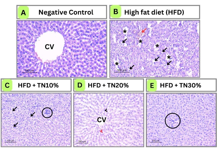
The photomicrograph of liver histopathology. The negative control group (A) showed normal architecture of hepatocytes emerging from the central vein (CV). HFD group (B) showed liver microvesicular steatosis (black arrows), macrovesicular steatosis (black asterisks), and dilated, congested blood sinusoids (red arrows). HFD + TN (10%) (C) showed microvesicular steatosis (black arrows) and chronic inflammatory infiltrate (black circle). HFD + TN (20%) group (D) showed normal hepatocytes (black arrowhead) radiating from the central vein (CV) and enclosed blood sinusoids (red arrowhead). HFD + TN (30%) group (E) showed normal hepatocytes with little lobular inflammation (black circle).
Hepatocytes in the HFD-fed group C (P) had dilated blood sinusoids and macro- and microvesicular steatosis, indicative of the damage caused by the HFD they were given (Figure 1B).
HFD + TN (10%) showed a lower proportion of hepatocytes with microvesicular steatosis alone. In addition, it exhibited a chronic inflammatory infiltrate, reflecting TN’s effect on initiating immunity (Figure 1C). The HFD + TN (20%) group showed the most normal histology of hepatocytes arising from the central vein and containing blood sinusoids between them (Figure 1D).
On the other hand, increasing the TN dose in HFD + TN (30%) improved hepatic injury caused by HFD, but lobular inflammation persisted (Figure 1E).
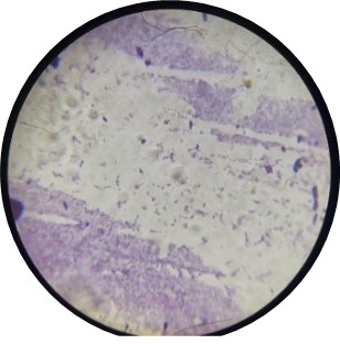
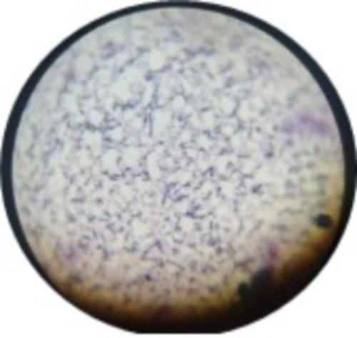
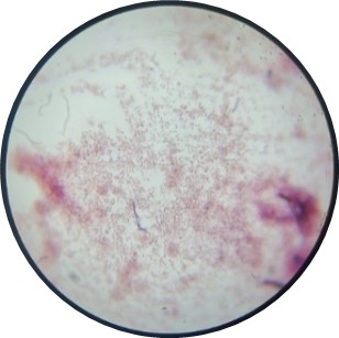
Table 5 summarizes the morphological, microscopic, and biochemical identification of the three bacterial isolates (Bifidobacterium, Firmicutes, and Akkermansia), confirming distinct phenotypic profiles consistent with their expected taxonomic characteristics. Bifidobacterium colonies were convex, white, and opaque, Gram-positive, non-motile, and negative for Voges-Proskauer (VP), indole, CAT, and H2S production, while positive for citrate utilization, starch hydrolysis, and the methyl red test. Firmicutes isolates also presented as convex, white, and opaque colonies and were Gram-positive and motile, with negative VP, indole, citrate, methyl red, and H2S production tests, but showed positive CAT activity and starch hydrolysis. In contrast, Akkermansia colonies were flat and off-white, Gram-negative, non-motile, and negative for VP, indole, citrate, CAT, methyl red, and H2S production, while demonstrating positive starch hydrolysis.
Macroscopic, microscopic, and biochemical analyses.
| Morphological characteristics/isolate | Bifidobacterium | Firmicutes | Akkermansia |
|---|---|---|---|
| Colony shape and colour | Convex/White Opaque | Convex/White Opaque | Flat/Off-white |
| Motility | Non-motile | Motile | Non-motile |
| Gram stain | Gram-positive | Gram-positive | Gram-negative |
| VP test | –ve | –ve | –ve |
| Indole | –ve | –ve | –ve |
| Citrate | +ve | –ve | –ve |
| Starch hydrolysis | +ve | +ve | +ve |
| Catalase | –ve | +ve | –ve |
| Methyl red | +ve | –ve | –ve |
| H2S production | –ve | –ve | –ve |
 |  |  | |
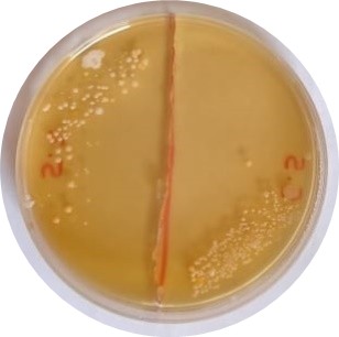 | 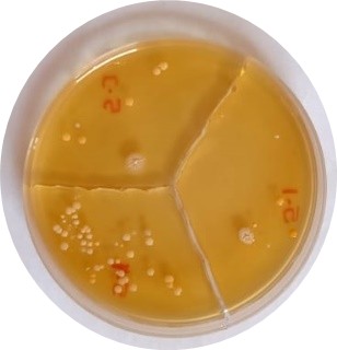 | 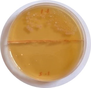 |
Culture-based microbiological assessment suggested that HFD feeding altered the profile of selected culturable intestinal bacteria compared with the normal control group. TN supplementation appeared to modify the occurrence of selected bacterial groups in a dose-dependent manner. However, because the analysis relied solely on culture-dependent methods, comprehensive characterization of gut microbial composition, diversity, and richness was not possible.
TN supplementation appeared to modify the distribution of selected bacterial groups in a dose-dependent manner. The TN-treated groups showed qualitative changes in the occurrence of selected bacteria compared with the HFD control group, suggesting a potential modulatory effect of TN supplementation on the intestinal microbial environment.
Table 6 shows that HFD feeding triggered severe gut dysbiosis in the C (P) group by drastically lowering culturable beneficial Bifidobacterium and Akkermansia populations while markedly increasing obesogenic Firmicutes, consistent with previous studies describing high-fat-diet-induced microbial imbalance.
Log10 CFU/g of target culturable gut bacteria across 5 groups (mean ± SD, n = 5).
| Bacterial taxon | C (N) normal control | C (P) HFD control | TN 10% (HFD + 10% TN) | TN 20% (HFD + 20% TN) | TN 30% (HFD + 30% TN) | p-value |
|---|---|---|---|---|---|---|
| Bifidobacterium | 8.72 ± 0.31ᵃ | 6.15 ± 0.28ᶜ | 7.43 ± 0.35ᵇ | 8.58 ± 0.29ᵃ | 7.81 ± 0.32ᵇ | < 0.001 |
| Firmicutes | 6.31 ± 0.22ᶜ | 8.94 ± 0.36ᵃ | 7.67 ± 0.31ᵇ | 6.45 ± 0.27ᶜ | 7.12 ± 0.29ᵇ | < 0.001 |
| Akkermansia muciniphila | 7.96 ± 0.25ᵃ | 4.82 ± 0.30ᶜ | 6.29 ± 0.33ᵇ | 7.84 ± 0.26ᵃ | 7.21 ± 0.28ᵃᵇ | < 0.001 |
Note: Different superscript letters denote statistically significant intergroup differences (p < 0.05).
TN powder exerted clear dose-dependent gut regulatory effects. The 10% TN treatment only partially restored beneficial bacteria, with their counts remaining significantly below those of the normal control group, while moderately reducing Firmicutes relative to the C (P) group. The 20% TN dose produced the most favorable microbial modulation by restoring Bifidobacterium and Akkermansia abundances to near-normal control levels and suppressing Firmicutes to levels comparable with healthy rats. This observation directly corresponds with the superior antioxidant, lipid-lowering, anti-inflammatory, and hepatoprotective biochemical and histopathological findings observed in this group. In contrast, the 30% TN dose increased beneficial bacterial populations compared with the HFD control but did not outperform the 20% TN group.
Moreover, mild Firmicutes overgrowth persisted, corresponding with the residual low-grade lobular inflammation observed in the liver histology. The new quantitative CFU colony count data further confirm that the abundant resistant starch, soluble fiber, and polyphenols in TN act as fermentable prebiotic substrates, selectively promoting the growth of mucin-degrading Akkermansia and lactic acid-producing Bifidobacterium while suppressing the expansion of obesity-associated Firmicutes. These findings strengthen the reliability of our original preliminary qualitative culture-based microbial observations.
The consumption of HFD is widely recognized as a major contributor to oxidative stress and metabolic dysfunction. Previous studies provided strong mechanistic evidence that HFD-induced oxidative stress is a central feature of diet-related metabolic disorders [23–25].
In addition, it can disturb the normal gut flora, which can lead to many pathological conditions because 70% of the body’s immune system resides in the gut. It has been reported that rats fed with HFD develop dysbiosis characterized by cecal mucosal damage, altered microbial composition, increased intestinal permeability, and elevated inflammatory responses [26].
TN have recently gained attention for their potential health benefits. Due to limited existing data, this study examined the effects of TN on antioxidant capacity and gut microbiota composition, with a specific focus on Bifidobacterium, Firmicutes, Akkermansia, and their metabolites in rats fed an HFD.
The findings indicate that HFD feeding induces significant oxidative stress, as evidenced by reduced antioxidant enzyme activities and increased lipid peroxidation, consistent with previous studies by Charradi et al. [27] (2013) and Noeman et al. [28] (2011). TN supplementation provided protective effects against these changes in a dose-dependent manner, with the 20% dose demonstrating the greatest efficacy.
TN significantly increased SOD, CAT, and GPx levels and reduced MDA, while GSH was significantly elevated only at the 30% dose. These effects are likely due to the high content of bioactive compounds in TN, such as polyphenols, vitamins, and unsaturated fatty acids, which function as direct free radical scavengers or enhance endogenous antioxidant systems.
Furthermore, TN supplementation ameliorated hyperlipidemia, as indicated by reductions in TC at the 20% supplementation level. This result aligns with previous reports by Ibitoye et al. [29] (2018), who found that TN oil lowered serum TC, triglycerides (TG), LDL, and VLDL while increasing HDL, thereby reducing cardiovascular risk.
The hypolipidemic effects of TN observed in the present study may be explained by several complementary mechanisms. First, TN is a rich source of dietary fiber, which can bind bile acids and cholesterol in the intestine, thereby reducing cholesterol absorption and increasing fecal sterol excretion, ultimately lowering circulating cholesterol concentrations [29].
Second, TN contains phytosterols that structurally resemble cholesterol and competitively inhibit its intestinal absorption, thereby reducing serum total and LDL-cholesterol levels [29]. Third, the high oleic acid content of TN may improve lipid metabolism by enhancing fatty acid oxidation, reducing hepatic lipogenesis, and improving plasma lipoprotein profiles [30].
Finally, the antioxidant constituents of TN, including phenolic compounds and tocopherols, may attenuate oxidative stress and hepatic lipid accumulation by preventing lipid peroxidation and improving hepatic metabolic function [13]. Collectively, these mechanisms may account for the improvements in serum lipid parameters and liver histology observed in TN-supplemented groups.
HFDs have been widely reported to alter gut microbial composition, contributing to intestinal dysbiosis and metabolic disturbances. Previous sequencing-based studies have demonstrated reductions in microbial diversity and changes in bacterial community structure following HFD feeding [19].
In addition, beneficial microorganisms such as Akkermansia and Bifidobacterium have been associated with improved gut barrier function and metabolic health. For example, Akkermansia muciniphila is a mucin-degrading bacterium that has been shown to stimulate mucus production and strengthen the intestinal barrier [31].
In the present study, gut microbial assessment was limited to culture-based isolation and phenotypic identification of selected bacterial groups. The findings suggest that TN supplementation may influence the intestinal microbial environment in HFD-fed rats. However, culture-dependent techniques capture only a limited proportion of the gut microbiota and do not provide comprehensive information on microbial diversity, community structure, or the relative abundance of bacterial taxa. Therefore, the observed microbial changes should be considered preliminary and interpreted with caution, and no definitive conclusions can be drawn regarding the restoration of gut dysbiosis or changes in overall microbial diversity.
The potential prebiotic effects of TN may be attributable to its high dietary fiber and resistant starch contents, which can serve as fermentable substrates for intestinal microorganisms [32]. Nevertheless, further studies employing high-throughput sequencing approaches, such as 16S rRNA gene sequencing or metagenomic analysis, together with microbial metabolite profiling, are required to confirm whether TN supplementation induces significant alterations in gut microbiota composition and function.
Although microbiota-specific studies remain limited, the modulatory effect of TN on gut microbiota observed in this study is supported by emerging human evidence. Selma-Royo et al. [9] (2022) reported no significant overall changes in the microbiota after short-term consumption of horchata, a beverage derived from TN tubers, in healthy individuals. However, they observed cluster-dependent shifts toward beneficial bacteria, including Akkermansia and Bifidobacterium, and microbial profiles associated with butyrate production and Mediterranean dietary patterns. Moreover, Selma-Royo et al. [9] (2022) noted that the response to such prebiotic components depends on baseline microbiota composition, which may explain the pronounced, dose-dependent restoration observed in this HFD-induced dysbiosis model. Additionally, polyphenols in TN may contribute by selectively modulating microbial growth and metabolism [33].
Moreover, TN supplementation reduced systemic inflammation, as indicated by decreased CRP levels, particularly at the 20% supplementation level. This reduction in systemic inflammation is closely correlated with the dose-dependent increase in Akkermansia richness, especially in the TN 20% group. Akkermansia is recognized as essential for maintaining gut permeability [34]. These findings suggest that TN-mediated enrichment of Akkermansia decreases intestinal permeability, thereby attenuating the inflammatory cascade triggered by HFD and resulting in elevated CRP.
The resistant starch and dietary fiber in TN serve as a selective substrate for Akkermansia and Bifidobacterium [9]. It is noteworthy that while TN 30% showed the highest diversity indices and Akkermansia enrichment, the TN 20% group exhibited the most normalized CRP levels and hepatic architecture.
Despite these benefits, TN supplementation did not reduce body weight gain; instead, it increased it at higher doses. This effect is likely due to TN’s nutritional composition, which is rich in lipids (primarily oleic acid) and digestible carbohydrates [35]. Consequently, TN supplementation likely contributed to additional caloric intake, particularly at 30% inclusion levels.
The diet’s increased palatability with higher TN content may also explain the lack of weight reduction despite metabolic improvements. This observation highlights the concept of metabolically healthy obesity (MHO), where excess fat is associated with a normal metabolic profile, including insulin sensitivity, low inflammatory markers, and reduced ectopic fat storage [36].
TN supplementation significantly reduced the risk of liver damage at the studied doses, as demonstrated by histopathological findings. The TN (20%) group exhibited near-normal hepatic architecture, while TN (10%) showed partial improvement with residual inflammation, and TN (30%) demonstrated improved hepatic tissue but still with residual inflammation. These results suggest that 20% is the optimal protective dose, beyond which no additional histological benefit is observed. According to Onuoha et al. [37] (2017), TN’s phytochemicals may contribute to these benefits by acting as direct antioxidants or by promoting GSH production and modulating gut-derived inflammatory signals.
The superior efficacy observed with the 20% TN supplementation suggests the existence of an optimal biological dose rather than a linear dose-response relationship. Although TN is rich in dietary fiber, resistant starch, oleic acid, tocopherols, flavonoids, phenolic acids, and phytosterols, increasing the supplementation level beyond 20% did not produce additional metabolic benefits.
One possible explanation is that moderate amounts of fermentable fiber and resistant starch provide sufficient substrate for beneficial intestinal bacteria and SCFA production, whereas excessive fiber intake may alter intestinal transit, fermentation kinetics, or nutrient availability, thereby attenuating these beneficial effects. In addition, TN is an energy-dense food containing approximately 20–35% lipid and 30–50% carbohydrate.
Consequently, the 30% supplementation level likely increased the overall caloric density and palatability of the experimental diet, which may have partially counteracted the beneficial effects of its bioactive constituents. Similar non-linear or hormetic dose-response relationships have been described for several plant-derived phytochemicals, in which moderate doses maximize antioxidant and anti-inflammatory responses, whereas higher doses do not confer additional benefit and may even diminish biological efficacy. Therefore, the findings of the present study suggest that the beneficial actions of TN result from a balanced interaction among its bioactive compounds rather than from simply increasing the administered dose [10, 13].
HFD-induced gut dysbiosis, characterized by depletion of beneficial Bifidobacterium and Akkermansia alongside expansion of obesogenic Firmicutes, is a well-recognized hallmark of obesity and metabolic dysfunction. In the present study, TN powder ameliorated these microbial alterations in a dose-dependent manner, with the 20% supplementation producing the most favorable gut microbial profile. The restoration of Bifidobacterium and Akkermansia to near-normal levels, coupled with suppression of Firmicutes, likely contributed to the marked improvements observed in oxidative stress, lipid metabolism, hepatic inflammation, and liver histopathology. These beneficial effects are biologically plausible because TN is naturally rich in resistant starch, soluble dietary fiber, and polyphenolic compounds that serve as fermentable prebiotic substrates. Fermentation of these substrates promotes the growth of beneficial bacteria, enhances SCFA production, strengthens intestinal barrier integrity, and suppresses inflammatory signaling, thereby reducing metabolic endotoxemia and improving host metabolic homeostasis. Interestingly, although the 30% TN diet also improved microbial composition compared with the HFD control, it did not provide additional benefits beyond the 20% dose, suggesting that maximal prebiotic efficacy may be achieved at moderate supplementation levels. Collectively, these findings support the concept that modulation of the gut microbiota represents an important mechanism underlying the hepatoprotective and metabolic benefits of TN and reinforce its potential as a functional food for preventing obesity-associated metabolic disorders [9].
The present study provides a comprehensive evaluation of TN supplementation in an HFD-induced rat model by integrating biochemical analyses, liver histopathology, and culture-based assessment of selected intestinal bacterial groups. The inclusion of three supplementation levels (10%, 20%, and 30%) enabled evaluation of dose-dependent responses and identified the 20% TN supplementation as the most effective level for improving oxidative stress, inflammatory, lipid, and histopathological parameters.
Despite these strengths, several limitations should be considered when interpreting the findings. First, gut microbiota assessment was restricted to culture-based isolation and phenotypic identification of selected bacterial groups. Because culture-dependent methods detect only a limited proportion of the intestinal microbiota, comprehensive characterization of microbial community composition, relative abundance, and taxonomic diversity was not possible. Consequently, the microbiological findings should be regarded as preliminary, and conclusions regarding modulation of the gut microbiota or potential prebiotic effects should be interpreted with caution.
Second, the intervention diets were not isocaloric because TN powder replaced portions of the HFD, resulting in differences in dietary fiber, macronutrient composition, and estimated energy density among the experimental groups. In addition, food intake was not monitored throughout the intervention. Therefore, it was not possible to distinguish the effects of TN bioactive constituents from those attributable to differences in dietary composition, caloric intake, or diet palatability.
Another limitation is the absence of comprehensive phenotypic measurements, including weekly body weight progression, liver index (liver-to-body weight ratio), visceral adipose tissue weights, and body composition analyses. Consequently, it was not possible to determine whether the observed metabolic improvements were accompanied by changes in fat distribution or body composition. These measurements should be incorporated in future studies to provide a more comprehensive assessment of the anti-obesity effects of TN supplementation.
Furthermore, microbial metabolites, particularly SCFAs, were not quantified, limiting the ability to establish functional links between alterations in the intestinal microbial environment and metabolic outcomes. Finally, as this study was conducted in an experimental animal model, caution should be exercised when extrapolating the findings to humans.
Another limitation of the present study, which should be acknowledged, is the relatively small sample size (n = 5 animals per group), which may have reduced the statistical power to detect modest treatment effects and limited the precision of some estimates. Consequently, the findings should be interpreted with appropriate caution. Although statistical analyses were selected according to the distribution of the data, and individual data points have been provided to improve transparency, larger studies are warranted to confirm the reproducibility and generalizability of the present findings.
Future studies should employ high-throughput sequencing techniques, such as 16S rRNA gene sequencing or shotgun metagenomic sequencing, in combination with metabolomic analyses to comprehensively characterize the gut microbiome and its functional activity. Monitoring food intake, using isocaloric or pair-fed experimental designs, assessing body composition and adipose tissue distribution, and measuring microbial metabolites would further clarify the mechanisms through which TN supplementation influences metabolic health. Ultimately, well-designed long-term clinical trials are warranted to confirm the efficacy and safety of TN supplementation in humans.
In conclusion, the present study demonstrates that TN (Cyperus esculentus L.) supplementation, particularly at the 20% inclusion level, ameliorated several metabolic disturbances induced by a HFD in rats.
TN supplementation improved antioxidant defense by enhancing endogenous antioxidant enzyme activities, reduced lipid peroxidation and inflammatory markers, improved serum lipid profile, and attenuated hepatic histopathological alterations. These beneficial effects may be attributed to the combined actions of dietary fiber, monounsaturated fatty acids, and naturally occurring antioxidant phytochemicals, including phenolic compounds, flavonoids, tocopherols, and phytosterols, present in TN.
Culture-based microbial assessment suggested that TN supplementation may favorably influence selected intestinal bacterial indicators in HFD-fed rats. However, because comprehensive sequencing-based microbiome analyses were not performed, no definitive conclusions can be drawn regarding gut microbiota composition, microbial diversity, gut dysbiosis restoration, or enrichment of specific bacterial taxa. Therefore, the microbiological findings should be regarded as preliminary.
Overall, TN shows promise as a functional food ingredient for improving metabolic health in the context of diet-induced obesity. Future studies employing high-throughput sequencing approaches, such as 16S rRNA gene sequencing or shotgun metagenomics, together with metabolomic analyses and well-controlled dietary interventions, are warranted to confirm its potential prebiotic effects, elucidate the mechanisms underlying its interactions with the gut microbiota, and evaluate its translational potential in human populations.
ALT: alanine aminotransferase
ANOVA: analysis of variance
CAT: catalase
CRP: C-reactive protein
GPx: glutathione peroxidase
GSH: glutathione
HFD: high-fat diet
MDA: malondialdehyde
ROS: reactive oxygen species
SCFAs: short-chain fatty acids
SOD: superoxide dismutase
TC: total cholesterol
TG: triglycerides
TN: tiger nut
The authors gratefully acknowledge the valuable contributions of the students of the Nutrition Department, Faculty of Applied Health Sciences Technology, Pharos University in Alexandria, for their assistance in experimental procedures and laboratory work. Their dedication and support were instrumental in the successful completion of this study. Artificial intelligence-based tools were used solely to improve the clarity and language of the manuscript. Specifically, ChatGPT was used for language editing. The authors take full responsibility for the scientific content, data interpretation, and conclusions presented in this manuscript. No artificial intelligence tool was used to generate original scientific content, analyze data, or draw conclusions.
DMAZ: Conceptualization, Methodology, Supervision, Validation, Formal analysis, Writing—original draft, Writing—review & editing. SS: Investigation, Data curation, Formal analysis, Visualization, Writing—original draft. BHE: Methodology, Resources, Validation, Writing—review & editing. BAB: Investigation, Data curation, Visualization. AAS: Methodology, Validation, Resources, Writing—review & editing. All authors have read and approved the final manuscript and agree to be accountable for all aspects of the work.
The authors declare that there is no conflict of interest.
All animal experimental procedures were approved by the Unit of Research Ethics Approval Committee (UREAC) of Pharos University, Alexandria, Egypt (Approval No.: 215). The study was conducted in accordance with the ARRIVE guidelines and the National Institutes of Health (NIH) Guide for the Care and Use of Laboratory Animals. Appropriate measures were taken to minimize animal suffering, and all procedures were performed under anesthesia where applicable.
Not applicable.
Not applicable.
The datasets generated and/or analyzed during the current study are available from the corresponding author upon reasonable request.
This research did not receive any specific grant from funding agencies in the public, commercial, or not-for-profit sectors.
© The Author(s) 2026.
Open Exploration maintains a neutral stance on jurisdictional claims in published institutional affiliations and maps. All opinions expressed in this article are the personal views of the author(s) and do not represent the stance of the editorial team or the publisher.
Copyright: © The Author(s) 2026. This is an Open Access article licensed under a Creative Commons Attribution 4.0 International License (https://creativecommons.org/licenses/by/4.0/), which permits unrestricted use, sharing, adaptation, distribution and reproduction in any medium or format, for any purpose, even commercially, as long as you give appropriate credit to the original author(s) and the source, provide a link to the Creative Commons license, and indicate if changes were made.



View: 141
Download: 6
Times Cited: 0


