 Original Article

Affiliation:
1Department of Computer Science & Engineering, Graphic Era Hill University, Haldwani 263139, Uttarakhand, India
ORCID: https://orcid.org/0000-0001-7258-4461
Affiliation:
2Department of Applied Sciences, MIET Kumaon, Haldwani 263139, Uttarakhand, India
Email: tarunksj@gmail.com
ORCID: https://orcid.org/0000-0001-9132-2395
Affiliation:
3Department of Chemistry, Swami Shraddhanand College (University of Delhi), Delhi 110036, India
ORCID: https://orcid.org/0000-0002-1603-0971
Affiliation:
4Department of Chemistry, Government Degree College Chaubattakhal, Pauri Garhwal 246162, Uttarakhand, India
ORCID: https://orcid.org/0000-0003-3701-3377
Affiliation:
5Department of Chemistry, College of Basic Sciences and Humanities, G.B. Pant University of Agriculture and Technology, Pantnagar 263145, Uttarakhand, India
ORCID: https://orcid.org/0009-0006-3419-5119
Affiliation:
6Laboratory of Heterocyclic Chemistry, Faculty of Sciences Monastir, University of Monastir, Monastir 5000, Tunisia
ORCID: https://orcid.org/0000-0003-2650-2634
Affiliation:
7Laboratory of Pharmacology of Inflammation and Behavior, Institute of Health Sciences, Federal University of Pará, Belém 66075110, Brazil
ORCID: https://orcid.org/0000-0002-6186-9581
Affiliation:
5Department of Chemistry, College of Basic Sciences and Humanities, G.B. Pant University of Agriculture and Technology, Pantnagar 263145, Uttarakhand, India
ORCID: https://orcid.org/0000-0002-0296-5231
Affiliation:
7Laboratory of Pharmacology of Inflammation and Behavior, Institute of Health Sciences, Federal University of Pará, Belém 66075110, Brazil
Email: mozaniel.oliveira@yahoo.com.br
ORCID: https://orcid.org/0000-0002-4076-2443
Explor Foods Foodomics. 2026;4:1010146 DOI: https://doi.org/10.37349/eff.2026.1010146
Received: September 29, 2025 Accepted: March 30, 2026 Published: May 17, 2026
Academic Editor: Miguel Herrero, Institute of Food Science Research (CIAL-CSIC), Spain
Aim: This study aimed to investigate the fruit of Berberis asiatica as a potential source of bioactive anthocyanins and to evaluate their antioxidant and antimicrobial properties with insights into molecular docking studies.
Methods: Crude extracts were prepared using solvents of varying polarity and characterized by liquid chromatography-tandem mass spectrometry (LC-MS/MS) and Fourier-transform infrared spectroscopy (FT-IR) analyses. The total anthocyanin content was quantified, and antioxidant activity was assessed using the DPPH radical scavenging assay and total antioxidant capacity. Antimicrobial activity was evaluated against selected bacterial and fungal strains. Additionally, in silico molecular docking studies were performed to examine ligand-target interactions.
Results: LC-MS/MS analysis identified eight compounds, including cyanidin-3-O-glucoside, cyanidin-3,5-diglucoside, malvidin-3-O-arabinoside, pelargonidin-3-O-glucoside, peonidin-3-O-glucoside, petunidin-3-O-glucoside, catechin, and epicatechin, indicating a pigment profile dominated by mono- and diglycosylated anthocyanins. The total anthocyanin content was 128.72 mg/g dry fruit, exceeding previously reported values for related species. Methanolic (80% v/v) and hydroalcoholic (50% v/v) extracts showed strong antioxidant activity (DPPH IC50 = 10.13 and 12.56 µg/mL, respectively), whereas nonpolar fractions were less active. At 200 µg/mL, these extracts exhibited significant antimicrobial activity, with inhibition zones up to 42 mm against Escherichia coli and 41 mm against Micrococcus luteus, along with antifungal effects against Aspergillus niger and Candida albicans. Docking studies revealed favorable binding energies (–7.3 to –8.0 kcal/mol) for key compounds against selected microbial and enzymatic targets.
Conclusions: The findings demonstrate that Berberis asiatica fruit is a rich source of anthocyanin-based pigments with potent antioxidant and antimicrobial activities. These results highlight its potential as a sustainable source of multifunctional bioactive compounds for nutraceutical and therapeutic applications.
Botany is integral to the pharmacological industries of India, China, and Egypt. As a result, there is a growing demand for herbs in the pharmaceutical, medical, and wellness sectors, especially in Asian countries and some African nations [1]. India’s global importance is highlighted by its cultivation and utilization of nearly 45,000 species of medicinal plants across 16 agro-climatic zones. Indian traditions are enriched by Vedic literature and comprehensive Sanskrit texts on ailments, healthcare, and holistic treatment, which emphasize the significance of 15,000–20,000 plant species [2, 3]. The wild edible fruits of the Kumaon region in Uttarakhand hold untapped potential for economic development. These geo-specialized fruits can positively impact local livelihoods through sustainable harvesting, value addition, and commercialization. Promoting the collection and utilization of indigenous wild edible fruits can generate new income sources, create employment opportunities in rural communities, and support ancillary services related to processing and marketing. This is particularly valuable in regions where conventional agriculture is constrained by limited resources or climate variability. Additionally, the rich diversity of wild edible fruits in Kumaon offers opportunities to enhance eco-tourism and agro-tourism, attracting visitors interested in traditional foods and nutraceutical products [4].
Berberis asiatica Roxb., commonly known as Kilmore, is a wild berry traditionally used in Ayurvedic medicine for treating diarrhoea, rheumatism, jaundice, syphilis, urinary disorders, and skin diseases. The species grows at altitudes between 1,500 and 2,400 m, flowers during March–April, and bears fruit from May to June. Due to habitat degradation, unregulated harvesting, and increasing anthropogenic pressure, natural populations of B. asiatica are declining in several Himalayan regions. This underscores the need for scientific valorization and sustainable utilization of this species to support biodiversity conservation while simultaneously strengthening local and indigenous economies dependent on medicinal plant resources.
Phytochemical investigations have shown that B. asiatica is rich in bioactive compounds, including alkaloids, phenolics, anthocyanins, tannins, vitamins, and minerals, and exhibits hepatoprotective, antidiabetic, antioxidant, and antilipidemic properties. Studies on related Berberis species further report anticancer, cardioprotective, anticonvulsant, and antioxidant activities [5, 6]. Anthocyanins are well documented for their antioxidant and antimicrobial activities, with studies demonstrating that structure activity relationships, particularly hydroxylation and methoxylation patterns of the B-ring, as well as glycosylation and acylation of the flavylium core, critically influence their radical-scavenging capacity, membrane interactions, and antimicrobial efficacy [7, 8].
Despite these documented bioactivities, detailed information on the anthocyanin composition and characterization of B. asiatica fruits remains limited. Therefore, the present study aims to characterize the anthocyanin profile of B. asiatica fruit extracts using both polar and non-polar solvent systems and to evaluate their antioxidant and antimicrobial activities. Advanced analytical techniques, including liquid chromatography-tandem mass spectrometry (LC-MS/MS) and Fourier-transform infrared spectroscopy (FT-IR), were employed for accurate identification and structural elucidation of anthocyanin compounds.
The fresh berries of B. asiatica were collected from the Ghorakhal District in Nainital, Uttarakhand, India, a region situated in a forested area. After the harvest, the fruits were quickly taken to the laboratory and preserved at –20°C for subsequent analysis. Sample collection was guided by ethnopharmacological and ethnobotanical literature [9] and authenticated by taxonomist Dr. D. S. Rawat, Department of Biological Sciences, GBPUA&T, Pantnagar. Specimens of the fruits were archived in the institution’s herbarium for future reference.
All chemicals and reagents used in the study were of analytical grade and procured from HiMedia Laboratories Pvt. Ltd., Mumbai, India. Wherever available, catalogue/item numbers have been provided.
Fresh fruits were thoroughly washed with distilled water and shade-dried in the dark at room temperature for 5–10 days. The dried fruits were powdered and used for extraction following previously reported methods [10, 11] with slight modifications. Briefly, 20 g of fruit powder was separately soaked in four solvents of increasing polarity, hexane, chloroform, 80% methanol (v/v), and 50% hydroalcohol (v/v), for 72 h. The mixtures were stirred intermittently every 24 h to enhance extraction efficiency and then filtered. Filtrates were concentrated at 30°C using a water bath, and the resulting extracts were stored at 4°C for further analysis.
We utilized an automated FT-IR spectrometer from PerkinElmer, Germany, for the identification of bioactive molecule groups. The FT-IR spectrometer analysis was carried out in the range of 4,000–400 cm–1, employing the KBr pellet technique. Identification of bioactive molecules within the 80% crude methanol extract of B. asiatica fruit was achieved by comparing the spectral FT-IR analysis with published literature.
TAC in the methanol extract (80%) followed the procedure proposed by Aguila and McCann [12] with slight adjustments. The samples were diluted with a solution of ethanol:water:HCl (conc.) in a 70:30:1 ratio, and the absorbance was recorded at 540 nm. The assessment of TAC was executed, and the findings were expressed as malvidin-3-glucoside equivalents through the utilization of the following formula:
Where d = dilution.
The identification of bioactive compounds, specifically anthocyanins, was conducted in the 80% crude methanol extracts of the sample fruit. This was achieved through the utilization of the LC-MS/MS analytical technique, provided by Agilent Technologies India Pvt. Ltd., Bangalore. The methodology employed a single quadrupole (SQ) LC/mass selective detector (MSD), and the LC-MS/MS system used in the analysis was equipped with an autosampler featuring a photo-diode array detector. For this investigation, a reversed-phase RP C18 column (150 × 3.0 mm, 2.5 µm) was selected, maintaining a constant temperature at 40°C. Mobile phase A: methanol, mobile phase B: acetonitrile. Chromatographic separation conditions: 0 min, 5% B; 1 min, 5% B; 15 min, 97% B; 20 min, 97% B; 21 min, 5% B; 25 min, 5% B. A consistent flow rate of 0.4 mL/min was consistently maintained during the entire analysis. The fruit extracts from B. asiatica in 80% v/v methanol were first dissolved in a water and methanol mixture (60:40 v/v) to create a stock solution with a concentration of 1 mg/mL. For liquid chromatography-electrospray ionization mass spectrometry (LC-ESI-MS) analysis, a 2 µL aliquot from the stock solution was utilized. The analysis utilized a Triple Quad mass spectrometer with an ESI source, specifically the Waters Quattro Premier XE from the USA.
The parameters were set as follows: electrospray capillary voltage at 3.5 kV, source temperature at 100°C, desolvation gas flow at 1,000 L/h, cone gas flow at 60 L/h, desolvation temperature at 350°C, nitrogen and cone voltage at 30 V, was employed in the ESI source, and the multiplier voltage was adjusted to 650 V. LC-MS analysis was performed in both positive and negative ionization modes using a full scan range of m/z 50–1,400. The recorded results included the total ion chromatogram along with the corresponding spectra.
Different solutions of each solvent extract from the fruits were prepared for the DPPH test following the procedure outlined [13]. This method included dissolving 0.2 g of each dehydrated extract in 10 mL of the specific solvent. A DPPH working solution of 0.025 g 2,2-diphenyl-1-picrylhydrazyl/1,000 mL CH3OH. In subsequent steps, 2 mL of the DPPH solution was combined with 40 μL of each fruit extract and then transferred to a cuvette. Following a 30 min incubation at room temperature. The reaction solution underwent analysis at 515 nm using a UV-Vis spectrophotometer (Systronics, India). The calculation of the percentage inhibition for the absorbance of the DPPH solution was determined using the following formula.
The notation “AbsT (0 min)” signifies the absorbance of DPPH at the initial 0 min mark, while “AbsT (30 min)” denotes the absorbance of DPPH after 30 min of incubation.
In the course of this procedure, methanol was employed to dissolve ascorbic acid (0.5 mM), functioning as a standard for assessing the inhibitory capacity of the fruit extract solution in terms of ascorbic acid equivalence. Furthermore, the IC50 concentration of the sample, signifying the amount needed to neutralize 50% of DPPH free radicals, was ascertained.
The overall antioxidant potential of the solvent extracts from the fruit was assessed using the methodology detailed in established antioxidant assays [14, 15]. As a positive control reference, ascorbic acid (0.5 mM) was employed. Each 0.1 mL extract was mixed with 1 mL of a reagent solution (0.6 M sulfuric acid, 28 mM sodium phosphate, and 4 mM ammonium molybdate). The tubes were sealed, and incubation took place at 95°C for 90 min. Upon cooling to 25°C, the absorbance against the blank, containing no test samples or extracts, was measured at 695 nm. The overall antioxidant activity was evaluated by measuring the absorbance at 695 nm, where higher values indicated increased antioxidant activity.
The evaluation of superoxide anion radical scavenging activity followed the methodology outlined in established procedures [13, 15–17]. A sample was created by combining fruit extract with 3 mL of a reaction buffer solution at a pH of 7.4, which included 1.3 μM riboflavin, 0.02 M methionine, and 5.1 μM nitroblue tetrazolium (NBT). The reaction mixture was subjected to 30 W fluorescent lamps for 20 min, and the absorbance was recorded at 560 nm utilizing a spectrophotometer. Ascorbic acid was employed as the positive control, while the reaction mixture lacking the sample functioned as the negative control. The calculation for the percentage of above said activity was determined as follows.
Where Ao = absorbance of positive control and As = absorbance of sample.
The assessment of H2O2 scavenging percentage followed the procedure [17]. A 40 mM H2O2 solution was created in phosphate buffer with a pH of 7.4. The concentration of the solution was assessed by gauging its absorbance at 230 nm through a UV-Vis spectrophotometer. The fruit solvent extracts were incorporated into the H2O2 solution, and the absorbance of H2O2 was recorded at 230 nm following a 10 min incubation period, juxtaposed with a blank solution containing phosphate buffer devoid of H2O2. As a positive control, ascorbic acid was utilized. The percentage calculation for scavenging H2O2 was carried out using the following formula:
The symbols employed in the computation are defined as follows: Ao corresponds to the absorbance of the positive control, while As denotes the absorbance of the sample.
In the antibacterial test, nutrient agar medium (NAM)/broth from HiMedia Laboratories Pvt. Ltd., and Sabouraud’s dextrose agar/broth were utilized. These materials were sourced from Bombay, India.
Bacterial inoculation involved placing the bacteria into the nutrient broth at 37°C for 18 h, ensuring a suspension with approximately 105 CFU/mL. The fungal strain underwent a comparable procedure, involving inoculation into Sabouraud’s dextrose broth. Subsequently, the fungal broth cultures were permitted to incubate for a period ranging from 48 to 72 h.
In this study, the fungal test organisms, namely Aspergillus niger (ATCC 16404) and Candida albicans (ATCC 10231), were obtained from the National Chemical Laboratory in Pune, Maharashtra, India.
The bacterial strains utilized in the research, including Micrococcus luteus (ATCC 9341), Salmonella abony (ATCC 6017), Escherichia coli (ATCC 8739), and Staphylococcus epidermidis (ATCC 12228), were acquired from the National Chemical Laboratory in Pune, Maharashtra, India.
The agar well diffusion method was modified [14], and bacterial cultures were cultured on NAM, with individual bacterial suspensions introduced into the nutrient broth. Fungal cultures were implemented using Sabouraud’s dextrose agar/broth. The fungus was inoculated separately into Sabouraud’s dextrose broth. In the agar, wells with a diameter of 8 mm were established and then filled with extracts from fruits and solvent blanks. Negative controls encompassed methanol (80% v/v), hydroalcohol (50% v/v), chloroform, and hexane solvents. Concurrently, a standard antibiotic (azithromycin, 1 mg/mL) served as the positive control. The incubation of fruit samples took place at 37°C for a duration of 18 h.
To evaluate the antibacterial activity, we measured the diameter of the inhibition zone observed. Evaluating the antifungal activity in fruit extracts was conducted using plates containing Sabouraud dextrose agar/broth medium. The identical procedure employed to assess antibacterial characteristics was utilized, and the inhibition zone’s diameter was measured within a timeframe of 48 to 72 h. Fluconazole (1 mg/mL) was employed as the benchmark to evaluate antifungal activity.
Molecular docking simulations were performed via AutoDock 4.2 program package [18]. The crystal structures of (PDB: 4ewp) [19], (PDB: 1kzn) [20], (PDB: 7bly) [21], and (PDB: 3nrz) [22] were procured from the RCSB protein data bank (https://www.rcsb.org/). First, water molecules were removed, and then missing hydrogens and Gasteiger charges were added to the system during the preparation of the receptor input file. Then, AutoDock Tools was used to prepare the ligand and protein files (PDBQT). Pre-calculation of grid maps was performed with AutoGrid to save time during docking. Geometric optimization of all compounds was performed using ACD (3D viewer) software (http://www.filefacts.com/acd3d-viewer-freeware-info) and the visualization and analysis of interactions were performed using Discovery Studio 2017R2 (https://www.3ds.com/products/biovia/reference-center) and PyMOL 0.99rc6 [23].
All experiments were performed in triplicate, and results are expressed as mean ± standard deviation (SD). Differences among solvent extracts and the standard reference (ascorbic acid) for antioxidant assays were analysed using one-way analysis of variance (ANOVA) followed by Tukey’s post hoc test. Statistical significance was considered at p < 0.05.
The functional group positions and characterization of molecules in the crude methanol extract of B. asiatica, determined by FT-IR spectroscopy, are shown in Figure 1 and Table 1. FT-IR spectroscopic analysis of the crude methanolic extract of B. asiatica revealed the presence of several functional groups associated with bioactive secondary metabolites. The broad band observed at 3,983.78 cm–1, typical of O-H stretching, together with the signal at 1,384.26 cm–1 (O-H deformation), indicates a high concentration of hydroxyl groups, characteristic of phenolic compounds and alcohols, commonly associated with antioxidant activity. The bands at 2,852.79 cm–1 and 2,924.55 cm–1, corresponding to C-H stretching of aliphatic chains, suggest the presence of lipophilic compounds such as fatty acids. The intense absorption at 1,721.07 cm–1 is related to carbonyl stretching (C=O), frequently present in carboxylic acids, ketones, and esters, while the signals at 1,600.11 and 1,514.08 cm–1 indicate the presence of conjugated aromatic rings, supporting the occurrence of flavonoids and berberine-type alkaloids.
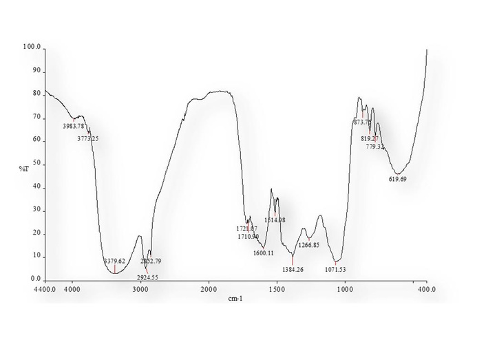
Fourier-transform infrared spectroscopy (FT-IR) spectra of crude methanol extract of B. asiatica.
Fourier-transform infrared spectroscopy (FT-IR) spectroscopic analysis of the crude methanolic extract of B. asiatica and correlation with identified bioactive compounds.
| Wave number (cm–1) | Functional group/vibration | Tentative assignment to compounds identified by liquid chromatography-tandem mass spectrometry (LC-MS/MS) |
|---|---|---|
| 3,983.78 | O-H stretching | Hydroxyl groups in phenolics and anthocyanins (cyanidin-3-O-glucoside, pelargonidin-3-O-glucoside) |
| 2,924.55–2,852.79 | C-H stretching (aliphatic) | Aliphatic chains in catechin, epicatechin, and fatty acid derivatives |
| 1,721.07 | C=O stretching | Carbonyl groups in glycosidic linkages, esters, or ketones of glycosylated flavonoids and anthocyanins |
| 1,600.11–1,514.08 | Aromatic C=C stretching | Conjugated aromatic rings in flavonoids and berberine-type alkaloids |
| 1,384.26 | O-H bending/deformation | Hydroxyl groups in phenolic compounds and anthocyanins |
| 1,266.85–1,071.53 | C-O stretching | Glycosidic bonds in glycosylated flavonoids and sugars |
| 873.75–779.33 | Aromatic C-H out-of-plane bending | Substituted aromatic rings in isoquinoline alkaloids and complex phenolics |
| 619.69 | C-C skeleton vibration | Aromatic ring backbone, supporting polyphenolic structures |
In the region between 1,300 and 1,000 cm–1, the bands at 1,266.85 cm–1 and 1,071.53 cm–1 are attributed to C-O vibrations, consistent with primary and secondary alcohols, and glycosidic bonds, correlating with glycosylated flavonoids and anthocyanins identified by LC-MS/MS (e.g., cyanidin-3-O-glucoside, pelargonidin-3-O-glucoside). The signals at 873.75, 819.27, and 779.33 cm–1, associated with out-of-plane aromatic C-H deformations, indicate tri- or tetra-substituted aromatic rings, common in complex phenolic compounds and isoquinoline alkaloids. The band at 619.69 cm–1 is attributed to the vibration of the C-C skeleton, also typical of aromatic structures.
The quantification of the anthocyanin content in the 80% methanolic extract of B. asiatica using a spectrophotometric method revealed a significant value of 128.72 mg/g of plant material Table 2. However, when compared to studies of other species in the same genus, such as Berberis vulgaris, the results differ significantly.
Total anthocyanin content in 80% v/v methanolic extract of B. asiatica via spectrophotometric method.
| B. asiatica fruits (s) extract | Anthocyanin content (mg/g of fruit material) |
|---|---|
| 80% v/v methanolic extract of B. asiatica | 128.72 ± 0.045*** |
***: p < 0.001, highly significant.
The LC-MS/MS analysis of the methanolic extract of B. asiatica fruit revealed a phytochemical core consisting of eight glycosylated anthocyanins: cyanidin-3-O-glucoside (Cy3G), cyanidin-3,5-diglucoside (Cy3diG5G), malvidin-3-O-arabinoside (M3A), petunidin-3-O-glucoside, peonidin-3-O-glucoside (P3G), and pelargonidin-3-O-glucoside (Pg3G), catechin and epicatechin as shown in Table 3. Their retention times (2.53–9.65 min) and diagnostic fragments (m/z 271–331) confirm the dominance of 3- and 3,5-substituted anthocyanins. The co-elution of catechin and epicatechin at ~5.3–7.8 min indicates a secondary flavanol pool that can reinforce antioxidant molecules in the crude methanolic extract of B. asiatica. Figure 2 displays the LC-MS/MS chromatogram, illustrating how compounds are separated based on their retention time as they elute from the LC column. Each peak primarily represents an intact (parent) molecule detected during the chromatographic run. In contrast, Figure 3 showcases the MS/MS fragmentation spectra, where selected parent ions are further fragmented into product (fragment) ions. This process reveals the characteristic splitting pattern of each molecule. A greater number of compounds appear in Figure 3 because a single parent compound generates multiple fragment ions during MS/MS analysis. These fragments are detected as additional signals, providing structural information and confirming compound identity, rather than representing new or distinct compounds. Figure 4 represents the structure of compounds identified in the methanolic extract of B. asiatica.
Liquid chromatography-tandem mass spectrometry (LC-MS/MS) profile of molecules identified in crude methanol extract of B. asiatica.
| Peak | Retention time (min) | Anthocyanin class | Molecular weight (g/mol) | Fragment (m/z) | Molecular formula |
|---|---|---|---|---|---|
| 1 | 2.531 | Cyanidin-3-O-glucoside | 449.0 | 287.0 | C21H21O11 |
| 2 | 2.809 | Pelargonidin-3-O-glucoside | 433.4 | 271.0 | C21H21O10 |
| 3 | 4.924 | Peonidin-3-O-glucoside | 463.4 | 301.0 | C22H23O11 |
| 4 | 5.358 | Catechin | 290.26 | 245.0 | C15H14O6 |
| 5 | 5.860 | Cyanidin-3,5-diglucoside | 611.5 | 287.0 | C27H30O16 |
| 6 | 7.796 | Epicatechin | 290.27 | 247.12 | C15H14O6 |
| 7 | 8.892 | Malvidin-3-O-arabinoside | 493.4 | 331.0 | C23H25O12 |
| 8 | 9.648 | Petunidin-3-O-glucoside | 479.4 | 317.23 | C22H23O12 |
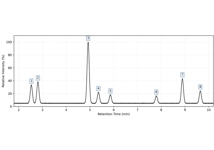
Liquid chromatography-tandem mass spectrometry (LC-MS/MS) chromatogram of molecules separated and identified in crude methanolic extract of B. asiatica.
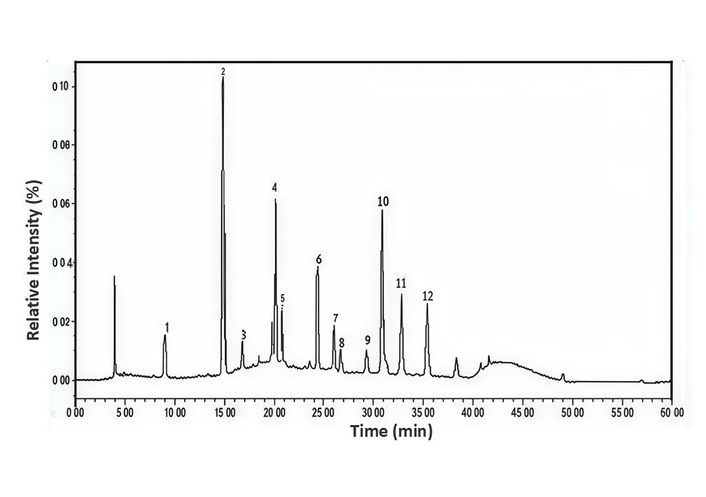
Tandem mass spectrometry (MS/MS) fragmentation spectra of compounds in the crude methanolic extract of B. asiatica. Peaks 1 to 8 correspond to anthocyanins identified in this study, while peaks 9 to 12 represent fragment ions detected during MS/MS analysis that were not fully characterized. These additional peaks provide structural information rather than indicating new, separate compounds.
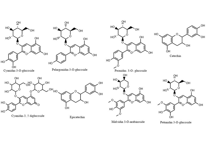
Structures of compounds identified in the methanolic extract of B. asiatica. Created from ChemDraw 18.2 software. © The Author(s) 2026.
The antioxidant characteristics of solvent extracts obtained from B. asiatica were assessed through traditional methods, including DPPH free radical scavenging activity, superoxide anion radical scavenging activity, H2O2 inhibition, and total antioxidant activity. The findings indicated that the 80% methanol extract exhibited noteworthy antioxidant activity, with subsequent rankings observed for the hydroalcoholic, hexane, and chloroform extracts. These findings held statistical significance when compared to the standard reference control, ascorbic acid at 1 mg/mL (Table 4).
Antioxidant activity of different solvent extracts of B. asiatica.
| Extract/Standard (1 mg/mL) | DPPH free radical scavenging activity (IC50, µg/mL) | Total antioxidant activity (A695 nm) | Superoxide anion radical scavenging (inhibition %) | Hydrogen peroxide scavenging (inhibition %) |
|---|---|---|---|---|
| 80% methanolic extract | 10.13 ± 0.028a | 2.56 ± 0.022a | 72.34 ± 0.035b | 63.23 ± 0.027b |
| Hydroalcoholic extract | 12.56 ± 0.056c | 1.82 ± 0.035b | 67.18 ± 0.047c | 55.45 ± 0.040c |
| Hexane extract | 15.67 ± 0.056d | 0.86 ± 0.086c | 56.23 ± 0.057d | 46.67 ± 0.040d |
| Chloroform extract | 20.56 ± 0.087e | 0.34 ± 0.092d | 32.45 ± 0.078e | 34.57 ± 0.040e |
| Standard (ascorbic acid) | 11.08 ± 0.034bc | 1.85 ± 0.047b | 86.56 ± 0.038a | 85.23 ± 0.030a |
Values are expressed as mean ± standard deviation (SD) (n = 3). Different superscript letters within the same column indicate statistically significant differences (p < 0.05) according to one-way analysis of variance (ANOVA) followed by Tukey’s post hoc test. For DPPH IC50 values, lower values indicate higher antioxidant activity; lettering (a–e) means with different letters indicate significant differences, and means with the same letters are not significantly different.
The potential utilization of B. asiatica as a natural source of antioxidants opens avenues for developing products or supplements enriched with these beneficial compounds. This application holds the promise of offering health benefits to the public. By harnessing the antioxidant properties identified in B. asiatica, it becomes possible to create formulations that contribute to improving overall health and well-being. These natural antioxidants can act as a safeguard against oxidative stress, addressing health challenges associated with the harmful effects of free radicals in the body. Incorporating B. asiatica-derived antioxidants into various products or supplements aligns with the growing interest in holistic and natural approaches to health, providing individuals with a proactive and natural solution to support their well-being.
The antimicrobial efficacy of B. asiatica solvent extracts was assessed at a concentration of 200 µg/mL through the well diffusion method. The study revealed that the methanol extracts (80% v/v) and hydroalcoholic extracts (50% v/v) of B. asiatica demonstrated pronounced antimicrobial activity, surpassing the effectiveness of hexane and chloroform extracts, particularly against M. luteus, S. epidermidis, E. coli, A. niger, S. abony, and C. albicans. Interestingly, chloroform extracts exhibited no antimicrobial activity against S. abony, A. niger, and C. albicans. The observed inhibition zones indicate a clear trend of higher antimicrobial potency for polar solvent extracts when compared descriptively with the positive controls. The detailed antimicrobial response of all tested extracts and controls is presented in Table 5 and Figure 5.
Antimicrobial activity of solvent extracts of B. asiatica.
| Diameter of zone of inhibition (mm) | ||||||
|---|---|---|---|---|---|---|
| Plant fruit extracts/controls | M. luteus (mm) | E. coli (mm) | S. abony (mm) | S. epidermidis (mm) | A. niger (mm) | C. albicans (mm) |
| 80% methanolic extract | 41.0 ± 0.82a | 42.0 ± 0.75a | 38.0 ± 0.91a | 37.0 ± 0.88a | 26.0 ± 0.64a | 22.0 ± 0.71a |
| Hydroalcoholic extract | 39.0 ± 0.94b | 38.0 ± 0.86b | 33.0 ± 0.79b | 35.0 ± 0.92b | 28.0 ± 0.68a | 18.0 ± 0.74b |
| Hexane extract | 23.0 ± 0.88c | 27.0 ± 0.91c | 25.0 ± 0.96c | 18.0 ± 0.83c | 12.0 ± 0.65b | 15.0 ± 0.72c |
| Chloroform extract | 18.0 ± 1.12d | 15.0 ± 1.08d | NA | 12.0 ± 1.15d | NA | NA |
| Positive control (azithromycin, 1 mg/mL) | 45.0 ± 0.68 | 45.0 ± 0.72 | 32.0 ± 0.84 | 38.0 ± 0.79 | NT | NT |
| Positive control (fluconazole, 1 mg/mL) | NT | NT | NT | NT | 36.0 ± 0.81 | 32.0 ± 0.76 |
| Methanol (solvent blank) | NA | NA | NA | NA | NA | NA |
| Hydroalcohol (solvent blank) | NA | NA | NA | NA | NA | NA |
| Hexane (solvent blank) | NA | NA | NA | NA | NA | NA |
| Chloroform (solvent blank) | NA | NA | NA | NA | NA | NA |
Values are expressed as mean ± standard deviation (SD) (n = 3). Different superscript letters within the same column indicate significant differences among treatments [p < 0.05, one-way analysis of variance (ANOVA) followed by Tukey’s post hoc test where means with different letters indicate significant differences, and means with the same letters are not significantly different]. Positive controls: azithromycin (1 mg/mL) for bacteria, fluconazole (1 mg/mL) for fungi. Solvent blanks did not show any activity. NA: no activity; NT: not tested.
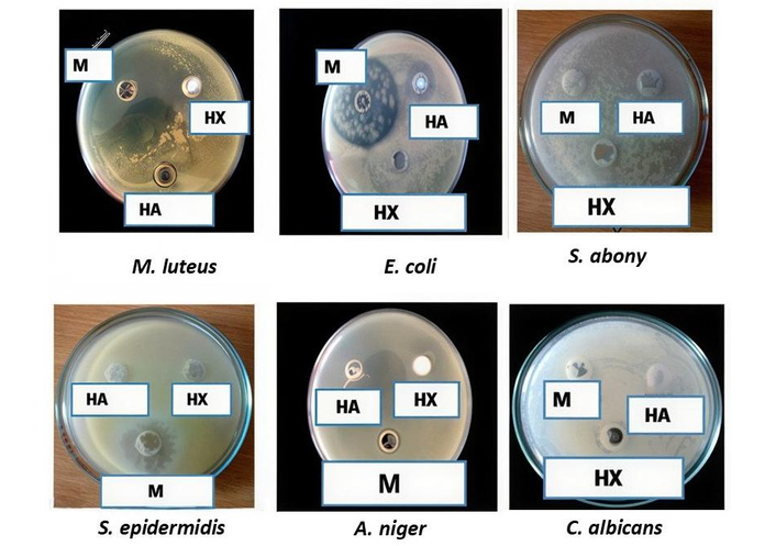
Antimicrobial activity of solvent extracts of B. asiatica fruits evaluated by the agar well diffusion assay. The figure shows zones of inhibition produced by 80% methanolic extract (M), hydroalcoholic extract (HA), and hexane extract (HX) against M. luteus, E. coli, S. abony, S. epidermidis, A. niger, and C. albicans.
Molecular docking is an established in silico structure-based technique widely used in drug discovery [24]. Motivated by this information, herein, molecular docking was used to identify a possible mechanism of action that correlated with the recorded antimicrobial and antioxidant potentials by modeling the identified phytocompounds (Cy3G, M3A, catechin, Cy3diG5G, epicatechin, petunidin-3-O-glucoside, P3G and Pg3G). In the current studies, among the strains used in the in vitro tests, the most sensitive strain against the phytomolecules was chosen to select the target receptors for carrying out the docking. ‘FabH from M. luteus’ (PDB: 4ewp) was taken as the target receptor to predict the sensitivity of a Gram-positive bacterium and ‘E. coli 24 kDa Domain’ (PDB: 1kzn) was used to conjecture the sensitivity of a Gram-negative bacterium against the selected phytocompounds in addition to ‘chitin deacetylase AngCDA from A. niger’ (PDB: 7bly) that used for the antifungal activity. Furthermore, ‘Xanthine Oxidase’ (PDB: 3nrz) was used as a receptor for the antioxidant potential.
Against M. luteus (PDB: 4ewp), the results presented in Table 6 show that the majority of docked phytocompounds have acceptable scores. Indeed, the most effective ligand is ‘Cy3diG5G’, which exhibited the best docking score (binding energy value = −7.3 kcal/mol). The 2D model in Figure 6 exhibits that this ligand fits well in the binding cavity of the targeted enzyme. The involved interactions depicted in Figure 6, which demonstrate the formation of five H-bonds with His96, Ala97, Trp197, and Ile200, reduced the binding energy score and consequently stabilized the docking complex. Further, other interactions are included, such as: Pi-donor hydrogen bond with Ser120, Pi-Pi T-shaped with Trp197.
Binding energies of the docked compounds.
| Compound | Free binding energy of PDB: 4ewp (kcal/mol) | Free binding energy of PDB: 1kzn (kcal/mol) |
|---|---|---|
| Cyanidin-3-O-glucoside | –6.8 | –7.4 |
| Malvidin-3-O-arabinoside | –6.7 | –8.0 |
| Catechin | –6.4 | –7.8 |
| Cyanidin-3,5-diglucoside | –7.3 | –5.0 |
| Epicatechin | –6.4 | –7.7 |
| Petunidin-3-O-glucoside | –6.8 | –6.9 |
| Peonidin-3-O-glucoside | –6.9 | –6.9 |
| Pelargonidin-3-O-glucoside | –6.8 | –7.3 |
| Azithromycin (standard) | –7.7 | –6.4 |
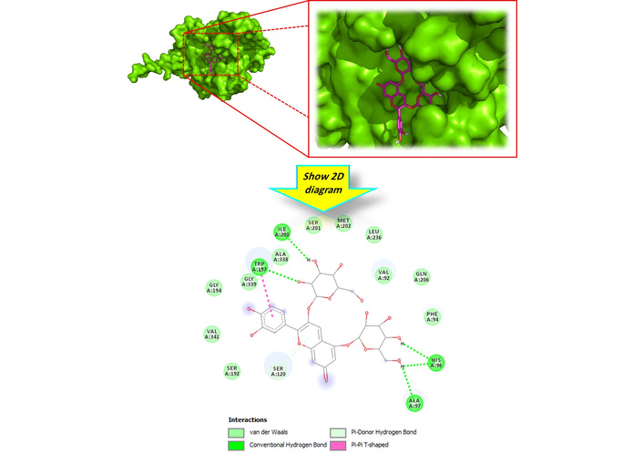
2D model of different interactions formed by the most active compound ‘cyanidin-3,5-diglucoside’ within the active site of FabH from M. luteus (PDB: 4ewp). BIOVIA, Dassault Systèmes, Discovery Studio 2021, San Diego: Dassault Systèmes, 2026.
Regarding the docking complex: ‘E. coli 24 kDa Domain’ (PDB: 1kzn), as Table 6 displays, all docked phytocompounds showed binding energy values ranging from –8 to –5 kcal/mol, have more interesting scores than the standard (azithromycin) except ‘Cy3diG5G’. For more details, ‘M3A’ was considered as the most bioactive ligand by exhibiting a conventional H-bond with Val43 and carbon hydrogen bond with Asp73, Pi-Cation with Arg76, Pi-Anion with Glu50, Pi-donor hydrogen bond with Asn46 and Pi-Alkyl with Ile78 and Pro79 (Figure 7). All other docked molecules exhibited different interactions, especially H-bonds.
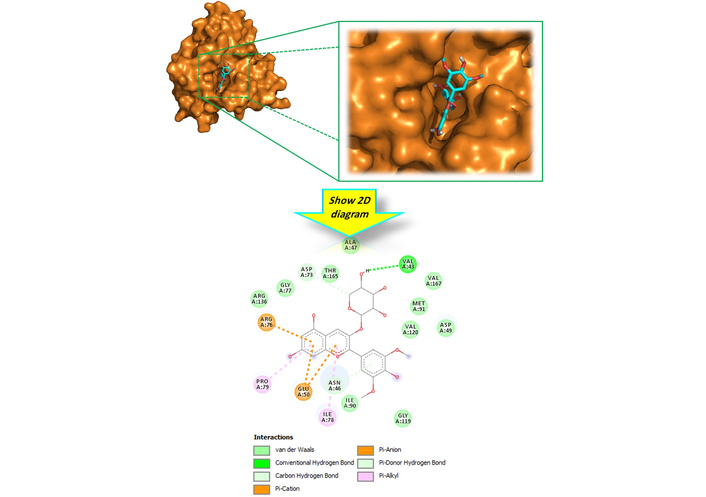
2D model of different interactions formed by the most active compound ‘malvidin-3-O-arabinoside’ within the active site of E. coli 24 kDa Domain (PDB: 1kzn). BIOVIA, Dassault Systèmes, Discovery Studio 2021, San Diego: Dassault Systèmes, 2026.
Toward ‘chitin deacetylase AngCDA from A. niger’, Table 7 displays that all docked phytoligands have more interesting scores than the standard (fluconazole). Indeed, Figure 8 shows that the most effective ligand ‘Cy3G’ (–7.8 kcal/mol) is involved in three H-bonds with Asp47 and His97, Pi-donor hydrogen bond with Tyr166, Pi-Pi stacked with Tyr166, Pi-Pi T-shaped with Tyr138, Phe139, His195, Amide Pi-Stacked with Tyr138, as well as Pi-Alkyl with Leu193.
Binding energy of the docked compounds in the binding cavity of chitin deacetylase AngCDA from A. niger (PDB: 7bly).
| Compound | Binding energy (kcal/mol) |
|---|---|
| Cyanidin-3-O-glucoside | –7.8 |
| Malvidin-3-O-arabinoside | –6.9 |
| Catechin | –6.4 |
| Cyanidin-3,5-diglucoside | –7.1 |
| Epicatechin | –6.9 |
| Petunidin-3-O-glucoside | –7.7 |
| Peonidin-3-O-glucoside | –7.0 |
| Pelargonidin-3-O-glucoside | –7.1 |
| Fluconazole (standard) | –6.2 |
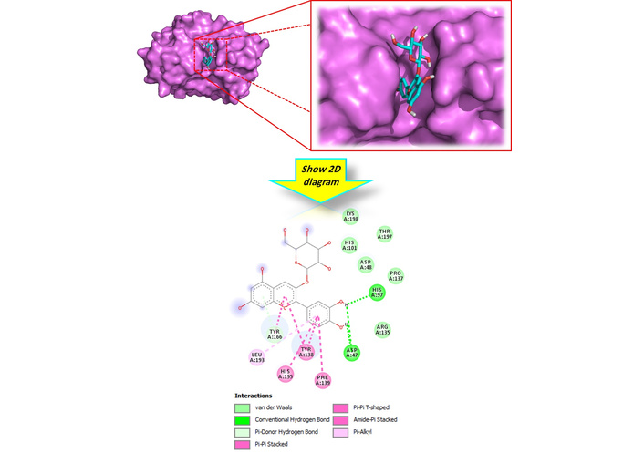
2D model of different interactions formed by the most active compound ‘cyanidin-3-O-glucoside’ within the active site of chitin deacetylase AngCDA from A. niger (PDB: 7bly). BIOVIA, Dassault Systèmes, Discovery Studio 2021, San Diego: Dassault Systèmes, 2026.
Against ‘Xanthine Oxidase’, it can be observed from the docking outcomes depicted in Table 8 that ‘catechin’, exhibiting the lowest binding energy (−7.3 kcal/mol) was found to be the most bioactive phytoligand compared to its analogues. Catechin was nicely bound to the target receptor via three H-bonds with Arg880, Thr1010, and Glu1261 through its hydroxy groups in addition to Pi-Sigma (Leu1014 and Ala1079), Pi-Pi stacked (Phe914), Pi-Pi T-shaped (Phe1009), and Pi-Alkyl (Leu873, Val1011, and Ala1078) (Figure 9).
Binding energy of the docked compounds in the binding cavity of ‘Xanthine Oxidase’ (PDB: 3nrz).
| Compound | Binding energy (kcal/mol) |
|---|---|
| Cyanidin-3-O-glucoside | –5.7 |
| Malvidin-3-O-arabinoside | –5.6 |
| Catechin | –7.3 |
| Cyanidin-3,5-diglucoside | –5.0 |
| Epicatechin | –7.2 |
| Petunidin-3-O-glucoside | –5.2 |
| Peonidin-3-O-glucoside | –6.0 |
| Pelargonidin-3-O-glucoside | –6.0 |
| Ascorbic acid (standard) | –3.5 |
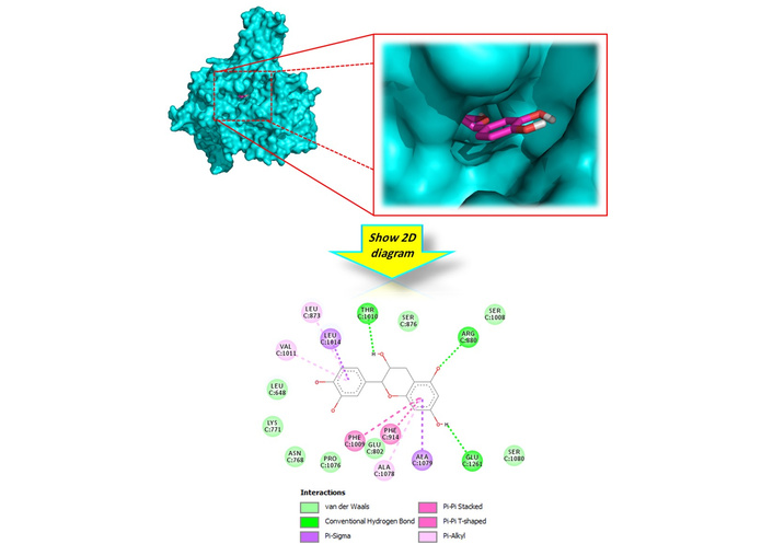
2D model of different interactions formed by the most active compound ‘catechin’ within the active site of ‘Xanthine Oxidase’ (PDB: 3nrz). BIOVIA, Dassault Systèmes, Discovery Studio 2021, San Diego: Dassault Systèmes, 2026.
All the above results show that these docked phytoconstituents of the B. asiatica fruit extract have a significant tendency to inhibit FabH from M. luteus, E. coli 24 kDa Domain, chitin deacetylase AngCDA from A. niger and Xanthine Oxidase.
Correlating the FT-IR spectral features with LC-MS/MS data provides stronger structural evidence for the identified bioactive compounds. The hydroxyl and carbonyl vibrations correspond to phenolic and glycosylated flavonoid structures, while C-O and C-H bands confirm glycosidic linkages and aliphatic chains in catechins and anthocyanins. Taken together, the FT-IR data confirm a diverse phytochemical matrix, composed primarily of phenolic compounds, alkaloids, and lipophilic constituents, supporting the bioactive potential of B. asiatica and its traditional use in medicinal practices. These results are consistent with those reported by Jara et al. [25]. Dara et al. [26] reported anthocyanin content ranging from 256.32 to 279.64 mg/L in extracts obtained using intensified methods, such as pulsed electric fields, cold plasma, and enzymatic treatment. Considering a common extraction ratio of 1 g of fruit to 10 mL of solvent, these values equate to approximately 25.6 to 27.9 mg/g of fruit, which is considerably lower than the value observed for B. asiatica in the present study. Therefore, even using a conventional technique, the B. asiatica extract had an anthocyanin density that was approximately four to five times higher than that recorded for B. vulgaris. Furthermore, Berberis lycium, another species of the same genus, had a chemical profile dominated by anthocyanins, such as cyanidin-3-glucoside (47.2%) and delphinidin-3-glucoside (35.3%), which exhibited potent antioxidant activity (IC50 = 25.3 μg/mL) [27]. While the present study did not identify individual anthocyanins, the high total concentration in B. asiatica indicates a rich composition and comparable or superior antioxidant potential. This comparison of species within the same genus highlights intraspecific phytochemical variability and reinforces B. asiatica potential as a promising source of anthocyanins for functional applications in the food, cosmetics, and pharmaceutical industries as both a bioactive agent and a high-performance natural colorant. The LC-MS/MS analysis of the methanolic extract of B. asiatica fruit revealed a phytochemical core consisting of eight glycosylated anthocyanins: Cy3G, Cy3diG5G, M3A, petunidin-3-O-glucoside, P3G, and Pg3G, catechin and epicatechin. This profile is similar to those described for other pigmented berries rich in Cy3G and Pg3G [28], setting the biochemical framework for the biofunctional comparisons below. Cy3G is the best-documented constituent and exhibits gastroprotective, antithrombotic, and epigenetic actions. Its efficacy against peptic ulcer-like injury has been demonstrated in both in vitro and rodent models. In these models, the aglycone cyanidin and Cy3G inhibited proinflammatory cytokines and curtailed lipid peroxidation [29]. The diglucoside Cy3diG5G has the same flavylium core, but its higher molecular mass reduces tissue penetration. In a sheep model, only 6.1% of the circulating Cy3diG5G pool crossed the blood-cerebrospinal fluid barrier versus 6.73% for Cy3-galactoside [30]. Nevertheless, a process involving enzymes that convert acyl-anthocyanins from red cabbage waste into high-purity Cy3diG5G (137.7 mg/100 g FW) has been reported. This provides a sustainable source for future bioactivity tests and is applicable [31].
Among malvidin analogues, M3A reversed ethyl carbamate-induced oxidative damage in intestinal epithelial cells by activating AMPK and restoring lysosomal autophagy, while M3G mitigated free fatty acid-driven steatosis through TFEB-mediated lysosomal biogenesis and Nrf2/ARE signaling [32]. A recent review positions malvidin glycosides as multipotent antioxidants capable of cardiometabolic and neuroprotective modulation [33]. Although less studied, petunidin-3-O-glucoside differs from malvidin by an additional methoxy group. This substitution typically enhances lipophilicity and may favor membrane interactions. Therefore, its co-occurrence broadens the redox-active spectrum of the extract. P3G and Pg3G occupy different pharmacological niches. P3G increases osteoblast differentiation and simultaneously curtails RANKL-induced osteoclastogenesis in a transgenic medaka model, demonstrating dual anabolic and anti-resorptive bone activity [34]. Molecular docking further predicts that P3G binds to the TNF-α receptor, which could interrupt inflammatory cascades [35]. Pg3G is inherently unstable but attained functional hepatoprotection when nano-encapsulated in pectin-chitosan liposomes. This encapsulation suppressed palmitate-induced ROS accumulation and mitochondrial dysfunction in L-02 hepatocytes [36]. Interestingly, a Cy3G/P3G-rich fraction from black rice bran inhibited NF-κB and NLRP3 inflammasome activation triggered by the SARS-CoV-2 spike protein in lung epithelial and macrophage cell lines. This finding highlights the anti-inflammatory synergy of these two anthocyanins [37]. The detected flavan-3-ols reinforce the extract’s biological potential. Catechin forms covalent adducts with ohmic-heated soy protein isolate, generating edible films with superior DPPH and ABTS radical-scavenging capacity. This indicates its intrinsic redox strength and protein-binding versatility [38]. Meanwhile, epicatechin down-modulates myostatin and atrogenes, activates AKT/mTOR, and stimulates mitochondrial biogenesis, positioning it as a candidate adjuvant for muscle wasting disorders [39]. Together, the colocalization of structurally diverse anthocyanins with potent flavanols suggests complementary mechanisms, such as radical scavenging, lysosomal/mitochondrial regulation, and cytokine suppression, that justify the advancement of B. asiatica extracts into targeted in vitro and in vivo models of gastrointestinal, hepatic, inflammatory, and musculoskeletal pathologies. The discovery in this research not only expands our scientific knowledge but also paves the way for the creation of innovative antimicrobial agents with tangible applications in both medicine and agriculture. This breakthrough holds the potential to make a substantial contribution to the overall well-being of society. By identifying and demonstrating the antimicrobial efficacy of B. asiatica solvent extracts, the study suggests practical uses in combating various pathogens that pose threats to human health and agricultural productivity. This newfound knowledge can inspire the development of new and improved antimicrobial treatments and agricultural practices, ultimately benefiting individuals, communities, and the broader society by addressing critical challenges in healthcare and food security.
The present investigation has revealed that the methanol extract of B. asiatica comprises eight bioactive compounds, which appear for the first time in the above-mentioned fruits, viz., cyanidin-3-O-glucoside, Pg3G, catechin, Cy3diG5G, epicatechin, M3A, petunidin-3-O-glucoside, and P3G, as determined via LC-MS/MS and FT-IR techniques. The findings of the study align with those of previous research [40–45]. In the study [46], the crude aqueous extract of B. asiatica stem bark exhibited antimicrobial activity against 11 out of 20 tested microorganisms using the disk diffusion method, with notable sensitivity shown by Staphylococcus aureus, Enterococcus faecalis, and Pseudomonas aeruginosa. The aqueous extract produced inhibition zones of up to 16 mm and inhibited 8 microorganisms. Comparatively, the alcoholic extract demonstrated the broadest antimicrobial spectrum, inhibiting 12 organisms. E. faecalis was most sensitive to the alcoholic extract and non-quaternary alkaloids. C. albicans was most effectively inhibited by the alcoholic extract.
The study [47] reported that the methanolic extract of B. asiatica demonstrated potent antibacterial activity against both S. aureus and Klebsiella pneumoniae. Disc-diffusion assays revealed inhibition zones of 14 mm (S. aureus) and 19 mm (K. pneumoniae). Minimum inhibitory concentration (MIC) values were 0.39 mg/mL for S. aureus and 3.125 mg/mL for K. pneumoniae, confirming strong growth suppression, with corresponding minimum bactericidal concentration (MBC) of 6.25 mg/mL for both species. B. asiatica methanol extract also showed remarkable antifungal activity (85%) against Aspergillus flavus, with the MIC value of 0.31 µg/mL [48]. These consistent antimicrobial activities underscore the extract’s potential as a natural antimicrobial agent. The study [47] conducted a study on B. asiatica, revealing significant levels of bioactive compounds. The plant exhibited a total phenolic content (TPC) of 37.686 ± 2.728 mg GAE/g and a total flavonoid content (TFC) of 115.568 ± 8.012 mg QE/g. Additionally, the DPPH free radical scavenging assay demonstrated strong antioxidant activity, with an IC50 value of 205.7 ± 5.353 µg/mL. The TAC found in B. asiatica, viz. 128.72 mg/g of fruit material. The findings were established as statistically significant, indicated by a p-value below 0.05. The findings indicated that both 80% v/v methanol extracts and 50% v/v hydroalcoholic extracts of B. asiatica exhibit noteworthy antimicrobial activity compared to hexane and chloroform extracts. However, it was observed that chloroform extracts lack antimicrobial activity against A. niger, S. abony, C. albicans. The polar extracts exhibited significant antioxidant activity when compared to non-polar extracts. The obtained results align with earlier findings [49–54]. This study demonstrates that B. asiatica fruits possess an unusually dense anthocyanin profile, dominated by cyanidin- and malvidin-type glycosides, and complemented by the flavanols catechin and epicatechin. The high TAC (128.72 mg/g) correlates with potent antioxidant activity. Additionally, polar extracts exhibit broad-spectrum antibacterial and antifungal activity, comparable to conventional reference drugs. Molecular docking analyses identify plausible targets, including fatty-acid synthase FabH, the E. coli 24-kDa gyrase domain, A. niger chitin deacetylase, and Xanthine Oxidase, providing mechanistic insight into both the antimicrobial and redox activities. Collectively, these findings position B. asiatica fruit as a promising candidate for the development of natural colorants, functional foods, and phytotherapeutic agents. Potential applications include natural food colorants and preservatives due to high anthocyanin and antimicrobial content, antioxidant dietary supplements targeting oxidative stress-related disorders, topical antimicrobial agents for skin infections or wound care, and functional food ingredients with both nutraceutical and redox-modulating properties. Future research should focus on scalable green extraction methods, stability, bioavailability, and in vivo validation in metabolic, infectious, and inflammatory disease models to fully harness the translational potential of this endangered Himalayan resource.
Cy3diG5G: cyanidin-3,5-diglucoside
Cy3G: cyanidin-3-O-glucoside
ESI: electrospray ionization
FT-IR: Fourier-transform infrared spectroscopy
LC-MS/MS: liquid chromatography-tandem mass spectrometry
M3A: malvidin-3-O-arabinoside
MIC: minimum inhibitory concentration
P3G: peonidin-3-O-glucoside
Pg3G: pelargonidin-3-O-glucoside
TAC: total anthocyanin content
Authors acknowledge the support and cooperation to Analytical and Instrumentation Facility, NCS Group, Nagpur, Maharashtra for LC-MS/MS and FT-IR analysis.
MK and TK: Conceptualization, Formal analysis, Writing—original draft. NV and PB: Formal analysis, Methodology, Writing—review & editing. Rajesh K: Formal analysis, Methodology, Writing—review & editing, Formal analysis, Methodology. MH and Ravendra K: Project administration, Supervision, Writing—review & editing. EdAFJ: Formal analysis, Methodology. MSdO: Writing—review & editing. All authors read and approved the submitted version.
The authors declare that they have no conflicts of interest.
Not applicable.
Not applicable.
Not applicable.
The authors, Dr. Mozaniel Santana de Oliveira mozaniel.oliveira@yahoo.com.br and Dr. Tarun Kumar tarunksj@gmail.com, will make the data of this research available to any researcher.
Not applicable.
© The Author(s) 2026.
Open Exploration maintains a neutral stance on jurisdictional claims in published institutional affiliations and maps. All opinions expressed in this article are the personal views of the author(s) and do not represent the stance of the editorial team or the publisher.
Copyright: © The Author(s) 2026. This is an Open Access article licensed under a Creative Commons Attribution 4.0 International License (https://creativecommons.org/licenses/by/4.0/), which permits unrestricted use, sharing, adaptation, distribution and reproduction in any medium or format, for any purpose, even commercially, as long as you give appropriate credit to the original author(s) and the source, provide a link to the Creative Commons license, and indicate if changes were made.

View: 818
Download: 45
Times Cited: 0
