 Case Report


Affiliation:
Department of Anesthesiology, Loma Linda University Medical Center, Loma Linda, CA 92354, USA

Affiliation:
Department of Anesthesiology, Loma Linda University Medical Center, Loma Linda, CA 92354, USA
Affiliation:
Department of Anesthesiology, Loma Linda University Medical Center, Loma Linda, CA 92354, USA
Affiliation:
Department of Anesthesiology, Loma Linda University Medical Center, Loma Linda, CA 92354, USA
ORCID: https://orcid.org/0000-0001-7317-5395
 ,
,
Affiliation:
Department of Anesthesiology, Loma Linda University Medical Center, Loma Linda, CA 92354, USA
Email: ccullom@llu.edu
ORCID: https://orcid.org/0000-0003-4741-262X

Explor Cardiol. 2026;4:1012119 DOI: https://doi.org/10.37349/ec.2026.1012119
Received: March 09, 2026 Accepted: July 01, 2026 Published: September 02, 2026
Academic Editor: Réda Ibrahim, Institut de Cardiologie de Montreal, Canada
The article belongs to the special issue Cardiovascular Risk for Mothers and Offspring Resulting from Complicated Pregnancy
Parturients with pulmonary arterial hypertension (PAH) face an increased risk of morbidity and mortality surrounding the peripartum period. Patients with severe PAH and Eisenmenger syndrome have the highest risk for perioperative mortality and provide a significant perioperative challenge for anesthesiologists, necessitating a multidisciplinary approach throughout pregnancy. We present a case of an urgent cesarean section at 37 weeks of gestation in a patient with Eisenmenger syndrome, a large atrial septal defect, and supra-systemic pulmonary hypertension. Due to the patient’s severity of PAH, cardiothoracic surgery was present and placed femoral arterial and venous catheters in preparation for possible extracorporeal membrane oxygenation (ECMO) cannulation in the event of hemodynamic collapse. The procedure was performed under a carefully titrated epidural, thus avoiding general anesthesia and the dangers associated with inducing a patient with her comorbid conditions. An arterial line, internal jugular central line, and Swan-Ganz catheter were placed while the patient was awake to aid in monitoring. Additionally, transthoracic echocardiography (TTE) was used to assist in cardiac monitoring while the epidural level was gradually increased. She was maintained on vasopressin, epinephrine, and phenylephrine infusions in addition to inhaled nitric oxide. She tolerated the procedure well, and the fetus was delivered safely. Careful management across pre-epidural, post-epidural, intraoperative, and postoperative stages illustrates the dynamic adjustments and multidisciplinary communication required in this patient population. Advanced medical therapy optimization, careful neuraxial anesthesia titration, and early consideration of extracorporeal cardiopulmonary resuscitation (ECPR) together capture the critical elements of management in this complex setting.
Pregnant individuals with pulmonary arterial hypertension (PAH) have an increased risk of peripartum morbidity and mortality. Patients with severe PAH and Eisenmenger syndrome have the highest risk for perioperative mortality and warrant multidisciplinary care coordination throughout pregnancy. For most patients with congenital heart disease (CHD) with left-to-right shunt, PAH is a late stage finding and portends a poor prognosis. Pregnancy in patients with PAH is associated with high risk of maternal and neonatal mortality [1–4]. These patients are at an elevated risk of pulmonary hypertensive crisis, pulmonary embolism, and cerebrovascular accident [1]. For the anesthesiologist, whether the choice is made to deliver via vaginal delivery or cesarean section, the anesthetic plan must consider the increased risks and abnormal maternal physiology. Cesarean delivery (CD) requires achieving surgical anesthesia without significant decreases in systemic vascular resistance (SVR), avoiding increases in pulmonary vascular resistance (PVR), and preserving right heart function. For vaginal delivery, dense neuraxial analgesia and assisted second stage of labor avoid increases in PVR due to pain and decreased venous return caused by increased intrathoracic pressure while pushing.
Furthermore, patients with severe PAH and long-standing left-to-right shunt may develop Eisenmenger syndrome, wherein shunt flow reverses or becomes bidirectional due to the increase in PVR. Parturients with Eisenmenger syndrome are more likely to experience severe heart failure, pulmonary hypertensive crisis, or peripartum death [5]. We present the management of an urgent CD for a parturient with Eisenmenger syndrome, a large atrial septal defect (ASD), and supra-systemic pulmonary artery pressure who presented with acute decompensated right heart failure at 37 weeks gestation. The patient’s severe PAH placed her at high risk for right heart failure and pulmonary hypertensive crisis, with potential for hemodynamic collapse and impaired placental circulation. Accordingly, the cardiothoracic surgery team placed femoral arterial and venous catheters in preparation for possible extracorporeal cardiopulmonary resuscitation (ECPR). The procedure was performed under a carefully titrated epidural, thus avoiding general anesthesia and the associated risks.
The timeline of the case is summarized in Table 1.
Timeline of clinical events.
| Event | Details/Findings |
|---|---|
| Previous pregnancy three years prior | Diagnosed with pulmonary hypertension during her fourth pregnancy and underwent an emergency cesarean section. Transthoracic echocardiography (TTE) revealed a large secundum atrial septal defect (ASD) and severe pulmonary hypertension with a right ventricular systolic pressure (RVSP) of 94 mmHg. |
| Two weeks prior to hospital admission | Presented to an outside hospital for shortness of breath and palpitations. TTE RVSP was 105 mmHg. |
| Admission to labor and delivery service | Presented at 37 weeks gestation with worsening dyspnea, dizziness, and pre-syncopal episodes with exertion. |
| Placement of epidural catheter | After activation of the epidural, she developed hypotension requiring vasopressor support and eventual discontinuation of the epidural. |
| Transfer to the intensive care unit (ICU) | On vasopressor support and inhaled nitric oxide. TTE showed a 2.4 cm ostium secundum ASD with intermittent right-to-left shunting, RVSP 80 mmHg with systemic systolic pressure of 95 mmHg. A radial arterial and central venous catheter were placed. |
| Cesarean section delivery | Epidural incrementally loaded in the operating room. Extracorporeal cardiopulmonary resuscitation (ECPR) catheters were placed. Required vasopressin, phenylephrine, and epinephrine infusions. Her baby was delivered by cesarean section. |
| Post-operative day one | Remained on inhaled nitric oxide, a vasopressin infusion, and treprostinil. |
| Post-operative day eleven | Weaned off vasoactive support. Treprostinil was continued and she was started on goal directed therapy with tadalafil, furosemide, and spironolactone. Discharged on postoperative day 11 with supplemental oxygen (2–4 L/min via nasal cannula). |
| Six weeks after discharge | Readmitted six weeks after discharge with a pulmonary hypertensive crisis, ventricular fibrillation arrest, and acute hypoxic respiratory failure. |
| Nine months after discharge | She unfortunately passed away 9 months later due to an out of hospital cardiac arrest. |
A 31-year-old gravida 5 para 4 woman at 37 weeks gestation was admitted with worsening dyspnea, dizziness, and pre-syncopal episodes with exertion. Her cardiac history included a large ostium secundum ASD, Eisenmenger syndrome, severe pulmonary hypertension (WHO group-I, NYHA class 4), and chronic right ventricular failure (Figure 1). Her social history was significant for current tobacco smoking as well as former use of marijuana and methamphetamine. All previous obstetric deliveries occurred at other hospitals. Her first two deliveries were vaginal deliveries with no other outside hospital records. She was diagnosed with pulmonary hypertension during her fourth pregnancy three years prior, during which she developed progressive shortness of breath and hypoxemia and underwent an emergency cesarean section at an outside hospital. Prior to delivery, her transthoracic echocardiography (TTE) revealed a large secundum ASD and severe pulmonary hypertension with an estimated right ventricular systolic pressure (RVSP) of 94 mmHg. Systemic systolic blood pressures at that time were in the 90–110 mmHg range. Her postpartum therapy included selexipag, macitentan, and sildenafil. Cardiology planned for a repeat heart catheterization after three months of maintenance pulmonary hypertension therapy before possible closure of her ASD. However, she stopped taking her medications due to financial constraints. Prior workup did not identify any additional shunts, such as congenital malformations besides the ASD.
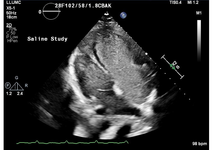
Trans-thoracic echocardiography visualizing an apical four chamber view with saline contrast from 1 year prior to presentation. Demonstrates right to left shunt as contrast flows from dilated right heart to left heart via atrial septal defect.
The patient was lost to follow up after her prior CD and was not taking any medications for pulmonary hypertension at the time of admission. She sought care at an outside hospital for shortness of breath and palpitations two weeks prior to admission; at that time her TTE estimated RVSP was 105 mmHg with systemic systolic pressures of 110 mmHg. On admission, her oxygen saturation was 90–95% on room air with desaturations to 84–85% on exertion.
A multidisciplinary team including maternal fetal medicine, anesthesiology, pulmonology, cardiology, cardiothoracic surgery, and intensivists convened to discuss her care. There was prevailing concern that vaginal delivery with repeated Valsalva could precipitate pulmonary hypertensive crisis, acute right ventricular failure, and hemodynamic collapse that could compromise maternal and fetal circulation. There was also concern for rapid decline and post-delivery cardiovascular complications in the event of uncontrollable bleeding during a vaginal delivery and potentially larger fluid shifts after delivery compared to a cesarean section. Alternatively, a planned cesarean section would provide controlled timing to ensure all necessary support staff were immediately available for escalation of care and reduce the risk of an emergent cesarean section. Overall, cesarean section created the most controlled environment for the multitude of potential complications. The consensus was that a repeat cesarean section combined with femoral sheath placement to facilitate ECPR was the safest option for delivery.
She was initially admitted to the labor and delivery unit. An epidural catheter was placed in anticipation of eventual CD. After a test dose of 3 mL of 1.5% lidocaine, the epidural was activated with an infusion of 0.15% ropivacaine and fentanyl 2.5 µg/mL at 2 mL/hour. She developed hypotension with systolic pressures in the low 90s. A vasopressin infusion was started at 0.04 units/min due to its ability to increase SVR with minimal effect on pulmonary vasculature. In addition, she required boluses of vasopressin and epinephrine to maintain her systolic blood pressure above 105 mmHg. The epidural infusion was discontinued, and she was transferred to the intensive care unit (ICU), where she continued to receive vasopressin at 0.04 units/min, inhaled nitric oxide by high flow nasal cannula (HFNC), and a Treprostinil intravenous infusion. TTE redemonstrated a large ~2.4 cm ostium secundum ASD with intermittent right-to-left shunting, RVSP 80 mmHg with systemic systolic pressure of 95 mmHg, a severely dilated and hypertrophied right ventricle, severely decreased right ventricular function, and preserved left ventricular function (Figure 2, Figure 3).
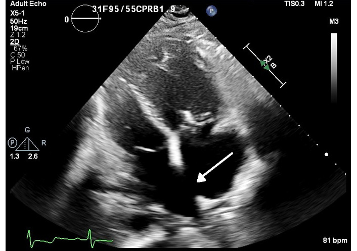
Trans-thoracic echocardiography visualizing an apical four chamber view that demonstrates a 2.4 cm ostium secundum atrial septal defect (ASD). Arrow identifies the apical septal defect. Study performed at time of current admission.
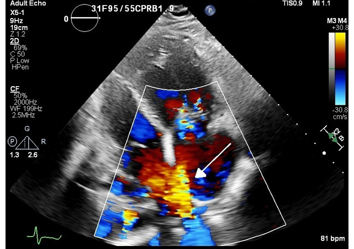
Trans-thoracic echocardiography visualizing an apical four chamber with color flow Doppler demonstrating a left to right shunt. Arrow denotes the shunt through the atrial septal defect. Study performed at time of current admission.
A radial arterial and central venous catheter was placed in the ICU, and she was transferred to the operating room. She arrived still on vasopressin at 0.04 units/min and inhaled nitric oxide. Her initial vitals were: systemic blood pressure 125/67 mmHg, heart rate 85 bpm, and oxygen saturation 99% with HFNC (80% FiO2 at 18 L/min). A pulmonary artery catheter was placed in the operating room via the existing 9 French central venous catheter using sterile technique. TTE was used to guide the catheter placement in the setting of an ASD. The initial pulmonary artery pressure was 101/28 mmHg and reached a maximum of 114/55 mmHg during the surgery. Given concern for further hypotension with epidural activation, vasopressin infusion was maximized to 0.06 units/min, and a phenylephrine infusion was started at 50 mcg/min. Phenylephrine was chosen to maintain SVR, thus supporting biventricular perfusion at the expense of increasing PVR. Her epidural was incrementally loaded with 2% lidocaine with epinephrine in 3 mL aliquots until a T4 sensory level was obtained. She developed persistent hypotension with epidural loading, and low dose epinephrine infusion was initiated at 3 mcg/min to support right heart function and to provide mild augmentation of SVR. She was positioned supine, and catheters were placed under epidural analgesia in the common femoral artery and common femoral vein to facilitate ECPR. Her baby was delivered by cesarean section with APGAR scores of 7 and 7 at 1 and 5 minutes, respectively. Her baby weighed 2.5 kg and was initially admitted to neonatal ICU (NICU) for continued need of continuous positive airway pressure (CPAP). Her baby was discharged home on day of life 9. Her pulmonary arterial systolic pressures were supra-systemic throughout much of the procedure with mean systemic pressures maintained between 65 and 70 mmHg. Although her estimated blood loss was 1,000 mL, she did not acutely decompensate during delivery, and ECPR was not initiated. She was transferred to the ICU postoperatively on pressor support including vasopressin at 0.06 units/min, phenylephrine at 50 mcg/min, and epinephrine at 3 mcg/min.
She remained on inhaled nitric oxide, a vasopressin infusion, and a treprostinil infusion at 8 ng/kg/min on postoperative day one. She was weaned off vasoactive support by postoperative day two. Treprostinil was continued, and she was started on goal directed therapy with tadalafil, furosemide, and spironolactone. She had persistent hypoxemia, and she was discharged on postoperative day 11 with supplemental oxygen (2–4 L/min via nasal cannula). She was readmitted six weeks after discharge with a pulmonary hypertensive crisis, ventricular fibrillation arrest, and acute hypoxic respiratory failure. Her urine drug screen resulted positive for amphetamine and cannabinoids. She unfortunately passed away 9 months later due to an out of hospital cardiac arrest.
TTE findings are summarized in Table 2. Images of the TTE findings are shown in Figure 1 and Figure 2.
Transthoracic echo findings throughout the patient’s course.
| Transthoracic echocardiography (TTE) occurrence | Findings | Systemic systolic pressure (mmHg) |
|---|---|---|
| Fourth pregnancy (three years prior) | Large secundum atrial septal defect (ASD) and severe pulmonary hypertension with an estimated right ventricular systolic pressure (RVSP) of 94 mmHg | 90–110 |
| Two weeks prior to admission at outside hospital | Estimated RVSP of 105 mmHg | 110 |
| On admission to intensive care unit (ICU) | Large ~2.4 cm ostium secundum ASD with intermittent right-to-left shunting, RVSP 80 mmHg, a severely dilated and hypertrophied right ventricle, severely decreased right ventricular function, and preserved left ventricular function | 95 |
For most patients with CHD with left-to-right shunt, PAH is a late stage finding and portends a poor prognosis. Pregnancy in patients with PAH is associated with high risk of maternal and neonatal mortality. These patients are at an elevated risk of pulmonary hypertensive crisis, pulmonary embolism, and cerebrovascular accident.
The physiologic changes of pregnancy present formidable risk for individuals with Eisenmenger syndrome, especially when accompanied by severe PAH. Normally, PVR and SVR decrease as blood volume and cardiac output increase during pregnancy. In response to this, the pulmonary system undergoes pulmonary vasodilation to prevent pulmonary pressures from markedly rising during pregnancy. However, PVR remains relatively fixed in patients with PAH. As blood volume and cardiac output increase, the right ventricle struggles to accommodate additional volume, and pulmonary arterial pressures may actually increase [6]. Therefore, pregnant individuals with PAH are susceptible to dyspnea, heart failure, and syncope [7]. Moreover, in patients with Eisenmenger syndrome, the pregnancy associated decrease in SVR may further promote right-to-left intracardiac shunting and hypoxemia. Not surprisingly, the risk of cardiopulmonary decompensation, thrombosis, and sudden death is significant in pregnant individuals with Eisenmenger syndrome [8]. Normally, cardiac output, heart rate, and stroke volume return to pre-labor values within 24–72 hours postpartum and reach non-pregnant levels by 6–8 weeks after delivery [9]. However, approximately 70% of maternal deaths occur between postpartum days 2 and 30, primarily due to right ventricular failure. This elevated peripartum mortality risk is attributed to the combined effects of hemodynamic stress, bleeding complications, and use of general anesthesia, superimposed on the physiological cardiovascular changes of the peripartum period; all of which can precipitate right heart failure. Postpartum management of patients with Eisenmenger syndrome requires careful optimization of medical therapies and continued postpartum monitoring [10, 11].
These physiological risks complicate decisions about the method of delivery in pregnant individuals with PAH, where premature delivery is common. Spontaneous labor carries unpredictable timing and may result in inexperienced teams managing labor and delivery without sufficient time to coordinate essential multidisciplinary care. Vaginal delivery itself introduces significant physiologic changes; uterine contraction diverts blood from the engorged uterus to the heart and can augment cardiac output by as much as 25% [10]. Increased cardiac output may be accompanied by additional increases in pulmonary arterial pressure and susceptibility to arrhythmia and right heart failure [10]. Furthermore, repeated Valsalva can impair venous return and rapidly precipitate hemodynamic deterioration [12]. Moreover, vaginal delivery limits access to the femoral vessels because of stirrup positioning, making an urgent transition to ECPR difficult if care must be escalated. Nonetheless, current studies suggest parturients with well controlled PAH can endure an assisted second stage of labor with adequate analgesia [6]. Comparatively, CD may circumvent the physiologic changes associated with vaginal delivery, and operative site staffing allows for rapid care escalation. In severe forms of PAH, including Eisenmenger’s syndrome, a cesarean section is generally advised [13]. A planned CD allows the care team to carefully initiate an anesthetic and analgesic plan to minimize the risks associated with CD and general anesthesia. Emergent CD is best avoided due to the increased risk of hemorrhage as well as the potential necessity of general anesthesia, which is particularly deleterious for individuals with Eisenmenger syndrome [10]. General anesthesia and positive pressure ventilation pose significant risk. Laryngoscopy and endotracheal intubation can be associated with significant sympathetic stimulation and acute increases in PVR, while positive pressure ventilation reduces preload. Moreover, volatile anesthetics decrease SVR and depress myocardial function [14, 15]. SVR is not only important for supporting right ventricular perfusion when RVSP is elevated but when SVR exceeds PVR the burden of right-to-left shunting is reduced. While no consensus exists on the optimal timing and delivery method, scheduled CD with careful planning offers the best opportunity for coordinated management and timely escalation of care.
Labor analgesia is imperative for limiting pain mediated increases in PVR. Neuraxial techniques offer rapid, reliable analgesia, but may be associated with an undesirable decrease in SVR in patients with Eisenmenger syndrome [14]. Incremental loading of a spinal, combined spinal epidural, and epidural analgesia alone have been described as strategies for mitigating the reduction in SVR associated with neuraxial techniques [14, 16–18].
Delivery may also warrant administration of uterotonic agents to augment uterine tone and mitigate postpartum hemorrhage. Oxytocin is first line therapy for augmenting uterine tone in most parturients. Bolus administration of oxytocin is associated with tachycardia and hypotension, and acute pulmonary edema has also been reported [19]. However, the hemodynamic effects of oxytocin can be reduced by administering a slow infusion and avoiding a rapid bolus [19, 20]. Carboprost and methylergonovine are second line agents for parturients with refractory uterine atony or postpartum hemorrhage; both are associated with increased PVR [20–23] and are best avoided in the parturient with PAH. Although administration may not be avoidable when postpartum hemorrhage occurs. Rectal misoprostol, though generally less effective, is safe in this population [22].
Medical management of pulmonary hypertension has been shown to significantly lower the risk of death in non-pregnant patients with Eisenmenger syndrome [5]. Medical therapies include a variety of endothelin receptor antagonists, prostaglandins, and phosphodiesterase-5 inhibitors and initiation of pulmonary hypertension therapy before delivery contributes to favorable outcomes for pregnant individuals with pulmonary hypertension [7]. A recent retrospective review of pregnant individuals with Eisenmenger syndrome found mortality was lower than previously reported (16.7% vs. 30–75%). Notably, 86.7% of the pregnant individuals in this review received therapies for pulmonary hypertension (sildenafil or tadalafil, and prostaglandins) during pregnancy [5, 10]. The Pulmonary Vascular Research Institute recommends parenteral prostaglandins such as epoprostenol or treprostinil for pregnant individuals with pulmonary hypertension and significant RV dysfunction; the benefit of prostaglandins likely outweighs the potential risks to the fetus [21]. Oral phosphodiesterase-5 inhibitors have been used in pregnant individuals with pulmonary hypertension, though the impact on peripartum outcomes remains unclear [11]. Endothelin receptor antagonists and guanylate cyclase stimulators, however, are teratogenic and should be avoided once the patient becomes pregnant [24]. Once discontinued, it is recommended to switch to another therapy if these treatments are not already being used [11]. Unfortunately, medical therapy optimization is not always feasible prior to delivery, especially for individuals with limited healthcare access. For some individuals, peripartum care may present the first opportunity to introduce therapies for pulmonary hypertension.
Despite appropriate management, the hemodynamic changes associated with delivery, high risk of thromboembolic events, risk of right ventricular failure, right ventricular hypoperfusion, and pulmonary hypertensive crisis are significant and may result in hemodynamic collapse and cardiac arrest [25]. In this case, ECPR is a viable option for emergent management. Both veno-venous (VV) and veno-arterial (VA) extracorporeal membrane oxygenation (ECMO) have been used to support patients with Eisenmenger syndrome in the peripartum period [26]. The criteria for ECPR initiation are similar for pregnancy and nonpregnant patients, though specific conditions such as severe PAH, peripartum cardiomyopathy, or decompensated CHD in pregnancy may predispose to rapid cardiopulmonary decline and justify early care escalation. Current ECPR guidelines from ELSO do not identify separate indications for pregnancy, underscoring the need for individualized risk assessment [27, 28].
Given the increasing prevalence of CHD among adults and the fact that cardiovascular conditions account for more than one third of pregnancy-related deaths in the United States, early identification of high-risk patients and referral to centers capable of advanced cardiopulmonary support is essential [29, 30]. Formal criteria for selecting candidates for ECPR standby in pregnancy have not been established, and data regarding the impact of delayed ECPR standby or activation on maternal outcomes are limited [31]. Existing cardiac risk stratification models, such as the mWHO Pregnancy Risk Classification and the Cardiac Disease in Pregnancy II (CARPREG II) tool, can help identify those at highest risk [31]. Although these tools are designed for pregnant patients with known cardiac disease, their incorporation into multidisciplinary team discussions may aid in determining whether a patient should be considered for ECPR standby and transferred to a tertiary care center with ECPR capabilities.
In those at very high risk for peripartum cardiovascular complications, such as those with severe right heart dysfunction, severe PAH, or Eisenmenger’s syndrome, preparation for elective cesarean section should be considered to facilitate cannulation and a timely transition to ECPR should the need arise [22]. Elective planning in a controlled operating room environment allows for the appropriate personnel and equipment to be ready for expeditious cannulation and initiation of ECMO [22]. This proactive planning allows for rapid vascular access and enables deliveries to occur while the patient is on ECMO support. Available data does suggest there may be an advantage to ECPR. A systematic review of studies investigating maternal survival with ECPR had an overall survival rate of 87.7% compared to 58.9% in a large retrospective study of peripartum cardiac arrest in general [25, 26]. This improved outcome is often attributed to pregnant patients being younger and benefiting from this aggressive and timely management for an acute indication [26]. Early placement of venous and arterial sheaths in the antepartum period for potential rapid initiation of ECPR in patients with severe PAH may improve outcomes in these high risk patients [26]. Preparing for ECPR early in complex maternal heart disease allows for the necessary coordination of specialized team members and increases the chances of stabilizing the situation and increasing survival. Decisions regarding ECPR standby should be individualized based on the patient’s clinical status, anticipated risk of cardiopulmonary deterioration, and institutional capabilities.
Pregnancy in individuals with Eisenmenger syndrome and PAH is associated with peripartum mortality and poor fetal outcomes. Individuals with Eisenmenger syndrome and pulmonary hypertension should be counseled about the risk of pregnancy. Care of the pregnant individual with CHD warrants multidisciplinary coordination between obstetricians, obstetric and cardiac anesthesiologists, cardiology, cardiothoracic surgery, and neonatologists. ECPR has emerged as an important approach to support parturients with significant cardiac disease through delivery.
This case represents a unique comprehensive approach to the management of pregnant patients with severe pulmonary hypertension and Eisenmenger syndrome with careful medical management and ECPR standby. The team balanced the extremely high surgical and anesthetic risk of her suprasystemic pulmonary pressures, intermittent right-to-left shunting, and absence of chronic pulmonary vasodilator therapy against the risks of ECPR itself, including bleeding, anticoagulation, and circuit related complications. Careful management across pre-epidural, post-epidural, intraoperative, and postoperative stages illustrates the dynamic adjustments and multidisciplinary communication required in this patient population. Advanced medical therapy optimization, careful neuraxial anesthesia titration, and early consideration of ECPR together capture the critical elements of management in this complex setting.
ASD: atrial septal defect
CD: cesarean delivery
CHD: congenital heart disease
ECMO: extracorporeal membrane oxygenation
ECPR: extracorporeal cardiopulmonary resuscitation
HFNC: high flow nasal cannula
ICU: intensive care unit
PAH: pulmonary arterial hypertension
PVR: pulmonary vascular resistance
RVSP: right ventricular systolic pressure
SVR: systemic vascular resistance
TTE: transthoracic echocardiography
SD: Conceptualization, Investigation, Methodology, Validation, Visualization, Writing—original draft, Writing—review & editing. JC: Conceptualization, Investigation, Methodology, Validation, Visualization, Writing—original draft, Writing—review & editing. DK: Conceptualization, Investigation, Methodology, Validation, Visualization, Writing—review & editing. MM: Conceptualization, Investigation, Methodology, Validation, Visualization, Writing—review & editing. CC: Conceptualization, Investigation, Methodology, Project administration, Supervision, Validation, Visualization, Writing—original draft, Writing—review & editing. All authors read and approved the submitted version.
The authors declare that they have no conflicts of interest.
Case studies are not overseen by our institutional IRB. No identifying pictures, no PHI used, and written consent was also obtained from the patient. To summarize, no ethical approval is needed due to the de-identified nature of this case report. And this study was in compliance with the Declaration of Helsinki.
Informed consent to participate in the study was obtained from all participants/ participants’ guardians.
Informed consent to publication was obtained from relevant participants.
The data of this manuscript could be available from the corresponding authors upon reasonable request (ccullom@llu.edu).
Not applicable.
© The Author(s) 2026.
Open Exploration maintains a neutral stance on jurisdictional claims in published institutional affiliations and maps. All opinions expressed in this article are the personal views of the author(s) and do not represent the stance of the editorial team or the publisher.
Copyright: © The Author(s) 2026. This is an Open Access article licensed under a Creative Commons Attribution 4.0 International License (https://creativecommons.org/licenses/by/4.0/), which permits unrestricted use, sharing, adaptation, distribution and reproduction in any medium or format, for any purpose, even commercially, as long as you give appropriate credit to the original author(s) and the source, provide a link to the Creative Commons license, and indicate if changes were made.



View: 355
Download: 5
Times Cited: 0
Johanna A. van der Zande ... Jerome M. J. Cornette
Kazim Raza Talpur ... Muhammad Waleed Abdullah
Jingwen Guo, Xueying Wang
Fatima Farrukh ... Zainab Samad


