 Review


Affiliation:
School of Sports Science, Qufu Normal University, Jining 273165, Shandong province, China
Email: jianda0426@163.com
ORCID: https://orcid.org/0000-0003-1783-918X


Explor Neurosci. 2026;5:1006142 DOI: https://doi.org/10.37349/en.2026.1006142
Received: May 10, 2026 Accepted: July 06, 2026 Published: August 03, 2026
Academic Editor: Ayan Mohamud Yusuf, University of Duisburg-Essen, Germany
The article belongs to the special issue Depression: From Pathophysiology to Treatment Innovation
Exercise is an effective non-pharmacological intervention for depressive symptoms, but the biological mechanisms underlying its intensity-dependent and symptom-specific effects remain incompletely defined. This review proposes the “lactate window” hypothesis as a testable mechanistic model, not as an established clinical prescription strategy. Lactate is a plausible candidate link because it is tightly related to exercise intensity, functions as an oxidative substrate and signaling molecule, and participates in brain energy metabolism, astrocyte-neuron metabolic coupling, neuroplasticity, neurovascular signaling, and glial-immunometabolic regulation. Major depressive disorder (MDD) is associated with altered brain energy metabolism, mitochondrial dysfunction, pH abnormalities, and disrupted glial-neuronal support, which may be particularly relevant to fatigue, anhedonia, low motivation, psychomotor slowing, and impaired effort-based decision-making. However, lactate should not be interpreted as uniformly beneficial. Preclinical work suggests that acute L-lactate can produce antidepressant-like effects, whereas human neuroimaging findings indicate that regional lactate in motivation-related cortical circuits may be associated with reduced willingness to exert physical effort. We therefore distinguish three evidence levels: established physiological findings, plausible mechanistic pathways, and hypothesis-generating clinical applications. The proposed lactate window is operationally defined by dynamic features of the response, including blood lactate peak, area under the curve (AUC), time-to-peak, clearance, recovery kinetics, an exploratory lactate-to-rating of perceived exertion (RPE) index, affective response, next-day fatigue, sleep, and adherence. The clinical goal is not to maximize lactate, but to identify a tolerable, recoverable metabolic challenge that may support adaptive brain remodeling. Future trials should test whether lactate kinetics predict antidepressant response beyond conventional exercise dose and whether such effects are strongest for energy-, motivation-, and effort-related symptom dimensions. Because direct clinical evidence in MDD remains limited, lactate-informed exercise prescription should currently be framed as a research agenda requiring prospective validation.
Major depressive disorder (MDD) is a leading contributor to global disability and disease burden, and available treatments are often limited by delayed onset, incomplete remission, relapse, adverse effects, and poor acceptability in some patients [1, 2]. In the Sequenced Treatment Alternatives to Relieve Depression (STAR*D) study, remission rates decreased across successive treatment steps, illustrating the need for complementary, scalable, and mechanistically informed interventions [2]. Exercise has therefore attracted increasing attention as a non-pharmacological intervention for depressive symptoms. A recent systematic review and network meta-analysis of randomized controlled trials found that several exercise modalities, including walking or jogging, yoga, strength training, and mixed aerobic activity, reduce depressive symptoms, with some evidence that intensity may modify benefit [3].
The central mechanistic question is not whether exercise can improve depressive symptoms, but why different patients, exercise modalities, and intensity prescriptions produce different responses. Much of the clinical exercise literature still treats exercise mainly as a behavioral exposure defined by frequency, duration, and target heart rate. This approach is useful, but incomplete. Exercise is also a biological stimulus with quantifiable metabolic outputs. Among these outputs, lactate is especially relevant because it changes dynamically with exercise intensity, muscle recruitment, glycolytic flux, training status, and recovery capacity [4–12].
The rationale for focusing on lactate is strengthened by increasing evidence that depression cannot be fully explained by monoaminergic or cognitive-affective models alone. MDD has also been linked to altered glucose metabolism, mitochondrial dysfunction, pH abnormalities, glial pathology, and impaired metabolic coupling between astrocytes and neurons [13–25]. A post-mortem systematic review and meta-analysis reported decreased brain pH in MDD, suggesting that acid-base balance and energy metabolism may be clinically relevant, although pH is not a direct measure of lactate metabolism and post-mortem data require cautious interpretation [13].
Lactate was once described primarily as a byproduct of glycolysis or a marker of anaerobic metabolism. It is now recognized as a mobile oxidative substrate, a gluconeogenic precursor, and a cell-to-cell signaling molecule [4–6, 26]. In the brain, lactate transport is mediated by monocarboxylate transporters (MCTs), and lactate participates in astrocyte-neuron metabolic coupling [27–34]. Experimental work also links lactate to plasticity-related pathways involving brain-derived neurotrophic factor (BDNF), cAMP response element-binding protein (CREB), and sirtuin 1 (SIRT1) [28, 34–38], as well as receptor-mediated neurovascular signaling through hydroxycarboxylic acid receptor 1 (HCAR1) and vascular endothelial growth factor (VEGF) [39]. Importantly, cyclic adenosine monophosphate (cAMP)-dependent protein synthesis has also been implicated in acute lactate-induced antidepressant-like effects in preclinical work [40].
This article does not argue that “more lactate is better.” Such a linear interpretation would be biologically weak and clinically unsafe. Instead, we propose a depression-specific, symptom-dimensional, recovery-based model: the “lactate window” hypothesis. In this model, a short-lived and recoverable lactate pulse may act as an adaptive metabolic cue, whereas prolonged, excessive, poorly cleared, or regionally atypical lactate may reflect metabolic stress, mitochondrial limitation, acid-base imbalance, or dysfunctional circuit activity. The distinctive contribution of this review is therefore not the claim that lactate biology matters for exercise, which is already established, but the proposal that lactate dynamics may help organize future research on energy-, motivation-, and effort-related depressive symptoms.
This article is a narrative mini-review and hypothesis paper rather than a systematic review or meta-analysis. To improve transparency, this manuscript specifies the literature identification strategy. Relevant literature was identified through targeted searches of PubMed, Web of Science, Scopus, and Google Scholar from database inception to June 2026. Search domains combined terms related to depression and symptoms (“major depressive disorder,” “depression,” “anhedonia,” “fatigue,” “psychomotor slowing,” “effort-based decision-making,” “motivation”), exercise (“exercise,” “physical activity,” “aerobic,” “resistance training,” “high-intensity interval training,” “lactate threshold”), lactate biology (“lactate,” “blood lactate,” “lactate kinetics,” “lactate shuttle,” “monocarboxylate transporter,” “MCT1,” “MCT2,” “MCT4”), and brain mechanisms (“astrocyte-neuron lactate shuttle,” “brain metabolism,” “mitochondria,” “pH,” “BDNF,” “CREB,” “SIRT1,” “HCAR1,” “VEGF,” “microglia,” “histone lactylation,” “magnetic resonance spectroscopy”).
Studies were prioritized when they met at least one of the following criteria: (i) human clinical or epidemiological relevance to MDD, depressive symptoms, fatigue, anhedonia, psychomotor slowing, or effort-based decision-making; (ii) exercise trials or exercise physiology studies that measured lactate kinetics or intensity-dependent metabolic responses; (iii) human magnetic resonance spectroscopy (MRS), positron emission tomography (PET), or physiological studies relevant to brain metabolism or lactate uptake; (iv) preclinical studies directly testing lactate administration, lactate transport, astrocyte lactate metabolism, or depression-like behaviors; and (v) molecular or cellular studies clarifying mechanisms that could plausibly connect lactate to neural plasticity, neurovascular remodeling, glial-immunometabolic regulation, or pH/mitochondrial function. Narrative reviews and meta-analyses were used to contextualize broader evidence, but primary studies were emphasized where mechanistic specificity was important.
The synthesis is organized around three evidence levels. Established findings include that exercise increases peripheral lactate in an intensity- and modality-dependent manner [4–12], lactate can cross the blood-brain barrier (BBB) through MCT-mediated transport [29, 30, 41], and MDD is associated with abnormalities in brain metabolism, mitochondrial function, pH, and glial support [13–25]. Plausible mechanistic inferences include the possibility that exercise-induced lactate influences astrocyte-neuron lactate shuttle (ANLS) function [27–34], BDNF/CREB/SIRT1-related plasticity [28, 34–38], HCAR1-VEGF signaling [39], mitochondrial oxidation and pH context [4–6, 13, 19, 26, 31], and glial-immunometabolic state [42–46]. Hypothesis-generating clinical applications include prospective testing of lactate curves, area under the curve (AUC), clearance and recovery kinetics, and an exploratory lactate-to-rating of perceived exertion (RPE) index as candidate physiological feedback variables in depression exercise trials. These applications have not been prospectively validated in MDD and should be interpreted as a research agenda rather than a current clinical standard.
Traditional accounts of MDD emphasize monoaminergic, neuroendocrine, inflammatory, cognitive-affective, and psychosocial mechanisms. These models remain important, but they do not fully explain clinical features such as fatigue, psychomotor retardation, low motivation, and impaired effort allocation. A complementary interpretation is that some depressive phenotypes involve reduced neurometabolic flexibility, defined here as the capacity of neural circuits to match changing cognitive, affective, and motivational demands with appropriate substrate supply, mitochondrial oxidation, vascular delivery, glial-neuronal metabolic cooperation, and recovery.
Glucose metabolism provides one entry point into this framework. A recent review synthesized evidence that MDD is associated with systemic and localized impairment of glucose metabolism across the illness course [14]. Earlier PET work also suggested regional alterations in cerebral glucose metabolism in depression, including prefrontal hypometabolism [17]. To provide more concrete context rather than relying on generalized statements, it is worth noting that a longitudinal fluorodeoxyglucose (FDG)-PET study by Kennedy and colleagues [18] examined 13 male patients with MDD before and after 6 weeks of paroxetine treatment and compared resting scans with 24 healthy male controls; successful treatment was associated with increased glucose metabolism in dorsolateral, ventrolateral, and medial prefrontal regions, parietal cortex, and dorsal anterior cingulate cortex (dACC). Although this study was small, male-only, and treatment-specific, it illustrates that depression-related symptoms can be accompanied by measurable changes in regional metabolic activity.
Mitochondrial dysfunction adds a second layer. Mitochondria regulate adenosine triphosphate production, redox balance, calcium signaling, apoptosis, and inflammatory responses, and abnormalities in these processes have been repeatedly implicated in MDD and related psychiatric disorders [15, 16]. If mitochondrial oxidative capacity is limited, neural circuits may struggle to meet the energetic demands of cognitive control, emotion regulation, reward pursuit, and physical effort. Under such conditions, lactate may have divergent meanings: it can serve as a useful oxidative substrate when transport and oxidation are efficient, but it may also accumulate when glycolytic flux exceeds mitochondrial use or when acid-base regulation is impaired.
Brain pH and lactate abnormalities further support a neurometabolic interpretation of depression. Post-mortem studies and animal model work suggest that reduced brain pH and altered lactate levels may occur across psychiatric conditions, but these findings are vulnerable to confounding by agonal state, medication exposure, brain activity before death, tissue handling, and post-mortem interval [13, 19]. These limitations do not invalidate the signal; they require cautious interpretation. The relevant point is that pH, lactate, glycolysis, mitochondrial oxidation, and excitation-inhibition balance form an interconnected metabolic system rather than independent variables.
Astrocytes are central to this system. They regulate glucose uptake, glycogen storage, lactate production, neurotransmitter recycling, potassium and proton buffering, BBB function, and neurovascular coupling [20, 21]. Post-mortem and experimental studies have reported reductions or abnormalities in astrocytic markers, glial density, and astrocyte-dependent support of prefrontal and hippocampal circuits in MDD [22–25]. Because astrocytes are major producers and distributors of brain lactate, glial pathology provides a plausible bridge between lactate metabolism and depressive symptoms. The key question is not whether lactate is simply “high” or “low,” but whether it is produced, transported, oxidized, and cleared at the right time, in the right region, and under a metabolic context that permits adaptation rather than burden.
A symptom-dimensional approach is necessary because MDD is heterogeneous. Global scores on the Hamilton Depression Rating Scale, Montgomery-Asberg Depression Rating Scale, Patient Health Questionnaire-9, or Beck Depression Inventory can mix affective, cognitive, somatic, sleep, appetite, guilt, and motivational symptoms. Lactate-related mechanisms are unlikely to explain all depressive symptoms equally. They are most plausible for symptom dimensions that require sustained coordination of neural and bodily energy systems, including fatigue, anergia, psychomotor slowing, anhedonia, reduced motivation, and impaired effort-based decision-making [47–57].
Anhedonia should not be reduced to the absence of pleasure. Contemporary models distinguish consummatory pleasure, anticipatory pleasure, reward learning, motivational drive, and willingness to expend effort for reward [54, 55]. Effort-based decision-making studies indicate that individuals with MDD may be less likely to choose high-effort/high-reward options, suggesting altered cost-benefit valuation rather than a purely hedonic deficit [47–49]. Dopaminergic cortico-striatal and cingulate circuits are central to this process, but metabolic state is also relevant because effort valuation implicitly asks whether the organism can afford energetic investment [50, 51].
Fatigue and psychomotor slowing are also strongly aligned with a neurometabolic framework. Psychomotor retardation is associated with fronto-striatal and cingulate motor circuits [52, 53]. Fatigue, anergia, and reduced behavioral activation likely arise from interactions among inflammation, dopamine signaling, mitochondrial function, sleep disturbance, metabolic health, and substrate availability [50–57]. This matters for lactate because exercise-induced lactate is not merely a marker of external work rate; it may also index the ability to generate, tolerate, transport, oxidize, and recover from a controlled metabolic challenge.
Recent conceptual work on exercise, inflammation, dopamine, and motivation supports this symptom-level perspective, proposing that exercise may improve interest-activity symptoms through interactions among movement, inflammatory signaling, and motivational circuitry [58]. This does not imply uniform benefit across all depressed patients. A patient with fatigue-dominant, sedentary, metabolically unhealthy depression may have different exercise tolerance and lactate dynamics than a patient whose depression is dominated by guilt, rumination, grief, interpersonal stress, or severe insomnia. The lactate-window model should therefore be framed as most relevant to metabolic-motivational depressive phenotypes rather than to depression as a unitary syndrome.
Many exercise-induced molecules have been proposed as antidepressant-relevant mediators, including BDNF, irisin, kynurenine pathway metabolites, inflammatory cytokines, endocannabinoids, and myokines. Lactate is distinctive because its appearance in blood is tightly coupled to exercise intensity, muscle recruitment, glycolytic flux, training status, nutrition, and clearance capacity [4–12]. It therefore links an external prescription, such as session duration or target heart rate, with an internal metabolic response.
The lactate shuttle framework replaced the older view of lactate as useless waste. Lactate is now understood as a mobile substrate and signaling molecule exchanged across cells, tissues, and organs [4–6]. During exercise, skeletal muscle-derived lactate enters the circulation and can be taken up by the heart, liver, skeletal muscle, and brain [4–6, 41, 59–65]. Because lactate changes over time, a single value is less informative than a curve. Peak lactate, AUC, time-to-peak, clearance half-time, and recovery may carry different biological information, especially when interpreted with RPE, affective response, sleep, and next-day fatigue.
Blood lactate concentration increases when lactate appearance exceeds lactate clearance. This occurs progressively as exercise intensity increases, particularly when glycolytic flux rises, fast-twitch fibers are recruited, sympathetic drive increases, and glycogenolysis accelerates [4–12]. Lactate accumulation is therefore not a simple marker of oxygen deficiency. Lactate can be produced under aerobic conditions and can be oxidized by active tissues during and after exercise [4–6].
Exercise modality strongly influences lactate dynamics. Moderate continuous exercise may produce a modest and relatively stable lactate elevation, whereas threshold-based training or high-intensity interval training (HIIT) can produce larger but shorter lactate pulses [8–12]. Lactate threshold and maximal lactate steady state have long been used to guide endurance training intensity [7, 8]. Interval training can improve oxidative capacity, mitochondrial biogenesis, lactate transport, and lactate clearance, meaning that the same external workload can produce different lactate profiles after training adaptation [9–12].
This individual variability is highly relevant to depression trials. Two patients can complete the same 30-minute session at the same target heart-rate range and yet show different lactate responses because of baseline fitness, sex, age, nutrition, sleep, medication, anxiety sensitivity, metabolic health, cardiovascular function, and exercise modality. Conversely, different activities can produce similar lactate curves. Lactate therefore offers a way to move from generic exercise exposure toward biologically anchored dosing, but only if interpreted as a dynamic response and not as a universal threshold.
This point should not be misread as a recommendation that HIIT is broadly superior for depression. Meta-analytic evidence supports exercise as an intervention for depressive symptoms, and some evidence suggests intensity may matter, but high-intensity protocols vary substantially in supervision, tolerability, dropout, and certainty of evidence [3, 66–69]. Low-intensity exercise can be clinically valuable for patients with severe fatigue, low fitness, cardiovascular risk, pain, anxiety sensitivity, obesity, or poor adherence. The lactate framework is meant to refine progression, not to force every patient toward maximal effort.
During exercise, skeletal muscle-derived lactate enters the circulation and can become available to the brain. Human arteriovenous and MRS studies show brain lactate uptake or increased brain lactate-related signals during exercise and elevated arterial lactate [41, 59–64]. During vigorous exercise, lactate may contribute substantially to cerebral carbohydrate metabolism [60–63]. These findings establish a physiological foundation for considering peripheral lactate as a potential signal to the brain.
The BBB does not prevent lactate movement. MCTs, including MCT1, MCT2, and MCT4, mediate lactate transport across endothelial cells, astrocytes, neurons, and other cells [29, 30, 41]. Transport is influenced by concentration gradients, transporter expression, local metabolic state, pH gradients, blood flow, and tissue demand. Exercise can increase the arterial-to-brain lactate gradient [64], whereas endurance training may enhance brain lactate transport capacity in animal models [65].
However, blood lactate is an imperfect proxy for brain lactate exposure. Peripheral lactate concentration is shaped by muscle production, hepatic and renal clearance, cardiac and skeletal muscle uptake, plasma volume, nutrition, temperature, recent activity, sleep, and timing of sampling. Brain lactate depends additionally on local glycolysis, astrocytic metabolism, neuronal uptake, vascular delivery, transporter expression, regional neural activity, mitochondrial oxidation, and pH regulation [29–31, 41, 59–65]. Therefore, a single post-exercise blood lactate value should not be interpreted as a direct measure of brain lactate. This limitation is central to the translational model and should guide future trial design.
A more rigorous framework would treat blood lactate as a peripheral exposure signal requiring central and clinical bridging measures. These may include MRS-based estimates of regional brain lactate or related metabolic signals, neurovascular imaging, actigraphy, sleep metrics, inflammatory markers, metabolic markers, and symptom-dimensional outcomes. The aim is not to claim a linear blood-brain correspondence, but to test whether peripheral lactate kinetics covary with central neurometabolic adaptation and symptom improvement under standardized conditions.
The ANLS model provides a mechanistic bridge between lactate metabolism and neural function. In this framework, neuronal activity increases glutamate uptake by astrocytes, which stimulates astrocytic glycolysis and lactate production. Lactate is then exported and taken up by neurons, where it can be oxidized to support activity-dependent energy demands [20, 21, 27–34]. Although aspects of the ANLS model remain debated, there is strong evidence that astrocyte-neuron metabolic cooperation is essential for neural activity, memory, and plasticity [20, 21, 28, 31–34].
The relevance to MDD is plausible but not clinically proven. If astrocytes are reduced, dysfunctional, or metabolically inflexible in depression, then lactate production, transport, oxidation, or clearance may be impaired [22–25]. Such impairment could reduce the ability of prefrontal, hippocampal, cingulate, and striatal circuits to sustain activity during cognitive control, emotion regulation, reward pursuit, and effortful action. Preclinical work supports this possibility: astrocytic lactate dehydrogenase A regulates neuronal excitability and depressive-like behaviors through lactate homeostasis in mice, and astrocyte-derived lactate has been implicated in passive coping and stress-related behavior [70–72].
This section prevents the hypothesis from becoming a peripheral exercise-metabolism argument only. Lactate is relevant to depression only if it can plausibly affect brain circuits. MCT transport, ANLS mechanisms, astrocytic pathology, and regional neurometabolic demands provide such a bridge. Still, the correct interpretation is conditional: lactate may be beneficial when it supports activity-dependent substrate availability and plasticity, but maladaptive when it reflects impaired mitochondrial oxidation, excessive glycolysis, local acidification, inflammation, or glial dysfunction [13, 19, 31, 73].
Lactate may influence antidepressant-relevant biology through several mechanisms. First, lactate can serve as an oxidative substrate during increased energy demand [29–33, 41, 59–65]. This role is relevant for prefrontal, anterior cingulate, hippocampal, and striatal circuits that support emotion regulation, memory, reward processing, motivated behavior, and effort valuation. Second, lactate can participate in synaptic plasticity. Astrocyte-neuron lactate transport is required for long-term memory formation, and lactate can regulate plasticity-related genes such as Arc, c-Fos, and Zif268 through mechanisms involving N-methyl-D-aspartate receptor signaling and redox-sensitive pathways [28, 34, 36, 37]. Third, exercise-induced lactate has been linked to hippocampal BDNF signaling through SIRT1-dependent mechanisms [35, 38]. BDNF is not the only downstream pathway of exercise, but it is a major plasticity-related mechanism that provides a plausible link between metabolic challenge and neural adaptation. Fourth, lactate may engage receptor-mediated neurovascular and neurogenic pathways. Exercise-induced lactate has been shown to signal through HCAR1 to induce cerebral VEGF and angiogenesis in preclinical work [39], while HCAR1 signaling has also been linked to neurogenesis and microglial activation in another experimental context [42]. Fifth, lactate may influence glial and immune-metabolic states through microglial activity, inflammatory signaling, phagocytosis, and histone lactylation [43–46]. Direct antidepressant relevance is supported mainly by preclinical studies. Peripheral L-lactate administration produced antidepressant-like effects in rodent models and was associated with hippocampal lactate changes and gene expression patterns related to neurogenesis, astrocytic function, serotonergic signaling, nitric oxide synthesis, and cAMP signaling [40, 74]. These findings are important because they suggest lactate can influence depression-relevant behaviors, but the term “antidepressant-like” should be used in preclinical contexts. These studies do not demonstrate that lactate-guided exercise improves clinical MDD outcomes in humans.
Table 1 summarizes the evidence map and distinguishes established physiological findings, plausible mechanistic inferences, and hypothesis-generating translational applications.
Evidence map for lactate-related mechanisms relevant to exercise-based antidepressant research.
| Mechanistic domain | Core evidence | Depression-relevant inference | Evidence strength and key limitation | Key refs |
|---|---|---|---|---|
| Peripheral lactate kinetics | Exercise intensity, modality, training status, and recovery influence lactate peak, AUC, time-to-peak, and clearance. | Links external exercise dose to internal metabolic response. | Established exercise physiology; not depression-specific and influenced by nutrition, sleep, sex, age, fitness, medication, and metabolic health. | [4–12] |
| Brain lactate uptake | Human physiology and MRS studies show brain lactate uptake during exercise or elevated arterial lactate. | Supports peripheral-central metabolic communication. | Human physiological evidence; mostly non-depressed samples and not direct proof of antidepressant mechanism. | [41, 59–65] |
| MCT transport | MCT1, MCT2, and MCT4 mediate lactate transport across BBB, astrocytes, and neurons. | Provides a transport route for lactate movement between blood, glia, and neurons. | Strong molecular/translational evidence; transporter expression and function may vary by region, disease state, and training. | [29, 30, 41] |
| ANLS and glial-neuronal coupling | Astrocyte-derived lactate supports neural activity, memory, and plasticity. | Relevant to depression-related circuits requiring metabolic support during cognitive control and effort. | Mechanistic evidence is substantial but direct clinical evidence in MDD remains limited. | [20, 21, 27, 28, 31–34, 70–72] |
| Plasticity signaling | Lactate can influence BDNF, CREB, SIRT1, Arc, c-Fos, Zif268, and NMDA-related signaling. | May support activity-dependent remodeling after exercise. | Mostly preclinical/cellular evidence; direction and magnitude in human MDD are uncertain. | [28, 34–38] |
| HCAR1-VEGF neurovascular pathway | Exercise-induced lactate can signal through HCAR1 to induce VEGF and angiogenesis in experimental models. | Links exercise metabolism to vascular and neurogenic adaptation. | Preclinical evidence; clinical relevance and dose-response in depression are unproven. | [39] |
| Glial-immunometabolic regulation | Lactate affects microglia, inflammation, phagocytosis, and histone lactylation. | May modulate neuroinflammatory aspects of fatigue and motivational symptoms. | Context-dependent mechanisms; lactate can have divergent immune effects depending on concentration, duration, cell type, and disease state. | [43–46] |
| Antidepressant-like behavior | Peripheral or acute lactate administration produces antidepressant-like effects in animal models. | Direct preclinical support for behavioral relevance. | Animal evidence; cannot be interpreted as direct clinical antidepressant efficacy. | [40, 74] |
| Lactate paradox | Regional dmPFC/dACC lactate is associated with physical effort-based decision-making in humans. | Warns against the claim that more lactate is always beneficial. | Human neuroimaging evidence is correlational; directionality, task demand, and confounding remain unresolved. | [73] |
| Translational exercise dosing | Exercise physiology studies show that blood lactate responses vary with exercise intensity, modality, training status, and recovery. | We propose that lactate kinetics be prospectively evaluated as candidate physiological feedback variables for characterizing internal metabolic response, tolerability, and putative responder phenotypes in future MDD exercise trials. | Hypothesis-generating only; prospective validation in MDD is required before lactate-informed dosing can be considered a clinical prescription strategy. | [4–12] |
ANLS: astrocyte-neuron lactate shuttle; AUC: area under the curve; BBB: blood-brain barrier; BDNF: brain-derived neurotrophic factor; CREB: cAMP response element-binding protein; dACC: dorsal anterior cingulate cortex; dmPFC: dorsomedial prefrontal cortex; HCAR1: hydroxycarboxylic acid receptor 1; MCT: monocarboxylate transporter; MDD: major depressive disorder; MRS: magnetic resonance spectroscopy; NMDA: N-methyl-D-aspartate; SIRT1: sirtuin 1; VEGF: vascular endothelial growth factor.
Figure 1 integrates these evidence levels into the proposed lactate-window framework, emphasizing dynamic exposure and recovery rather than a universal lactate threshold.
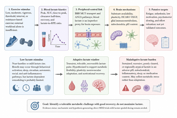
Lactate-window hypothesis linking exercise intensity, peripheral lactate kinetics, peripheral-to-brain communication, brain neurometabolic mechanisms, and putative depression-relevant symptom targets. The figure deliberately avoids displaying symptom improvement as established clinical fact. Instead, fatigue, anhedonia, low motivation, psychomotor slowing, and effort valuation are presented as hypothesized target domains requiring prospective validation. Blood lactate is represented as a dynamic exposure signal defined by peak, AUC, time-to-peak, clearance, recovery, and exploratory lactate-to-RPE index, not as a single static biomarker. The model distinguishes low lactate stimulus, adaptive lactate window, and maladaptive lactate burden. The clinical aim is not to maximize lactate but to identify a tolerable and recoverable metabolic challenge. ANLS: astrocyte-neuron lactate shuttle; AUC: area under the curve; BBB: blood-brain barrier; HCAR1: hydroxycarboxylic acid receptor 1; MCT: monocarboxylate transporter; MDD: major depressive disorder; RPE: rating of perceived exertion; VEGF: vascular endothelial growth factor.
The strongest version of the lactate-window hypothesis must explicitly include a paradox. On one side, exercise-induced lactate and experimentally administered lactate can support substrate availability, synaptic plasticity, BDNF-related signaling, neurovascular remodeling, neurogenesis, and antidepressant-like behavior in preclinical models [34–40, 42–46, 74]. On the other side, depression and neuropsychiatric models can show reduced brain pH and altered lactate levels, suggesting that lactate accumulation may sometimes index metabolic stress, mitochondrial limitation, acid-base disturbance, or abnormal neural activity [13, 19].
A recent human 7T MRS and functional MRI study sharpened this paradox by reporting that higher lactate concentrations in the dorsomedial prefrontal cortex (dmPFC)/dACC were associated with reduced motivation for physical effort, and that plasma lactate correlated with lactate in this cortical region [73]. This finding is important because the dmPFC/dACC is implicated in effort valuation and action selection. However, the result is correlational. It does not prove that lactate causes reduced motivation, nor does it establish that regional lactate has the same meaning during exercise recovery, depressive episodes, inflammatory states, or clinical intervention.
Confounding variables must therefore be considered. Regional lactate may be influenced by task demand, recent activity, sleep, inflammatory state, systemic metabolic health, medication exposure, arousal, anxiety, vascular delivery, mitochondrial oxidative capacity, lactate-pyruvate balance, pH context, and baseline fitness. The same lactate concentration may have different meanings depending on whether it occurs as a transient physiological pulse after tolerable exercise or as persistent accumulation in a stressed or inefficient circuit. This state-dependence is the reason lactate should be modeled dynamically and contextually.
To address ambiguity, the revised model defines the lactate window operationally rather than by a universal blood lactate concentration. A single threshold is unlikely to apply across patients because lactate kinetics are shaped by fitness, sex, age, exercise modality, nutrition, sleep, medication, metabolic disease, anxiety sensitivity, and cardiovascular capacity. The window should instead be defined by the pattern of exposure and recovery.
Low lactate stimulus refers to exercise that produces near-baseline or only mildly elevated blood lactate, usually during low-intensity walking, cycling, stretching, gentle resistance work, or early behavioral activation. This should not be dismissed as ineffective. Low-intensity exercise can reduce depressive symptoms through non-lactate-dominant pathways, including increased routine, improved sleep, circadian entrainment, reduced sedentary behavior, social exposure, autonomic regulation, self-efficacy, and anti-inflammatory effects [3, 58, 66, 67]. For patients with severe fatigue, panic symptoms, low fitness, pain, cardiovascular risk, post-viral fatigue, or poor adherence, low-intensity exercise may be the correct starting point.
Adaptive lactate window refers to a moderate-to-vigorous, threshold-based, interval-based, or resistance-based stimulus that produces a short-lived, tolerable, and recoverable lactate pulse. Proposed operational indicators may include a measurable lactate peak above baseline, limited AUC, a demonstrable post-peak decline across prespecified recovery sampling, acceptable RPE, preserved or improved affective response, no excessive next-day fatigue, stable or improved sleep, and maintained adherence. These indicators are hypothesis-generating rather than validated clinical thresholds. In this zone, lactate may function as a time-dependent metabolic cue that supports substrate availability and plasticity [26–28, 34–40, 74], while HCAR1-mediated and glial-immunometabolic pathways provide additional plausible mechanisms [39, 42–46].
Maladaptive lactate burden refers to a response characterized by excessive peak, prolonged elevation, slow clearance, disproportionate RPE, adverse affective response during exercise, worsened sleep, prolonged next-day fatigue, pain flare, dropout risk, or regional brain accumulation in a context of mitochondrial dysfunction, inflammation, acidosis, poor sleep, medication effects, or low metabolic fitness. In this state, lactate may reflect impaired regulation rather than useful signaling. This does not contradict preclinical antidepressant-like effects of acute lactate; it means that lactate biology is conditional on duration, clearance, region, pH, lactate-pyruvate balance, mitochondrial oxidative capacity, inflammatory state, training status, and depressive phenotype.
The lactate-window hypothesis is valuable only if it generates testable predictions. The model therefore states the following falsifiable predictions.
Prediction 1: In exercise trials for MDD, antidepressant response will correlate more strongly with lactate dynamics, including peak, AUC, clearance, and recovery half-time, than with external exercise dose alone.
Prediction 2: Peak lactate alone will be less predictive than combined indices that include recovery kinetics, RPE, affective response, next-day fatigue, sleep, and adherence.
Prediction 3: Lactate-related benefits will be stronger for fatigue, anergia, motivational anhedonia, psychomotor slowing, and effort-based decision-making than for all depressive symptoms equally.
Prediction 4: Blood lactate kinetics will show stronger relationships with symptom change when central bridging markers, such as MRS-derived regional metabolism or neurovascular indices, indicate adaptive brain response.
Prediction 5: Slow clearance, disproportionate RPE, negative affect during exercise, worsened sleep, or prolonged next-day fatigue will predict poor adherence or weaker symptom improvement, even if peak lactate is high.
Prediction 6: Baseline phenotype will moderate response. Patients with sedentary behavior, fatigue, anhedonia, metabolic dysfunction, or impaired effort valuation may show stronger lactate-linked symptom changes than patients whose depressive symptoms are dominated by non-metabolic dimensions.
Current exercise prescriptions for depression often specify frequency, duration, modality, and target intensity using heart rate, percentage of maximal oxygen uptake (VO2max), or perceived exertion. These parameters are useful but do not fully capture internal metabolic response. A lactate-informed framework would supplement external dose with physiological response. The practical question becomes not only whether a patient completed 30 minutes of exercise, but what metabolic challenge the session produced and how well the patient recovered.
This framework remains a research agenda. It should not be presented as an already validated clinical tool, and it should not be interpreted as recommending high-intensity exercise for all patients with depression. Its translational value is to generate hypotheses about why exercise intensity may matter, why some patients respond poorly to fixed prescriptions, and how future trials could personalize exercise dose using recovery-sensitive biomarkers.
Baseline assessment should include physical activity level, cardiorespiratory fitness, fatigue severity, anhedonia and motivational impairment, sleep quality, medication exposure, metabolic health, cardiovascular risk, pain, anxiety sensitivity, history of post-exertional symptom exacerbation, exercise preference, and feasibility. Exercise could then be progressed toward a tolerable metabolic stimulus rather than a fixed high-intensity target. For some patients, the first therapeutic goal is adherence and behavioral activation. For others, especially those already capable of exercise, the research target may be a controlled near-threshold lactate pulse with rapid recovery.
Figure 2 translates the hypothesis into a prospective research workflow spanning baseline phenotyping, stepwise exercise dosing, serial lactate sampling, response characterization, and multidimensional outcomes.
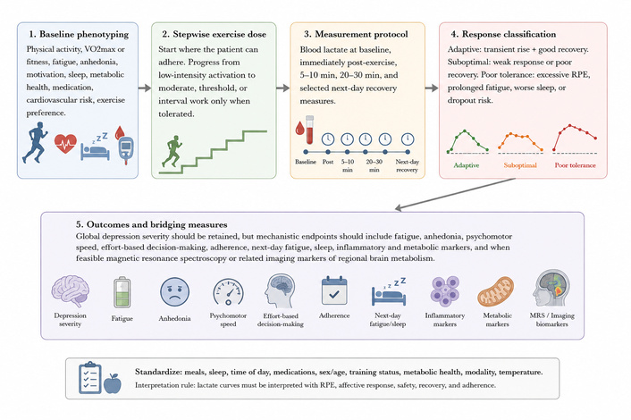
Lactate-informed exercise research framework for depression. The figure reframes lactate-informed exercise prescription as a prospective research framework rather than a validated clinical algorithm. Baseline phenotyping should include activity level, VO2max or cardiorespiratory fitness, fatigue, anhedonia, motivation, sleep, metabolic health, medication, cardiovascular risk, and exercise preference. A candidate serial sampling schedule may include baseline, immediate post-exercise (5–10 minutes and 20–30 minutes), with selected next-day recovery indicators when relevant. Outcomes should include global depression severity but should prioritize mechanistic symptom dimensions such as fatigue, anhedonia, psychomotor speed, effort-based decision-making, adherence, sleep, and functional recovery. Standardization of meals, sleep, time of day, medication, sex, age, training status, metabolic health, exercise modality, temperature, and sampling method is required before clinical interpretation. MRS: magnetic resonance spectroscopy; RPE: rating of perceived exertion; VO2max: maximal oxygen uptake.
The lactate-window hypothesis is unlikely to apply equally across all forms of depression. It may be most relevant to patients with fatigue-dominant depression, anhedonia, low motivation, effort avoidance, sedentary behavior, obesity, insulin resistance, metabolic syndrome, or inflammatory/metabolic signatures [47–58]. In these patients, lactate dynamics might help distinguish deconditioning, poor recovery, and abnormal metabolic tolerance. Combining lactate measures with effort-based decision-making tasks may clarify whether changes in metabolic response track willingness to work for reward.
Safety boundaries are equally important. Evidence on high-intensity exercise in depression and mental illness remains heterogeneous, and adherence barriers are common [68, 69, 75, 76]. As proposed safety considerations for future trials, patients with severe fatigue, panic symptoms, cardiovascular disease, severe obesity, post-viral fatigue, unstable medical illness, chronic pain, low baseline fitness, or high anxiety sensitivity should undergo appropriate clinical screening and stepwise progression rather than default early high-intensity protocols. Low-intensity walking, cycling, or appropriately dosed resistance exercise can be used to establish routine and confidence before progression toward moderate or threshold-based sessions. In severe depression, suicidality, or marked functional impairment, exercise should complement, not replace, standard psychiatric care.
Adherence is a core endpoint, not an afterthought. Barriers to exercise in depression include low energy, impaired motivation, negative affect, fear of failure, discomfort, lack of social support, cost, and difficulty organizing behavior [75, 76]. A lactate profile that looks physiologically interesting but produces aversion, symptom worsening, or dropout has no practical antidepressant value. The success criterion is better metabolic adaptability and sustained participation, not higher lactate.
Future trials should directly test whether lactate kinetics predict antidepressant response and whether lactate-informed progression adds explanatory or clinical value beyond standard exercise prescription. Practical designs could compare low-intensity continuous exercise, moderate-to-vigorous continuous exercise, lactate-threshold-based training, interval training, and resistance circuits while matching or statistically adjusting for session duration, energy expenditure, supervision, expectancy, social contact, and adherence.
Blood lactate should be measured as a curve. Candidate time points include pre-exercise baseline, immediately after exercise, 5–10 minutes post-exercise, 20–30 minutes post-exercise, and selected next-day recovery measures in studies focused on fatigue. Sampling density should be sufficient to capture delayed post-exercise peaks. Prespecified kinetic variables may include peak lactate, baseline-corrected AUC over a defined interval, time-to-peak, clearance indices when sampling density permits, recovery measures, and an exploratory lactate-to-RPE index. Measurements should be standardized for meal timing, carbohydrate intake, hydration, caffeine, sleep, time of day, menstrual cycle or hormonal status when relevant, medication, recent activity, ambient temperature, exercise modality, sampling site, and device calibration.
Clinical endpoints should include global depressive symptom severity, but mechanistically relevant outcomes should include fatigue, anhedonia, psychomotor speed, effort-based decision-making, affective valence during exercise, next-day fatigue, sleep, actigraphy, and functional recovery [47–58]. Adherence should be treated as a core outcome and interpreted with literature on exercise barriers and adherence in psychiatric populations [75, 76]. Candidate biomarker panels may include BDNF, inflammatory markers, metabolic indices, cortisol, mitochondrial measures, and sleep or activity measures, selected according to prespecified hypotheses [14–16, 38, 42–46, 56–57]. When feasible, neuroimaging studies should use MRS or related methods to assess regional brain lactate, pH, glutamate/glutamine, or metabolic signals in regions such as the prefrontal cortex, anterior cingulate cortex, hippocampus, and striatum [31, 59, 62, 73]. However, MRS lactate measurement is technically difficult and should not be treated as a simple clinical readout.
The primary trial question should be explicit: does antidepressant response depend on achieving an adaptive lactate response rather than simply completing a prescribed exercise volume? Secondary questions should test whether lactate dynamics predict changes in fatigue, anhedonia, motivation, psychomotor slowing, or effort avoidance. A negative result would also be informative. If lactate dynamics do not predict symptom changes when measured rigorously, then the lactate-window hypothesis should be revised or rejected.
A conceptual extension involves pharmacokinetic-pharmacodynamic (PK-PD) thinking. Although exercise-induced lactate is not a drug exposure, PK-PD principles provide a useful analogy for distinguishing time-dependent exposure, downstream response, recovery, and interindividual variability. Recent discussion of mechanistic clinical pharmacology emphasizes that PK-PD modeling and dynamic biomarkers can link exposure, target engagement, and downstream biological effects in a translational framework [77]. Applied cautiously, this perspective supports treating lactate as a dynamic exposure signal rather than a static biomarker.
The analogy has limits. Exercise induces multiple simultaneous signals, including temperature, catecholamines, myokines, hemodynamic changes, ventilation, affective experience, and behavioral reinforcement. Peripheral lactate kinetics do not define brain lactate exposure in the way plasma drug concentration might approximate systemic drug exposure. Therefore, PK-PD concepts should be used to improve study design and temporal modeling, not to imply that lactate alone is the active “dose” of exercise.
Several limitations should be stated explicitly. First, this is a narrative mini-review and hypothesis paper, not a systematic review or meta-analysis. Although the manuscript describes a transparent literature search approach, the synthesis remains selective and conceptually driven. Second, much of the mechanistic evidence comes from preclinical, cellular, molecular, or non-depressed human exercise physiology studies. These studies are essential for mechanism building but do not establish clinical efficacy in MDD. Third, direct evidence that blood lactate kinetics predict antidepressant response in patients with MDD is currently insufficient, and prospective validation is required before lactate-informed exercise dosing can be considered superior to or more informative than standard exercise prescription. Fourth, blood lactate is an imperfect proxy for brain lactate exposure. Peripheral kinetics are influenced by muscle metabolism, clearance organs, cardiovascular function, nutrition, sleep, and sampling conditions, whereas brain lactate depends on local neural activity, astrocytic metabolism, MCT expression, vascular delivery, mitochondrial oxidation, and pH regulation. Fifth, neuroimaging measures of brain lactate are technically challenging. MRS sensitivity, field strength, voxel placement, spectral overlap, timing, and motion can limit interpretability. Sixth, depression is heterogeneous. Lactate-related mechanisms may apply mainly to energy-, motivation-, and effort-related symptom profiles rather than to all depressive symptoms. Seventh, exercise tolerance varies widely. High-intensity or lactate-threshold approaches may be inappropriate for patients with severe fatigue, panic symptoms, cardiovascular disease, post-viral fatigue, severe obesity, pain, unstable medical illness, or low adherence. Finally, lactate should be studied as part of a broader physiological feedback system that includes RPE, affective response, sleep, inflammation, metabolic health, and adherence.
Exercise-induced lactate provides a useful framework for linking exercise intensity, neurometabolic adaptation, and depression-relevant symptom dimensions. Lactate is biologically plausible because it connects peripheral exercise metabolism with brain substrate availability, MCT transport, ANLS mechanisms, plasticity-related signaling, HCAR1-VEGF pathways, glial-immunometabolic regulation, mitochondrial oxidation, and pH context. Yet lactate is not inherently therapeutic. Its meaning depends on timing, duration, clearance, regional distribution, metabolic state, training status, inflammatory context, sleep, medication, and depressive phenotype. The lactate-window hypothesis therefore distinguishes low lactate stimulus, adaptive lactate signaling, and maladaptive lactate burden. The adaptive zone is not defined by a universal lactate concentration; it is defined by a transient, tolerable, and recoverable metabolic challenge. The maladaptive zone is characterized by excessive, prolonged, poorly cleared, or regionally atypical lactate in an adverse biological context. This model should currently be understood as a falsifiable mechanistic framework and research agenda, not as a validated clinical prescription tool. Future studies should test whether lactate dynamics predict antidepressant response beyond external exercise dose, whether these relationships are strongest for fatigue, anhedonia, motivation, psychomotor slowing, and effort-based decision-making, and whether central neurometabolic markers bridge peripheral lactate curves with symptom change. The clinical goal is not to maximize lactate, but to identify a safe, individualized, and recoverable metabolic stimulus that may support adaptive brain remodeling and sustained functional benefit.
ANLS: astrocyte-neuron lactate shuttle
AUC: area under the curve
BBB: blood-brain barrier
BDNF: brain-derived neurotrophic factor
cAMP: cyclic adenosine monophosphate
CREB: cyclic adenosine monophosphate response element-binding protein
dACC: dorsal anterior cingulate cortex
dmPFC: dorsomedial prefrontal cortex
HCAR1: hydroxycarboxylic acid receptor 1
HIIT: high-intensity interval training
MCT: monocarboxylate transporter
MDD: major depressive disorder
MRS: magnetic resonance spectroscopy
PET: positron emission tomography
PK-PD: pharmacokinetic-pharmacodynamic
RPE: rating of perceived exertion
SIRT1: sirtuin 1
VEGF: vascular endothelial growth factor
VO2max: maximal oxygen uptake
JK: Conceptualization, Investigation, Writing—original draft, Writing—review & editing. The author read and approved the submitted version.
The author declares that there are no conflicts of interest.
Not applicable.
Not applicable.
Not applicable.
Not applicable.
Not applicable.
© The Author(s) 2026.
Open Exploration maintains a neutral stance on jurisdictional claims in published institutional affiliations and maps. All opinions expressed in this article are the personal views of the author(s) and do not represent the stance of the editorial team or the publisher.
Copyright: © The Author(s) 2026. This is an Open Access article licensed under a Creative Commons Attribution 4.0 International License (https://creativecommons.org/licenses/by/4.0/), which permits unrestricted use, sharing, adaptation, distribution and reproduction in any medium or format, for any purpose, even commercially, as long as you give appropriate credit to the original author(s) and the source, provide a link to the Creative Commons license, and indicate if changes were made.



View: 693
Download: 15
Times Cited: 0
Oluwagbenga Mayowa Adu ... Shukurat Joy Ajenikoko


