 Review


Affiliation:
Molecular Oncology and Angiogenesis Unit, IRCCS Ospedale Policlinico San Martino, 16132 Genoa, Italy
Email: alessandro.poggi@edu.unige.it
ORCID: https://orcid.org/0000-0002-1860-430X


Explor Immunol. 2026;6:1003262 DOI: https://doi.org/10.37349/ei.2026.1003262
Received: April 10, 2026 Accepted: June 25, 2026 Published: August 09, 2026
Academic Editor: Shubhada Chiplunkar, Tata Memorial Centre, Kharghar, India
Human γδ T cells represent a minor subset of lymphocytes present in the peripheral blood. This lymphocyte subset is mainly localized within the mucosae of airways and gut. In the latter context, γδ T cells can represent a key immune cell subset involved both in regulating intestinal homeostasis and in responding to pathogens and colorectal carcinoma (CRC) growth. γδ T cell subsets such as the Vδ2+ respond to phosphate antigens produced by bacteria, while Vδ1+ cells can exert an immune response after mucosal stress stimuli. γδ T cells do not recognize as classical αβ+ T cells the peptide antigens in the context of major histocompatibility complex (MHC). γδ T cells may play a complementary role with αβ+ T cells in mucosal immunity at the gastrointestinal barrier. Colon γδ T cells can exhibit antitumor properties and regulatory functions. Indeed, human γδ T cell subsets present in the gut bear some activatory receptors, such as NKG2D and DNAX Accessory Molecule (DNAM)-1, leading to the elimination of CRC cells. By contrast, γδ T cells producing interleukin (IL)-17, transforming growth factor β, and amphiregulin show pro-tumor activity. This dual property of γδ T cells poses challenges for their use as an immunotherapeutic tool, while the MHC-independent recognition of antigens can support their use as off-the-shelf allogeneic cells.
The molecular features of the T cell receptor (TCR) of γδ T cells were identified in the mid-1980s (roughly 1984–1987) by several investigators, before the identification of lymphocytes bearing this TCR [1–4]. These peripheral blood lymphocytes expressing a putative alternative TCR lacked CD4 and CD8 molecules. This discovery suggested that γδ T cells could exhibit diverse antigen-recognition capabilities independently of the co-stimulatory function of CD4+ and CD8+ accessory molecules. Currently, γδ T cells are considered a key lymphocyte subset that is highly abundant in the airways, gastrointestinal mucosa, and skin [5–7]. This localization clearly indicates the role of γδ T cells in patrolling interfaces between the host and infectious agents, such as bacteria and viruses [5–7]. It is well established that γδ T cells exhibit characteristics of both innate and adaptive immunity [8]. Indeed, they express many receptors that sense pathogen-associated molecular patterns (PAMPs) and danger-associated molecular patterns (DAMPs) and are involved in innate immune activation, as well as a TCR that recognizes non-peptide antigens in an MHC-independent manner [8]. Indeed, γδ T cells can recognize phosphoantigen (pAg) such as (E)-4-hydroxy-3-methyl-but-2-enyl pyrophosphate (HMB-PP), isopentenyl pyrophosphate (IPP), heat shock protein (Hsp), and lipid antigens presented in the context of CD1d molecules [8].
Peripheral blood-derived γδ T cells are usually subdivided into Vδ1 and Vδ2 subsets (Figure 1) that can show migratory properties leading to their localization in mucosal tissues and skin [4, 5]. Furthermore, γδ T cells express numerous natural killer (NK) human leukocyte antigen (HLA) class I receptors [9]. These receptors recognize certain HLA-I allele subgroups on healthy and tumor cells and can deliver a negative signal that inhibits γδ T cell activation [9, 10]. The specific localization as intraepithelial cells within the gastrointestinal mucosa suggests a key role for γδ T cells in responding to tumor cell growth in this region [7, 8, 11]. Furthermore, the potential use of γδ T cells as effectors for adoptive immunotherapies has been initially considered in some ongoing clinical trials. Herein, we summarize and discuss recent advances in characterizing γδ T cells in colorectal carcinoma (CRC), and their current and future use as immunotherapeutic tools to address the most aggressive tumors.
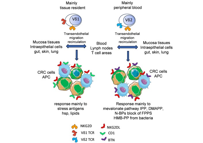
Peripheral blood γδ T cell subsets with putative roles in the control of CRC cell growth. Human peripheral blood γδ T cell subsets can be identified because of the expression of specific Vδ chains as Vδ1 (the minority) or Vδ2 (the majority) T cells. It appears that γδ T cell subsets are localized mainly in different tissues, and they may be recruited from the peripheral blood using different adhesion molecules and molecular mechanisms to cross the endothelium and penetrate the tissue matrix. Several chemokines and adhesion molecules are involved in the homing and recirculation of γδ T cells. Importantly, the Vδ1 T cells can respond with TCR to antigens such as heat shock protein (Hsp) or lipids expressed in the context of the CD1 molecule on tumor cells and through NKG2D interacting with the NKG2DL overexpressed by stress stimuli on CRC cells. On the other hand, the Vδ2 T cells respond to small antigen pyrophosphate (pAgs) presented by tumor cells in association with butyrophilin (BTN) molecules. The pAgs can derive from commensal bacteria or from the mevalonate pathway. Note that Vδ2 T cells can respond, like Vδ1, to NKG2DL, but Vδ1 T cells do not respond to pAgs. CRC: colorectal carcinoma; APC: antigen presenting cell. Several other molecules are involved in γδ T cells-tumor cells cross-talk (see next chapters of this review).
γδ T cells are present in the small and large gastrointestinal tracts [10–15], and they interact with gut microbiota at different anatomical sites, such as the epithelial layer, lamina propria, and Peyer’s patches [15]. The most important feature of gut γδ T cells is the expression of the CD8αα homodimer, in contrast to the absence of CD4 and CD8 in peripheral blood γδ T cells. This suggests that γδ T cells present in the gut may respond to microbiota antigens using the CD8 accessory molecules, thereby maintaining gut homeostasis [15–20]. In mice, gut γδ T cells are predominantly Vγ5 TCRs, whereas in humans they are Vδ1Vγ2+ TCRs [21–23]. It has been shown that γδ intraepithelial lymphocyte (IEL) can release antimicrobial agents such as interferon (IFN)-γ upon cross-talk with intestinal epithelium. These γδ-IEL can protect epithelial cells from invasion by resident bacteria, particularly during initial interactions with microorganisms, suggesting that γδ-IEL are a key component of maintaining intestinal microbiota homeostasis. The key biological significance of γδ-IEL and αβ-IEL has been shown in mice. The localization of murine Vγ5 T cells is associated with the expression of CCR9 and integrin αEβ7, the CCR9 ligand CCL25, and E-cadherin, which is expressed by epithelial cells, thereby favoring their localization in the gut and differentiation in lymphoid tissues [24–30]. Importantly, the integrin αE expression is regulated by transforming growth factor beta (TGF-β) and RUNT related transcription factor 3 (RUNX3) [24, 25]. The use of antibodies against α4β7 or mucosal addressin cell adhesion molecule (MAdCAM)-1, or β7-deficient mice, strongly proposes that the homeostatic recruitment of lymphocytes to the gut epithelium is strictly linked to β7 integrins. Indeed, the development of the gut-associated lymphoid tissue (GALT) is impaired in mouse models of β7–/– lymphocytes. This indicates that the absence of this integrin limits lymphocyte extravasation and localization in gut tissues, such as the lamina propria and IEL [26–33]. Importantly, αEβ7 improves the retention of effector and memory lymphocytes within epithelial cells and interacts with E-cadherin and mucosal dendritic cells (DCs) [33] similarly to the integrin alpha4beta7 [34, 35]. It is of note that mucosal gut DCs can express retinal dehydrogenase (RALDH) involved in the metabolism of retinoic acid (RA). RA deficiency in mice can reduce α4β7 integrin expression on effector cells. In contrast, this deficiency does not affect the expression of E-selectin or L- or P-selectin ligands on effector cells, nor does it influence skin tropism [34]. Furthermore, the pharmacological inhibition of RALDH with citral and the RA receptor antagonist LE135 can impact the gut-tropism of lymphocytes [34]. In addition, it has been reported that only a subset of murine and human DCs expresses the enzymes (RDH10 and RALDH2) and the cellular transporter (CRABP2) required for all-trans RA (ATRA) to elicit an effective signal. Furthermore, this molecular mechanism appears to be regulated by the fatty acid-sensing nuclear receptor peroxisome proliferator-activated receptor γ (PPARγ) [35]. Altogether, these findings strongly support the idea that IELs are localized and retained within the gut mucosa by a complex network of adhesion molecules and soluble factors dependent on the presence of hormones such as RA. Pharmacological regulation of RA-mediated signaling may modulate the number and function of effector T cells and, consequently, the extent and nature of a mucosal immune response.
Tissue-resident immune cells, including γδ T cells, have evolved phenotypic and functional features that enable stable tissue residence without recirculation [36]. Some of these cells differentiate within the tissue from immature precursors; for example, γδ T cells mature in the tissue without passing through the thymus for MHC-related selection. However, the definitive contribution of extrathymic IEL development remains controversial [36–38]. The γδ T cells expressing chemokine receptors for epithelial-derived chemokines, such as CCL20 and CXCL16, can migrate to mucosal tissues [39–41]. In addition, some cytokines favor the localization and retention within intestinal tissue. The role of transforming growth factor (TGF)-β in regulating integrin expression has already been mentioned [24, 25]. Moreover, interleukin (IL)-7 and IL-15 contribute to the differentiation, survival, and functional maturation of γδ T cells in the intestinal tract [15, 40, 41]. IL-15 can be expressed on the cell surface of intestinal epithelial cells (IECs) and lamina propria DCs. The presentation of IL-15 by IECs to γδ T cells is essential for the migration and localization of this lymphocyte subset in the intestine, as demonstrated in both in vitro and in vivo experiments [16]. Similarly, IL-7 administration in adult C57BL/6J mice significantly affected IEL phenotype and function, and close interaction between IEL and IEC is associated with IL-7 expression in IEC [42]. Involved in the location of γδ T cells in the intestine are also the aryl hydrocarbon receptors (AhRs) and orphan G-protein coupled receptors GPR18 and GPR55 [43–45]. The AhR is a transcription factor that is a sensor of xenobiotic chemicals, such as aromatic compounds, influencing enzymes involved in their metabolism [45]. In detail, AhR deficiency or the absence of AhR ligands impacts IEL maintenance and the composition and load of the microbiota. This favors the increase of immune activation, dominated by a Th1 pro-inflammatory response, leading to damage to intestinal epithelial cells. Importantly, the absence of AhR does not impact the number of general lymphoid populations or their ability to develop, home to their target organ, or proliferate. However, in IELs without AhR, mice lacking one or both AhR alleles are no longer localized to the intestine; instead, they migrate away (more than 95% of γδ T cells were absent from the intestine). The AhR ligands are derived from cruciferous vegetables. Typically, AhR expression was higher in TCR Vγ5 skin and in TCR Vγ7 intestinal γδ T cells, respectively. Consistent with the reduction of IELs in AhR-deficient mice, intestinal epithelial turnover was reduced compared with that of control mice. The adoptive transfer of AhR+ γδ T cells into AhR-deficient mice induced EC proliferation, suggesting a direct link and interaction between γδ T cells and IECs.
Furthermore, the absence of AhR IEL was associated with reduced expression of granzymes, C-type lectins, and matrix metalloproteinase (MMP)-7, possibly indicating diminished control of the intestinal microbial load in mice with reduced or absent AhR activity. Thus, AhRs regulating the cytochrome P450 further influence the immune response to external stimuli [45]. How AhR-mediated signals maintain IELs at epithelial sites is currently unknown and is the subject of further investigation.
The presence of γδ T cells in the gastrointestinal tract should be considered when looking at homeostatic maintenance, pro-inflammatory diseases, and neoplastic transformation. Thus, the biological role of γδ T cells in the gut should be analyzed keeping in mind that this lymphocyte cell population shares several features with innate and adaptive immune cells, as already mentioned [15, 30, 41]. Herein, we will focus our attention on the γδ T cells present in CRC, while the role and significance of γδ T cells in inflammatory bowel diseases have been reviewed elsewhere [46, 47].
The detailed analysis of the phenotypic and functional characterization of γδ T cells present in CRC can provide a scenario to understand the biological significance of this lymphocyte subset in this disease. This analysis can be performed by immunohistochemistry (IHC), flow cytometry, and molecular single-cell mRNA techniques [11, 48–54]. The cross-talk of γδ T cells with the diverse components of the tumor microenvironment (TME) is the context in which the engagement of specific receptors on γδ T cells may lead to pro-tumor and anti-tumor effects [52–55]. Schematically, the γδ T cells producing IL-17 represent the main subset with pro-tumor effects, while γδ T cells have an antitumor effect through tumor cell elimination [56–58]. This last effect is mediated by the release of perforins and granzymes, surface and soluble death receptors such as Fas and tumor necrosis factor (TNF)-related apoptosis-inducing ligand (TRAIL), and triggering of antibody-dependent cellular cytotoxicity (ADCC) [30, 39, 41, 59]. IL-17-producing γδ T cells are usually Vδ1, whereas killing properties are shared by both Vδ1 and Vδ2 T cells [30, 39, 41, 59]. However, this skewed subdivision is instrumental in trying to understand the complex interactions between the TME and the immune system. IL-17 is a pleiotropic cytokine that has pro-inflammatory effects linking the innate and the adaptive immune response, favoring the recruitment of neutrophils, and its dysregulation can provoke some autoimmune diseases [57]. In principle, the triggering of autoimmunity should be considered as a positive event in the context of a tumor [60, 61]. Indeed, the anti-tumor therapy with the so-called immune checkpoint blockers (ICB) can induce the exacerbation or appearance of autoimmune reactivity and diseases as an undesired side effect in association with an optimal antitumor response [62, 63]. It has been demonstrated that Vδ1 T cells can recruit myeloid cells in the tumor [58]. These cells can differentiate in the TME as myeloid-derived suppressor cells (MDSCs), inhibiting the activation of CD8+ T cells through many inhibitory mechanisms, such as arginase 1 (ARG-1) and indoleamine 2,3-dioxygenase (IDO) [64, 65]. Notably, IDO expression is triggered by pro-inflammatory cytokines such as IFN-γ [66, 67]. On the other hand, IFN-γ can increase the expression in tumor cells and tumor-associated fibroblasts/mesenchymal cells of the programmed cell death protein ligand 1 (PD-L1) ligand of programmed cell death protein 1 (PD-1) [68–70]. The interaction between the PD-1 on cytotoxic T lymphocytes (CTLs) and PD-L1 present on the TME leads to further inhibition of CD8+ T cell antitumor activity. In conclusion, the IL-17 produced by Vδ1 T cells can strongly impair the adaptive response against tumors [58].
Although reported in breast adenocarcinoma and prostate cancer, instead of CRC, tumor-infiltrating Vδ1 T cells exerted a potent suppressor function on DC maturation and function controlled by the TLR8 signaling pathway. Indeed, the use of Poly-G3 and ssRNA40 as ligands for TLR8 expressed on Vδ1 reversed their suppressive functions. These findings suggest that innate ligands can shape the suppression activity of Vδ1, further complicating the cross-talk among the different components of the TME [71].
Rodin and coworkers [51] used mass and flow cytometry, mRNA quantification, and TCR sequencing to show that the composition of γδ T cells in the macroscopically healthy and CRC mucosa is heterogeneous among patients, and it is composed of Vδ1, Vδ2, and non-Vδ1-Vδ2 γδ T cells. This double-negative cell population expresses mainly the Vδ3 gene and minority Vδ4, Vδ5, Vδ7, and Vδ8 TCR genes [51]. Vδ2 T cells paired with Vγ9 mainly, although some Vδ2 T cells were Vγ9 negative. Overall, this analysis of TCR repertoire indicates a wide variety of γδ T cells in the CRC among the diverse patients. The key feature in CRC, compared to healthy colon mucosa, is the detection of an increase in non-Vδ1-Vδ2 T cells expressing Vδ3. However, in the relatively small group of patients analyzed (n = 49), there was no statistically significant correlation between the proportion of non-Vδ1-Vδ2 cells and microsatellite status, grade of differentiation, stage, location (right vs. left-sided), or patient age.
The mass spectrometry analysis indicated that peripheral blood and CRC mucosa γδ T cells formed distinct clusters. Furthermore, Vδ1 and Vδ2 cells express markers associated with cytotoxic effector functions. On the other hand, the non-Vδ1-Vδ2 cells appeared to express genes for proteins with tumor-promoting functions, such as neutrophil-recruiting chemokines, Galectin 3, and transforming growth factor-beta [51]. Importantly, the functional analysis of non-Vδ1-Vδ2 cells (mainly Vδ3) indicated that these cells express lower levels of IFN-γ and TNF than Vδ2 cells. Furthermore, IL-17 was expressed in non-Vδ1-Vδ2 cells in line with what was reported by other authors [72] and in murine systems [54, 73]. Altogether, these findings suggest that subsets of γδ T cells may play different roles in the CRC TME. It should also be noted that the number of γδ T cells present in the CRC mucosa is limited compared to other T cells, such as CD4+ Th17 [74]. This finding would indicate that CD4+ Th17 may have a preeminent role in pro-tumor effects compared to γδ T cells expressing IL-17. Importantly, the intraepithelial, but not stromal, localization of IL-17-producing CD4+ T cells positively correlated with improved survival [74]. Furthermore, IL-17 may trigger CRC angiogenesis [75]. On the other hand, IL-17 may recruit cytotoxic CD8+ T lymphocytes into the tumor [74, 76]. These findings indicate that it is not correct to assign a specific pro- or anti-tumor activity to IL-17. IL-17-producing cells may show different effects on the growth of CRC tumors depending on their localization, frequency, and intrinsic features.
It has been reported in CRC that high levels of the TRDV1 gene were not associated with a positive prognosis. Furthermore, gene expression correlation indicated that γδ T cells were more closely related to NK and innate lymphoid cells (ILCs) than CD4+ and CD8+ T cells. The Vδ1 T cells expressed low levels of some inhibitory receptors, such as PD-1, LAG-3, and T-cell immunoglobulin and mucin-domain containing-3 (TIM-3), but were positive for T cell immunoreceptor with Ig and ITIM domains (TIGIT) [72]. Importantly, the Vδ1 T cells in CRC can produce IFN-γ or amphiregulin (AREG). AREG is an EGF-family ligand that plays a key role in epithelial cell proliferation, differentiation, and tissue regeneration via the epidermal growth factor receptor (EGFR)/mitogen activated protein kinase (MAPK) signaling pathway. This factor was found to be associated with microsatellite stability (MSS) compared to microsatellite instability (MSI) and the consensus molecular subtype 2 (CMS2) of CRC or a wound-healing phenotype of CRC. It is of note that γδ T cells were the only leukocyte population increasing the expression of AREG in CRC compared to healthy tissue. In addition, the AREG produced by Vδ1 T cells can trigger the proliferation of the two CRC cell lines SW480 and HCT116, as well as migration in a wound healing assay. These effects were inhibited with the anti-EGFR antibody cetuximab (Cet), which in turn blocks the interaction of AREG with the EGFR. It is of note that some Vδ1 T cells present in CRC can produce both AREG and IFN-γ or IFN-γ only. This would suggest that Vδ1 in the TME can show pro- and/or anti-tumor features. It is of note that the use of IL-15 can downregulate the production of AREG, increasing that of IFN-γ by Vδ1 T cells, while IL-1β is a potent stimulator for AREG production. These findings indicated that the cultivation of tumor-infiltrating and peripheral blood Vδ1 T cells with some specific cytokines can skew the type of Vδ1 T cells. Indeed, the possible use of Vδ1 T cells for antitumor therapy should avoid the presence of Vδ1 T cells producing AREG and favoring the growth of the tumor, and this can be accomplished with cultivation in IL-15 [72]. These considerations may explain the heterogeneity of function of Vδ1 T cells in CRC and highlight the role of the cytokine microenvironment in determining the properties of infiltrating cells. In the cohort of CRC analyzed, the Vδ2 T cells were reduced in CRC mucosa compared to the healthy one, and some of them can produce AREG, although the majority produced IFN-γ and a minority both AREG and IFN-γ. This would suggest that Vδ2 T cells may produce AREG, and this property should be considered for adoptive immunotherapy.
However, not all reports are in line with the apparent role as suppressor/pro-tumor cells of Vδ1 T cells. Indeed, the expression of the activating receptor NKp46 identified the largest fraction of IEL subset [77]. Importantly, the gut tropism and the ontogeny of these cells are linked to the ontogeny and gut-tropism of NKp46+ Vδ1 IELs depends on gut-environmental factors and some specific features. Low frequencies of the NKp46+ Vδ1 T cells in healthy gut specimens of patients affected by CRC were associated with faster tumor progression and development of metastatic diseases. These contrasting results with the other reports above can be explained by the different technical approaches, reagents, and gene or protein level to analyze the IL-17+ T cells as well as the intra- and inter-donor heterogeneity (IDH) of CRC tumors.
Besides Vδ1 T cells, the Vδ2 T cells have been described as important anti-CRC effector cells associated with a better prognosis in MSI CRC [78, 79]. Indeed, using a deconvoluted CIBERSORT matrix to investigate the abundance of Vγ9Vδ2 T cells, it has been reported that CRC with a high infiltration of this cell subset had better survival than those with a low infiltration. Importantly, it appears that a better overall survival (OS) was also associated with αβ T cells. In this case, a clear enrichment in gene sets for “response to IFN-α”, “inflammatory response”, “antigen processing and presentation”, “T cell activation”, and “cytolytic activity” was found with αβ T cells but not with γδ T cells. These findings suggest that the infiltration with either αβ or γδ T cells can always lead to a better anti-CRC response, but in a different immunological context.
Looking at what is described in this review regarding Vδ1, Vδ2, non-Vδ1-Vδ2, and Vδ3 lymphocytes, it appears that some subsets of CRC mucosa γδ T cells may have protumor and antitumor effects (Figure 2).
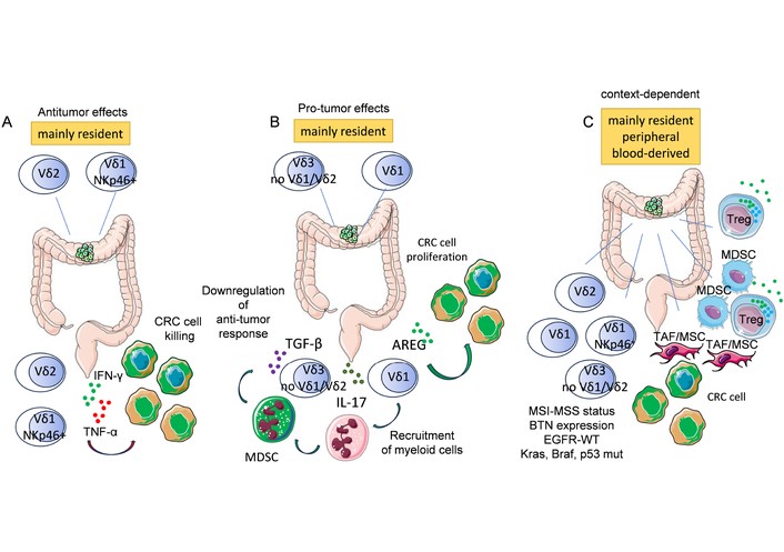
γδ T cell subsets and their putative roles in the CRC tumor microenvironment (TME). Human γδ T cell subsets present in the gut can be identified because of the expression of specific Vδ chains of the γδ TCR, including Vδ1, Vδ2, Vδ3, and non-Vδ1/Vδ2. It appears that γδ T cell subsets are localized mainly in different anatomical sites such as intraepithelial cells, lamina propria cells, and lymphocytes associated with lymphoid aggregates. γδ T cells may also be recruited from the peripheral blood, as shown in Figure 1. A. Vδ1 and Vδ2 colon resident T cells can show potent antitumor properties by killing CRC cells and producing pro-inflammatory cytokines such as IFN-γ and TNF-α. B. On the other hand, resident Vδ1, Vδ3, and non-Vδ1/Vδ2 T cells can produce TGF-β, IL-17, and amphiregulin (AREG). These cytokines exert various pro-tumor effects. Indeed, TGF-β is a potent immunosuppressor of antitumor immune response; IL-17 can recruit myeloid cells into the tumor, and this event favors their differentiation into myeloid-derived suppressor cells (MDSCs). AREG is a ligand of the epidermal growth factor receptor, leading to proliferation of CRC. C. The subsets of γδ T cells can exert their anti-tumor or pro-tumor effects depending on the TME composition and the molecular/phenotypic features of CRC cells. Typically, some subgroups of microsatellite stability (MSS) CRC show an immunosuppressive milieu composed of MDSCs, tumor-associated fibroblasts (TAFs) or mesenchymal stromal cells (MSC), regulatory T cells (Treg), and immunosuppressive cytokines. This TME favors the presence of γδ T cells producing IL-17 and other immunosuppressive factors, leading to tumor escape. The presence of BTN and pAgs on CRC can stimulate the response of Vδ2 T cells. Microsatellite instability (MSI) CRC can show a greater immune infiltration than MSS, together with a less immunosuppressive TME. This can favor the antitumor immune response. Furthermore, some mutations of oncogenes such as Kras and Braf or oncosuppressor genes such as p53 can influence CRC proliferation. The therapeutic approach is different depending on the presence of WT or mutated EGFR-mediated biochemical pathways. Overall, γδ T cells can play opposite roles in CRC only partially related to the expression of a specific TCR Vδ chain.
The pro-tumor effects are associated with IL-17 expression in mucosal Vδ1/Vδ3 T cells, while anti-tumor effects are associated with Vδ2 T cells. This clear-cut subdivision is challenged by what has been recently reported by Ran and colleagues [49]. These authors analyzed nine published gene datasets of single-cell RNA from 18,483 γδ T cells out of 951,785 total cells from the neoplastic or adjacent healthy tissue of 165 human CRCs. The bioinformatics analysis revealed that γδ T cells overall exhibited a cytotoxic-related transcriptional pattern in both CRC and healthy mucosa. Unexpectedly, none of the γδ T cell clusters showed an IL-17-related pattern, including master transcription factor RAR-related orphan receptor gamma (RORC), IL-23R, CCR6, and MAF, suggesting the inability to produce IL-17. Different subsets of γδ T cells could be defined as effector, tissue-resident memory, progenitor exhausted, and exhausted cells, and the authors noted an increased expression of cytotoxic molecules [49]. To identify γδ T cells from unsorted whole tissue, the combination of CD3D, CD3E, CD3G, CD247, and TRDC was considered as the minimal set of markers to be applied [49]. Overall, these findings propose that datasets from mice or humans may differ in their typical features for identifying specific T cell subsets or that the purity of selected lymphocyte subsets may be insufficient for single-cell analysis, potentially yielding misleading results [49, 80].
Notably, γδ T cells infiltrating different types of tumors do not show the exhausted functional behavior typical of CD8+ TCR αβ T cells, although they express several immune checkpoint molecules [81, 82]. Furthermore, it is to be noted that there are many possible explanations for these inconsistent findings. Firstly, the methods of detecting IL-17-producing cells may have different sensitivities, as well as detecting a different step for showing the relevance of IL-17. Typically, single-cell RNA sequencing (sc-RNA-seq) can detect tiny cell subsets comprised in the whole cell population analyzed, as in the case of γδ T cells. The mRNA presence does not mean the protein presence, and on the other hand, the antibodies used in IHC and flow cytometry (FC) can lack the good avidity and affinity for the low amount of IL-17 present in the CRC TME. The isolation procedures to analyze either sc-mRNA or IHC/FC can select some subsets of a given population, thus leading to misinterpretation of data. Finally, the huge amount of data coming from omics analyses needs well-established and controlled pipelines. To avoid any misleading message, it would be necessary to validate each other’s data obtained with advanced molecular or more classical protein-based approaches.
Table 1 is a summary of the main features of γδ T cells in healthy colon and CRC both in mice and humans, to point out the experimental model and the major differences among γδ T cells in these two species.
Main features of γδ T cells in humans and mice with pro- or anti-tumor activity.
| γδ T cell population | Species | Model study | Main features | Molecules on lymphocytes | Molecules/Factors on epithelial cells | Ref. |
|---|---|---|---|---|---|---|
| Vγ5 | Mouse | Healthy colon β7–/– mice mAb to α4β7 or MAdCAM-1 RALDH deficiency | Impaired localization | CCR9, αEβ7 | CCL25, E-cadherin | [24, 25] |
| γδ T cellsVγ7+ | Mouse | Healthy colon, AhR deficient alleles | Impaired localization | Granzymes, C-type lectins, GPR18, GPR55 | IL-7, IL-15, MMP-7 | [43–45] |
| non-Vδ1-Vδ2Vγ7Vγ6 | Mouse/Human | Suppressor antitumor cells | CD8α+ PD-1− | [54, 73] | ||
| Vδ1Vγ2+Vδ1 | Human | CRC, FC, IHC | IL-17 producer, MDSC recruitment | ARG1–2, IDO | [48, 50, 52–55] | |
| Vδ1 in breast and prostate cancers | Human | FC, regulatory activity in vitro and in vivo NOD SCID mice | Suppressor cellsIKKα, IKKβ mediated | TLR11, TLR7, TLR8, MyD88 | TLR8L Poly-G3ssRNA40Reversed suppression | [71] |
| Vδ1 | Human | CRC, healthy mucosa, FC, TCR seq, IHC, functional in vitro experiments | Potent cytotoxic cellsIFN-γ | NKp46+ NKp44+ NKp30+ NKG2C+NKG2D CD8α+ CD56 CD16low | Ligands for activatory receptorsIL-2IL-15 | [78] |
| non-Vδ1-Vδ2Vδ1, Vδ4Vδ5, Vδ7Vδ8 | Human | Humanized CRC, MS, FC, TCR seq | TGF-β, Galectin 3, AREG | Low production of IFN-γ | No correlation with stage, MSS or MSI | [51, 72] |
AhR: aryl hydrocarbon receptor; αEβ7: integrin αE integrin β7; ARG1–2: arginase 1, 2; CCL25: chemokine (C-C motif) ligand 25; CCR9: CC chemokine receptor type 9; FC: flow cytometry; GPR18: G-protein coupled receptor 18; GPR55: G-protein coupled receptor 55; IDO: indoleamine 2,3-dioxygenase; IHC: immunohistochemistry; IKKα: inhibitor of nuclear factor-κB (IκB) kinase α; IKKβ: inhibitor of nuclear factor-κB (IκB) kinase β; MAdCAM: mucosal addressin cell adhesion molecule; MDSC: myeloid derived suppressor cells; MMP-7: matrix metalloproteinase 7; MS: mass spectrometry; MSI: microsatellite instability; MSS: microsatellite stability; Poly-G3: a guanosine-rich, phosphorothioate-modified oligodeoxynucleotide; NOD-SCID: non obese diabetic severe combined immunodeficiency; RALDH: retinal dehydrogenase; ssRNA40: GU-rich single-stranded RNA; TCR-seq: T cell receptor sequencing; TGF: transforming growth factor; TLR8L: toll-like receptor 8 ligand.
Immunotherapy of cancer is characterized by the attempt to trigger the immune response of the patient against the tumor cells and the TME [81–83]. A possible advantage of using γδ T cells for immunotherapy is the ability of this cell population to recognize tumor cells independently of the presence of HLA-I antigens [84, 85]. Importantly, it should be pointed out in evidence that γδ T cells can express several HLA-I receptors typical of innate cells, such as NK cells [86–89]. The mainstream opinion on this point is that γδ T cells express inhibitory receptors for HLA-I, such as killer-cell immunoglobulin-like receptor (KIR) and C-type lectin inhibitory receptor (CLIR) [86–89]. These receptors linking the corresponding HLA-I antigen allele expressed on self-cells, should deliver, in principle, an inhibitory signal that can impair the activating signals delivered through the activating receptors of γδ T cells [86, 89]. To clarify this scenario better, it is necessary to describe in detail the features of both inhibitory and activating receptors on γδ T cells [90–92]. In addition, it is conceivable that suitable numbers of γδ T cells can be obtained by culturing peripheral blood mononuclear cells (PBMC) [10, 30, 40, 93].
The frequency of γδ T cells is usually low in the PBMC, ranging from 1–10% roughly. The two main subsets in humans are represented by Vδ2 (the majority) and Vδ1 T cells (the minority). To generate a good enough number of γδ T cells from PBMC, it is necessary to expand them using appropriate culture conditions [94, 95]. Ideally, the use of an antigen could be a better means to achieve this aim. It is unclear whether γδ T cells can respond to specific polymorphic antigens; at least they do not respond to classical antigen peptides presented by antigen presenting cells (APCs) as αβ T cells [30, 41, 96, 97]. The γδ T cells recognize monomorphic molecules that function as the superantigens for some groups of αβ T cells [98, 99]. Among the molecules recognized by the TCR of γδ T cells are microbial metabolites such as HMB-PP, isoprenoid precursors such as IPP or dimethylallyl pyrophosphate (DMAPP), Hsps (Hsp60 in humans and Hsp65 in mice, Hsp70, and Hsp90), and lipid antigens presented in the context of CD1d molecules [100–109] and other specific molecules among which EphA2 [97, 110–117] (Table 2).
Antigens recognized by human γδ T cells and main molecules involved in CRC cell-γδ T cell interaction.
| Localization | γδ TCR main activatory molecules | TCR ligand | Main molecules involved in presentation/activation | Putative molecules involved in inhibition |
|---|---|---|---|---|
| Blood | Vγ9Vδ2NKG2DDNAM-1 | HMB-PP, IPP, DMAPP TGM1hMSH2 | BTN2A1, BTN3A1 MR1 MICA/MICB ULBP1-6 | PD-1 TIGITTIM-3 |
| Blood | Vδ1 NKG2DDNAM-1 | Hsp lipids | CD1a, b, dMICA/MICB ULBP1-6 | PD-1 TIGITTIM-3 |
| Intraepithelial | Vγ4Vδ1 NKG2DDNAM-1 | BTNL3, BTNL8 | MICA/MICB ULBP1-6 | PD-1 TIGITTIM-3 |
| Lamina propria | Vδ1 Vγ9Vδ2NKG2DDNAM-1 | HspLipidspAg | IL23 MICA/MICBULBP1-6 | PD-1 TIGITTIM-3 |
| Tumors, CRC, medulloblastomaTissue resident | Vγ9Vδ1 | EphA2 | MICA/MICB ULBP1-6 | PD-1 TIGITTIM-3 |
| TumorsTissue resident | Vγ8Vδ3Non-Vγ9Vδ2 | MR1Annexin A2 | PD-1 TIGITTIM3 | |
| Tumors (kidney) | Vγ4Vδ5Non-Vγ9Vδ2 | EPCRAnti-CMV response | CD16 | PD-1 TIGITTIM-3 |
BTN: butyrophilin; CMV: cytomegalovirus; CRC: colorectal carcinoma; DMAPP: dimethylallyl pyrophosphate; EPCR: endothelial protein C receptor; EphA2: ephrin type-A receptor 2; HMB-PP: (E)-4-hydroxy-3-methyl-but-2-enyl pyrophosphate; hMSH2: human MutS homologue 2 (DNA mismatch repair enzyme); Hsp: heat shock protein; IPP: isopentenyl pyrophosphate; MICA: MHC class I chain-related protein A; MR1: MHC class I-related protein 1; pAg: phosphoantigen; PD-1: programmed cell death protein 1; TCR: T cell receptor; TGM1: protein-glutamine γ-glutamyl transferase K; TIGIT: T cell immunoreceptor with Ig and ITIM domains; TIM-3: T-cell immunoglobulin and mucin-domain containing-3; ULBP1-6: UL-16 binding proteins 1-6.
It is well known that human Vδ2+ T cells can be expanded using IPP or DMAPP. These phosphate antigens (pAg) are intermediates of the cholesterol synthesis that can be produced in epithelial cells, monocytes, and mesenchymal stromal cells (MSC) [118–120]. These pAgs can trigger both the activation and consequent proliferation of Vδ2+ T cells as well as the triggering of cytolysis directed to the pAg-producing cells or the production and release of pro-inflammatory cytokines such as IFN-γ and TNF-α [121, 122]. For presentation, some molecules belonging to the butyrophilin (BTN) family are necessary [123–126]. In particular, it has been demonstrated that BTN3A1 and BTN2A1 are the cell surface ligands presenting pAgs undergoing structural rearrangement. Specifically, BTN2A1 binds the Vγ9 domain, and BTN3A1 binds the Vδ2 and Vγ9 chains of the Vγ9Vδ2 TCR, rapidly triggering activation [123–126].
Conceivably, the stimulation of Vγ9Vδ2 T cells with pAgs will take place when BTN2A1 and BTN3A1 are expressed [120, 127–129]. Thus, the two components of the stimulatory armamentarium should be present: pAgs and BTN [124]. Practically, Vγ9Vδ2 T cells can be expanded from unseparated PBMC by adding IPP to the culture medium and exogenous IL-2 [118, 130]. This system, which needs the use of a purified chemical compound such as IPP, can be easily substituted by culture with micromolar concentrations of aminobisphosphonates (N-BPs) such as zoledronic acid (ZA) or risedronic acid (RIS) [131–134]. The N-BPs can interfere with cholesterol biosynthesis, inhibiting the enzyme farnesyl pyrophosphate synthase (FPPS) in the mevalonate pathway, preventing the prenylation of small GTPases. The effects of N-BPs are mainly of two types: an increase in the intracellular content of IPP and DMAPP, together with interference with the cytoskeleton arrangements by affecting the activation of Rho, Rac, and Cdc42 proteins [135–137]. Starting from unseparated PBMCs with a low percentage of Vγ9Vδ2 at the onset of the culture, the use of ZA and the addition of low doses of IL-2 (30 IU/mL) can induce a strong proliferation of Vγ9Vδ2. This can reach 60–90% of the culture after 10–12 days and more than 95% on day 14. This can be an easy method to obtain enough Vγ9Vδ2 for making immunotherapy. The expansion of Vγ9Vδ2 can be triggered using other cytokine cocktails containing IL-15 and other stimuli [138, 139]. In principle, TCR triggering by pAgs can be considered an easy way to generate large numbers of Vγ9Vδ2 T cells for immunotherapy. Importantly, the type of Vγ9Vδ2 T cell expanded cells is mainly composed of effector memory (EM) CD45RA–CD27– and central memory (CM) CD45RA–CD27+/CCR7+ cells, while naive (N) and terminally differentiated effector memory (TEMRA) cells CD45RA+CD27–/CCR7– represent a minority [95, 140, 141]. These cells express functional activating receptors such as Natural Killer Group 2, member D (NKG2D, CD314), and DNAX Accessory Molecule (DNAM)-1 involved in the killing of tumor target cells in different cancers [142–145]. The ligands of NKG2D are the major histocompatibility complex class I (MIC)-related molecules A and B, as well as UL-16 binding proteins 1-6 (ULBP1-6) [146–148]. These molecules are upregulated on tumor cells, and the NKG2D-NKG2DL interaction can deliver signals leading to proliferation, cytotoxic granule exocytosis, and cytokine production [146–148]. Also, NKG2DL can be shed through the action of a disintegrin and metalloproteinase (ADAM) 10 or MMP downregulating the immune function [148]. Furthermore, DNAM-1 is physically and functionally linked to lymphocyte function associated antigen 1 (LFA1) adhesion molecule [149]. It can interact with CD155 (poliovirus receptor) and CD112 (Nectin-2) expressed on stressed tumor cells, leading to similar effects mediated by NKG2D engagement [150]. Importantly, CD112 can interact with poliovirus receptor related immunoglobulin domain containing (PVRIG/CD112R) and TIGIT, while CD155 can interact with TIGIT and T cell activation, increased late expression (TACTILE/CD96). TIGIT, TACTILE, and PVRIG are potent inhibitory receptors expressed on effector lymphocytes, and consequently their engagement can strongly inhibit the signal mediated by activating receptors such as TCR, NKG2D, and DNAM-1 [151–154]. Overall, the final results after the interaction between γδ T cells [88, 89, 91, 92] and tumor cells may depend on the balance between the positive and negative signals delivered into effector cells such as NK cells [154] (Figure 3).
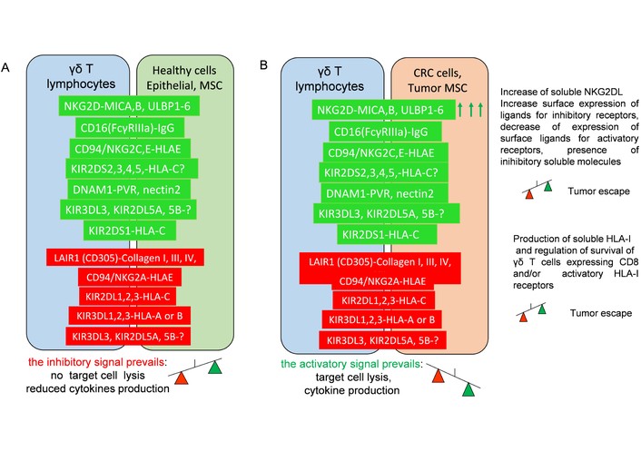
Either activatory or inhibitory receptors are expressed on γδ T cells, and their ligands on healthy colon or CRC cells. Schematic representation of some surface molecules involved in the cross-talk among γδ T cells and healthy colon cells or CRC cells. The activatory (green) and inhibitory (red) receptor-ligand pairs are shown. A. γδ T cell interaction with healthy cells in colon mucosa leads to signals in γδ T cells that do not induce killing and release of pro-inflammatory cytokines, triggering the damage of healthy colon mucosa. Instead, γδ T cells may be involved in supporting tissue homeostasis. B. On the other hand, the overexpression of some ligands for activatory molecules on CRC cells, such as NKG2D ligand (indicated by the green arrows), can trigger the activation of γδ T cells, leading to CRC cell killing and release of pro-inflammatory cytokines. The release by CRC cells of NKG2DL can interfere with the recognition of CRC cells. CRC cells can escape from γδ T cell-mediated killing by upregulating inhibitory molecules and releasing inhibitory cytokines. The presence of soluble HLA-I can influence the survival of γδ T cells. The induction of expression of ligands for activating receptors triggered by stress signals such as tumor transformation is one of the molecular mechanisms responsible for the specific killing of CRC cells instead of healthy colon mucosa cells. Note that just some examples of activating or inhibitory molecules are shown, and this representation is not exhaustive of all the known interactions demonstrated.
It is to be noted that Vγ9Vδ2 shows a very low level of expression of PD-1 with a peak at days 3 and 4, with a consequent decrease to baseline levels after 7 days [155]. On day 14, the large majority of Vγ9Vδ2 T cells are negative for PD-1 expression [156]. The upregulation of PD-1 and TIM-3 appeared to be related to the dose of ZA used, but this inhibitory molecule was not induced using the mycobacterium Bacillus Calmette-Guérin (BCG) [157]. Importantly, the cytotoxic activity of Vγ9Vδ2 T cells expanded with 10 µM ZA was impaired, but not that of those expanded with low doses such as 0.5–1 µM ZA [157]. In our hands, the optimal stimulation and expansion of Vγ9Vδ2 T cells can be achieved only with low doses of ZA (0.5–1 µM), while higher doses (more than 5 µM) can result in reduced expansion of Vγ9Vδ2 T cells [158]. These findings suggest that the optimal dose to trigger proliferation of Vγ9Vδ2 T cells is quite low and is associated with very low, transient PD-1 expression. This supports the idea that the expansion with pAgs is a good choice to plan adoptive immunotherapy [159–163]. As already mentioned, γδ T cells express HLA-I receptors involved in the regulation of activation and tumor cell killing [164–166]. While the CLIR type of inhibitory receptors NKG2A/CD94 complex is expressed on a large fraction of γδ T cells [89, 164–167], the expression of KIR is more discrete on peripheral blood γδ T cells [88, 167, 168]. Importantly, the functional role of KIR on γδ T cells is controversial [10, 93]. The presence of inhibitory receptors such as KIR should deliver a negative signal blocking activation [9, 10, 88, 91, 93]. However, it is evident that Vδ1 and Vδ2 T cells can express isoforms of KIR that do not deliver an inhibitory signal into γδ T cells but can trigger activation [93]; likewise, it can happen in NK cells [169–171]. In detail, we have shown that γδ T cells can express KIR2DS1 and KIR2DS2 based on reactivity with monoclonal antibodies (mAbs) that recognize the extracellular portion of both inhibitory and activating discrete HLA-I alleles. The presence of the activating isoform is functionally demonstrated by the activation of target cell killing of the mouse FcγR (such as the murine mastocytoma P815, Figure 4) using the specific murine anti-human KIR mAb [93].
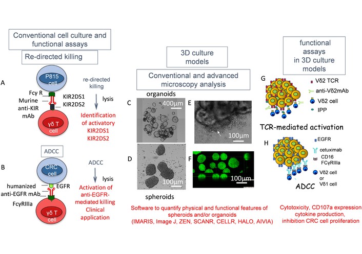
Experimental models to study anti-tumor activity of γδ T cells and 3D culture systems. The identification of activatory molecules at the cell surface of γδ T cells can be achieved by testing murine antibodies in a redirected killing assay (A). This assay is based on the ability of an antibody to make a bridge between the receptor on γδ T cells and the tumor target expressing the FcγR for murine immunoglobulins. If this bridge leads to the killing of the target, the receptor involved is of the activating type. This assay has allowed the identification of several HLA-I receptors specific for some MHC alleles such as activating KIR and CLIR. This assay is different from the ADCC (B) in which a human mAb is specific for a target molecule expressed on tumor cells and the FcγR is expressed on effector γδ T cells. In this case, the FcγR triggers the killing of the target cell. This assay can be used to determine whether a humanized antibody such as the therapeutic anti-EGFR cetuximab can trigger the killing of EGFR+ CRC cells. 3D culture systems such as organoids (C) or spheroids (D) can be used in vitro to study the interaction with γδ T cells. These models can be employed to define the sensitivity to therapeutic drugs. In the case of patient-derived organoids, it is possible to identify and select appropriate drugs to tailor the therapy to a specific patient. These studies can be performed by applying imaging systems. (E) Organoid + γδ T cells, with indicated by the arrow the destructive effect of γδ T cells at the periphery of the organoid. (F) Spheroid analysis with confocal microscopy after labelling with specific probes for nuclei. The analysis of images is performed using specific software, some of which is associated with artificial intelligence, to appropriately define any interaction between lymphocytes and tumor cells. The use of specific mAbs to TCR Vδ2 can be added to the 3D culture system and determine whether the recognition of small antigen pyrophosphates (pAg) is downregulated (G and H). The use of the therapeutic anti-EGFR humanized mAb can show the sensitivity of CRC cells to ADCC in 3D models.
It is of note that the γδ T cell clones (either Vδ1 or Vδ2) expressing KIR2DS1 or KIR2DS2 can interact with the soluble HLA-I molecule alleles Cw4 or Cw3, respectively. This interaction with their natural ligands can deliver an apoptotic signal leading to the death of γδ T cells [93]. Overall, these findings would indicate that the presence of activating KIR on γδ T cells can regulate their survival [93]. In addition, sizeable fractions of γδ T cells express the CD8 receptor for the invariant portion of HLA-I [93]. Similarly to activating KIR, this receptor engaged with soluble HLA-I can trigger apoptosis of γδ T cells through the Fas-L-Fas interaction [93]. Altogether, these findings suggest that γδ T cells bear several HLA-I receptors that can regulate their activation and survival [93]. There is no direct evidence that these receptors can play a role in the context of CRC. It is possible to speculate that within the TME, the soluble (s) or surface-expressed HLA-I on CRC cells, MSCs, or regulatory monocytic cells may regulate the γδ T cell antitumor response. Theoretically, the function of sHLA-I, possibly derived from CRC cell killing, interacting with either activating or inhibitory receptors on γδ T cells may influence, depending on the rate of positive and negative signals delivered, their anti-tumor effect by interfering with the direct binding to CRC cells. This effect could be present with MSCs and other stromal cells such as monocyte-derived suppressors or dendritic cells. It is still undefined whether the amount of sHLA-I may indeed deliver a signal in the CRC TME. However, the lower physical interaction with tumor cells and TME stromal cells can reduce the signals delivered upon the engagement of activating or inhibitory receptors expressed on γδ T cells, modifying the outcome of γδ T cell-mediated activities. Finally, the results of the whole signaling mediated by either sHLA or surface HLA-I may be heterogeneous in different sites of the CRC mass. This heterogeneity can lead to the associated production of different cytokines, which in turn may influence the behavior of the TME. These hypotheses further complicate a very intricate scenario. The idea of blocking the activity of γδ T cells expressing receptors for HLA-I can be a solution to reduce the undesired effects of apoptosis mediated by CD8 and/or KIR2DS1/S2 and other activatory HLA-I receptors. This can be accomplished using γδ T cells lacking these receptors for immunotherapy or by blocking the HLA-I-γδ TCRs with monovalent anti-KIR2DS1/2 antibody-derived fragments to limit the apoptotic signal delivered by the receptor crosslinking with HLA-I [93]. However, all these considerations need an experimental demonstration to support tools targeting molecules that can influence at the same time the antigen-specific activity of αβ T cells.
Some phenotypic and functional features of γδ T cells are different in murine models from what happens in humans [8, 172–176]. Overall, the role of Vδ1 T cells is similar in mice and humans, while it is difficult to mimic in a mouse model the role of Vδ2 T cells, as this cell subset is almost absent in mice [174]. Furthermore, the human Vδ2 T cells, compared to Vδ1 T cells, can be more easily expanded and used as effectors in vitro, and they can be considered the more suitable subset of γδ T cells for immunotherapy. The functional features of Vδ2 T cells during the interaction between CRC epithelial cells can be studied in the three-dimensional (3D) culture systems such as patient-derived tumor spheroids and organoids [120, 129, 134, 158, 177–183] (Figure 3). These two 3D culture systems show some important differences to consider [184–187]. Spheroids are usually composed by the aggregation of a single cell type in a few days, and they generally do not need the presence of extracellular matrix to grow. On the other hand, organoids are more complex 3D structures forming crypts and a lumen and require specific matrix proteins for growth, besides several peculiar medium components. The organoids are composed of different cell types coming from stem cells differentiating into epithelial cells lining intestinal crypts, enterochromaffin neuroendocrine cells, and goblet cells. Intuitively, the growth of an organoid is slow and typical for each donor if they are derived from CRC mucosa specimens, and it is more difficult to standardize the culture conditions of organoids than those of spheroids. Importantly, both CRC spheroids from established cell lines and CRC organoids from several patients can be used as targets for γδ T cells. These models are ideal to study the molecular mechanisms of target cell recognition and the means of killing exerted by cytotoxic effector cells. In addition, these 3D structures can be used to select the drugs that are efficient both in single and combo administration and to analyze the mechanisms of insurgence of resistant cells [184–187]. It is of note that these analyses need advanced microscopy tools and appropriate bioinformatic analysis of many images to study these interactions. The inter-tumor heterogeneity (ITH) is related to the large variety in phenotype and treatment response of each patient. While the IDH is due to the cell products derived from PBMC of different donors. These two heterogeneities have been studied using “phenoscaping” during the interaction between CRC organoids and γδ T cells [177]. This advanced experimental approach uses high-dimensional single-cell analysis employing sc-RNA-seq and/or cytometry to determine how perturbation of a cellular system leads toward a specific state.
Applying thiol-reactive organoid barcoding in situ mass cytometry [188–192], it has been studied the posttranslational modification signaling, cell activation state, apoptosis, and immunophenotype of γδ T cells interacting with patient-derived organoids (PDO) at the single-cell level resolution [177]. Overall, the analysis of more than 1,000 γδ T cells in these 3D culture systems showed that tumor cells can suppress γδ T cell-mediated cytotoxicity, but γδ T cells can overcome this immune suppression by using multimodal tumor cell killing. Indeed, phenoscaping indicated that unmodified γδ T cells showed poor cytotoxicity against CRC PDO. On the other hand, γδ T cells expressing an IL-15Rα–IL-15 fusion protein can efficiently kill PDO, but this cytotoxicity can be downmodulated by specific PDO, indicating an ITH-specific immune modulation. This effect was overcome through anti-B7-H3 mAb obtained from a murine single-chain variable fragment (scFv) phage-display library [177]. This was associated with an hIgG1 format using a G1m1, 17 heavy chain allotype and produced as a full antibody able to trigger ADCC. Furthermore, the engineered γδ T cells can kill PDO enriched in cancer stem cells refractory to chemotherapy. Overall, the ADCC can overcome the immunomodulation due to PDO, and it suggests that γδ T cells can kill PDO CRC targets through different molecular mechanisms. This notion is further supported by several experimental evidence obtained using co-cultures of γδ T cells and PDO from CRC mucosa [129]. In fact, it has been demonstrated that CRC PDO can trigger the expansion of Vδ2 T cells when exposed to 5 µM ZA starting from highly purified peripheral blood T lymphocytes. Furthermore, this effect was evident using the antibody-drug conjugate (ADC) composed of the anti-EGFR therapeutic antibody Cet conjugated with ZA (Cet-ZA) [129]. The Vδ2 T cells obtained in the co-cultures with PDO killed PDO cells in the absence of any additional stimulus at quite a high effector-target cell ratio (E:T ratio 20:1), while at a low E:T ratio Vδ2 T cells did not kill PDO cells spontaneously. However, at a low E:T ratio, the addition to the cell cultures of either soluble ZA, Cet, or Cet-ZA can trigger the CD107a degranulation and killing of tumor cells. Importantly, the effect of the Cet-ZA can be partly inhibited through anti-TCR mAb specific for the Vδ2 chain and anti-CD16 antibody. These findings strongly suggest that the Vδ2 T cells can use both TCR and FcγR to target CRC PDO, supporting again the multimodal ability of Vδ2 T cells to kill tumor cells [129]. Importantly, the single-cell analysis has allowed the identification in a murine model of the key role of IL-17-producing γδ T cells [178]. This was associated with the analysis of organoids at different stages of evolution of damage at anorectal junctions representing hyperplasia, dysplasia, and carcinoma stages [178].
On the other hand, the use of spheroids from well-established CRC cell lines can provide important information on the functional features of tumor cells in 3D masses. Indeed, we have shown that CRC cell lines can generate spheroids with peculiar physical features by evaluating the spheroid mass density. This mass density was relevant during the killing of tumor cells exerted by NK cells [193].
The killing of CRC tumor cells induced upon the interaction with γδ T cells was mediated by both the engagement of TCR and ADCC. Furthermore, the CRC spheroids and organoids can be used as a target of nanoparticles carrying N-BPs to study novel putative therapeutic tools to trigger γδ T cells’ anti-tumor activity [181]. Figure 3 shows some of the features and applications of spheroids and organoids.
γδ T cells maintain gut homeostasis by interacting with the microbiome, as reported in the Introduction, and the dysbiosis present in CRC can influence the response of γδ T cells [6, 14–19]. The finding that commensal microbiota can promote the development of lung cancer in mouse models indicates that the role of γδ T cells is essential in cancer cell growth [194]. Indeed, local airway mucosal bacteria, through the Myd88-dependent IL-1β and IL-23 production from myeloid cells, can trigger proliferation and activation of Vγ6+Vδ1+ γδ T cells that produce IL-17. Furthermore, these γδ T cells expressed CXCL2, a neutrophil chemoattractant, and prostaglandin-endoperoxide synthase 2 (PTGS2) to promote inflammation and tumor cell proliferation [194]. It is of note that γδ T cells and resident memory T cells expressing CD39 regulatory molecules have been found at lower frequencies in the colonic tissue of CRC donors compared to healthy controls [195]. Furthermore, it has been reported in murine models that γδ T cells are selected by microbiota to promote mucosal tolerance. On the other hand, T cell secretion of fecal miRNAs such as miR-let-7f may be linked to restoration of mucosal immune responses [196]. These findings, together with the different reported properties of γδ T cell subsets (Introduction, and γδ T cells in healthy gastrointestinal mucosa chapters and Table 1), support that microbiome-γδ T cell cross-talk can greatly influence the dysregulation of immune response present in CRC.
The administration of γδ T cells in solid tumors extrapolated from the number of clinical trials present on https://clinicaltrials.gov/ is limited (Table 3). This is in comparison with the large use of other kinds of immune cells, such as αβ T cells [50, 197–200]. Similarly, the use of γδ T cells in treating CRC in a clinical setting has been reviewed recently [200], and it is evident that this immune cell subset is not intensively employed and is under study. This could be related to the above-mentioned pro-tumor and anti-tumor effects of γδ T cells in the context of the gut. However, it is clear that the Vδ2 T cells are highly cytotoxic against CRC cell lines and primary CRC cells, as shown before.
Administration of γδ T cells in clinical trials as an immunotherapeutic tool for CRC and some solid tumors.
| Study title | NCT number conditions | Phase of study status | Mechanism of action | Interventions, route of administration, lymphodepletion, enrollment estimated | Sponsor, study type, first posted, last verified | Primary and secondary endpoints |
|---|---|---|---|---|---|---|
| A Safety and Efficacy Study of Allogeneic CAR Gamma-Delta T Cells in Subjects with Relapsed/Refractory Solid Tumors | NCT06150885Solid tumorsCRC, TNBC, GBM, NSCLC | Phase IPhase IIaRecruiting | Nanobodies CAR BiTe targeting PD-L1, HLAGElimination of tumor cells bearing inhibitory receptors PD-L1 and/or HLA-G | CAR001: HLA-G-CAR. BiTE allo γδ T cellsIntravenousLD: NRN = 60 | Ever Supreme Bio Technology Co., Ltd.Interventional2023-11-292024-09 | SafetyPotential efficacy |
| ACE2016 in Adult Subjects With Locally Advanced or Metastatic Solid Tumors Expressing Epidermal Growth Factor Receptor (EGFR) | NCT06415487Locally advanced metastatic solid tumors | Phase IRecruiting | Killing tumor cells and inhibition ICB | ACE2016: allo γδ T cellsPembrolizumab: anti-PD-1IntravenousLD: Cyclo, FludN = 30 | Acepodia Biotech, Inc.Interventional2024-05-162025-06 | Safety |
| Haplo/Allogeneic NKG2DL-targeting Chimeric Antigen Receptor-grafted γδ T Cells for Relapsed or Refractory Solid Tumour | NCT04107142CRCTNBCGCSarcoma | Phase IUnknown status | NKG2DL (MICA/B, ULBP1-6)Killing tumor cells | CTM-N2D: alloNKG2DL-CAR-γδ T cellsIntravenousLD: NRN = 10 | CytoMed Therapeutics Pte Ltd.Interventional2019-09-272019-09 | SafetyDLTAdverse effects |
| Study of SUPLEXA in Patients With Metastatic Solid Tumours and Haematologic Malignancies | NCT05237206Metastatic solid tumors CRC, PC, others | Phase ICompletedWITH RESULTSSerious AE n = 6/35Other AE n = 25/35 | Antitumor activity | Biological: SUPLEXAA cell mixture comprised predominantly of NK, NK-T, αβ T and γδ T cells stored in cryogenic mediaIntravenousLD: NRN = 46 | Alloplex Biotherapeutics Inc.Interventional2022-02-142025-08 | Safety and tolerabilityefficacy |
| First-in-Human Study of ICT01 in Patients With Advanced Cancer | NCT04243499Solid tumorCRC, bladder cancer, BC, GC, melanoma, PDAC hematological malignancies | Phase IPhase IIActively enrolledN = 292 | Activation of γδ T cells and inhibition of ICB | IV ICT01hBTN3A mAb plus anti-PD-1 pembrolizumabIntravenousLD: NRN = 292 | ImCheck TherapeuticsInterventional2020-01-282026-01 | SafetyTolerabilityDCR using RECISTControl rate using RECIL |
| Phase 1/2a Study of ICT01 Plus Low Dose SC IL-2 in Patients With Advanced Solid Tumors (EVICTION-2) | NCT05307874Solid tumorCRC, bladder cancer, BC, GC, melanoma, PDAC hematological malignancies | Phase IPhase IIActively enrolled | Activation of γδ T cells and inhibition of ICB PD-1 | IV ICT01hBTN3A mAb plus low doses of IL-2 subcutaneously plus anti-PD-1 pembrolizumabIntravenousLD: NRN = 56 | ImCheck TherapeuticsInterventional | SafetyAEDCR |
| Safety Study for a Gamma Delta T Cell Product Used With Low Dose Radiotherapy in Patients With Locally Advanced or Metastatic NSCLC or Solid Tumors With Bone Metastases | NCT06069570Carcinoma, NSCLC, bone metastasis | Phase IRecruiting | Killing of tumor cells | KB-GDT-01 allo-γδ T cellsLow dose of radiotherapyIntravenousLD: NRN = 48 | Kiromic BioPharma Inc.Interventional2023-10-062025-03 | Safety tolerabilityAE, MTD, MADORR, PFSOS, TTPTTR, DCR |
| Allogeneic NKG2DL-targeting CAR γδ T Cells (CTM-N2D) in Advanced Cancers (ANGELICA) | NCT05302037Refractory cancersSolid tumor hematological malignancies | Phase IRecruiting | Killing of tumor cells expressing NKG2DL (MICA/B, ULBP1-6) | Allogeneic NKG2DL-targeting chimeric antigen receptor-grafted γδ T cells (CTM-N2D)IntravenousLD: NRN = 12 | CytoMed Therapeutics Pte Ltd.Interventional2022-03-312024-11 | SafetyDLTAEPFSOS |
| A Study of PF-08046052/SGN-EGFRd2 in Advanced Solid Tumors | NCT05983133CRCHNSCCNSCLCPDAC | Phase IRecruiting | Killing of EGFR+ tumor cells activating specifically Vδ2 TCR | Bispecific γδ T-cell engagerLAVA1223 anti-EGFR-Vδ2 TCRIntravenousN = 68 | Seagen, a subsidiary of Pfizer2023-08-092026-04 | SafetyPFSOS |
| UTAA06 Injection for Treatment of Advanced Malignant Solid Tumors | NCT06372236Advanced solid tumors | Phase I | Targeting B7-H3+ tumor cells | B7-H3 CAR-Vδ1 T cellsIntravenousN = 10 | Peking University2024-04-172025-05 | SafetyMTDPK |
The table has been obtained from the https://clinicaltrials.gov/ website, accessed on 26th May 2026. AE: adverse events; BC: breast carcinoma; Cyclo: cyclophosphamide; DCR: disease control rate; DLT: dose limiting toxicity; Flud: fludarabine; GC: gastric cancer; GBM: glioblastoma; HNSCC: head and neck squamous cell carcinoma; ICB: immune check point; LD: lymphodepletion; MTD: maximum tolerated dose; MDA: maximum administered dose; NSCLC: non-small cell lung cancer; NR: not reported; ORR: objective response rate; PDAC: pancreatic adenocarcinoma; PC: prostate carcinoma; PFS: progression free survival; PK: pharmacokinetics; OS: overall survival; TTR: Time to Treatment Response; TTP: Time to Progression; TNBC; triple negative breast carcinoma; MICA/B: major histocompatibility complex class I (MIC) related molecules A and B; ULBP1-6: UL-16 binding proteins 1-6.
The NCT04107142 clinical trial is a phase I study aimed at investigating the safety and tolerability of allogeneic/haploidentical NKG2DL-targeting chimeric antigen receptor (CAR)-grafted γδ T cells (CTM-N2D) in refractory/relapsing solid tumors, including patients suffering from CRC. As CTM-N2D is obtained starting from PBMC-derived γδ T cells (see https://w2.cytomed.sg/car-gamma-delta-t-cell/), which are highly purified and transduced with an NKG2DL-specific CAR, it is conceivable that the large majority of these CAR γδ T cells are Vδ2+. Importantly, the CTM-N2D could recognize on tumor cells several molecules, such as NKG2DL, DNAM-1L, and pAgs overexpressed by stress signals [201, 202], while these molecules are expressed at a lower level on healthy cells. In addition, this product is coming from PBMC of healthy donors, and it may be considered applicable to many patients as an affordable off-the-shelf product pending manufacturing scale-up and regulatory approval. The target dose is 3 × 10⁸–3 × 10⁹ transduced cells, and four doses are scheduled for each patient at an interval of a week. While a 3 + 3 dose regimen is used to determine the safe regimen on the incidence of dose limiting toxicity (DLT). At present, data regarding this trial have not been published, and it is still to be defined whether γδ T cells with CAR specific for NKG2DL will counteract the progression of solid tumor cells as demonstrated in murine models [202]. However, the safety profile is good without cases of cytokine release syndrome (CRS), immune effector cell-associated neurotoxicity syndrome (ICANS), and tumor lysis syndrome. To employ Vδ1 T cells, it has been reported that using the erythroleukemia K562 expressing IL-21 at the cell surface as feeder cells, it is possible to activate and induce a strong proliferation of Vδ1 T cells [203]. The transduction with an anti-HER2 CAR increased the antitumor activity of these Vδ1 T cells in vitro and in vivo murine models. Importantly, it appeared that the blocking of NKG2D, DNAM-1, and γδ TCR did not affect the cytotoxicity of these Vδ1 T cells, supporting the idea of the involvement of other activating molecules in the anti-tumor cytotoxicity. Altogether, it is evident that both Vδ1 and Vδ2 T cells could be transduced with specific CAR and potentially used as potent immunotherapeutic tools, as shown in several other reports using bispecific antibodies or nanobodies [204–206].
However, the clinical efficacy of γδ T cells in CRC as well as other solid tumors is not reported (Table 3). The clinical translation of the different therapeutic tools based on γδ T cells or on activation of this immune cell subset should face several obstacles. The number of γδ T cells is low in PBMC, while isolation and expansion from solid tumors is a challenge, and we have already reported the methods to expand them quickly [118, 130]. The γδ T cells expanded in vitro should be infused and migrate through the endothelial cells, progressing into the extracellular matrix components to reach tumor tissues. Surface receptors such as CD31 and NKRP1A can help this process, and at the same time the expression of very late antigens (VLA) can increase the binding with matrix components, modulating the γδ T cell progression into the tissue. In the TME, γδ T cells find surface and soluble enzymes such as ADAMs and MMPs of tumor and stromal cells that can trigger the shedding of ligands for activating receptors such as NKG2DL. This may lead to a competition for the engagement of the same ligand expressed at the tumor cell surface. In addition, the direct tumor cell recognition through the γδ TCR may be reduced due to the heterogeneity of expression of accessory molecules such as BTN and BTN-like members on CRC cells.
It is unsurprising that the clinical efficacy of γδ T cells is restricted in light of these hindrances [207]. Some results from the Suplexa-101 trial (NCT05237206) on both hematological and solid malignancies are encouraging (http://dx.doi.org/10.1136/jitc-2024-SITC2024.0608) [208]. Indeed, a certain efficacy in some solid tumors comprising CRC-MSI-H with complete or partial remission besides stable disease maintenance for a long time beyond 80 weeks have been reported. However, the composition of the autologous cell preparations infused in patients is not only of γδ T cells but also quite heterogeneous, with the large majority of NK and NKT cells besides some CD8+ or CD4+ αβ T cells. This would imply that cells with innate features and not dependent on the expression of HLA-I can respond efficiently to advanced refractory tumors. If this interpretation of results is correct, the use of γδ T cells would be appropriate in refractory/resistant CRC.
CRC is the third most commonly diagnosed cancer and the second leading cause of cancer-related mortality worldwide, with increasing rates in young adults and developing countries [207–214]. It is evident that the mortality together with the incidence is increasing in adults younger than 65 years. This identifies the CRC burden in individuals born after 1950, suggesting the exposure to still undefined triggers for the generation of CRC. The CMS identification and the study of the TME composition have provided insights into the possibility of using immunotherapy in CRC patients [215–219]. Typically, CRCs are subdivided into MSI and MSS, with either a clear pro-inflammatory lymphocyte infiltrate in the former or more non-inflammatory/regulatory immune cells in the latter [218–220]. It is difficult to well-define the biological significance and the immunotherapeutic role of γδ T cells in either MSI or MSS. Focusing on MSS CRC, because the αβ T cell-mediated response in MSI is well-established, it is unclear which is the more suitable target to increase the immune response [218–223]. The broad and HLA-I-independent tumor cell recognition would suggest that γδ T cells are good antitumor effectors against MSS CRC expressing ligands for the activating molecules listed in Inhibitory and activating receptors of γδ T cells potentially relevant in CRC. On the other hand, it is conceivable that a good immune response in MSS CRC could also be related to the presence of a plethora of inhibitory receptors.
Besides the general subdivision into MSI and MSS CRC, the CMS subclassification into CMS1 (MSI, immune-enriched), CMS2 (canonical), CMS3 (metabolic), and CMS4 (mesenchymal, high TGF-β/stroma) possesses different clinical value regarding the therapy applied [224]. In addition, they may show different types and amounts of infiltrating γδ T cells as well as different influences on adoptive γδ T cell immunotherapy [225, 226]. Generally, the better clinical outcome is associated with CMS1, while the worse one is associated with CMS4. The CMS1 subtype is characterized by strong immune infiltration together with MSI status. On the contrary, CMS4 has plenty of stromal cells, collagen extracellular matrix, and cytokines delivering inhibitory signals in immune cells, impairing the localization and function of antitumor effector cells. The use of γδ T cells is conceivable in CMS1 to further support the β T cell response and limit the generation of resistant CRC cells with low expression of HLA-I and impaired antigen presentation [227]. The finding that several CMS1 CRCs can downregulate HLA-I both in a reversible and irreversible manner is relevant to support the reconstitution with IFN-γ in the former situation [227]. This IFN-γ can be produced by γδ T cells upon engagement with activating receptors. On the other hand, CMS2, CMS3, and CMS4 are mainly represented by MSS types and can downregulate HLA-I by similar molecular mechanisms to CMS1 [227]. In these CRCs, the antitumor effects of γδ T cells may be markedly impaired by the hostile TME associated with inhibitory cytokines and immunosuppressive cells. Moreover, the metabolic state of CRC TME, typical of the CMS3 subtype, can play a role in regulating the γδ T cells’ functions. Indeed, in glioblastomas (GBMs), γδ T cells need oxygen to function, as low oxygen levels downregulate NKG2D expression, impairing tumor cell killing, while the use of inhibitors of HIF1α and metformin can reinvigorate the elimination of tumors [228, 229]. It is conceivable that the γδ T cells should be used in appropriate combinations with pharmacological inhibition of some immunosuppressive cellular and molecular mechanisms responsible for the refractoriness to killing of these CRCs. Conceivably, hypoxia-associated acidosis, glucose reduction, and the alteration of amino acid metabolism can further contribute to the impairment of tryptophan and/or glutamine metabolisms in innate cells, IEL, and γδ T cells [230–234].
It has been shown that the heterogeneous expression of PD-L1 in both MSI and MSS CRC, and that PD-L1 expression was an independent factor for a better prognosis of CRC [222]. In addition, some subtypes of CRC, such as the mesenchymal subtype CMS4 with prominent stromal components associated with high TGF-β and angiogenesis expression, would propose that several regulating molecular mechanisms are relevant in CRC [215]. Overall, these findings strongly suggest that combo therapies using different drugs hitting several and not overlapping target molecules would support the rationale of using γδ T cells in immunotherapy.
Furthermore, this is increasing the interest in other regulatory receptors other than classical immune checkpoints PD-1 and cytotoxic T-lymphocyte antigen 4 (CTLA-4) [153, 154, 235]. We have shown that MSCs as tumor-associated fibroblasts can express BTN and trigger the expansion of Vδ2 T cells using soluble or anti-EGFR mAb Cet-ZA [120]. The expanded Vδ2 T cells can kill CRC MSCs, potentially relieving their immunosuppressive behavior. In addition, stromal cells in the TME express and secrete collagens, besides vasculogenic growth factors such as VEGF. Both collagen and VEGF, in turn, may have potent anti-immune cell effects by interacting with collagen receptors and favoring tumor cell vascularization. In detail, the Vδ2 T cells can express the leukocyte-associated immunoglobulin-like receptor (LAIR)-1 collagen receptor [236, 237]. LAIR-1 is an inhibitory receptor able to downregulate different activation signals in various leukocyte subsets. The possible direct involvement of LAIR-1 in downregulating the anti-tumor immune response in CRC has not been demonstrated. However, it is conceivable that this receptor can play a role in the immune suppression mediated by the CRC TME as well as in other kinds of tumors [238, 239]. For instance, MSC can impair the upregulation of NKG2D induced by IL-2 in NK cells, and this impairment can downregulate the NKG2D-mediated recognition of CRC cells [240]. Furthermore, the LAIR-1 engagement can markedly inhibit the proliferation of peripheral blood γδ T cells triggered with anti-Vδ2 mAb and, to a low extent, the cytolytic activity of γδ T cell clones [240]. Altogether, these findings would suggest that LAIR-1 expressed on γδ T cells can deliver an inhibitory signal impairing, at least in part, the antitumor activities of this cell subset. This suggests that other unconventional immune checkpoint receptors on γδ T cells should be targeted to increase their antitumor effect. Table 4 provides some features for the selection of CRC patients grouped in molecular subtypes for future immunotherapy approaches with γδ T cells based on the findings reported in this review.
Features of CRC cells and TME to select patient immunotherapy with γδ T cells and possible combos with drugs or immune cells.
| CRC subtypes, main features | γδ T cell infiltration (degree and features) | Targeting molecules for γδ T cell activation on CRC cells | Combinations of drugs/cells with γδ T cells |
|---|---|---|---|
| CMS1: MSI immuneMSI, SCNAlow, BRAFm, TIL | Good | EGFRBTN and BTN-like, NKG2DL, DNAM-1L functional APC | Anti-EGFR-mAb linked to N-BPs or pAgs |
| CMS2: canonicalWNT, MYC, SCNAhigh | Medium/Low | EGFRBTN and BTN-like, NKG2DL, DNAM-1L functional APC | Anti-EGFR mAb |
| CMS3: metabolicSCNAlow, KRAS | Low/Impaired by TME | EGFRBTN and BTN-like, NKG2DL, DNAM-1L functional APC | Anti-EGFR mAb, metformin, HIF1α inhibitors |
| CMS4: mesenchymalTGF-β, stromal, EMT, SCNAhigh | Low/Strongly impaired by TME | EGFRBTN and BTN-like, NKG2DL, DNAM-1L functional APC | Anti-VEGF mAb, anti-TGF mAb, anti-MSC mAb, anti-LAIR-1 mAb |
BRAFm: mutation gene for serine/threonine-protein kinase B-Raf; CMS: consensus molecular subtype; N-BPs: aminobisphosphonates; MSI: microsatellite instability; SCNA: somatic copy number alteration; EMT: epithelial-mesenchymal transition; TGF: transforming growth factor; TIL: tumor infiltrating lymphocyte.
Another key point to be considered it is the localization and persistence of immune effector cells within the TME to induce a good immunotherapeutic effect [241–243]. Several experimental approaches have been attempted for improving CAR T cells, such as peculiar genetic manipulation, association with drugs, selection of the origin of T cells, and improving culture conditions, well reviewed elsewhere [242]. Regarding γδ T cells, it is to note that Vδ1 and Vδ2 T cell clones may use different surface receptors, chemokine receptors such as C-X-C motif chemokine receptor 3 (CXCR3) and CXCR4, and biochemical pathways to migrate through endothelial cells [244–246]. These properties should be considered when using γδ T cell populations for adoptive immunotherapy, either as unmodified or genetically modified cells.
It should also be considered the ability of γδ T cell subsets to recognize the MHC class I-related gene protein (MR1), CD1b, CD1c, and CD1d. This, together with the possible interaction with regulatory T cells of a TME characterized by the heterogeneous expression of several transcription factors [115, 247–254]. Importantly, MR1 can present bacterial metabolites of vitamins B2 and B9 to mucosal-associated invariant T (MAIT) cells and T cells expressing different Vδ and Vγ chains [115, 250, 252, 253]. Likewise, CD1 can trigger γδ T cells [115], suggesting that polyclonal γδ T cells used for immunotherapy can find at the CRC mucosa several antigens and presenting molecules able to trigger their functional activities. This should be considered, as the response can possibly have the desired antitumor effect but also protumor stimulation, as described in this review and reported elsewhere [49, 54, 56] (see Tables 1 and 5). Figure 5 shows two of the possible immunotherapeutic applications of γδ T cells for different CRC subtypes.
Pros and cons of the human γδ T cells.
| Feature | Vδ1 | Vδ2 | Vδ3, non-Vδ1- Vδ2 | Context dependency |
|---|---|---|---|---|
| Friends | Cytotoxic, NKp46+ | Cytotoxic, IFN-γ, TNF-α, pAg, ADCC | MSI, high immune infiltrationIL-15 | |
| Foes | IL-17, AREG, TGF-β, Galectin 3LAIR-1, other inhibitory receptors | LAIR-1, other inhibitory receptors | IL-17, AREG, TGF-β, Galectin 3 | Stromal-rich CRC (CMS4)Low infiltrationIL-1β |
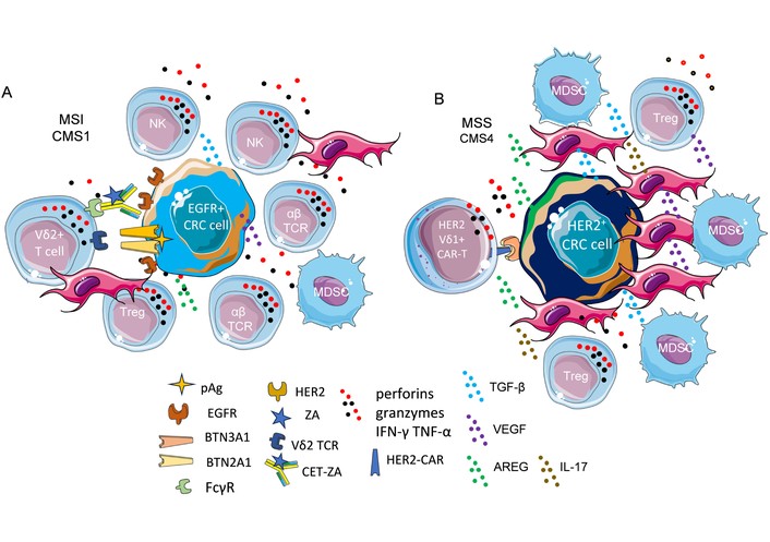
Possible future applications of γδ T cells for immunotherapy. The use of humanized (cetuximab) therapeutic mAbs directed to the EGFR linked to the aminobisphosphonate zoledronic acid (ZA) can trigger the proliferation and activation of anti-tumor activity of γδ T cells (A). This activation will depend on the expression of BTN and EGFR on CRC cells. It is of note that cetuximab can trigger ADCC and release of pro-inflammatory anti-tumor cytokines. The TME of an MSI/CMS1 subtype CRC can only partly inhibit the immune response, as the immunosuppressive components (Treg, MDSC, TAFs) are less represented. (B) In the case of the MSS/CMS4 subtype, the γδ T CAR-T cells should target the EGFR family (EGFR or HER2; the case of HER2 is shown) or other targeting molecules expressed on CRC cells. In this case, the TME is strongly immunosuppressive, as the components such as MDSCs, Tregs, TAFs, and immunosuppressive cytokines are well represented. The use of anti-EGFR therapeutic antibodies conjugated to ZA is conceivable if these CRCs express BTN and EGFR. It is to be noted that γδ T cells show both innate and adaptive immunity features. To plan their application for cell therapy, it is necessary to better know their functional features, such as whether they show anti-tumor or pro-tumor activities and in which context these properties can be elicited.
A leading problem for therapeutic approaches to CRC is present for local unresectable and metastatic forms [255, 256]. Surgery, radiotherapy, and chemotherapy, either alone or in combination, are the standard and conventional therapeutic tools for resectable forms in early stages, while immunotherapy has potential for late stages of CRC disease [254–256]. Although the therapeutic armamentarium is wide and advanced, CRC is still incurable in a large proportion of patients [254–256]. Immunotherapy of CRC comprises mAbs and their derivatives, immune checkpoint inhibitors, adoptive T cell therapy with CAR-T cells or tumor infiltrating lymphocyte (TIL), vaccines, and oncolytic viruses [255, 256]. What is the place of γδ T cells in this context? The gut microbial community may play a role, together with environmental factors and genetic/epigenetic specific features of a patient, in CRC pathogenesis, response to immunotherapy, and generation of resistant tumor cells [257, 258]. γδ T cells can sense gut microbes and modulate their function in response to bacterial products, suggesting a key role for γδ T cells in shaping a correct response of colon mucosa to external stimuli [7, 109, 250]. γδ T cells possess innate and adaptive features, and they are important for intestinal homeostasis [6, 7]. Unfortunately, our present knowledge on γδ T cell biology is limited, and the murine models do not correspond to what can happen in humans [21, 252, 254]. Ideally, γδ T cells can respond to tumor growth without HLA-I specificity, and the use of γδ T cells can be easier than that of αβ T cells [259], showing fewer adverse effects than CAR-T cells [260, 261]. The use of mAbs linked to either pAgs or N-BPs or dual-payload ADC could be the first step to analyze in human beings the effect of therapeutic mAbs in CRC eliciting a γδ T cell response in patients [129, 262]. These mAbs can trigger both the TCR-mediated killing of BTN+ CRC cells and the ADCC through the CD16 receptor expressed on Vδ2 T cells. This antitumor activity can complement the αβ T cell antigen-specific response in MSI. The same approach can be applied for MSS CRC, as anti-EGFR mAb linked to N-BPs can kill both EGFR+ CRC and also TAFs/MSCs, limiting their immunosuppressive effect. The main matter to be resolved is represented by the correct identification of the markers and functional behavior of γδ T cells within the colon in health and CRC, which could help to design and plan the appropriate immunotherapeutic means based on the use of these cells. The concept that inhibitory receptors are present to downregulate antitumor response should be challenged with the possibility that inhibitory signals through these inhibitory receptors can promote effector cell survival, as happens with sHLA-I interacting with KIR. Indeed, it is conceivable that inhibitory receptors are present to favor the switch-off of the inflammatory reaction, favoring repair and limiting autoreactivity. Overall, the γδ T cells playing a role in the homeostasis of the colon can play a role in checking CRC cell growth. However, little is known about their plasticity and molecular mechanisms to enhance antitumor activities while conserving their protective function of colon mucosa.
ADAM: a disintegrin and metalloproteinase
ADC: antibody-drug conjugate
ADCC: antibody-dependent cellular cytotoxicity
AE: adverse events
AhRs: aryl hydrocarbon receptors
APCs: antigen presenting cells
AREG: amphiregulin
ARG-1: arginase 1
BC: breast carcinoma
BCG: Bacillus Calmette-Guérin
BTN: butyrophilin
CAR: chimeric antigen receptor
Cet: cetuximab
CLIR: C-type lectin inhibitory receptor
CMS2: consensus molecular subtype 2
CRC: colorectal carcinoma
CTLA-4: cytotoxic T-lymphocyte antigen 4
CTLs: cytotoxic T lymphocytes
CXCR3: C-X-C motif chemokine receptor 3
Cyclo: cyclophosphamide
DAMPs: danger-associated molecular patterns
DCR: disease control rate
DLT: dose limiting toxicity
DMAPP: dimethylallyl pyrophosphate
DNAM: DNAX Accessory Molecule
EGFR: epidermal growth factor receptor
EMT: epithelial-mesenchymal transition
FC: flow cytometry
FcγR: receptor for the Fc (fragment crystallizable) of γ chain immunoglobulin
Flud: fludarabine
GALT: gut-associated lymphoid tissue
GBM: glioblastoma
GC: gastric cancer
HLA: human leukocyte antigen
HMB-PP: (E)-4-hydroxy-3-methyl-but-2-enyl pyrophosphate
Hsp: heat shock protein
IDH: inter-donor heterogeneity
IDO: indoleamine 2,3-dioxygenase
IEL: intraepithelial lymphocyte
IFN: interferon
IHC: immunohistochemistry
IKKα: inhibitor of nuclear factor-κB (IκB) kinase α
IKKβ: inhibitor of nuclear factor-κB (IκB) kinase β
IL: interleukin
ILCs: innate lymphoid cells
IPP: isopentenyl pyrophosphate
ITH: inter-tumor heterogeneity
KIR: killer-cell immunoglobulin-like receptor
mAbs: monoclonal antibodies
MAdCAM: mucosal addressin cell adhesion molecule
MAIT: mucosal-associated invariant T
MAPK: mitogen activated protein kinase
MDSCs: myeloid-derived suppressor cells
MICA/B: major histocompatibility complex class I (MIC) related molecules A and B
MMP: matrix metalloproteinase
MSI: microsatellite instability
MSS: microsatellite stability
N-BPs: aminobisphosphonates
NK: natural killer
OS: overall survival
pAg: phosphoantigen
PAMPs: pathogen-associated molecular patterns
PBMC: peripheral blood mononuclear cells
PD-1: programmed cell death protein 1
PD-L1: programmed cell death protein ligand 1
PDO: patient-derived organoids
PFS: progression free survival
PVRIG: poliovirus receptor related immunoglobulin domain containing
RA: retinoic acid
RALDH: retinal dehydrogenase
RIS: risedronic acid
RORC: RAR-related orphan receptor gamma
RUNX3: RUNT related transcription factor 3
scFv: single-chain variable fragment
SCID: severe combined immunodeficiency
SCNA: somatic copy number alteration
sc-RNA-seq: single-cell RNA sequencing
TACTILE: T cell activation, increased late expression
TCR: T cell receptor
TGF: transforming growth factor
TIGIT: T cell immunoreceptor with Ig and ITIM domains
TIL: tumor infiltrating lymphocyte
TIM-3: T-cell immunoglobulin and mucin-domain containing-3
TME: tumor microenvironment
TNBC: triple negative breast carcinoma
TNF: tumor necrosis factor
TTP: Time to Progression
TTR: Time to Treatment Response
ULBP 1-6: UL-16 binding proteins 1-6
VLA: very late antigens
ZA: zoledronic acid
All the figures have been designed from the templates of Anatomy and Human Body and Cellular Biology series of Smart Servier Medical Art (https://smart.servier.com/).
AP: Conceptualization, Investigation, Validation, Supervision, Writing—original draft, Writing—review & editing. The author read and approved the submitted version.
The author declares that he has no conflicts of interest.
Not applicable.
Not applicable.
Not applicable.
Not applicable.
The manuscript has been supported by the grant AIRCIG21648 to AP. The funders had no role in study design, data collection and analysis, decision to publish, or preparation of the manuscript.
© The Author(s) 2026.
Open Exploration maintains a neutral stance on jurisdictional claims in published institutional affiliations and maps. All opinions expressed in this article are the personal views of the author(s) and do not represent the stance of the editorial team or the publisher.
Copyright: © The Author(s) 2026. This is an Open Access article licensed under a Creative Commons Attribution 4.0 International License (https://creativecommons.org/licenses/by/4.0/), which permits unrestricted use, sharing, adaptation, distribution and reproduction in any medium or format, for any purpose, even commercially, as long as you give appropriate credit to the original author(s) and the source, provide a link to the Creative Commons license, and indicate if changes were made.



View: 1253
Download: 28
Times Cited: 0


