 Review


Affiliation:
Independent Researcher, Mountain View, CA 94040, USA
Email: Moawiah.Naffaa@proton.me
ORCID: https://orcid.org/0000-0003-0451-5901


Explor Immunol. 2026;6:1003261 DOI: https://doi.org/10.37349/ei.2026.1003261
Received: March 16, 2026 Accepted: June 14, 2026 Published: August 04, 2026
Academic Editor: Elisabetta Bolognesi, IRCCS Fondazione Don Carlo Gnocchi, ONLUS, Italy
Neuroimmune disorders are increasingly understood not as the consequence of dysfunction in isolated cell types, but as dynamic diseases shaped by coordinated transitions across interacting neural and immune cell states. This narrative review synthesizes current evidence showing how microglia, astrocytes, neural stem cells, vascular elements, and infiltrating peripheral immune cells shift between homeostatic, inflammatory, reparative, and disease-associated states in response to injury, infection, degeneration, and metabolic stress. We highlight how cytokine signaling, damage-associated molecular patterns, oxidative and metabolic stress, and transcriptional and epigenetic reprogramming reshape neuroimmune behavior across these cellular populations, thereby influencing inflammation, synaptic remodeling, tissue repair, and disease progression. By framing neurological disorders as state transition networks rather than static cellular abnormalities, this review integrates emerging insights from single-cell and spatial profiling with systems-level neuroimmunology and identifies cellular plasticity as both a mechanistic principle and a therapeutic opportunity. This perspective provides a unifying conceptual framework for understanding neuroimmune pathology in disorders such as Alzheimer’s disease, multiple sclerosis, stroke, and traumatic brain injury, while also pointing toward next-generation strategies that selectively modulate maladaptive cellular programs and promote regenerative neuroimmune states.
Neuroimmune signaling is increasingly recognized as an important component of many neurological disorders, including neurodegenerative diseases, autoimmune conditions of the central nervous system, traumatic brain injury, and stroke [1–4]. In these conditions, interactions between resident brain cells and immune populations influence inflammatory responses, cellular injury, and tissue repair processes. These interactions occur across multiple cellular compartments of the central nervous system and can shape both acute pathological responses and longer-term structural and functional changes in neural tissue.
Historically, studies in neuroimmunology have focused largely on the roles of specific cell types. Microglia are widely regarded as the principal innate immune cells of the brain and are involved in immune surveillance, synaptic remodeling, and clearance of cellular debris [5, 6]. Astrocytes also participate in immune signaling through the production of cytokines, chemokines, and metabolic mediators that influence neuronal function and vascular regulation [3, 7, 8]. In addition to these resident cells, peripheral immune populations such as macrophages and lymphocytes can enter the central nervous system during pathological conditions and interact with local cellular environments.
Recent technological advances have substantially expanded our understanding of the diversity of cellular responses within the brain. High-resolution approaches including single-cell transcriptomics, spatial transcriptomics, and epigenomic profiling have revealed that many neural and immune cell populations exhibit considerable heterogeneity and can adopt multiple functional states [2, 3, 9, 10]. These studies indicate that microglia, astrocytes, neural stem cells, and infiltrating immune cells can shift between transcriptionally and functionally distinct states in response to environmental signals, injury, or inflammatory cues [2, 3, 10, 11].
The concept of cellular state transitions has therefore emerged as an important framework for understanding neuroimmune responses during disease. Rather than representing fixed cell identities, many immune-related processes in the central nervous system involve dynamic changes in cellular behavior that evolve over time. Such transitions may influence inflammatory signaling, synaptic remodeling, regenerative responses, and tissue degeneration depending on the local context and duration of immune activation [3, 6, 7].
A substantial body of literature has examined individual aspects of neuroimmune signaling, including microglial activation, astrocyte reactivity, cytokine signaling pathways, and immune cell infiltration into the central nervous system [2, 4, 7]. While these studies have provided important insights into specific molecular and cellular mechanisms, relatively few reviews have synthesized these findings into a unified framework describing neurological disease as networks of interacting cellular state transitions across multiple neural and immune cell populations.
In this narrative review, we synthesize current evidence describing how transitions between cellular states contribute to the development and progression of neuroimmune disorders and organize this literature within a cellular state transition framework in which neurological disease can be interpreted as networks of interacting neural and immune cell states. We first discuss baseline cellular states that support homeostasis in the healthy brain. We then examine signals that trigger immune-related state transitions during injury and disease. Subsequent sections review state dynamics in key cellular populations—including microglia, astrocytes, neural stem cells, and infiltrating immune cells—and consider how interactions among these states influence inflammatory responses and tissue remodeling within the central nervous system. By organizing the literature around cellular state transitions rather than static cell identities, this review aims to provide a framework that integrates emerging insights from single-cell technologies and systems-level analyses to better understand neuroimmune mechanisms in neurological disease [2, 3, 9, 10].
This article was designed as a narrative review rather than a systematic review or meta-analysis. The literature was curated to synthesize mechanistic and conceptual evidence relevant to cellular state transitions in neuroimmune disorders. Literature searches were conducted using PubMed/MEDLINE, Web of Science, Scopus, Google Scholar, and publisher databases, with emphasis on studies published from 2010 through early 2026. Earlier landmark studies were also included when they established foundational concepts in neuroimmunology, glial biology, blood-brain barrier regulation, adult neural stem-cell biology, or single-cell and spatial profiling.
Search terms included combinations of “neuroimmune,” “cellular state,” “cell state transition,” “microglia,” “astrocyte reactivity,” “disease-associated microglia,” “neural stem cells,” “blood-brain barrier,” “neurovascular unit,” “peripheral immune infiltration,” “cytokines,” “DAMPs,” “single-cell RNA sequencing,” “spatial transcriptomics,” “Alzheimer’s disease,” “multiple sclerosis,” “stroke,” and “traumatic brain injury.” Articles were prioritized when they provided mechanistic evidence, high-resolution cellular profiling, translational relevance, or conceptual frameworks linking cellular plasticity to neurological disease.
Studies were included if they addressed one or more of the following: homeostatic cellular states in the central nervous system; molecular triggers of neuroimmune activation; microglial, astrocytic, vascular, neural stem-cell, or infiltrating immune-cell state transitions; disease-associated cellular remodeling; or therapeutic modulation of maladaptive neuroimmune states. Alzheimer’s disease, multiple sclerosis, stroke, and traumatic brain injury were selected because they represent distinct initiating contexts—chronic neurodegeneration, autoimmune demyelination, acute ischemic injury, and traumatic tissue disruption—while also sharing convergent neuroimmune remodeling processes. This selection allowed comparison of state-transition principles across chronic, autoimmune, ischemic, and traumatic neurological conditions.
Because this review integrates evidence from experimental models, human tissue studies, omics-based profiling, and translational therapeutic studies, conflicting findings were weighed according to biological context, disease stage, model system, and strength of evidence. Human data and causal experimental studies were prioritized when available. Findings derived primarily from animal models, correlative transcriptomic analyses, or marker-based state definitions were interpreted more cautiously. When evidence differed across studies, the manuscript emphasized the context-dependent nature of cellular state transitions rather than treating individual cellular states as universal or fixed disease categories.
Understanding cellular states in the healthy central nervous system provides an essential baseline for interpreting how neuroimmune responses evolve during disease. Under physiological conditions, the brain maintains a tightly regulated environment in which multiple cell populations coordinate to preserve tissue integrity, support neuronal function, and regulate immune surveillance. These interactions involve neurons, glial cells, vascular elements, and immune-related cells that collectively maintain metabolic balance, synaptic stability, and structural organization within neural circuits [12–14]. Rather than representing static cellular identities, many of these cell populations exist in functional states that enable them to respond dynamically to physiological signals while maintaining overall homeostasis.
The central nervous system can be viewed as a complex cellular ecosystem composed of interconnected neuronal, glial, vascular, and immune components. Neurons form the primary signaling networks responsible for information processing, while glial cells—including microglia and astrocytes—provide structural, metabolic, and regulatory support. Vascular cells, including endothelial cells and pericytes, maintain the blood-brain barrier and regulate the exchange of molecules and immune cells between the brain and the systemic circulation. Together, these cellular populations create a coordinated environment that supports neural activity while maintaining controlled immune surveillance, forming an integrated neuroimmune network within the healthy central nervous system (Figure 1).
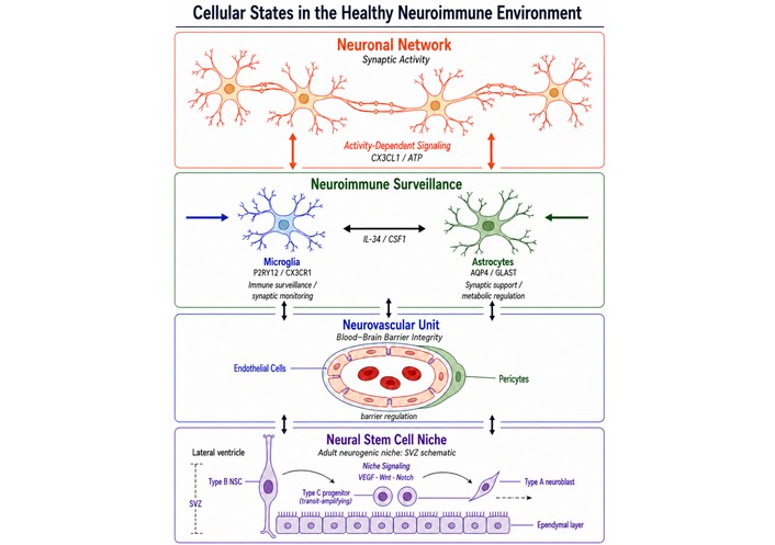
Cellular organization of the healthy neuroimmune environment. Neuronal networks interact with microglia, astrocytes, the neurovascular unit, and neural stem cell niches to maintain CNS homeostasis through activity-dependent signaling, immune surveillance, and vascular regulation.
Historically, the brain was described as an immune-privileged organ because immune-cell entry and inflammatory activation within the central nervous system are more tightly regulated than in many peripheral tissues [12, 15, 16]. However, this concept should not be interpreted as immune isolation. Modern neuroimmunology recognizes the central nervous system as a site of continuous but highly controlled immune surveillance, supported by resident immune cells, vascular and perivascular interfaces, meningeal immune compartments, and lymphatic drainage pathways [15, 17–20]. Within this regulated environment, cellular communication among neurons, glia, vascular elements, and immune-related cells coordinates synaptic function, metabolic support, barrier regulation, and immune signaling. These interactions allow the brain to preserve tissue stability while retaining the capacity to detect injury, infection, or systemic inflammatory signals.
The principal cellular populations that compose the neuroimmune environment and selected representative features of their homeostatic and disease-associated state tendencies are summarized in Table 1. These entries are intended as an orientation framework rather than a complete classification of discrete or fixed cellular states.
Major cellular populations and representative state tendencies in the neuroimmune environment.
| Cell type | Primary physiological functions | Representative molecular markers | Role in CNS homeostasis | Representative roles during neurological disease |
|---|---|---|---|---|
| Microglia | Immune surveillance, phagocytosis of apoptotic cells and debris, synaptic remodeling, regulation of neuronal activity | P2RY12, CX3CR1, TMEM119 | Continuous monitoring of the neural microenvironment, maintenance of synaptic integrity, clearance of damaged cellular components [21] | Context-dependent shifts toward inflammatory, phagocytic, metabolic, repair-associated, or disease-associated programs, including TREM2-associated responses to lipid-rich debris, amyloid pathology, and neurodegenerative tissue damage [21, 22] |
| Astrocytes | Regulation of neurotransmitter levels, metabolic support for neurons, potassium buffering, neurovascular coupling | S100β, AQP4, GLAST/EAAT1; GFAP in reactive states or selected astrocyte populations | Maintenance of extracellular ion balance, modulation of synaptic transmission, metabolic support of neurons, regulation of blood-brain barrier stability [8, 23] | Context-dependent astrocyte remodeling programs that may support neuroprotection, inflammatory signaling, synaptic dysfunction, barrier regulation, scar formation, or tissue remodeling depending on timing and disease context [24] |
| Neural stem cells | Generation of neurons and glial cells, maintenance of neurogenic niches, contribution to neural plasticity | SOX2, Nestin, GFAP (subset), DCX (progenitors) | Maintenance of quiescent stem-cell pools and controlled neurogenesis within specialized niches such as the subventricular zone and dentate gyrus [25, 26] | Altered activation, proliferation, and lineage decisions in response to inflammatory signaling, injury, or neurodegenerative conditions [27] |
| Endothelial cells | Formation of the blood-brain barrier, regulation of molecular transport, vascular signaling | Claudin-5, VE-cadherin, PECAM-1, GLUT1 | Maintenance of vascular integrity and selective permeability between the systemic circulation and neural tissue [28, 29] | Barrier dysfunction, increased permeability, and recruitment of immune cells during neuroinflammatory and neurodegenerative disorders [29] |
| Pericytes | Structural support of microvessels, regulation of vascular stability and permeability | PDGFRβ, NG2, CD146 | Stabilization of capillary structure and maintenance of blood-brain barrier function [28, 30] | Vascular dysregulation and blood-brain barrier disruption contributing to inflammation and neurodegeneration [30] |
| Peripheral immune cells | Systemic immune surveillance and inflammatory signaling | CD45, CD3 (T cells), CD68 (macrophages), Ly6G (neutrophils) | Limited immune monitoring under tightly regulated vascular control [15, 31] | Infiltration into the CNS during pathological conditions, contributing to inflammatory signaling, tissue damage, or repair responses [31] |
Note: The markers and functions listed in this table are representative rather than exhaustive. Cellular states in the central nervous system exist along dynamic continua that vary according to developmental stage, anatomical region, disease context, species, timing of injury, and local microenvironment. Therefore, the listed markers should not be interpreted as defining fixed cellular identities, and the disease-associated roles should be viewed as context-dependent state tendencies rather than mutually exclusive or stable phenotypes.
Microglia serve as the principal innate immune cells of the central nervous system and play a critical role in maintaining tissue homeostasis. In many regions of the healthy brain, microglia exist in a surveillant state characterized by a highly branched, ramified morphology and continuous extension and retraction of cellular processes that monitor the surrounding microenvironment [14, 32]. However, microglial morphology is regionally heterogeneous, and less ramified morphologies can occur under physiological conditions in specialized CNS regions such as circumventricular organs [33]. Through this dynamic surveillance behavior, microglia detect changes in neuronal activity, extracellular signaling molecules, and cellular damage.
Beyond immune surveillance, microglia contribute to several physiological processes essential for neural circuit maintenance. These include the removal of apoptotic cells and cellular debris, regulation of synaptic remodeling during development and adulthood, and modulation of neuronal activity through interactions with synaptic structures [13, 21, 34, 35]. Such functions highlight the role of microglia not only as immune responders but also as active participants in maintaining neural circuit stability.
At the molecular level, homeostatic microglial states are associated with characteristic transcriptional signatures that distinguish them from activated or inflammatory states. Several markers have been widely used to identify homeostatic microglia under physiological conditions, including purinergic receptor P2RY12, fractalkine receptor CX3CR1, and transmembrane protein TMEM119 [22, 36–38]. In contrast, triggering receptor expressed on myeloid cells 2 (TREM2) is more closely associated with microglial responses to lipid-rich debris, amyloid pathology, and disease-associated transitions, including the disease-associated microglia (DAM) program discussed below [39]. These signaling pathways contribute to microglial sensing of neuronal signals, regulation of immune responses, and maintenance of tissue integrity.
Together, these features illustrate that microglia occupy a specialized homeostatic state in the healthy brain. In this state, they support synaptic maintenance and tissue surveillance while remaining poised to respond rapidly to environmental changes. This capacity for dynamic responsiveness reflects the broader principle that cellular states in the central nervous system are stable under physiological conditions yet capable of transitioning when the tissue environment is altered.
Astrocytes represent one of the most abundant glial cell populations in the central nervous system and play essential roles in maintaining neuronal and vascular stability. In the healthy brain, astrocytes support neuronal function through multiple mechanisms that regulate the extracellular environment and facilitate communication within neural circuits. Among their key functions is the regulation of neurotransmitter levels within the synaptic cleft. Astrocytes actively uptake neurotransmitters such as glutamate and γ-aminobutyric acid, thereby preventing excessive synaptic signaling and maintaining excitatory–inhibitory balance within neural networks [8, 40, 41]. Because GFAP expression is low or regionally variable in many homeostatic astrocyte populations, particularly in the healthy cerebral cortex, GFAP should be interpreted cautiously as a general astrocyte marker and is often more informative in reactive states or specialized niches [42].
Astrocytes also provide metabolic support for neurons by supplying energy substrates and participating in metabolic coupling between neuronal activity and glucose utilization. Through astrocyte–neuron metabolic interactions, astrocytes help sustain neuronal energy demands during periods of high synaptic activity [8, 43, 44]. In addition, astrocytes play an important role in maintaining extracellular ion homeostasis. Their capacity for potassium buffering helps regulate neuronal excitability and stabilize electrical signaling within neural circuits [8, 45, 46].
Beyond these metabolic and synaptic functions, astrocytes contribute to the structural and physiological integrity of the neurovascular system. Astrocytic endfeet surround cerebral blood vessels and participate in the regulation of the blood-brain barrier, helping maintain vascular stability and control the exchange of molecules between the bloodstream and the neural environment [23, 47–49]. Through these interactions with vascular cells, astrocytes contribute to the maintenance of a controlled microenvironment that protects neural tissue from fluctuations in systemic conditions.
Astrocytes also participate in immune-related signaling within the central nervous system. Under physiological conditions, they can produce cytokines, chemokines, growth factors, and purinergic signaling cues that influence neuronal survival, glial communication, and vascular responses [8, 24, 38, 50, 51]. These effects are mediated through inflammatory, oxidative-stress, and cell-survival pathways, including signaling programs that regulate cytokine production, redox balance, mitochondrial stress, and programmed cell death. Comparative evidence from non-neural disease models further illustrates that inflammatory and survival-related cellular states can be modified by biochemical interventions, including lipoic acid-mediated immunomodulatory and anti-inflammatory effects in diabetic models and green tea polyphenol-associated modulation of apoptotic pathways [52]. Although these studies do not provide direct evidence for astrocyte state transitions in the central nervous system, they support the broader principle that inflammatory, antioxidant, and anti-apoptotic pathways can be pharmacologically modulated during cellular stress responses.
Taken together, astrocytes represent a multifunctional cellular system that integrates neural activity with metabolic and immune signaling pathways. Their diverse physiological roles allow them to support neuronal communication, regulate extracellular conditions, and maintain neurovascular stability within the healthy brain.
In addition to differentiated neural and glial populations, the adult brain contains specialized populations of neural stem cells that contribute to cellular plasticity and limited regenerative capacity. These stem cells are primarily located in two well-characterized neurogenic regions: the subventricular zone (SVZ) lining the lateral ventricles and the subgranular zone of the dentate gyrus within the hippocampus [27, 53, 54]. Within these niches, neural stem cells can generate progenitor cells with neuronal or glial lineage potential, although the extent of ongoing cellular turnover varies substantially across species, age, brain region, and disease context. In particular, the persistence and functional significance of adult human hippocampal neurogenesis remain actively debated [55].
Adult neural stem cells exist predominantly in a quiescent state characterized by low proliferative activity and stable maintenance within the niche environment. This quiescent state is considered essential for preserving the long-term regenerative potential of the stem-cell pool [25–27]. Periodically, subsets of these cells transition into an activated state in response to intrinsic molecular programs and signals from the surrounding microenvironment, including circuit-linked regulatory cues within neurogenic niches. In established adult neurogenic regions, activation can lead to the generation of transient amplifying progenitors and subsequently to neuronal or glial lineage progression, although the magnitude and functional significance of these processes vary by species, age, brain region, and disease context [25, 54, 56, 57]. More broadly, regenerative biology outside the nervous system also illustrates how stem-cell state transitions and lineage plasticity can be experimentally directed, including studies of bone marrow stem-cell differentiation toward β-cell-like phenotypes and biomaterial-based delivery systems designed to support tissue remodeling [58, 59].
The balance between quiescent and activated stem-cell states is tightly regulated by signals from the local niche, including interactions with neighboring astrocytes, vascular cells, extracellular matrix components, and circuit-derived regulatory inputs. Molecular cues such as growth factors, neurotransmitters, and inflammatory mediators can influence stem-cell activation and lineage progression [54, 60–62]. Through these regulatory mechanisms, the neurogenic niches maintain a controlled equilibrium between stem-cell maintenance and differentiation.
Although adult neurogenesis is well established in many experimental animal models, its extent and functional relevance in the adult human hippocampus remain unresolved. Therefore, claims regarding adult neural stem-cell contribution to plasticity, learning, or tissue repair should be interpreted in a species- and context-dependent manner. Maintaining neural stem cells in a predominantly quiescent state therefore represents a critical mechanism that preserves regenerative capacity while preventing excessive or uncontrolled proliferation. This regulated balance between quiescence and activation provides an important example of how cellular state dynamics support homeostasis within the adult brain.
Vascular cells form a critical structural and regulatory interface between the central nervous system and the systemic circulation. In the healthy brain, endothelial and associated vascular cells maintain the integrity of the blood-brain barrier and regulate the movement of molecules and immune cells between the bloodstream and neural tissue [28, 29, 63]. Through these functions, the vascular system contributes to a stable but immunologically monitored microenvironment that supports neuronal activity while permitting controlled immune surveillance at vascular, perivascular, meningeal, and barrier-associated interfaces [64].
Endothelial cells lining cerebral blood vessels play a central role in the formation of the blood-brain barrier. These cells are connected by specialized tight junctions that restrict paracellular diffusion and help maintain selective permeability across the vascular wall [29, 65, 66]. Tight junction proteins, together with transport systems within endothelial cells, regulate the controlled passage of nutrients, metabolites, and signaling molecules into the brain.
In addition to endothelial cells, other structural components contribute to vascular stability and barrier function. Pericytes, which are embedded within the vascular basement membrane, provide structural support to endothelial cells and participate in the regulation of vascular tone, permeability, and barrier maintenance [28, 30, 67]. The basement membrane itself forms a specialized extracellular matrix that supports the vascular wall and provides a scaffold for interactions among endothelial cells, pericytes, and astrocytic endfeet.
Astrocytes further contribute to the regulation of the neurovascular interface through specialized endfeet that closely surround cerebral blood vessels. These structures help coordinate signaling between neural activity and vascular responses while contributing to the maintenance of blood-brain barrier integrity [23, 49, 68]. Together, endothelial cells, pericytes, basement membrane components, and astrocytic processes form a highly coordinated vascular unit that maintains structural stability and regulates the exchange of signals and molecules across the barrier.
Under physiological conditions, the neurovascular system regulates immune-cell trafficking and immune surveillance at the central nervous system interface. Although unrestricted leukocyte entry into the brain parenchyma is prevented, controlled immune monitoring can occur through vascular, perivascular, meningeal, and barrier-associated compartments, including immune-cell surveillance along CNS vascular interfaces. The vascular barrier therefore functions not as an immunological wall but as a selective regulatory interface that modulates communication between the systemic immune system and the neural environment [31, 63, 64, 69].
Through these mechanisms, vascular cells maintain a controlled interface between the central nervous system and peripheral immune compartments. This regulatory function is essential for preserving tissue homeostasis while allowing appropriate immune monitoring of the neural environment.
Although individual cellular populations contribute distinct functions within the central nervous system, brain homeostasis ultimately depends on coordinated interactions among multiple cell types. Neurons, glial cells, vascular elements, and immune-related cells form interconnected signaling networks that regulate metabolic balance, synaptic stability, and tissue integrity. Within these networks, communication among cellular populations allows the brain to maintain a stable internal environment while responding to physiological demands.
Interactions between microglia and astrocytes represent one important component of this homeostatic network. Microglia monitor the neural environment and respond to subtle changes in tissue conditions, while astrocytes regulate metabolic support, neurotransmitter levels, and extracellular ion balance. Communication between these cell types helps coordinate local immune surveillance and metabolic regulation within neural circuits [13, 70, 71].
Neuron–glia communication also plays a central role in maintaining homeostasis. Neurons release signaling molecules that influence glial cell activity, while glial cells regulate synaptic function and neuronal excitability through the control of neurotransmitter uptake, metabolic support, and extracellular ion concentrations [8, 11, 43, 72]. These bidirectional interactions allow neural circuits to maintain functional stability during ongoing synaptic activity.
In addition, vascular cells participate in the regulation of immune signaling within the brain by controlling the exchange of molecules and immune cells between the systemic circulation and the neural environment. Through the coordinated actions of endothelial cells, pericytes, and astrocytic endfeet, the neurovascular interface maintains barrier integrity while permitting regulated communication between central and peripheral immune systems [15, 23, 29, 30].
Taken together, these interconnected cellular interactions illustrate that brain homeostasis is maintained through a dynamic equilibrium among multiple cellular states. Although these states remain relatively stable under physiological conditions, they retain the capacity to adapt to changes in neural activity, metabolic demands, and environmental signals. This balance between stability and adaptability enables the central nervous system to preserve tissue integrity while remaining responsive to physiological and environmental challenges.
Transitions from homeostatic cellular states toward inflammatory, repair-associated, metabolic, phagocytic, or disease-associated programs in the central nervous system are typically initiated by signals that indicate tissue disturbance or physiological stress. Under normal conditions, the cellular networks described in the previous section maintain a stable environment through coordinated signaling among neurons, glial cells, vascular elements, and immune-related cells. However, this equilibrium can be disrupted by a variety of stimuli, including tissue injury, infection, metabolic imbalance, or cellular stress. These disturbances generate molecular signals that initiate immune responses and alter the functional states of multiple cellular populations within the brain [13, 73, 74].
Such signals activate intracellular pathways that modify gene expression, cellular metabolism, and signaling behavior, thereby initiating transitions away from homeostatic states. Importantly, these responses do not occur in isolated cell types. Instead, immune activation often involves coordinated changes across several interacting populations, including microglia, astrocytes, vascular cells, and infiltrating immune cells. Through these interconnected responses, local disturbances can propagate across the neuroimmune environment and lead to broader changes in tissue physiology [13, 70, 75].
Homeostatic cellular states in the central nervous system are relatively stable but remain sensitive to changes in the local tissue environment. A range of molecular and physiological signals can disrupt this balance and trigger cellular reprogramming. These signals may arise from tissue injury, infection, metabolic disturbances, or cellular stress and can rapidly alter signaling networks within neural tissue.
When such disturbances occur, the resident brain cells detect changes in extracellular molecules, metabolic conditions, or the structural integrity of the surrounding tissue. These signals activate intracellular pathways that initiate immune responses and alter cellular behavior. As a result, cells that normally perform supportive or regulatory functions may transition toward states associated with inflammation, tissue remodeling, or repair. Because many cell populations within the brain communicate extensively with one another, these signals frequently initiate coordinated responses across multiple cellular systems [13, 74–76].
The major categories of molecular and physiological signals that trigger cellular state transitions in the central nervous system are summarized in Table 2.
Triggers of cellular state transitions in the central nervous system.
| Trigger category | Example molecules/signals | Source | Cellular targets | Functional consequence |
|---|---|---|---|---|
| Damage-associated molecular patterns (DAMPs) | HMGB1, extracellular ATP, extracellular DNA fragments, heat-shock proteins | Injured neurons, damaged glial cells, mitochondrial damage, extracellular matrix breakdown | Microglia, astrocytes, endothelial cells | Activation of pattern-recognition receptors, induction of inflammatory signaling, initiation of neuroimmune responses [77, 78] |
| Pro-inflammatory cytokines | IL-1β, TNF, IL-6, IFN-γ | Microglia, astrocytes, endothelial cells, infiltrating immune cells | Microglia, astrocytes, neurons, immune cells | Activation of inflammatory signaling pathways, modulation of gene-expression programs, promotion of cellular activation states [79, 80] |
| Chemokines | CCL2, CXCL10, CX3CL1 and related chemokines | Activated glial cells, endothelial cells, infiltrating immune cells | Microglia, monocytes/macrophages, T cells | Regulation of immune-cell recruitment, migration, and positioning within neural tissue [81] |
| Metabolic stress signals | Altered glucose metabolism, mitochondrial dysfunction, lipid metabolism shifts | Neurons, glial cells experiencing metabolic imbalance | Microglia, astrocytes, neural stem cells | Activation of immunometabolic pathways and modulation of inflammatory signaling [82, 83] |
| Oxidative stress | Reactive oxygen species, mitochondrial oxidative products | Mitochondrial dysfunction, inflammatory signaling, cellular injury | Neurons, microglia, astrocytes | Modification of signaling proteins, activation of inflammatory transcriptional pathways, amplification of immune responses [84, 85] |
| Hypoxia | Reduced oxygen availability, hypoxia-inducible signaling pathways | Ischemia, vascular dysfunction, tissue injury | Microglia, astrocytes, endothelial cells | Activation of hypoxia-responsive transcription programs and metabolic adaptation pathways [86, 87] |
One of the earliest triggers of immune activation in the central nervous system is the release of DAMPs, commonly referred to as DAMPs. These endogenous molecules are released from stressed, injured, or dying cells and serve as signals indicating disruption of tissue integrity [77, 78, 88]. Within the brain, DAMPs can originate from multiple sources, including injured neurons, degenerating glial cells, damaged mitochondria, and degradation of extracellular matrix components.
Several well-characterized molecules function as DAMPs in neural tissue. These include high mobility group box 1 (HMGB1), extracellular adenosine triphosphate (ATP), extracellular DNA fragments, and heat-shock proteins. When released into the extracellular space, these molecules act as endogenous danger signals that alert surrounding cells to tissue damage [38, 77, 88–90].
DAMPs are detected by pattern-recognition receptors expressed on resident immune-related cells such as microglia and astrocytes. These receptors include members of the Toll-like receptor family, nucleotide-binding oligomerization domain-like receptors, and receptors for advanced glycation end products [73, 78, 91, 92]. Activation of these receptors initiates intracellular signaling cascades that promote inflammatory gene expression, cytokine production, and cellular activation.
Through these mechanisms, DAMPs represent an important early signal that converts local tissue injury into broader neuroimmune responses. By activating pattern-recognition pathways in resident glial cells, these endogenous danger signals initiate inflammatory signaling networks that can drive transitions from homeostatic cellular states toward activated or disease-associated states within the central nervous system.
Cytokines are central regulators of immune activity within the central nervous system and play an important role in coordinating cellular responses to injury, infection, and physiological stress. These signaling molecules are produced by multiple cell types, including microglia, astrocytes, endothelial cells, and infiltrating immune cells, and act through receptor-mediated pathways to influence cellular behavior across the neuroimmune environment [13, 51, 74]. Through these signaling networks, cytokines regulate inflammatory responses, cellular communication, and tissue adaptation within the brain.
Several cytokines are particularly important in neuroimmune signaling. Interleukin-1β (IL-1β), tumor necrosis factor (TNF), interferon-γ (IFN-γ), and IL-6 are among the most extensively studied mediators of inflammatory signaling in the central nervous system [79, 80, 93]. These molecules influence multiple aspects of cellular physiology, including the activation of glial cells, modulation of neuronal signaling, and regulation of immune responses within neural tissue. In glial populations, cytokine signaling can promote changes in transcriptional programs that shift cells toward activated or inflammatory states. In neurons, cytokines may influence synaptic transmission, excitability, and cellular survival under certain conditions.
Chemokines represent a related class of signaling molecules that regulate the movement and positioning of immune cells. Within the central nervous system, chemokine signaling helps coordinate immune cell recruitment and migration during inflammatory responses. Chemokines also influence the activation and functional states of resident immune cells such as microglia, thereby contributing to the regulation of neuroimmune signaling networks [13, 80, 81].
The effects of cytokines and chemokines are mediated through intracellular signaling pathways that rapidly modify gene-expression programs. Key pathways involved in cytokine signaling include the nuclear factor κB (NF-κB), Janus kinase-signal transducer and activator of transcription (JAK-STAT), and mitogen-activated protein kinase (MAPK) pathways [79, 87, 94, 95]. Activation of these pathways alters transcriptional networks within cells, enabling rapid shifts in cellular function in response to environmental signals.
Through these signaling mechanisms, cytokines and chemokines function as critical mediators that link extracellular immune signals to changes in cellular behavior. By modulating gene-expression programs and intracellular signaling networks, these molecules contribute to the reprogramming of cellular states that occurs during neuroimmune activation.
In addition to classical immune mediators such as cytokines and danger-associated signals, metabolic disturbances within cells can also influence immune activation in the central nervous system. Cellular metabolism is closely linked to immune signaling pathways, and alterations in metabolic homeostasis can promote inflammatory responses and changes in cellular behavior. Several forms of metabolic stress have been implicated in the regulation of neuroimmune activity, including mitochondrial dysfunction, accumulation of reactive oxygen species, altered cellular energy metabolism, and hypoxic conditions within neural tissue [82, 83, 86].
Mitochondria play a central role in maintaining cellular energy balance and regulating redox homeostasis. Disruptions in mitochondrial function can lead to increased production of reactive oxygen species and the release of mitochondrial components that act as inflammatory signals [84, 85, 89]. Elevated levels of reactive oxygen species can modify proteins, lipids, and nucleic acids, thereby altering intracellular signaling pathways and influencing inflammatory responses. These oxidative changes may also affect transcriptional regulators that control immune gene expression.
Changes in cellular metabolism can further contribute to immune activation by altering metabolic pathways involved in energy production and biosynthesis. For example, shifts in glucose utilization, mitochondrial respiration, and lipid metabolism have been shown to influence the activity of immune-related signaling pathways in glial and immune cells [82, 83, 96]. Hypoxia, which may occur in conditions such as ischemia or tissue injury, can also trigger metabolic adaptations that activate hypoxia-responsive transcriptional programs and inflammatory signaling networks.
Through these mechanisms, metabolic and oxidative stress act as important modulators of immune signaling within the brain. By altering redox balance, transcriptional regulators, and metabolic pathways, these stress signals can amplify inflammatory responses and contribute to transitions from homeostatic cellular states toward activated or pathological states. Studies in non-neural inflammatory and metabolic disease models further support the general principle that antioxidant and anti-inflammatory interventions can influence cellular stress responses and survival pathways, although such findings require CNS-specific validation before being applied directly to neuroimmune state transitions [52].
The molecular signals described above ultimately influence cellular behavior by altering gene-expression programs within affected cells. Cellular state transitions in the central nervous system are therefore closely linked to transcriptional and epigenetic mechanisms that regulate how genes are activated or repressed in response to environmental signals [97, 98]. When cells detect inflammatory mediators, metabolic disturbances, or danger-associated molecules, intracellular signaling pathways transmit these signals to the nucleus, where they modify transcriptional regulatory networks.
A key mechanism in this process involves activation of transcription factors that control the expression of immune and stress-response genes. Signaling pathways triggered by cytokines, pattern-recognition receptors, or metabolic stress can activate transcription factors that initiate the expression of genes associated with inflammatory signaling, cellular stress responses, and tissue remodeling [99–102]. Through these regulatory processes, transcription factors function as central mediators linking extracellular signals to coordinated changes in gene expression.
In addition to transcription factor activation, epigenetic mechanisms play an important role in shaping cellular responses to environmental stimuli. Epigenetic regulation includes processes such as chromatin remodeling, histone modifications, and DNA methylation changes that influence the accessibility of genes to transcriptional machinery [98, 103, 104]. These mechanisms can stabilize or modify transcriptional programs over time, allowing cells to maintain altered functional states in response to persistent environmental signals.
Together, transcriptional and epigenetic regulatory processes reshape the gene-expression landscape of cells within the central nervous system. By modifying how genes are expressed in response to injury, inflammation, or metabolic stress, these mechanisms enable cells to transition from homeostatic states toward activated, inflammatory, or disease-associated phenotypes. This cascade linking tissue disturbance to transcriptional and epigenetic reprogramming and neuroimmune network remodeling is summarized in Figure 2.
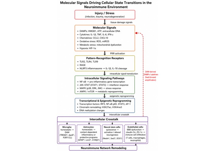
Molecular signals driving cellular state transitions in the neuroimmune environment. Injury, infection, or neurodegeneration triggers the release of molecular signals including damage-associated molecular patterns (DAMPs), cytokines, chemokines, and metabolic stress signals. These signals activate pattern-recognition receptors such as TLRs, RAGE, and the NLRP3 inflammasome, initiating intracellular signaling pathways including NF-κB, JAK-STAT, MAPK, and AMPK/mTOR. Downstream transcriptional and epigenetic reprogramming alters gene-expression programs that drive functional state transitions in microglia, astrocytes, neural stem cells, and endothelial cells. Intercellular crosstalk among these populations, together with infiltration of peripheral immune cells, contributes to neuroimmune network remodeling during neurological disease.
Microglia represent the principal immune cells of the central nervous system and play a central role in sensing and responding to disturbances in neural tissue. As discussed in previous sections, the healthy brain maintains a stable neuroimmune environment through coordinated interactions among neurons, glial cells, and vascular elements. Within this environment, microglia function as dynamic regulators of immune surveillance and tissue maintenance. However, in response to injury, infection, or neurodegenerative processes, microglia can shift from homeostatic surveillance programs toward diverse inflammatory, phagocytic, metabolic, IFN-responsive, repair-associated, or disease-associated programs. These transitions involve coordinated changes in cellular morphology, signaling pathways, metabolic activity, and gene-expression programs [22, 105–107].
Recent advances in molecular profiling techniques, particularly single-cell transcriptomic approaches, have revealed that microglial activation is not limited to a single inflammatory phenotype but instead involves multiple functional states that arise under different physiological and pathological conditions [22, 105, 106, 108]. Understanding these microglial state transitions is therefore essential for interpreting how neuroimmune responses contribute to both tissue protection and disease progression.
Microglia originate from primitive macrophage precursors derived from the embryonic yolk sac and populate the central nervous system early during development. Unlike many other immune cells, microglia maintain themselves through local self-renewal rather than continuous recruitment from circulating monocytes [109–111]. As long-lived resident immune cells, they remain distributed throughout the brain parenchyma and serve as primary sensors of changes within the neural environment.
In the healthy brain, microglia continuously survey the surrounding tissue through highly motile cellular processes that extend and retract to monitor neuronal activity and extracellular signals. This dynamic surveillance behavior enables microglia to rapidly detect subtle alterations in tissue integrity, including changes in neuronal signaling, extracellular molecules, or cellular damage [112–114]. Through this surveillance function, microglia contribute to maintaining the stability of neural circuits and tissue homeostasis.
Microglia also engage in extensive communication with other cellular populations in the brain. Interactions with neurons help regulate synaptic remodeling and neuronal activity, while communication with astrocytes and vascular cells contributes to the coordination of inflammatory responses and metabolic support. Through these interactions, microglia integrate signals from multiple sources to determine whether local conditions require maintenance of homeostasis or initiation of immune responses.
Taken together, these features position microglia as sentinel cells within the central nervous system. By detecting environmental changes and coordinating cellular responses, microglia play a critical role in initiating and regulating neuroimmune activity.
Under physiological conditions, microglia exist predominantly in homeostatic states that support tissue stability and neural circuit maintenance. In many CNS regions, these states are associated with a ramified morphology characterized by small cell bodies and extensively branched processes that continuously monitor the local microenvironment [109, 115, 116]. However, homeostatic microglial morphology is not uniform across the CNS; regional microenvironmental differences can shape microglial density, branching complexity, and baseline morphology even in the absence of overt pathology [33]. This surveillance behavior allows microglia to detect subtle changes in neuronal activity or tissue integrity while maintaining a non-inflammatory functional profile.
Homeostatic microglia contribute to several important physiological processes. These include the removal of apoptotic cells and cellular debris, regulation of synaptic pruning during development and adulthood, and modulation of neuronal signaling within active neural circuits [109, 112, 117]. Through these functions, microglia help maintain synaptic balance and support the structural organization of neural networks.
Molecular profiling studies have identified characteristic transcriptional signatures associated with homeostatic microglia. Common markers used to identify these cells include purinergic receptor P2RY12, fractalkine receptor CX3CR1, and transmembrane protein TMEM119 [107, 109, 118]. These molecules contribute to the ability of microglia to sense extracellular signals and maintain communication with neighboring cells. In addition, signaling through receptors such as TREM2 plays an important role in regulating microglial responses to environmental cues.
Together, these molecular and functional characteristics define a stable homeostatic microglial state that supports neural tissue maintenance. Importantly, this state does not represent a passive condition but rather a dynamic equilibrium in which microglia remain poised to respond rapidly when the surrounding environment changes.
When the neural environment is disrupted by injury, infection, or neurodegenerative processes, microglia can transition from homeostatic programs toward context-dependent inflammatory, phagocytic, antigen-presenting, metabolic, or repair-associated programs. A variety of signals may initiate this transition, including the release of DAMPs from injured cells, the presence of pathogen-associated molecules during infection, and the accumulation of abnormal proteins or cellular debris in neurodegenerative conditions [105, 108, 119]. In addition, cytokines and chemokines produced by neighboring cells can further amplify microglial activation.
Activated microglia exhibit several functional changes that distinguish them from homeostatic cells. These include increased production of pro-inflammatory cytokines, generation of reactive oxygen species, enhanced phagocytic activity, and in some cases the presentation of antigens to other immune cells [108, 119, 120]. These responses are mediated through intracellular signaling pathways that regulate inflammatory gene expression and cellular metabolism.
Several signaling cascades have been implicated in the regulation of inflammatory microglial activation. These include pathways mediated by NF-κB, MAPK, and inflammasome complexes that promote the maturation and release of inflammatory cytokines [95, 106]. Activation of these pathways alters transcriptional programs within microglia and drives functional changes that support immune responses within the brain.
Importantly, current evidence suggests that microglial activation does not occur as a simple binary transition between resting and activated states. Instead, microglial responses appear to exist along a continuum of functional phenotypes that vary depending on the nature, duration, and intensity of environmental stimuli. This spectrum of activation states reflects the broader principle that microglial behavior is dynamically regulated by the surrounding tissue environment during neuroimmune responses.
Recent advances in single-cell transcriptomic technologies have revealed additional microglial states that emerge during neurodegenerative disease. Among the most extensively studied of these states are DAM, a transcriptionally distinct population first described in mouse models of neurodegeneration [22, 105, 108]. These cells were identified through single-cell RNA sequencing analyses that revealed coordinated shifts in gene-expression programs as microglia respond to pathological changes within neural tissue.
DAM exhibit several molecular and functional features that distinguish them from homeostatic microglia. One characteristic change involves alterations in lipid metabolism pathways, which are thought to reflect the increased need for processing lipid-rich cellular debris during neurodegenerative processes [22, 108, 119]. DAM also display enhanced phagocytic activity, enabling them to engulf extracellular aggregates and damaged cellular components. At the transcriptional level, these cells frequently exhibit downregulation of genes associated with homeostatic microglial identity while simultaneously upregulating genes linked to immune activation, lipid handling, and cellular stress responses.
A central regulator of the DAM state is signaling through TREM2. Activation of TREM2-dependent pathways has been shown to promote microglial responses to tissue damage and to facilitate transitions toward disease-associated phenotypes [108, 119, 121]. Experimental studies suggest that this signaling pathway contributes to microglial responses to lipid accumulation and cellular debris in neurodegenerative conditions.
In models of Alzheimer’s disease, DAM have been frequently observed in close proximity to amyloid plaques. These plaque-associated microglia display transcriptional signatures consistent with the DAM state and appear to participate in local immune responses surrounding pathological protein aggregates [108, 119, 120, 122]. Although the functional consequences of DAM activation remain an area of active investigation, these observations highlight the importance of microglial state transitions in shaping immune responses during neurodegenerative disease.
Transitions between microglial states are regulated by multiple molecular mechanisms that integrate environmental signals with intracellular regulatory networks. These mechanisms include transcriptional regulation, epigenetic remodeling, metabolic adaptation, and signaling pathway activation. Together, these processes determine how microglia respond to changes in the neural environment and adopt new functional phenotypes.
Transcriptional regulation plays a central role in controlling microglial state transitions. Environmental signals such as cytokines, damage-associated molecules, and metabolic stress activate intracellular signaling pathways that modify transcription factor activity and alter gene-expression programs within microglia [105, 106, 108]. These transcriptional changes can promote inflammatory responses, enhance phagocytic activity, or initiate stress-response pathways depending on the nature of the stimulus.
Epigenetic mechanisms also contribute to the regulation of microglial identity and responsiveness. Processes such as chromatin remodeling, histone modifications, and changes in DNA methylation influence the accessibility of genomic regions involved in immune signaling and cellular metabolism [106, 123, 124]. Through these regulatory mechanisms, microglia can maintain stable transcriptional programs or transition toward alternative functional states in response to environmental signals.
Metabolic reprogramming represents another important component of microglial state transitions. Activated microglia often undergo shifts in energy metabolism that support increased biosynthetic and inflammatory activity. For example, inflammatory activation has been associated with metabolic changes that favor glycolytic pathways over oxidative phosphorylation [125, 126]. Such metabolic adaptations can influence signaling pathways and gene-expression programs that sustain immune responses.
Several signaling pathways have been implicated in regulating these transitions. TREM2 signaling has emerged as a key regulator of microglial responses to lipid accumulation and tissue damage, while IFN-related pathways contribute to microglial responses during viral infection and inflammatory conditions [127–129]. Together, these signaling networks integrate extracellular cues with transcriptional and metabolic regulation, enabling microglia to transition between functional states.
Much of the current understanding of microglial heterogeneity and state transitions has been enabled by recent technological advances in high-resolution molecular profiling. Single-cell RNA sequencing has allowed researchers to characterize transcriptional diversity within microglial populations and identify distinct cellular states that emerge under different physiological and pathological conditions [22, 105, 108]. These approaches have revealed that microglia exhibit a spectrum of transcriptional profiles rather than a single uniform phenotype.
Spatial transcriptomic technologies have further expanded these insights by enabling the mapping of gene-expression patterns within intact tissue architecture. By combining spatial information with molecular profiling, these methods have provided a more detailed understanding of how microglial states vary across different brain regions and in proximity to pathological features such as protein aggregates or sites of tissue injury [31, 130, 131].
Experimental models of neurodegenerative disease have also contributed substantially to the study of microglial state transitions. Mouse models of Alzheimer’s disease, Parkinson’s disease, and other neurodegenerative conditions have enabled investigators to examine how microglia respond to progressive neuronal damage and protein aggregation over time. These studies have helped define transcriptional programs associated with disease-related microglial states, including the emergence of DAM [108, 127, 130].
Complementary evidence has been obtained from analyses of human post-mortem brain tissue. Molecular profiling of microglia from individuals with neurodegenerative disorders has identified transcriptional signatures that overlap with disease-associated states described in experimental models [22, 132, 133]. Together, findings from single-cell technologies, experimental disease models, and human studies have revealed previously unrecognized heterogeneity within microglial populations and have highlighted the importance of cellular state transitions in shaping neuroimmune responses. However, transcriptional similarity between microglial states across models and human tissue should be interpreted cautiously, because shared molecular signatures do not necessarily establish equivalent function, disease causality, or therapeutic tractability.
Astrocytes are among the most abundant glial cells in the central nervous system and play essential roles in maintaining neural homeostasis. In addition to supporting neuronal activity under physiological conditions, astrocytes are highly responsive to changes in the neural environment and can undergo significant functional and molecular changes during injury and disease. These reactive transitions influence inflammatory signaling, neuronal function, and tissue remodeling in a wide range of neurological conditions. Understanding astrocyte reactivity and the diversity of astrocyte states that emerge during neuroimmune activation is therefore important for interpreting how glial responses contribute to both protective and pathological processes within the brain.
Under physiological conditions, astrocytes contribute to multiple processes that support neural circuit stability and metabolic balance. These cells extend elaborate processes that interact with synapses, neurons, and blood vessels, positioning them as key intermediaries between neural activity and the broader tissue environment. Through these structural and functional interactions, astrocytes help regulate synaptic transmission and maintain the extracellular conditions necessary for efficient neuronal signaling [134–136].
One important role of astrocytes involves the regulation of neurotransmitter levels within synaptic spaces. By actively taking up neurotransmitters such as glutamate and γ-aminobutyric acid, astrocytes help maintain excitatory–inhibitory balance and prevent excessive neuronal stimulation [40, 46, 136]. Astrocytes also provide metabolic support to neurons by supplying energy substrates and participating in metabolic coupling between neuronal activity and glucose metabolism.
In addition to their metabolic functions, astrocytes contribute to maintaining extracellular ion balance within neural tissue. Their ability to buffer potassium and regulate other ions is essential for stabilizing neuronal excitability and preventing abnormal electrical activity within neural circuits [46, 134, 137]. Astrocytes also participate in neurovascular regulation through specialized endfeet that interact with cerebral blood vessels, thereby contributing to the maintenance of blood–brain barrier function and vascular signaling.
Through these diverse physiological roles, astrocytes function as integrative regulators that coordinate neuronal activity, metabolic processes, and vascular interactions within the central nervous system. Although these cells maintain a stable functional state under normal conditions, they remain highly sensitive to environmental changes within neural tissue.
When the neural environment is disrupted by injury, infection, neurodegeneration, or inflammatory signaling, astrocytes can transition from homeostatic programs toward diverse context-dependent remodeling programs. This process, commonly referred to as astrogliosis, represents a broad spectrum of cellular responses rather than a single uniform or binary state [138, 139].
A variety of signals can trigger astrocyte reactivity. These include tissue injury that disrupts cellular integrity, neurodegenerative processes that generate abnormal protein aggregates or cellular debris, and infection that activates immune signaling pathways. Inflammatory cytokines produced by microglia and infiltrating immune cells also play an important role in initiating astrocyte responses. Through these signals, astrocytes detect changes in the local microenvironment and activate intracellular pathways that alter their functional state [140–142].
Reactive astrocytes exhibit several characteristic changes compared with their homeostatic counterparts, although these changes vary by brain region, disease context, and injury severity. Morphologically, astrocytes often display cellular hypertrophy and increased expression of structural proteins associated with cytoskeletal remodeling, including increased GFAP expression in many but not all reactive contexts. At the molecular level, reactive astrocytes undergo changes in gene-expression programs that influence inflammatory signaling, metabolic activity, neurotransmitter-related pathways, and interactions with neighboring cells [143]. Alterations in cellular metabolism and signaling pathways further contribute to the functional diversity of reactive astrocyte states.
These adaptive responses allow astrocytes to participate in protective processes such as limiting tissue damage, supporting neuronal survival, and contributing to tissue repair. However, under certain conditions, prolonged or dysregulated astrocyte activation may also contribute to inflammatory signaling and neuronal dysfunction. As a result, astrocyte reactivity represents a dynamic process in which multiple cellular states can emerge depending on the nature and duration of environmental stimuli.
Reactive astrocytes display considerable functional diversity, and accumulating evidence suggests that astrocyte reactivity cannot be fully described as a single uniform state. Instead, reactive astrocytes can adopt distinct functional programs that influence neural survival, inflammation, and tissue remodeling in different ways. Early conceptual frameworks proposed that reactive astrocytes could be broadly categorized into neuroprotective and neurotoxic tendencies [139, 144, 145]. However, these categories should be interpreted as simplified functional descriptors rather than discrete astrocyte identities. Current evidence supports a more continuous and context-dependent view of astrocyte remodeling, in which inflammatory, metabolic, synaptic, vascular, scar-forming, and repair-associated programs may overlap within the same tissue environment [146].
Neuroprotective astrocyte states are generally associated with processes that support neuronal survival and tissue recovery following injury. These astrocytes can enhance metabolic support to neurons, increase antioxidant defenses that limit oxidative damage, and help buffer excitotoxic neurotransmitters such as glutamate [46, 142, 144]. In addition, reactive astrocytes in protective states may promote tissue repair through the release of trophic factors and by contributing to structural stabilization within injured neural tissue.
In contrast, other reactive astrocyte states have been associated with inflammatory signaling and neuronal dysfunction. In these contexts, astrocytes may produce inflammatory mediators that amplify immune responses within the brain. These inflammatory signaling pathways can influence synaptic function, disrupt neuronal communication, and in some cases contribute to neuronal injury or degeneration.
It is important to note that these classifications represent functional tendencies rather than rigid cellular identities. Reactive astrocytes likely exist along a spectrum of states that vary depending on the nature of environmental stimuli, the duration of inflammatory signaling, and the local cellular context. As a result, astrocyte reactivity is increasingly viewed as a dynamic process in which multiple intermediate states can arise during neuroimmune responses.
Astrocyte reactivity is strongly influenced by signals from other cellular populations within the neuroimmune environment, particularly microglia. Because microglia serve as primary sensors of tissue injury and inflammation in the central nervous system, their activation often precedes and shapes subsequent astrocyte responses [38, 71, 140, 147]. Through the release of cytokines, chemokines, purinergic mediators, and other inflammatory signals, microglia can initiate signaling cascades that influence astrocyte gene-expression programs and functional states.
Several microglia-derived molecules have been identified as important regulators of astrocyte reactivity, highlighting the importance of microglia–astrocyte signaling networks in shaping reactive glial state transitions. Cytokines such as IL-1α and TNF, as well as components of the complement system, have been shown to influence astrocyte activation and promote the emergence of specific reactive states [71, 141, 148]. These signaling molecules can activate intracellular pathways within astrocytes that alter transcriptional programs, metabolic activity, and interactions with neighboring neurons.
Damage signals released from injured neurons may further contribute to astrocyte activation by providing additional inflammatory or stress-related cues within the tissue environment. Through these combined signals, astrocytes integrate information from multiple cellular sources when determining their functional responses to injury or disease.
These observations highlight the importance of intercellular communication in regulating astrocyte behavior. Rather than emerging independently, reactive astrocyte states often arise through microglia-driven signaling cascades that coordinate immune responses across different cell populations within the central nervous system.
Recent advances in molecular profiling technologies have greatly expanded the understanding of astrocyte heterogeneity. High-resolution approaches such as single-cell RNA sequencing have revealed that astrocytes exhibit diverse transcriptional programs that vary across brain regions, developmental stages, and disease conditions [149, 150]. These findings suggest that astrocyte populations contain multiple molecularly distinct states that may perform specialized functions within the neural environment.
Spatial transcriptomic techniques have further contributed to this understanding by enabling the mapping of gene-expression patterns within intact brain tissue. These approaches allow investigators to examine how astrocyte transcriptional states are distributed across different anatomical regions and how they change in response to local pathological conditions [147, 149, 151]. In combination with experimental disease models, these studies have provided insights into how astrocyte responses evolve during neurodegenerative and inflammatory disorders.
Analyses of human brain tissue have also supported the presence of diverse astrocyte states in neurological disease. Molecular profiling of astrocytes from post-mortem brain samples has revealed transcriptional signatures that reflect both inflammatory and protective cellular responses [139, 147, 152]. Together, these studies indicate that astrocyte reactivity encompasses a spectrum of transcriptional and functional states rather than a simple binary classification. This emerging view of astrocyte diversity highlights the complexity of glial responses within the neuroimmune environment and underscores the importance of cellular state transitions in shaping brain pathology and repair. As with microglia, astrocyte state labels derived from molecular profiling should be interpreted as context-dependent descriptors that require functional validation, particularly when extrapolating from experimental models to human disease. Major functional states of microglia and astrocytes discussed in Sections 4 and 5 are summarized in Table 3.
Functional states of microglia and astrocytes in the neuroimmune environment.
| Cell type | Functional state | Key molecular markers | Functional features | Associated conditions |
|---|---|---|---|---|
| Microglia | Homeostatic microglia | P2RY12, TMEM119, CX3CR1 | Immune surveillance, synaptic monitoring, clearance of cellular debris, maintenance of tissue homeostasis | Healthy brain physiology [22, 153] |
| Microglia | Activated inflammatory microglia | CD68, MHC-II, IL-1β, TNF | Production of inflammatory cytokines, reactive oxygen species generation, antigen presentation, enhanced phagocytosis | Infection, acute injury, neuroinflammation [97, 154] |
| Microglia | Disease-associated microglia (DAM) | TREM2, APOE, LPL, CST7 | Enhanced phagocytosis of protein aggregates, lipid metabolism adaptation, response to neurodegenerative pathology | Alzheimer’s disease, Parkinson’s disease, neurodegeneration [155, 156] |
| Astrocytes | Homeostatic astrocytes | GLT-1/EAAT2, Kir4.1; GFAP low or regionally restricted under basal conditions | Regulation of neurotransmitter levels, metabolic support to neurons, potassium buffering, neurovascular coupling | Healthy brain physiology [40, 46] |
| Astrocytes | Neuroprotective reactive astrocytes | Increased GFAP, antioxidant pathways, trophic factors | Support neuronal survival, limit oxidative stress, promote tissue stabilization and repair | Early injury response, recovery phases [142, 144] |
| Astrocytes | Neurotoxic reactive astrocytes | Complement component C3, inflammatory mediators | Amplification of inflammatory signaling, disruption of synaptic function, promotion of neuronal injury | Neurodegenerative disease, chronic neuroinflammation [139, 140] |
Adult neural stem cells represent an important component of cellular plasticity within the central nervous system. In addition to their role in generating new neurons and glial cells, these cells respond to changes in the tissue environment, including signals associated with inflammation and injury. Increasing evidence indicates that inflammatory signaling and neuroimmune interactions can influence stem-cell behavior, altering their activation, proliferation, and differentiation potential. Understanding how neural stem cells and progenitor populations respond to these signals is therefore important for interpreting how regenerative responses are regulated during neurological disease.
In the adult mammalian brain, neural stem cells are primarily located in two specialized neurogenic regions: the SVZ lining the lateral ventricles and the subgranular zone of the dentate gyrus within the hippocampus [157–159]. Within these niches, neural stem cells can give rise to progenitor populations with neuronal or glial lineage potential, but the magnitude of this contribution differs across species, anatomical regions, age, and disease states. Therefore, cellular turnover and structural plasticity mediated by adult neural stem-cell populations should be interpreted as context-dependent rather than uniform features of the adult brain [55, 160].
These stem cells do not function in isolation but instead exist within complex microenvironments that regulate their activity, integrating local cellular, vascular, immune, and circuit-derived cues that shape stem-cell behavior. The stem-cell niche integrates signals from multiple sources, including neighboring astrocytes, microglia, endothelial cells, extracellular matrix components, and circuit-linked inputs that regulate stem-cell activity. Through these interactions, neural stem cells receive regulatory cues that influence their proliferation, differentiation, and long-term maintenance [25, 57, 62, 159, 161].
Signals within the niche include growth factors, neurotransmitters, metabolic cues, and immune-related molecules. Together, these signals help maintain a balanced environment in which stem cells can remain largely quiescent while retaining the capacity to activate in response to physiological demands or tissue injury. This specialized microenvironment therefore plays a critical role in integrating neural, vascular, and immune signals that regulate stem-cell behavior.
Adult neural stem cells exhibit dynamic transitions between distinct functional states. Under physiological conditions, most stem cells remain in a quiescent state characterized by low proliferative activity and stable maintenance within the niche. This quiescent state is essential for preserving the long-term regenerative capacity of the stem-cell pool and preventing premature depletion of stem-cell populations [25, 162, 163].
In response to specific signals from the surrounding microenvironment, subsets of these quiescent cells can transition into an activated state, indicating that adult neural stem-cell activation is closely coupled to niche-derived and circuit-associated regulatory mechanisms [57]. Defined niche-associated signaling pathways, including cholinergic circuit-linked inputs, have been shown to participate in regulating this transition [164]. Activated stem cells enter the cell cycle and generate proliferating progenitor cells, which subsequently differentiate into neurons or glial cells depending on the signals present within the niche. This sequence of transitions—from quiescent stem cells to activated stem cells, proliferating progenitors, and ultimately differentiated cells—represents a fundamental process underlying adult neurogenesis.
These transitions reflect the broader concept that stem-cell behavior is governed by dynamic cellular states rather than fixed identities. Neural stem cells are increasingly understood as active components of regulatory niche networks that both receive and shape local signaling dynamics [62, 165]. Similar to immune and glial populations, stem cells respond to environmental cues by adjusting their functional state through changes in signaling pathways, transcriptional programs, and metabolic activity.
Inflammatory signals within the neural environment can significantly influence stem-cell activity and lineage decisions. Cytokines, chemokines, and DAMPs released during injury or disease can interact directly with stem cells or indirectly alter the niche environment through effects on neighboring cells such as microglia and astrocytes [166–168].
Microglia-derived signals are particularly important regulators of stem-cell behavior. Activated microglia can release cytokines and growth factors that influence stem-cell activation and differentiation. Similarly, astrocytes and vascular cells within the niche may respond to inflammatory cues and modify the molecular environment that governs stem-cell function. In parallel, niche-associated neurotransmitter and circuit-linked signaling pathways can also influence the transition of quiescent neural stem cells toward activation [164].
Through these signaling pathways, inflammatory environments can alter multiple aspects of stem-cell biology. In some contexts, immune-related signals promote stem-cell activation and proliferation, while in others they may suppress cell-cycle progression or shift differentiation toward specific lineages. These effects highlight the sensitivity of neural stem cells to immune-derived signals within their microenvironment.
The influence of inflammation on neural stem cells is not uniformly detrimental. Instead, the effects of inflammatory signaling often depend on the intensity and duration of the immune response. Mild or transient inflammatory signals can stimulate stem-cell activation and enhance neurogenesis in certain contexts [167, 169, 170]. Such responses may represent adaptive mechanisms that support tissue repair or functional plasticity following minor injury.
In contrast, chronic or sustained inflammation can have inhibitory effects on stem-cell populations. Prolonged exposure to inflammatory cytokines and oxidative stress may suppress stem-cell proliferation, impair neurogenic potential, and shift differentiation toward glial lineages rather than neuronal outcomes [167, 171–173]. These changes can reduce the regenerative capacity of neurogenic niches and contribute to long-term alterations in neural plasticity.
These observations illustrate that the effects of inflammation on stem-cell populations are highly context-dependent. The duration, magnitude, and cellular sources of inflammatory signals all influence how stem cells respond within the neural environment.
Stem-cell responses have been observed in a variety of neurological conditions associated with injury or degeneration. In experimental models of stroke and traumatic brain injury, neural stem cells within the SVZ and dentate gyrus can become activated and contribute to the generation of new neurons or glial cells during tissue repair processes [174, 175]. These responses may support limited structural remodeling and functional recovery following injury.
However, in chronic disease environments such as neurodegenerative disorders and aging, stem-cell function is often impaired. Persistent inflammatory signaling, metabolic stress, and alterations in the stem-cell niche can reduce the ability of neural stem cells to proliferate and generate new neurons [157, 161, 173]. As a result, regenerative responses may decline over time, contributing to reduced neural plasticity in aging and disease.
Together, these findings illustrate that neural stem cells represent an important cellular component of neuroimmune responses in the brain and may participate in broader feedback relationships with surrounding niche and circuit elements during disease-associated adaptation [165]. Through their capacity to transition between quiescent and activated states, stem cells integrate signals from the surrounding microenvironment and contribute to adaptive or maladaptive responses during neurological disease.
These observations should be interpreted within a broader regenerative-medicine context. Studies outside the central nervous system have shown that stem-cell state transitions and lineage plasticity can be experimentally manipulated, including bone marrow stem-cell differentiation toward β-cell-like phenotypes and hyaluronic acid-based delivery approaches for tissue remodeling [58, 59]. Although such extra-neural examples do not provide direct evidence for neural stem-cell reprogramming in the adult brain, they support the broader principle that cellular state transitions can be therapeutically guided when lineage potential, niche signals, and delivery systems are appropriately controlled.
While neural stem cells respond primarily to local signals within neurogenic niches, broader neuroimmune responses are also shaped by systemic immune activity. Under pathological conditions, inflammatory signals can alter vascular barrier properties and permit the entry of circulating immune cells into the central nervous system. These infiltrating immune populations further modify the neuroimmune environment through interactions with resident glial cells and local signaling networks. Understanding how peripheral immune cells enter and adapt to the central nervous system therefore represents an important component of the broader cellular state transition framework.
Peripheral immune cells can enter the central nervous system under pathological conditions and contribute to neuroimmune responses that influence disease progression and tissue remodeling. Although the brain is normally protected by specialized vascular barriers that limit immune cell entry, inflammatory signals generated during injury, infection, or neurodegeneration can alter these regulatory mechanisms. As a result, circulating immune cells may migrate into neural tissue where they interact with resident glial populations and participate in local immune responses. Understanding how peripheral immune cells enter the brain and adapt to the neural environment is therefore important for interpreting how systemic immune responses contribute to neurological disease.
Under physiological conditions, the blood-brain barrier restricts the entry of circulating immune cells into neural tissue. This barrier is formed by tightly connected endothelial cells supported by pericytes, basement membrane structures, and astrocytic endfeet, which together regulate molecular and cellular exchange between the bloodstream and the central nervous system [48, 176, 177]. Through these structural and molecular mechanisms, the blood-brain barrier maintains a controlled environment that limits unnecessary immune activation within the brain.
During inflammatory or pathological conditions, however, the properties of the vascular barrier can change. Inflammatory mediators released during tissue injury or infection can alter endothelial cell signaling and increase vascular permeability. These changes facilitate the recruitment and migration of leukocytes from the circulation into neural tissue [178–180]. Endothelial cells play a central role in this process by expressing adhesion molecules and chemotactic signals that guide immune cell trafficking.
Several adhesion molecules are particularly important for regulating leukocyte migration across the vascular wall. Intercellular adhesion molecule-1 (ICAM-1), vascular cell adhesion molecule-1 (VCAM-1), and selectin family proteins mediate interactions between circulating leukocytes and endothelial cells [181, 182]. Chemokines produced within inflamed neural tissue further promote directed migration of immune cells toward sites of injury or inflammation. Through these coordinated molecular interactions, vascular and inflammatory signals permit the controlled entry of immune cells into the central nervous system.
Multiple types of circulating immune cells can enter the central nervous system under pathological conditions. Among these, monocyte-derived macrophages represent an important population that participates in inflammatory signaling and tissue remodeling. After migrating from the circulation into neural tissue, these cells can differentiate into macrophages that contribute to the clearance of cellular debris and the production of inflammatory mediators [183, 184].
T lymphocytes also play important roles in neuroimmune responses. Different T-cell subsets perform distinct functions within the brain. CD4+ helper T cells regulate immune signaling through cytokine production, whereas CD8+ cytotoxic T cells can directly target infected or damaged cells. Regulatory T cells contribute to immune regulation by suppressing excessive inflammatory responses and promoting tissue protection under certain conditions [185–187].
Neutrophils represent another class of infiltrating immune cells, particularly during acute injury or infection. These cells are among the earliest responders in inflammatory responses and can release enzymes, reactive oxygen species, and inflammatory mediators that influence tissue damage and immune signaling [188, 189]. Together, these diverse immune cell populations contribute distinct functional roles within neuroimmune responses.
The major peripheral immune cell populations that infiltrate the central nervous system and their functional roles are summarized in Table 4.
Peripheral immune cells infiltrating the central nervous system during neuroinflammation.
| Immune cell type | Origin | Entry mechanism | Key functions of CNS | Associated diseases |
|---|---|---|---|---|
| Monocyte-derived macrophages | Circulating monocytes originating from bone marrow hematopoiesis | Migration across blood-brain barrier through adhesion molecules (ICAM-1, VCAM-1) and chemokine signaling | Phagocytosis of cellular debris, production of inflammatory mediators, contribution to tissue remodeling and repair | Multiple sclerosis, stroke, traumatic brain injury [183, 184] |
| CD4+ T cells | Peripheral lymphoid organs | Antigen-dependent migration across inflamed vascular endothelium via chemokine-directed trafficking | Regulation of immune responses through cytokine production, coordination of inflammatory signaling | Multiple sclerosis, viral CNS infections [187] |
| CD8+ T cells | Peripheral lymphoid organs | Recruitment through chemokine gradients and adhesion molecule interactions at the blood-brain barrier | Cytotoxic responses against infected or damaged cells, antigen-specific immune defense | Viral encephalitis, autoimmune neuroinflammation [190, 191] |
| Regulatory T cells (Tregs) | Peripheral immune system, thymus-derived T-cell lineage | Migration to inflamed CNS tissue through chemokine-mediated trafficking | Suppression of excessive inflammation, modulation of immune responses, support of tissue protection and repair | Multiple sclerosis, neurodegenerative disorders [186] |
| Neutrophils | Bone marrow-derived granulocytes | Rapid recruitment through endothelial adhesion molecules and chemokine signaling during acute inflammation | Release of proteases, reactive oxygen species, and inflammatory mediators; contribution to early inflammatory responses | Stroke, traumatic brain injury, infection [188, 192] |
After entering neural tissue, peripheral immune cells undergo functional changes that reflect the unique conditions of the central nervous system environment. The molecular signals present within the neural microenvironment—including cytokines, chemokines, metabolic cues, and interactions with resident cells—can alter gene-expression programs and functional behaviors of infiltrating immune cells [29, 193].
Monocyte-derived macrophages, for example, can adopt distinct polarization states that influence inflammatory responses and tissue repair. Some macrophage populations exhibit pro-inflammatory phenotypes that promote cytokine production and immune activation, whereas others display reparative properties associated with tissue remodeling and debris clearance [194, 195]. These polarization states reflect the ability of macrophages to adapt their functional roles in response to local environmental signals.
T lymphocytes also undergo functional modulation within the central nervous system. Effector T cells may become activated in response to antigen presentation and inflammatory signals, while regulatory T cells can suppress inflammatory responses and promote immune resolution. Through these adaptive responses, infiltrating immune cells integrate into the neuroimmune environment and contribute to local immune regulation.
Peripheral immune cells that enter the central nervous system interact extensively with resident glial populations. These interactions form part of broader neuroimmune networks that regulate inflammation, tissue repair, and cellular survival within neural tissue. Communication between infiltrating macrophages and resident microglia can influence inflammatory signaling and phagocytic activity, while interactions with astrocytes can modify the local inflammatory environment [13, 189, 196].
T lymphocytes also interact with microglia and astrocytes through cytokine signaling and antigen presentation pathways. These interactions can modulate glial activation states and influence downstream immune responses within the brain. Through these signaling networks, peripheral immune cells become integrated into the existing cellular communication systems that regulate neuroimmune activity.
Such interactions can have multiple functional consequences. In some contexts, these cellular networks promote inflammatory signaling that contributes to tissue damage. In other situations, coordinated interactions between immune cells and glia can support debris clearance, tissue remodeling, and recovery following injury.
Peripheral immune cell infiltration has been observed in a variety of neurological diseases and injury conditions. In multiple sclerosis, immune cell migration across the blood-brain barrier contributes to inflammatory attacks on myelin and neural tissue [181, 184]. Similarly, infiltration of macrophages and neutrophils occurs in response to ischemic injury during stroke and in traumatic brain injury, where these cells participate in inflammatory signaling and tissue remodeling.
Peripheral immune responses have also been implicated in neurodegenerative diseases. In these conditions, interactions between infiltrating immune cells and resident glia may influence the progression of neuronal damage and the regulation of inflammatory responses within the brain [178, 197, 198].
Importantly, the effects of peripheral immune cells in the central nervous system are not uniformly detrimental. While excessive or chronic immune infiltration can contribute to tissue damage, certain immune responses may also promote repair processes and support tissue recovery under specific conditions. These observations highlight the complex and context-dependent roles of peripheral immune cells in shaping neuroimmune responses and disease outcomes within the central nervous system.
Interactions among resident glial cells, infiltrating immune populations, neurons, vascular elements, and neural stem-cell niches form an integrated neuroimmune communication network within the central nervous system. These interconnected signaling relationships coordinate inflammatory signaling, synaptic remodeling, neurogenesis, and tissue repair processes. The major cellular components and bidirectional signaling pathways that regulate these neuroimmune interactions are summarized in Figure 3.
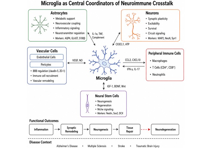
Neuroimmune cellular interaction network in the central nervous system. Microglia function as central immune regulators that coordinate bidirectional signaling with astrocytes, neurons, vascular cells, neural stem cells, and infiltrating peripheral immune cells. These interactions occur through cytokines, chemokines, complement signaling, growth factors, and metabolic mediators that regulate inflammatory responses, synaptic remodeling, neurogenesis, and tissue repair. Dysregulation of these neuroimmune networks contributes to the pathogenesis of multiple neurological disorders including Alzheimer’s disease, multiple sclerosis, stroke, and traumatic brain injury.
Inflammatory responses within the central nervous system are often followed by phases of structural and functional remodeling that influence the long-term outcome of neural injury or disease. During these phases, coordinated interactions among immune cells, glial populations, neurons, and vascular elements reshape the local tissue environment. These remodeling processes can support recovery and repair by removing damaged structures and restoring functional networks, but they may also contribute to persistent dysfunction when inflammatory signaling remains unresolved or when structural changes interfere with normal neural connectivity [199, 200].
Inflammatory responses typically represent an early phase of neuroimmune activation that precedes subsequent remodeling of neural tissue. As described in previous sections, activation of microglia, astrocytes, and infiltrating immune cells initiates signaling cascades that alter the molecular and cellular environment of the brain. These changes include the production of cytokines, chemokines, and growth factors that influence both immune responses and cellular repair mechanisms [200, 201].
As inflammatory signaling evolves, cellular state transitions within microglia, astrocytes, and other immune-related populations begin to shape the environment in which tissue remodeling occurs. These transitions influence processes such as debris clearance, extracellular matrix remodeling, and the release of trophic factors that regulate neuronal survival and structural reorganization. In many contexts, resolution of acute inflammation is accompanied by activation of repair-related pathways that promote tissue stabilization and regeneration [199, 201, 202].
Although inflammation and remodeling are closely connected processes, they represent distinct phases of neuroimmune responses. While inflammatory signaling primarily functions to detect and respond to tissue disturbance, subsequent remodeling processes determine whether the affected neural tissue undergoes repair, adaptation, or long-term dysfunction.
One of the major consequences of neuroimmune activation is the remodeling of synaptic structures and neural circuits. During inflammatory responses, microglia and astrocytes can influence synaptic stability and connectivity through mechanisms that regulate synapse elimination and formation [203, 204]. These processes are particularly important for removing damaged synapses and restoring functional neural networks following injury.
Microglia play a central role in synaptic pruning by recognizing and engulfing synaptic elements that are marked for removal. This process can occur during both developmental and pathological conditions and may be guided by immune-related signaling pathways that identify dysfunctional synaptic connections [204, 205]. Astrocytes also contribute to synaptic regulation by modulating neurotransmitter uptake, releasing signaling molecules that influence neuronal communication, and interacting directly with synaptic structures.
Through these coordinated interactions, immune signaling pathways can reshape synaptic architecture and modify the organization of neural circuits. In some cases, these processes promote adaptive circuit reorganization that supports recovery of function. In other contexts, excessive synaptic pruning or persistent inflammatory signaling may disrupt neural connectivity and contribute to neurological dysfunction.
Structural remodeling following neural injury frequently involves the formation of glial scars, which are composed primarily of reactive astrocytes and extracellular matrix components. After injury, astrocytes can proliferate and undergo morphological changes that lead to the formation of a dense cellular barrier surrounding the damaged region [201, 206]. This process is accompanied by deposition of extracellular matrix molecules that reinforce the structural integrity of the injured tissue.
Glial scar formation plays several protective roles within the injured brain. By isolating damaged areas, glial scars help contain inflammatory responses and limit the spread of cellular damage to surrounding healthy tissue [201, 207]. In addition, the structural framework created by reactive astrocytes can stabilize the affected region and contribute to the restoration of tissue integrity.
However, glial scars can also have inhibitory effects on neural regeneration. The extracellular matrix molecules present within scar tissue may restrict axonal growth and limit the ability of regenerating neurons to reconnect with their targets. As a result, glial scars represent a complex remodeling response that combines protective functions with potential barriers to long-term neural repair.
In addition to structural remodeling, inflammatory and injury-related signals can influence regenerative processes within the brain. Activation of neural stem cells within the SVZ and the dentate gyrus of the hippocampus may lead to the generation of new neurons and glial cells during periods of tissue repair [208, 209]. These newly generated cells can migrate, differentiate, and integrate into existing neural circuits under certain conditions, highlighting the functional relevance of adult neurogenesis for circuit adaptation and plasticity [210].
The contribution of neurogenesis to tissue repair remains an area of active investigation. In some experimental models, enhanced neurogenesis has been associated with improved functional recovery following injury. However, the extent of adult neurogenesis in the human brain is relatively limited compared with developmental stages, and the capacity for large-scale neuronal replacement appears constrained.
Nevertheless, stem-cell activation and progenitor responses may contribute to local plasticity and structural adaptation in injured neural tissue, consistent with broader links between neurogenesis and circuit-level plasticity in the adult brain [160]. Through these mechanisms, regenerative responses can participate in remodeling processes that support recovery after neuroimmune activation.
Another important component of tissue remodeling following neural injury is the reorganization of the neurovascular system. Angiogenesis, the formation of new blood vessels from existing vasculature, can occur in response to hypoxia, inflammatory signaling, and tissue damage [211, 212]. This process helps restore oxygen and nutrient delivery to injured regions of the brain and supports the metabolic demands of tissue repair.
Angiogenic responses involve coordinated interactions among several cellular populations, including endothelial cells, astrocytes, pericytes, and immune cells. Endothelial cells proliferate and migrate to form new vascular structures, while astrocytes and pericytes help stabilize these vessels and regulate their integration into the existing vascular network [202, 212]. Immune-related signaling molecules and growth factors released during inflammation further influence vascular remodeling processes.
Through these mechanisms, angiogenesis contributes to the restoration of metabolic support and structural stability within injured neural tissue. By improving blood supply and supporting the regeneration of cellular environments, neurovascular remodeling plays an important role in the broader process of tissue repair following neuroimmune activation.
Neurological diseases often begin from distinct initiating events, including protein aggregation, autoimmune demyelination, ischemia, traumatic injury, infection, or metabolic stress. However, these different triggers frequently converge on recurring cellular processes involving glial activation, vascular remodeling, immune-cell recruitment, metabolic adaptation, synaptic remodeling, and tissue repair. The cellular state-transition framework is useful because it links these processes across molecular, cellular, and tissue levels. Rather than treating microglia, astrocytes, vascular cells, neural stem cells, and infiltrating immune cells as independent contributors, this framework emphasizes how changes in one cellular population reshape the functional states of others and thereby influence disease trajectory. This systems-level cellular state-transition framework for neuroimmune disease is illustrated in Figure 4.
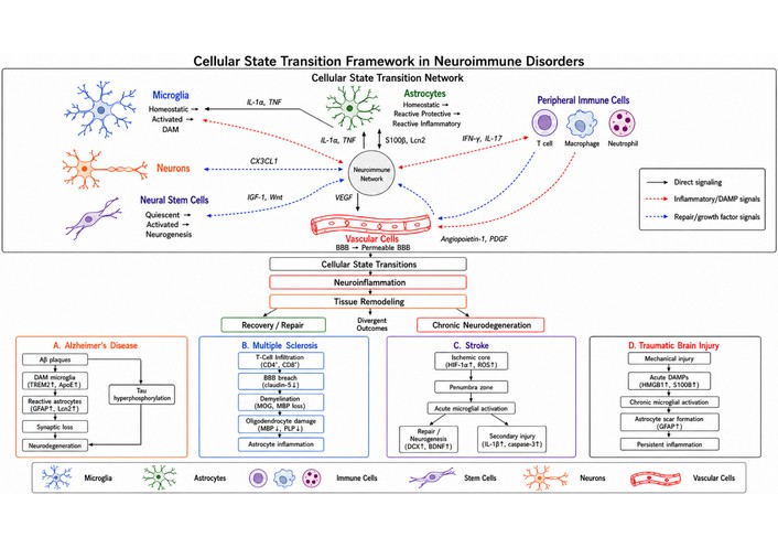
Cellular state transition framework in neuroimmune disorders. Neurological diseases can be conceptualized as dynamic networks of interacting cellular states within the central nervous system. Environmental perturbations such as injury, infection, or neurodegeneration trigger molecular signals that induce transcriptional and metabolic reprogramming across multiple cellular populations including microglia, astrocytes, neurons, neural stem cells, vascular cells, and infiltrating immune cells. Interactions among these cellular populations generate coordinated cellular state transitions that reshape neuroimmune networks and influence disease trajectories. These network dynamics regulate inflammatory responses, tissue remodeling, and neuronal survival, leading to divergent outcomes including repair and recovery or persistent neurodegeneration. The lower panels illustrate disease-specific manifestations of these state-transition networks in Alzheimer’s disease, multiple sclerosis, stroke, and traumatic brain injury.
Traditional models of neuroimmune disease have often emphasized specific pathological drivers, such as amyloid accumulation in Alzheimer’s disease, autoimmune demyelination in multiple sclerosis, ischemic injury in stroke, or mechanical tissue disruption in traumatic brain injury [213]. These models remain essential, but they do not fully explain why similar inflammatory mediators can produce different outcomes across diseases or why the same cell population may support tissue repair in one context while contributing to degeneration in another. A cellular state-transition framework adds explanatory value by shifting the focus from isolated pathological triggers to the dynamic cellular programs that emerge in response to those triggers.
This framework helps clarify three recurring features of neuroimmune disease. First, it accounts for temporal plasticity. Microglia, astrocytes, vascular cells, neural stem cells, and infiltrating immune cells do not maintain fixed pathological identities throughout disease. Instead, their functions change over time as injury signals, cytokines, metabolic stress, vascular dysfunction, and tissue-remodeling cues evolve [214, 215]. This is especially relevant in stroke and traumatic brain injury, where early inflammatory responses may support debris clearance and tissue stabilization, whereas persistent activation can contribute to secondary injury or chronic neurodegeneration [216, 217].
Second, the framework helps resolve apparent contradictions in glial biology. Microglial activation and astrocyte reactivity are often described as either protective or harmful, but this binary interpretation is insufficient. The same broad category of activation may include phagocytic, inflammatory, antigen-presenting, metabolic, synapse-remodeling, or repair-associated programs [218, 219]. Whether these programs promote recovery or pathology depends on disease stage, tissue context, cellular interactions, and the persistence of the triggering signal. The state-transition framework therefore interprets glial responses as context-dependent trajectories rather than fixed beneficial or detrimental phenotypes.
Third, the framework provides a way to compare diseases that differ in initiating cause but converge on shared cellular processes. Alzheimer’s disease, multiple sclerosis, stroke, and traumatic brain injury differ in their primary triggers, yet each involves coordinated changes across resident glia, vascular elements, and peripheral immune populations [213–215]. The generalizable principle is not that these diseases share identical cellular states, but that disease progression reflects the interaction of multiple state transitions across the neuroimmune network. At the same time, the framework has boundaries. State labels derived from transcriptomic studies do not always correspond to stable functional programs, and findings from animal models may not fully translate to human disease. Therefore, cellular state transitions should be interpreted as dynamic, context-dependent patterns rather than universal disease categories [215, 218].
The progression of many neurological disorders can be viewed as the result of coordinated transitions among multiple cellular populations within the neuroimmune environment. Microglia, astrocytes, infiltrating immune cells, neural stem cells, and vascular cells all respond to environmental disturbances through changes in signaling pathways, gene-expression programs, and functional behavior. These responses rarely occur in isolation. Instead, signals generated by one cell population often influence the state transitions of neighboring cells, creating interconnected networks of cellular responses.
Within these networks, microglial activation may initiate inflammatory signaling that influences astrocyte reactivity, while vascular changes and immune cell infiltration further modify the local tissue environment. Neural stem cells may also respond to these signals by altering their proliferative or differentiation states. Through these coordinated interactions, disease progression reflects a dynamic interplay among multiple cellular systems rather than isolated dysfunction of individual cell types [213].
The transition from homeostatic to inflammatory cellular states emerges from coordinated signaling across multiple neuroimmune cell populations (Figure 3).
Viewing neurological disease through the lens of cellular state transitions therefore highlights the importance of intercellular communication and coordinated signaling networks. Changes in one cellular population can propagate through the neuroimmune environment, influencing tissue remodeling, neuronal survival, and long-term disease outcomes.
Alzheimer’s disease provides a well-studied example of how cellular state transitions contribute to neurodegenerative pathology. In this condition, accumulation of amyloid-β plaques and other pathological protein aggregates triggers local immune responses within the brain. Microglia located near amyloid plaques frequently transition toward disease-associated states characterized by altered transcriptional programs and increased phagocytic activity [131, 220].
These microglial responses are accompanied by changes in astrocyte behavior. Astrocytes surrounding amyloid plaques often exhibit reactive phenotypes that involve altered metabolic activity, inflammatory signaling, and structural remodeling of neural tissue [131, 221]. Interactions between microglia and astrocytes within these plaque-associated environments can influence the local inflammatory milieu and affect neuronal survival.
Through these interactions, microglial–astrocyte signaling networks contribute to the regulation of plaque-associated inflammation and may influence the progression of neuronal injury. The cellular state transition framework therefore helps explain how multiple glial populations cooperate to shape the pathological environment surrounding amyloid deposits.
From a state-transition perspective, Alzheimer’s disease is therefore not simply an example of generalized neuroinflammation. Its distinctive feature is the formation of a chronic degenerative niche in which plaque-associated microglial and astrocytic states, synaptic remodeling, metabolic stress, and impaired clearance mechanisms reinforce one another over time. This differs from acute injury disorders, where glial activation may be temporally linked to damage containment and repair. In Alzheimer’s disease, the central disease-specific question is whether glial state transitions remain adaptive responses to accumulating pathology or become self-sustaining programs that contribute to progressive neurodegeneration.
Multiple sclerosis represents another example of a neurological disorder in which coordinated cellular state transitions play a central role in disease pathology. In this autoimmune condition, peripheral immune cells infiltrate the central nervous system and initiate inflammatory responses that target myelin and oligodendrocytes. The resulting immune activity leads to the formation of demyelinating lesions within the brain and spinal cord [222].
Within these lesions, microglia and infiltrating macrophages adopt activated states that contribute to inflammatory signaling and myelin degradation. Astrocytes also undergo reactive changes that influence the local inflammatory environment and contribute to structural alterations within affected tissue regions [223, 224]. At the same time, certain cellular responses may support remyelination and repair processes, particularly during phases of disease remission.
The interplay between infiltrating immune cells, resident microglia, and reactive astrocytes therefore shapes both destructive and reparative processes within demyelinating lesions. These interactions illustrate how transitions in immune and glial cell states influence the balance between demyelination and remyelination in multiple sclerosis.
Multiple sclerosis illustrates a different state-transition logic. Here, the dominant organizing feature is not chronic protein aggregation but immune-cell entry, demyelination, lesion evolution, and incomplete repair. The relevant cellular transitions therefore involve both peripheral immune populations and resident glial cells, including microglia/macrophage activation, astrocyte remodeling, and oligodendrocyte-lineage responses within inflammatory lesions. Compared with Alzheimer’s disease, the key disease-specific issue is how autoimmune inflammation reshapes local CNS cellular states and determines whether lesions progress toward tissue injury, stabilization, or remyelination.
Acute neurological injuries such as ischemic stroke and traumatic brain injury provide examples of conditions in which cellular state transitions unfold sequentially over time. Following injury, the affected brain regions undergo rapid inflammatory responses characterized by activation of resident microglia and recruitment of peripheral immune cells. These early immune responses help remove damaged cellular components and initiate signaling pathways that shape subsequent tissue remodeling [225, 226].
As the inflammatory phase progresses, astrocytes and vascular cells also undergo reactive changes that influence the structural and metabolic environment of injured tissue. Later phases of the response involve processes such as angiogenesis, synaptic remodeling, and activation of neural stem cells, which together contribute to tissue repair and functional adaptation.
Although these responses can support recovery, prolonged activation of microglia and sustained astrocyte reactivity may also contribute to persistent inflammatory signaling and secondary neuronal injury [225, 227]. In both stroke and traumatic brain injury, long-term alterations in glial cellular states can therefore influence the balance between tissue repair and chronic neurodegeneration.
Together, these examples illustrate how neurological diseases and injuries can be interpreted as networks of interacting cellular state transitions. Microglia, astrocytes, immune cells, and other cellular populations respond to pathological stimuli through coordinated changes in functional states, and these responses collectively shape the trajectory of disease progression and recovery.
As shown in Figure 4, different neurological diseases represent distinct configurations of cellular state transitions involving microglia, astrocytes, infiltrating immune cells, neural stem cells, and vascular elements.
Stroke and traumatic brain injury provide the clearest examples of temporally ordered state transitions after acute tissue disruption. In these disorders, cellular responses are shaped by the timing of injury, blood-brain barrier disruption, hypoxia or mechanical damage, and secondary inflammatory cascades. Early microglial, astrocytic, vascular, and infiltrating immune-cell responses may participate in debris clearance, containment of injury, and initiation of repair, whereas persistent or dysregulated activation can contribute to secondary injury and chronic neuroimmune remodeling. Thus, unlike Alzheimer’s disease or multiple sclerosis, the main analytical issue in stroke and traumatic brain injury is not only which cellular state emerges, but when it emerges and whether it resolves or persists.
The disease-specific examples above show that Alzheimer’s disease, multiple sclerosis, stroke, and traumatic brain injury do not share identical cellular states, but they do reveal several recurring principles of state-transition logic. The first is trigger-specific convergence. Although the initiating events differ, each disorder can activate overlapping inflammatory, metabolic, vascular, and transcriptional programs within resident and infiltrating cell populations [213, 214]. The second is temporal divergence. Similar inflammatory programs may have different consequences depending on when they occur. Acute activation may support containment of damage, debris clearance, or repair, whereas persistent activation may sustain tissue injury, synaptic disruption, or neurodegeneration [216, 217]. The third is intercellular propagation. State transitions in one population can alter the behavior of others; for example, microglial inflammatory signaling may shape astrocyte reactivity, vascular dysfunction may promote immune-cell entry, and infiltrating immune cells may reinforce local glial activation [140, 214]. The fourth is repair–degeneration competition. Many neuroimmune states cannot be classified as purely protective or pathological because they participate in both tissue stabilization and injury amplification depending on timing, magnitude, and context.
These cross-disease patterns are summarized in Table 5, which compares the initiating context, dominant state-transition pattern, shared principle, and interpretive boundary across Alzheimer’s disease, multiple sclerosis, stroke, and traumatic brain injury.
Comparative logic of cellular state transitions across neuroimmune disorders.
| Disorder | Primary initiating context | Dominant state-transition pattern | Shared principle | Boundary of interpretation |
|---|---|---|---|---|
| Alzheimer’s disease | Chronic protein aggregation and age-associated tissue stress | Plaque-associated microglial and astrocyte remodeling, metabolic adaptation, synaptic disruption [139, 228] | Chronic glial state transitions can reshape the local degenerative niche | Plaque-associated states may not generalize directly to acute injury or autoimmune disease |
| Multiple sclerosis | Autoimmune infiltration and demyelination | Infiltrating immune-cell activation, microglia/macrophage activation, astrocyte remodeling, remyelination failure or repair [214, 229] | Peripheral immune entry can reorganize resident glial states and lesion evolution | MS lesions differ by stage, region, and inflammatory activity; not all glial activation is damaging |
| Stroke | Acute ischemia, hypoxia, and tissue necrosis | Rapid microglial activation, peripheral immune recruitment, vascular disruption, later repair/remodeling states [230, 231] | Timing determines whether inflammatory states support clearance or amplify injury | Acute ischemic responses should not be directly equated with chronic neurodegeneration |
| Traumatic brain injury | Mechanical injury, barrier disruption, and secondary inflammatory cascades | Early damage sensing, glial activation, immune-cell recruitment, persistent astrocyte/microglial changes in some cases [227, 232] | Acute injury can transition into chronic neuroimmune remodeling | TBI is heterogeneous; focal, diffuse, mild, and severe injury may produce different state trajectories |
This comparison highlights both the usefulness and the limits of the cellular state-transition framework. The shared logic is not that these disorders involve identical cellular states, but that distinct initiating insults can converge on recurring forms of glial, vascular, immune, metabolic, and repair-related remodeling. At the same time, the timing, anatomical context, disease stage, and causal role of each state differ substantially across disorders. The framework should therefore be used as a comparative interpretive model rather than as a claim that cellular states are equivalent across neurodegenerative, autoimmune, ischemic, and traumatic conditions.
These principles are broadly useful but should not be overgeneralized. Alzheimer’s disease is dominated by chronic degenerative pathology and plaque-associated glial remodeling, whereas multiple sclerosis is shaped by autoimmune infiltration, demyelination, and incomplete remyelination. Stroke and traumatic brain injury involve acute tissue disruption followed by temporally ordered inflammatory and repair responses [216, 217]. Therefore, the same molecular marker or transcriptional signature may not have the same biological meaning across diseases. A state that appears reparative in acute injury may represent maladaptive persistence in chronic neurodegeneration. Similarly, transcriptomic similarity between cell populations does not necessarily imply functional equivalence. The value of the state-transition framework lies in identifying recurring organizational principles while preserving disease-specific mechanisms and temporal context.
Advances in neuroimmunology and cellular profiling have increasingly highlighted the importance of cellular state transitions in shaping neurological disease progression. As described throughout this review, microglia, astrocytes, neural stem cells, and infiltrating immune cells can adopt diverse functional states in response to environmental signals. While some of these transitions support protective responses and tissue repair, others contribute to persistent inflammation, neuronal dysfunction, and chronic pathology. Consequently, a growing area of research is focused on identifying strategies that can modulate pathological cellular states while preserving or promoting beneficial responses within the neuroimmune environment [233–235].
One potential therapeutic approach involves modulation of cytokine signaling pathways that regulate inflammatory responses. Cytokines such as ILs, TNFs, and IFNs play central roles in controlling immune activation and glial behavior within the central nervous system. Pharmacological interventions that alter cytokine signaling may help reduce excessive inflammatory responses and limit secondary tissue damage during neurological disease [236–238]. Such approaches are already being explored in several immune-mediated conditions and may provide insights into broader strategies for regulating neuroimmune activity. For example, therapeutic strategies targeting the complement cascade—particularly inhibition of complement component C1q or C3 signaling—are being investigated for their potential to reduce excessive synaptic pruning and inflammatory responses in neurodegenerative diseases [239, 240].
Selected therapeutic strategies that target cellular state transitions in the neuroimmune environment are summarized in Table 6.
Therapeutic strategies targeting cellular state transitions in neurological disease.
| Therapeutic strategy | Molecular target | Target cell population | Mechanism of action | Disease context |
|---|---|---|---|---|
| Complement pathway inhibition | Complement proteins C1q, C3 | Microglia, synapse-associated immune signaling pathways | Reduces complement-mediated synaptic pruning and inflammatory activation | Alzheimer’s disease, multiple sclerosis [241, 242] |
| Immune checkpoint modulation | PD-1/PD-L1 signaling axis | T lymphocytes, microglia | Modulates immune activation and inflammatory signaling by regulating immune checkpoint pathways | Stroke, neuroinflammatory disorders [243, 244] |
| Triggering receptor expressed on myeloid cells 2 (TREM2) modulation | TREM2 | Microglia | Enhances microglial metabolic activity, phagocytosis, and responses to neurodegenerative pathology | Alzheimer’s disease and related neurodegenerative diseases [245, 246] |
| Colony-stimulating factor 1 receptor (CSF1R) inhibition | CSF1R | Microglia | Alters microglial survival and proliferation, thereby modulating microglial population dynamics and inflammatory responses | Neurodegenerative disease models, neuroinflammation [247, 248] |
| Cytokine pathway inhibition | IL-6, TNF, interferon signaling pathways | Microglia, astrocytes, immune cells | Reduces inflammatory signaling and downstream neuroimmune activation | Stroke, neuroinflammatory disorders [249, 250] |
Another area of investigation involves targeting immune checkpoint pathways that regulate immune cell activation and tolerance. Immune checkpoints are regulatory mechanisms that help maintain balanced immune responses by controlling the activation and persistence of immune cells. Modulating these pathways may influence the activity of infiltrating immune cells and alter the inflammatory environment within the central nervous system [244, 251]. Although most studies of immune checkpoint modulation have focused on cancer immunotherapy, similar principles may be applicable to neurological diseases characterized by dysregulated immune responses. Emerging studies are also exploring the modulation of immune checkpoint molecules such as PD-1 and PD-L1 in neuroinflammatory conditions, where altering T-cell activity may influence neuroimmune signaling and tissue repair [243, 244, 251].
Metabolic pathways have also emerged as potential therapeutic targets for modulating cellular states within the neuroimmune system. As discussed earlier, immune activation is closely linked to metabolic reprogramming in glial and immune cells. Interventions that influence cellular metabolism—such as pathways involved in glycolysis, mitochondrial function, or lipid metabolism—may alter inflammatory signaling and reshape cellular responses within diseased neural tissue [246, 252]. By modifying metabolic programs, it may be possible to influence whether immune cells adopt inflammatory or reparative functional states. In microglial populations, modulation of TREM2 signaling has attracted considerable attention because of its role in regulating lipid metabolism, phagocytosis, and transitions toward disease-associated microglial states in neurodegenerative disorders [245, 246, 252].
A further strategy involves direct reprogramming of cellular states through approaches that alter gene-expression networks or signaling pathways within specific cell populations. Advances in molecular biology and gene-regulatory technologies have raised the possibility of shifting glial or immune cells from pathological states toward more protective or regenerative phenotypes. Although many of these approaches remain experimental, they illustrate the potential of targeting cellular state transitions as a therapeutic strategy. For instance, pharmacological inhibition of colony-stimulating factor 1 receptor (CSF1R), which regulates microglial survival and proliferation, has been explored as a strategy to modify microglial population dynamics and inflammatory states in experimental models of neurological disease [247, 248].
Recent technological developments are also expanding the ability to identify disease-associated cellular states that may serve as therapeutic targets. Techniques such as single-cell RNA sequencing and spatial transcriptomics have enabled detailed mapping of cellular heterogeneity within the brain and have revealed previously unrecognized disease-associated cell populations [122, 131, 233]. By identifying the transcriptional and molecular features that characterize these states, these technologies may help guide the development of therapies that selectively target pathological cellular programs while preserving normal cellular functions.
Together, these emerging approaches illustrate how understanding cellular state transitions can inform new therapeutic strategies for neurological disease. Rather than targeting single molecules or pathways in isolation, future therapies may increasingly focus on modulating broader cellular programs that regulate immune activation, tissue repair, and neural function within the complex neuroimmune environment.
Although cellular state-transition frameworks provide a useful way to organize neuroimmune disease mechanisms, the underlying evidence varies substantially in strength, biological context, and translational maturity. A first important distinction is between experimental animal models and human disease. Many disease-associated microglial, astrocytic, vascular, and immune-cell states have been defined in mouse models, where disease timing, genetic background, injury severity, and tissue sampling can be experimentally controlled [231, 232]. Human studies provide essential disease relevance, but they are often constrained by post-mortem tissue availability, clinical heterogeneity, medication exposure, agonal state, disease stage, and limited temporal resolution [215, 253]. As a result, cellular states identified in animal models should not be assumed to map directly onto human disease without functional and cross-species validation.
A second limitation concerns the difference between correlative molecular profiling and causal functional evidence. Single-cell and spatial transcriptomic approaches can identify transcriptional signatures, regional enrichment, and disease-associated cell populations, but these data do not by themselves establish whether a cellular state drives pathology, reflects tissue adaptation, or represents a secondary consequence of disease [253, 254]. Functional experiments, longitudinal analyses, perturbation studies, and integration with proteomic, metabolic, electrophysiological, and histopathological data are needed to determine whether a transcriptionally defined state has causal relevance. This distinction is especially important when using omics-derived states to infer therapeutic targets.
A third interpretive boundary involves the relationship between markers, transcriptional clusters, and stable biological programs. Markers such as P2RY12, TMEM119, GFAP, TREM2, APOE, C3, or CST7 can help identify cellular populations or disease-associated tendencies, but they do not define fixed or universal functional identities. The same marker may have different biological meaning depending on brain region, disease stage, species, injury timing, and cellular microenvironment. Similarly, transcriptomic clusters may shift across datasets because of differences in tissue processing, sequencing depth, analytical pipelines, annotation strategy, and disease model. Therefore, cellular state labels should be interpreted as context-dependent operational categories rather than stable disease entities [218, 254].
A fourth limitation concerns disease context. Acute injury states in stroke or traumatic brain injury should not be directly equated with chronic glial remodeling in Alzheimer’s disease or autoimmune lesion evolution in multiple sclerosis. Similar inflammatory mediators may participate in debris clearance, containment of tissue damage, repair, or chronic injury amplification depending on timing and local context. Thus, the framework is most useful when it compares patterns of transition logic across diseases while preserving disease-specific mechanisms, rather than assuming that a shared marker or transcriptional signature implies the same biological role across disorders.
These limitations have direct therapeutic implications. Identifying a disease-associated cellular state does not automatically make that state intervention-ready. A therapeutic target must be validated beyond transcriptomic association by showing that modulating the relevant pathway changes cellular function, disease trajectory, or tissue outcome in an appropriate model and, ultimately, in human disease [215, 254]. The translational value of the state-transition framework therefore depends on moving from descriptive state mapping toward causal testing, cross-species validation, temporal resolution, and functional perturbation.
Circadian and sleep-related processes may represent an additional layer of regulation for cellular state transitions in neuroimmune disorders. Sleep is increasingly understood not only as a behavioral state but also as a period during which neural, metabolic, immune, and systemic processes are reorganized. Circadian changes in pineal melatonin, hypothalamic–pituitary–adrenal axis activity, and cortisol rhythms may influence inflammatory tone, mitochondrial function, redox balance, and intercellular signaling across multiple tissues [255].
One mechanism through which sleep and circadian signaling may influence neuroimmune state dynamics is the regulation of mitochondrial function. Mitochondria are major sources of cellular energy and reactive oxygen species, and mitochondrial stress can shape inflammatory signaling, cytokine responses, and redox-sensitive transcriptional programs. Circadian regulation of mitochondrial bioenergetics, redox signaling, and mitochondrial dynamics may therefore influence whether cells maintain homeostatic programs or shift toward inflammatory, stress-responsive, or disease-associated states [256].
Astrocyte-associated glymphatic function provides another potential point of connection between sleep biology and neuroimmune remodeling. Aquaporin-4 localization on astrocytic endfeet contributes to glymphatic fluid movement and clearance processes, which are most active during sleep. Melatonin has been proposed to influence astrocytic and mitochondrial pathways that may support nocturnal tissue maintenance, although the extent to which these mechanisms directly regulate neuroimmune cellular state transitions requires further validation.
Aging and systemic inflammation may further modify this circadian-metabolic axis. Pineal melatonin production declines with aging, and inflammatory mediators, gut permeability-associated lipopolysaccharide, and cytokine signaling can suppress or alter melatonin synthesis through immune-pineal mechanisms [255, 257]. Because aging, mitochondrial dysfunction, oxidative stress, impaired sleep, and chronic inflammation are closely linked to neurodegenerative vulnerability, disrupted circadian regulation may contribute to the persistence or maladaptive amplification of neuroimmune cellular states.
These observations suggest that cellular state transitions should not be viewed only as local responses to injury, cytokines, or disease-associated molecular patterns. They may also be shaped by systemic temporal regulators, including sleep architecture, melatonin signaling, cortisol rhythms, mitochondrial resetting, and inflammatory signals arising from peripheral tissues. Future studies should clarify whether circadian disruption directly alters microglial, astrocytic, endothelial, or neural stem-cell state trajectories, and whether restoring circadian-mitochondrial regulation can reduce maladaptive neuroimmune remodeling in aging and neurological disease.
Neuroimmune disorders involve complex interactions among multiple cellular populations within the central nervous system. Increasing evidence indicates that disease progression is not driven solely by the dysfunction of individual cell types but is shaped by coordinated transitions between functional cellular states. These transitions occur across diverse cellular populations—including microglia, astrocytes, neural stem cells, infiltrating immune cells, and vascular elements—and collectively influence inflammatory responses, tissue remodeling, and neural circuit stability.
Viewing neuroimmune diseases through the framework of cellular state transitions provides a broader perspective that integrates molecular signaling pathways with cellular behavior and tissue-level outcomes. In this context, changes in gene-expression programs, metabolic activity, and intercellular communication contribute to dynamic shifts in cellular identity that influence both protective and pathological processes. The interplay among these state transitions determines whether neuroimmune responses promote tissue repair and adaptation or contribute to persistent inflammation and neuronal injury. This systems-level perspective emphasizes that neuroimmune pathology emerges from coordinated cellular networks rather than isolated cellular dysfunction.
Recent advances in high-resolution molecular technologies have significantly expanded the ability to characterize cellular heterogeneity within the brain. Approaches such as single-cell transcriptomics and spatial transcriptomic analysis have revealed previously unrecognized diversity in glial and immune cell states and have provided insights into how these states evolve during neurological disease [138, 147, 258, 259]. However, transcriptional state labels should not be interpreted as direct equivalents of stable functional programs. Cross-study differences in tissue processing, disease stage, species, sequencing platform, analytical pipeline, and annotation strategy can influence how cellular states are defined and compared. Integrating molecular profiling with functional perturbation, spatial validation, longitudinal sampling, and human disease studies will therefore be essential for distinguishing causal cellular programs from correlative or context-dependent signatures.
From this synthesis, several testable propositions emerge. First, neuroimmune disease progression should be better predicted by coordinated multi-cellular state trajectories than by isolated markers of microglial or astrocytic activation alone. Second, the timing of a cellular state transition should determine its functional meaning, such that similar inflammatory or reparative programs may be protective during early injury containment but maladaptive when persistent or reactivated during chronic disease. Third, disease-associated glial states should have different causal roles depending on whether they arise in chronic neurodegeneration, autoimmune demyelination, acute ischemic injury, or traumatic tissue disruption. Fourth, therapeutically useful state definitions should require functional validation, including evidence that targeted modulation of a proposed state alters cellular behavior, tissue pathology, or neurological outcome. These propositions provide a framework for testing whether cellular state transitions are not only descriptive signatures of disease but causal organizing principles in neuroimmune pathology.
Future research should move beyond descriptive state mapping toward causal and temporally resolved models of neuroimmune regulation. Priority areas include cross-species validation of cellular states, experimental testing of state-defining pathways, longitudinal analysis of acute-to-chronic transitions, and integration of transcriptomic signatures with proteomic, metabolic, electrophysiological, and histopathological evidence. Therapeutic strategies that selectively influence maladaptive cellular programs or promote beneficial neuroimmune states remain promising, but their translation will depend on demonstrating that targeted modulation of a defined state produces measurable functional benefit in disease-relevant contexts.
DAM: disease-associated microglia
DAMPs: damage-associated molecular patterns
IFN-γ: interferon-γ
IL-1β: interleukin-1β
MAPK: mitogen-activated protein kinase
NF-κB: nuclear factor κB
SVZ: subventricular zone
TNF: tumor necrosis factor
TREM2: triggering receptor expressed on myeloid cells 2
MMN: Conceptualization, Methodology, Investigation, Resources, Data curation, Formal analysis, Visualization, Writing—original draft, Writing—review & editing. The author read and approved the submitted version.
The author declares no conflicts of interest.
Not applicable.
Not applicable.
Not applicable.
Not applicable.
This research received no external funding.
© The Author(s) 2026.
Open Exploration maintains a neutral stance on jurisdictional claims in published institutional affiliations and maps. All opinions expressed in this article are the personal views of the author(s) and do not represent the stance of the editorial team or the publisher.
Copyright: © The Author(s) 2026. This is an Open Access article licensed under a Creative Commons Attribution 4.0 International License (https://creativecommons.org/licenses/by/4.0/), which permits unrestricted use, sharing, adaptation, distribution and reproduction in any medium or format, for any purpose, even commercially, as long as you give appropriate credit to the original author(s) and the source, provide a link to the Creative Commons license, and indicate if changes were made.



View: 1053
Download: 27
Times Cited: 0


