 Review


Affiliation:
1Neurology Service, “Dr. José E. González” University Hospital, Monterrey, NL 64460, Mexico
2Faculty of Medicine, Autonomous University of Nuevo Leon, Monterrey, NL 64460, Mexico
Email: luisfdz12@gmail.com
ORCID: https://orcid.org/0000-0003-2387-4643


 ,
,
Affiliation:
2Faculty of Medicine, Autonomous University of Nuevo Leon, Monterrey, NL 64460, Mexico
3Internal Medicine Department, “Dr. José E. González” University Hospital, Monterrey, NL 64460, Mexico
ORCID: https://orcid.org/0009-0003-8003-1690
,
Affiliation:
2Faculty of Medicine, Autonomous University of Nuevo Leon, Monterrey, NL 64460, Mexico
4Gastroenterology and Endoscopy Service, “Dr. José E. González” University Hospital, Monterrey, NL 64460, Mexico
ORCID: https://orcid.org/0000-0001-9534-2799
,
Affiliation:
5UDEM Health, University of Monterrey, San Pedro Garza Garcia, NL 66238, Mexico
Affiliation:
6Internal Medicine Department, Christus Muguerza High Specialty Hospital, Monterrey, NL 64060, Mexico
Affiliation:
7Neurology Service, Zambrano Hellion Hospital TecSalud, San Pedro Garza Garcia, NL 66260, Mexico
Affiliation:
1Neurology Service, “Dr. José E. González” University Hospital, Monterrey, NL 64460, Mexico
2Faculty of Medicine, Autonomous University of Nuevo Leon, Monterrey, NL 64460, Mexico
ORCID: https://orcid.org/0000-0002-7812-0462
,
Affiliation:
2Faculty of Medicine, Autonomous University of Nuevo Leon, Monterrey, NL 64460, Mexico
4Gastroenterology and Endoscopy Service, “Dr. José E. González” University Hospital, Monterrey, NL 64460, Mexico
ORCID: https://orcid.org/0000-0002-2080-1716
,
Affiliation:
5UDEM Health, University of Monterrey, San Pedro Garza Garcia, NL 66238, Mexico
Affiliation:
5UDEM Health, University of Monterrey, San Pedro Garza Garcia, NL 66238, Mexico
Explor Dig Dis. 2026;5:1005136 DOI: https://doi.org/10.37349/edd.2026.1005136
Received: March 12, 2026 Accepted: July 30, 2026 Published: September 18, 2026
Academic Editor: Pavel Strnad, University Hospital Aachen, Germany
The article belongs to the special issue Cirrhosis and Its Complications
The neurological burden of liver cirrhosis extends far beyond the classical paradigm of hepatic encephalopathy (HE). This comprehensive clinical review synthesizes the pathophysiology, diagnosis, and contemporary management of the broad spectrum of liver-brain axis complications. Historically, neurological deterioration in these patients has been centered on ammonia toxicity, astrocyte swelling, and neuroinflammation characteristic of HE. However, chronic liver failure and portosystemic shunting also lead to the accumulation of other potent neurotoxins, such as manganese, triggering severe and often irreversible motor syndromes, including acquired hepatocerebral degeneration and hepatic myelopathy. Additionally, peripheral neuropathy and dysautonomia represent profoundly underdiagnosed comorbidities driven by metabolic derangement, toxins, and chronic inflammation. As the disease progresses, systemic instability generates new threats: a precarious hemostatic rebalancing predisposes patients to devastating ischemic and hemorrhagic cerebrovascular complications, challenging the outdated paradigm of “auto-anticoagulation”. Concurrently, cirrhosis-associated immune dysfunction and bacterial translocation facilitate the development of atypical neuroinfections that frequently masquerade as refractory HE, demanding advanced diagnostic tools and prognostic scores like CLIF-SOFA rather than traditional sepsis criteria. Finally, we address neuromotor and circadian disorders—namely restless legs syndrome, debilitating muscle cramps, and insomnia—which drastically deteriorate patients’ daily quality of life. Mitigating this complex neurological morbidity requires a definitive paradigm shift: moving beyond an exclusive focus on hyperammonemia toward multidisciplinary strategies that precisely manage everything from structural and cognitive deficits to functional immunosuppression and acute neurovascular emergencies.
Chronic liver injury triggers a series of maladaptive responses, including progressive inflammation and fibrogenesis, which ultimately culminate in liver cirrhosis (LC) [1]. While chronic liver disease poses a significant global public health problem and remains a leading cause of mortality, its clinical impact extends far beyond hepatic impairment [1]. Decompensated cirrhosis is not a single-organ illness but a complex multi-system disorder, driven by mechanisms such as systemic inflammation that precipitate dysfunction in organs beyond the liver [2]. Within this systemic disease framework, neurological complications represent a critical prognostic watershed and are among the most debilitating manifestations [3].
LC is widely associated with central nervous system (CNS) involvement, driven by the increased systemic circulation of neurotoxins—such as ammonia (NH3) and manganese (Mn)—due to hepatic dysfunction and portosystemic shunting [4, 5]. Furthermore, the synergistic effect of these accumulating toxins and systemic inflammation compromises the integrity of the blood-brain barrier (BBB), facilitating local neuroinflammation and microglial activation [4]. This toxic microenvironment leads to complex neuroglial alterations, including astrocytic swelling, altered neurotransmission, and neuronal degeneration with concurrent loss of neuronal mass in the basal ganglia, cerebellum, thalamus, brainstem, and spinal cord [4, 5].
The neurological manifestations in these patients encompass a broad clinical spectrum. Historically, these can be divided into two main categories: those arising from the direct toxic effects of liver failure and portosystemic shunting on the nervous system, and those specifically related to the underlying etiology of the liver disease, such as Wernicke-Korsakoff syndrome in chronic alcohol use, or copper-induced neurotoxicity in Wilson’s disease [6]. Regarding the direct complications of cirrhosis, the clinical presentation ranges from subtle neuropsychiatric alterations (e.g., sleep disturbances, anxiety, and mild cognitive impairment) to profound motor and consciousness deficits seen in overt hepatic encephalopathy (HE) and acquired hepatocerebral degeneration (AHD) [4].
While HE is the most widely recognized neurological complication, the true burden of cirrhosis on the CNS is much more complex. Non-classical entities such as AHD, hepatic myelopathy (HM), and severe CNS infections driven by cirrhosis-associated immune dysfunction (CAID) remain significantly underrecognized [7, 8]. Furthermore, traditional diagnostic paradigms in the emergency setting—such as the routine use of neuroimaging for altered mental status—have been challenged by landmark cohort studies [9, 10]. The most significant transitions from these traditional practices toward contemporary evidence-based management are summarized in Table 1. Despite these paradigm shifts over the last decade, there is a lack of recent literature integrating these diverse clinical entities into a cohesive diagnostic and therapeutic framework. Therefore, this comprehensive clinical review aims to critically evaluate the current evidence regarding the pathophysiology, diagnosis, and management of the direct neurological complications of cirrhosis, providing updated, evidence-based algorithms to optimize patient care.
The paradigm shift in cirrhosis management.
| Clinical challenge | Outdated practice | Contemporary evidence-based paradigm | Clinical evidence | Key references |
|---|---|---|---|---|
| Nutritional support | Protein restriction to reduce NH3 load. | High-protein diet (1.2–1.5 g/kg/day) and late-night snacks. | Restriction exacerbates sarcopenia; skeletal muscle is vital for compensatory NH3 detoxification. | [11–13] |
| Neuroimaging in AMS | Routine head CT for all cirrhotic patients with AMS. | Deferred neuroimaging unless focal deficits or trauma are present. | Diagnostic yield is extremely low (0.3%) and does not correlate with the severity of coagulopathy. | [9, 10, 14] |
| Coagulation status | “Auto-anticoagulation” myth based on elevated PT/INR. | Precarious rebalanced hemostasis; risk for both ICH and ischemic stroke. | Standard tests fail to reflect the parallel decline of pro- and anticoagulant factors. | [15–17] |
| Muscle cramps | Use of quinidine derivatives. | Pickle juice, Baclofen, or Taurine. | Quinine is no longer recommended due to cardiac toxicity; pickle juice triggers inhibitory oropharyngeal neural reflexes. | [18–21] |
| Sleep management | Standard hypnotics (Benzodiazepines or Z-drugs). | Bright light therapy or hydroxyzine. | Benzodiazepines precipitate HE; Z-drugs are associated with high exposure/toxicity in cirrhosis. | [22–24] |
AMS: altered mental status; CT: computed tomography; ICH: intracranial hemorrhage; INR: international normalized ratio; PT: prothrombin time.
A comprehensive and systematic literature search was conducted across multiple electronic databases, including PubMed/MEDLINE, Embase, Scopus, the Cochrane Library, Web of Science, and Google Scholar, for articles published from inception through January 2026. The search strategy integrated Medical Subject Headings (MeSH) and specific keywords: “Liver Cirrhosis,” “Hepatic Encephalopathy,” “Acquired Hepatocerebral Degeneration,” “Hepatic Myelopathy,” “Cirrhosis-Associated Immune Dysfunction,” “Neuroinflammation,” and “Cerebrovascular Disorders in Cirrhosis.”
The primary focus was on peer-reviewed manuscripts published in English. Preference was given to high-quality evidence from systematic reviews, meta-analyses, randomized controlled trials, and updated clinical practice guidelines from the American Association for the Study of Liver Diseases (AASLD) and the European Association for the Study of the Liver (EASL). Additionally, a manual search of the reference lists of the retrieved articles (“snowballing” technique) was performed to identify further relevant studies that might have been missed in the initial electronic search. Two independent reviewers (LEFG, VAFG) conducted the screening of titles and abstracts to ensure academic rigor and clinical relevance to the pathophysiology, emerging diagnostic tools, and contemporary management strategies of the liver-brain axis.
HE is a severe clinical entity characterized by a broad spectrum of neuropsychiatric symptoms, ranging from mild cognitive impairment to coma, in patients with acute or chronic liver diseases [14].
Epidemiologically, HE imposes a massive global burden on healthcare utilization and severely impacts patients’ quality of life. The prevalence of covert (or minimal) HE is estimated to be highly variable, affecting between 20% and 80% of all cirrhotic patients, largely depending on the diagnostic tools employed [25]. Conversely, overt HE will develop in approximately 30% to 40% of patients with cirrhosis at some point during their disease course [25].
The onset of overt HE marks a critical prognostic transition in the natural history of liver disease; following a first episode, the median survival of cirrhotic patients is foreshortened substantially to approximately two years, and to just one year in patients over 65 years of age [26]. Furthermore, the contemporary epidemiology of HE is shifting. While traditionally associated with alcohol-related liver disease (ARLD) and viral hepatitis, the rising global prevalence of metabolic dysfunction-associated steatotic liver disease (MASLD) is transforming the landscape, with HE now frequently presenting as the first decompensating event in this growing demographic [26].
HE results from profound neuroglial impairment driven primarily by hyperammonemia, acting synergistically with neuroinflammation and altered neurotransmission [27, 28]. NH3, primarily generated by the bacterial breakdown of proteins in the colon, is normally detoxified by hepatocytes via the urea cycle. However, in patients with liver failure or portosystemic shunting, NH3 evades hepatic clearance, accumulates in the systemic circulation, and freely crosses the BBB (Figure 1) [28, 29].
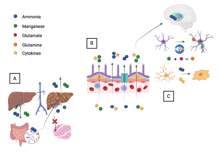
Integrated pathophysiology of the liver-brain axis in cirrhosis. (A) Systemic alterations: In the setting of liver cirrhosis and portal hypertension, the development of portosystemic shunts allows neurotoxins, primarily ammonia (NH3) from the gut microbiota and manganese (Mn), to bypass hepatic clearance. Concurrently, sarcopenia reduces the skeletal muscle’s compensatory capacity to detoxify NH3 via glutamine synthetase, while cirrhosis-associated immune dysfunction (CAID) promotes systemic inflammation with circulating cytokines (e.g., TNF-α, IL-6). (B) Blood-brain barrier (BBB) interface: Elevated systemic NH3 freely diffuses across the BBB. Cytokines increase BBB permeability, facilitating the entry of inflammatory mediators and Mn into the central nervous system. (C) Neuroglial damage and clinical syndromes: Within the brain parenchyma, pathophysiology diverges into two distinct pathways. In hepatic encephalopathy (HE), astrocytes rapidly metabolize NH3 and glutamate into glutamine via glutamine synthetase. Intracellular glutamine accumulation induces severe osmotic stress, astrocytic swelling (Alzheimer type II astrocytes), and subsequent oxidative/nitrosative stress, leading to neuronal dysfunction and altered neurotransmission (NMDA receptor overstimulation). Conversely, in acquired hepatocerebral degeneration (AHD), Mn selectively deposits within the basal ganglia, particularly the globus pallidus. This chronic intracellular accumulation causes mitochondrial toxicity and disrupts dopaminergic pathways, manifesting clinically as progressive Parkinsonism and movement disorders.
Astrocytes are highly vulnerable to hyperammonemia due to their exclusive expression of glutamine synthetase, an enzyme that condenses NH3 and glutamate into glutamine [28]. At this cellular level, the resulting osmotic stress defines a critical pathophysiological dichotomy between acute liver failure (ALF) (Type A HE) and cirrhosis (Type C HE). In ALF, the rapid and massive accumulation of NH3 prevents any metabolic adaptation, leading to severe cytotoxic edema, acute astrocyte swelling, and a life-threatening increase in intracranial pressure that can culminate in brain herniation [29, 30]. Conversely, in cirrhosis, the progressive nature of the disease allows for partial osmotic compensation [31]. Although astrocytes attempt to compensate by releasing myo-inositol through volume-sensitive anionic channels, this mechanism eventually fails, exacerbating astrocytic swelling [28, 32]. Consequently, patients with Type C HE develop a “low-grade” cerebral edema. While this low-grade edema rarely causes overt intracranial hypertension, advanced in vivo neuroimaging techniques, such as cerebral water content mapping via magnetic resonance imaging (MRI), consistently demonstrate subtle but functionally significant increases in brain water content, even in patients with covert HE [33, 34]. Chronically, these cells undergo morphological alterations, developing into Alzheimer-type II astrocytes, characterized by a pale, enlarged nucleus and a prominent nucleolus [27, 35].
Furthermore, the relationship between brain edema and HE is highly synergistic; low-grade edema alone is unlikely to be the sole mechanism for the clinical syndrome, but it acts as a primary catalyst for secondary neuronal damage [36]. Astrocytic swelling triggers oxidative and nitrosative stress (ONS) [28, 31, 37]. Hyperammonemia overstimulates N-methyl-D-aspartate (NMDA) receptors, leading to intracellular calcium influx and superoxide production via nicotinamide adenine dinucleotide phosphate (NADPH) oxidase activation [28, 38]. This toxic cascade impairs mitochondrial function, RNA oxidation, and protein homeostasis. Concurrently, systemic neuroinflammation plays a central role in disease progression. Pro-inflammatory states, predominantly mediated by Th17 CD4+ lymphocytes, promote microglial activation and the central release of cytokines (e.g., TNF-α, IL-1β, IL-6) (Figure 1). In fact, neuroinflammation and edema are mutually reinforcing; circulating inflammatory cytokines exacerbate astrocyte swelling and amplify NH3’s neurotoxic effects [39]. This inflammatory milieu further disrupts neurotransmission and exacerbates the cognitive and motor deficits hallmark of HE [40–42].
HE encompasses a broad spectrum of neuropsychiatric and motor impairments. To standardize its nomenclature, the International Society for HE and Nitrogen Metabolism (ISHEN) established a dichotomous classification—covert and overt HE—which aligns clinically with the traditional West Haven (WH) criteria [14, 43].
Covert HE encompasses minimal HE (WH grade 0) and WH grade 1. Minimal HE represents a subclinical stage identifiable only through specialized psychometric testing. Although these patients lack obvious clinical signs, minimal HE severely impacts their quality of life, manifesting as visuospatial and attentional deficits that increase the risk of falls and impair driving ability [27, 44]. In WH grade 1, subtle cognitive impairments (e.g., dyscalculia) and behavioral changes (e.g., euphoria or anxiety) emerge. This stage is often accompanied by circadian rhythm disturbances and sleep inversion, although the patient remains fully oriented in time and space [14, 45].
Overt HE is clinically defined by the onset of temporal or spatial disorientation and/or the presence of asterixis (a negative myoclonus). Within this category, WH grade 2 is characterized by lethargy, apathy, and inappropriate behavior [14]. WH grade 3 involves profound somnolence, stupor, and severe confusion. Finally, WH grade 4 denotes deep coma, which may be accompanied by absent pupillary reflexes and extensor posturing, indicating severe cerebral depression [14, 27]. Due to the high risk of aspiration and the inability to maintain airway reflexes, patients presenting with WH grades 3 and 4 require immediate intensive care admission and a low threshold for airway protection via endotracheal intubation [46].
The diagnosis of HE remains primarily clinical and fundamentally relies on being a diagnosis of exclusion. When a cirrhotic patient presents with altered mental status in the emergency setting, a rapid and structured clinical triage is required to differentiate structural brain lesions from systemic, infectious, or metabolic etiologies (Figure 2).
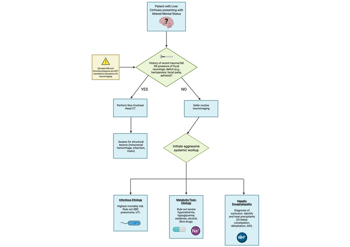
Evidence-based diagnostic algorithm for altered mental status (AMS) in cirrhosis. The initial evaluation of a cirrhotic patient presenting with AMS requires a rapid clinical triage to identify structural brain lesions versus systemic etiologies. The presence of focal neurologic deficits (e.g., hemiparesis, cranial nerve palsies) or a history of trauma strongly dictates the need for a non-contrast head computed tomography (CT). Conversely, in the absence of localizing signs or trauma, the diagnostic yield of a head CT is exceedingly low (0.3%), and routine neuroimaging is not recommended, regardless of the severity of coagulopathy (elevated INR) or thrombocytopenia. In these non-focal patients, clinical efforts must be immediately directed towards identifying infectious precipitants—which account for nearly a quarter of AMS cases and carry the highest mortality—as well as metabolic derangements and exogenous toxins. HE remains a diagnosis of exclusion once structural, infectious, and metabolic etiologies have been thoroughly ruled out. INR: international normalized ratio; HE: hepatic encephalopathy.
Historically, routine neuroimaging was performed to rule out intracranial hemorrhage (ICH) due to the misconception of cirrhotic “auto-anticoagulation.” However, landmark cohort studies have demonstrated that in the absence of a recent fall, trauma, or focal neurological deficits, the diagnostic yield of a head computed tomography (CT) scan is exceptionally low (~0.3%) and does not correlate with the severity of coagulopathy or thrombocytopenia [9, 10]. Therefore, in non-focal patients, routine neuroimaging should be deferred. Instead, initial diagnostic efforts must aggressively target the identification of infectious precipitants [e.g., spontaneous bacterial peritonitis (SBP), pneumonia], metabolic derangements, and exogenous toxins. HE is confirmed only once these life-threatening entities have been thoroughly ruled out (Figure 2) [14].
For the diagnosis of covert HE, the gold standard remains the Psychometric HE Score (PHES), a standardized battery of five subtests assessing psychomotor speed, executive function, and working memory [37]. A score of ≤ 4 standard deviations from a matched control population establishes the diagnosis, although its administration requires trained personnel [41, 47].
Regarding auxiliary testing, routine serum NH3 measurements correlate poorly with HE severity and are not recommended for diagnostic staging or prognostic evaluation [14]. While electroencephalogram (EEG) findings—such as diffuse slowing, theta activity, and classical triphasic waves—are common in advanced metabolic encephalopathies, their primary and most critical diagnostic utility in the emergency setting is to rule out non-convulsive status epilepticus in the comatose cirrhotic patient [27, 48]. Table 2 consolidates the primary clinical scores and severity scales used to stage both the neurological impairment and the underlying liver dysfunction.
Standardized clinical scores and severity scales for the assessment of neurological and systemic involvement in cirrhosis.
| Score/Scale | Domain assessed | Key clinical threshold & utility | Key references |
|---|---|---|---|
| WH | Over HE severity. | Grades 3–4: Significant risk of aspiration; requires intensive care and airway protection. | [27] |
| PHES | Cognitive function in HE. | A score of ≤ 4 SD establishes the diagnosis of covert HE; assesses psychomotor speed and attention. | [41, 47] |
| MELD Score | 3-month mortality and transplant priority. | Serves as the gold standard for OLT prioritization; poor correlation with RLS severity. | [49] |
| CTP | Liver disease severity and prognosis. | Class C: Associated with a 79.31% prevalence of neuropathy and higher postoperative mortality (63.1%). | [50, 51] |
| NEWS & CLIF-SOFA | Systemic infection and ACLF. | Validated tools for identifying occult sepsis and discriminating the risk of serious adverse events. | [52, 53] |
ACLF: acute-on-chronic liver failure; CLIF-SOFA: Chronic Liver Failure-Sequential Organ Failure Assessment; CTP: Child-Turcotte-Pugh; MELD: Model for End-Stage Liver Disease; NEWS: National Early Warning Score; OLT: orthotopic liver transplantation; PHES: Psychometric Hepatic Encephalopathy Score; RLS: restless legs syndrome; WH: West Haven.
The cornerstone of acute HE management involves the prompt identification and aggressive treatment of precipitating factors (e.g., systemic infections, gastrointestinal bleeding, electrolyte imbalances, and constipation), alongside securing the airway in severe cases (WH grades 3 and 4) [11, 54].
A major paradigm shift in contemporary HE management is the strict contraindication of protein restriction. Historically advised to limit nitrogenous load, protein restriction is now known to exacerbate sarcopenia. Because skeletal muscle plays a critical compensatory role in NH3 detoxification via glutamine synthetase, muscle wasting directly precipitates HE recurrence [11, 55]. Contemporary nutritional management strategies unequivocally mandate a high-protein diet (1.2–1.5 g/kg/day) and the implementation of a late-night complex carbohydrate snack to prevent overnight fasting and starvation-induced muscle catabolism [56, 57].
Pharmacologically, non-absorbable disaccharides (lactulose and lactitol) remain the cornerstone of first-line therapy for episodic overt HE [52]. Their colonic fermentation reduces luminal pH, thereby facilitating the conversion of NH3 into non-absorbable ammonium ions (NH4+) and promoting catharsis. For secondary prophylaxis—to prevent HE recurrence after a first overt episode—the addition of rifaximin is the established standard of care [58, 59]. Rifaximin, a minimally absorbed oral antibiotic, modulates the gut microbiome and reduces intestinal bacterial translocation, thereby decreasing endotoxemia [11]. When used concomitantly with lactulose, rifaximin significantly lowers the risk of recurrent HE and reduces HE-related hospital readmission rates [60].
In patients who remain refractory to standard dual therapy, NH3 scavengers offer targeted pathophysiological benefits. L-ornithine-L-aspartate (LOLA) provides essential substrates for the urea cycle and muscle glutamine synthesis, effectively lowering circulating NH3 and accelerating recovery from acute HE episodes [61]. Similarly, long-term supplementation with branched-chain amino acids (BCAAs) has demonstrated efficacy in improving manifestations of HE, including minimal HE, by promoting muscle protein synthesis and counteracting sarcopenia through enhanced NH3 fixation in skeletal muscle [62].
Emerging evidence also highlights the pleiotropic benefits of intravenous human albumin in the management of HE. Beyond its classic role in volume expansion, albumin mitigates CAID by neutralizing pro-inflammatory cytokines, binding endotoxins, and improving endothelial dysfunction, thereby reducing systemic inflammation and potentially improving neurocognitive outcomes [63]. Ultimately, for patients with recurrent, medically refractory HE and deteriorating hepatic function, liver transplantation remains the only definitive curative intervention [13].
AHD is a chronic, underrecognized neurological syndrome occurring in patients with advanced liver disease and extensive portosystemic shunting. It is clinically characterized by a complex movement disorder, predominantly atypical Parkinsonism, coupled with cognitive impairment [5]. Within contemporary literature, the term cirrhosis-related Parkinsonism (CRP) is frequently utilized to describe the predominant motor phenotype of this spectrum, whereas AHD encompasses the broader syndromic presentation, including psychiatric and cognitive manifestations. Recently, we proposed adopting the term “Acquired Hepatocerebral Syndrome” (AHS) to better reflect the dynamic and potentially reversible nature of this condition, especially in its early, metabolic-toxic stages [64]. Epidemiologically, the classic AHD syndrome is estimated to affect 1–2% of all patients with cirrhosis. However, subclinical or mild extrapyramidal signs can be detected in up to 50% of this population [65]. While bilateral T1-weighted MRI hyperintensities in the basal ganglia are incidentally found in the vast majority of cirrhotic patients, only a specific subset of these individuals (approximately 30–40%) actually develop Parkinsonism or the clinical phenotype of AHD, suggesting a complex, multi-hit pathophysiology [5].
The pathogenesis of AHD is highly complex and is best understood through a synergistic “multi-hit” hypothesis [66]. Under normal physiological conditions, the hepatobiliary system plays a central role in maintaining heavy metal homeostasis. However, in the presence of liver failure and portosystemic shunting—which epidemiological data suggest is present in up to 78% of individuals with CRP—this detoxifying capacity is severely compromised [67]. Consequently, neurotoxic substances, primarily Mn, bypass hepatic filtration and accumulate in the systemic circulation.
The first “hit” involves the massive influx of Mn across the BBB. This translocation is facilitated by an array of transporters, including the divalent metal transporter 1 (DMT1), zinc transporters (Zip8 and Zip14), and calcium channels [68, 69]. Once within the CNS, Mn preferentially deposits in the globus pallidus and striatum, directly disrupting dopaminergic signaling. At the synaptic level, Mn exerts a dual neurotoxic effect: it impairs both presynaptic dopamine transporters (DAT) and postsynaptic D2 receptors, depleting dopamine availability. Furthermore, Mn accelerates dopamine auto-oxidation, generating reactive oxygen species (ROS) that trigger mitochondrial dysfunction, cytochrome C release, and subsequent neuronal apoptosis via caspase-9 and caspase-3 activation [70, 71].
The second “hit” is characterized by severe astrocytic dysfunction and excitotoxicity. Astrocytes are uniquely vulnerable to Mn toxicity. Mn accumulation impairs astrocytic glutamine synthetase—an enzyme crucial for NH3 detoxification—leading to reduced glutamine production and a dangerous accumulation of extracellular glutamate. This imbalance triggers profound synaptic excitotoxicity through the overactivation of NMDA receptors [72].
The third and perhaps most critical “hit” for the irreversibility of AHD is the synergy between heavy metal toxicity, neuroinflammation, and protein misfolding. Chronic exposure to Mn, particularly when combined with systemic hyperammonemia, acts as a potent trigger for the NLRP3 inflammasome. This innate immune activation leads to the release of IL-1β and propagates widespread neuroinflammation that can spread intercellularly [73]. Compounding this inflammatory milieu, Mn-induced nitrosative and oxidative stress severely alters protein homeostasis. Notably, Mn promotes the misfolding, oligomerization, and prion-like aggregation of α-synuclein [74–76]. This crucial molecular event strips α-synuclein of its neuroprotective properties, irreversibly cementing the structural neurodegeneration and the atypical Parkinsonian phenotype seen in AHD.
The clinical hallmark of AHD is a complex movement disorder dominated by atypical Parkinsonism. Crucially, AHD-associated Parkinsonism differs markedly from idiopathic Parkinson’s disease (IPD): it is characterized by a rapidly progressive, symmetric presentation, early gait instability with frequent falls, and a predominantly postural or action tremor (often with a large amplitude) rather than a classic resting tremor [5, 77].
Furthermore, approximately 30.5% of AHD patients exhibit “ataxia-plus syndrome” (hepatic ataxia) [5]. This cerebellar syndrome is characterized by profound limb and gait ataxia, along with a distinctive ataxic dysarthria that presents as scanning, slurred, staccato, and explosive speech. Notably, despite the severe cerebellar involvement in the ataxia-plus syndrome of AHD, nystagmus is typically absent [5]. The unique clinical presentation is a key differentiating feature from other degenerative cerebellar ataxias, in which nystagmus is a frequent finding.
Other hyperkinetic movement disorders are also observed, including chorea (present in roughly 9.6% of patients) and dystonia (7.5%) [5]. Cranial dyskinesias represent a highly characteristic motor phenotype in AHD, characterized by repetitive protrusion and retraction of the tongue and lips, accompanied by facial grimacing [5, 78]. These cranial dyskinesias frequently co-occur with chorea [5]. Conversely, myoclonus and asterixis are less prominent than in acute HE, and ballism is exceptionally rare [5].
Beyond motor deficits, AHD encompasses significant neurocognitive and behavioral symptoms. While global cognitive function is largely preserved and focal cortical deficits (such as aphasia, apraxia, or agnosia) are usually absent, patients frequently develop subcortical and frontal impairments [5, 79]. These include marked psychomotor retardation, inattentiveness, and profound deficits in memory, verbal fluency, and visuospatial functions [5, 79]. The psychiatric spectrum is equally prominent, with patients frequently exhibiting apathy, disinhibition, aggression, and occasionally paranoia [5, 80].
The diagnosis of AHD and CRP presents a significant clinical challenge, relying on a composite of clinical, radiological, and functional findings rather than a single definitive test. While elevated Mn concentrations in whole blood and cerebrospinal fluid (CSF) are hallmark biochemical findings, they are not essential diagnostic criteria in isolation. Despite their consistency in affected individuals, Mn levels correlate poorly with the severity of neurological symptoms or the extent of radiological abnormalities, limiting their utility as standalone biomarkers [77, 81].
Structural neuroimaging with MRI is the primary diagnostic tool. Its main advantage is its high sensitivity for detecting the characteristic radiological signature of AHD: bilateral and symmetrical hyperintensities on T1-weighted images (T1WI). These changes predominantly involve the globus pallidus but frequently extend to the substantia nigra, striatum, dorsal pons, dentate nucleus, and subthalamic nucleus, while classically sparing the thalamus and ventral pons [5]. These abnormalities reflect the paramagnetic properties of focal Mn deposition, which shortens the T1 relaxation time and produces high signal intensity without significantly altering T2-weighted images [82]. However, the principal limitation of MRI is its low specificity; similar T1 signal hyperintensities are incidentally observed in up to 75–100% of cirrhotic individuals regardless of the presence of neurological symptoms, meaning structural MRI alone cannot definitively establish the diagnosis or predict clinical severity [5, 83].
To overcome these structural limitations, advanced functional neuroimaging is employed to assess the integrity of the dopaminergic system and differentiate AHD from IPD. Techniques such as single-photon emission CT (SPECT) to evaluate the DAT and positron emission tomography (PET) using tracers like 18F-DOPA provide crucial insights into presynaptic dopaminergic function [84]. The findings in AHD patients are notably heterogeneous, resulting in three distinct functional patterns: (1) normal DAT uptake, which strongly suggests a purely postsynaptic or non-dopaminergic pathology; (2) scattered or diffusely reduced uptake, indicating an atypical pattern of dopaminergic impairment; and (3) an abnormal, asymmetrical posterior-to-anterior gradient of uptake indistinguishable from IPD, which may represent true presynaptic dopaminergic neurodegeneration driven by severe Mn toxicity or a co-existing pathology [5, 84].
Finally, histopathological post-mortem studies provide the definitive diagnostic gold standard. Brain examinations in AHD reveal characteristic widespread, non-selective neurodegeneration, including neuronal cell loss, polymicrocavitation, and spongiform degeneration predominantly affecting cortical layers III–V of the frontal, parietal, and occipital lobes [85]. In deeper structures, Alzheimer type II astrocytosis and laminar necrosis profoundly affect the lenticular nucleus, caudate, thalamus, and cerebellum [86]. While these findings provide definitive diagnostic certainty, their clinical utility is limited to post-mortem confirmation.
Orthotopic liver transplantation (OLT) remains the most definitive and effective intervention for AHD, with the potential to halt or entirely reverse neurological deterioration [66, 87]. By restoring hepatic metabolic and detoxification functions, OLT effectively reduces systemic Mn levels, facilitating their gradual clearance from the basal ganglia [88]. Consequently, several observational studies report a dramatic reversal of both clinical motor symptoms and radiological hyperintensities in up to 61% of patients at 6-, 12-, and 24-month post-transplant follow-ups [81]. While OLT can successfully resolve these chronic deficits, a patient’s pre-operative neurological status remains a critical determinant of post-transplant prognosis. Indeed, severe acute neurological compromise, such as fulminant hepatic failure with sustained intracranial pressure > 50 mmHg, represents an absolute contraindication for OLT [89]. Otherwise, standard contraindications (e.g., active extrahepatic malignancy, severe cardiopulmonary disease) remain the same as for standard end-stage liver disease [66, 89].
For patients who are not immediate candidates for OLT, interventional radiological procedures aimed at reducing portosystemic shunting serve as a highly valuable bridging or alternative therapy. Techniques such as balloon-occluded retrograde transvenous obliteration (BRTO) and percutaneous transvenous embolization of large shunts have successfully improved both Parkinsonian/ataxic symptoms and MRI abnormalities [5, 90]. Crucially, these procedures do not directly alter the cellular metabolism of Mn; rather, their mechanism is purely hemodynamic. By obliterating major spontaneous shunts, they redirect a greater volume of portal blood flow through the remnant liver parenchyma, significantly enhancing the first-pass hepatic clearance of Mn and NH3, thereby starving the CNS of these accumulating toxins [5].
Pharmacological management of AHD remains largely symptomatic and challenging, as no universally accepted medical guidelines exist [71]. As demonstrated by functional neuroimaging, the structural loss of postsynaptic dopaminergic receptors renders standard levodopa therapy—the cornerstone of IPD—largely ineffective and unpredictable [71]. However, alternative dopaminergic strategies may offer targeted benefits. Notably, the dopamine agonist bromocriptine has demonstrated efficacy in improving mild to moderate Parkinsonian symptoms, particularly rigidity and bradykinesia, in a double-blind, placebo-controlled trial [67]. Furthermore, therapies targeting the underlying heavy metal and metabolic burden have shown promise in select cohorts. The administration of the Mn-chelating agent trientine has been reported to reduce symptoms and normalize MRI findings, while long-term supplementation with BCAAs may help restore the cerebral neurotransmitter balance and improve the overall neurological phenotype [91, 92].
HM is a rare, devastating, and frequently underdiagnosed neurological complication of advanced liver disease, primarily characterized by a progressive spastic paraparesis. Although historically termed a “myelopathy” based on early post-mortem findings of isolated spinal cord demyelination, modern neuroimaging has prompted a paradigm shift, recognizing the syndrome as a widespread upper motor neuron disorder (or encephalomyelopathy) affecting the entire corticospinal tract [93, 94]. While traditionally associated with chronic LC and the presence of extensive portosystemic shunting—whether spontaneous, surgically created, or iatrogenic via transjugular intrahepatic portosystemic shunts (TIPS)—HM can exceptionally manifest following ALF [93, 95].
Epidemiologically, the exact incidence remains elusive due to its insidious onset and subclinical phases. However, recent large-scale cohort analyses reveal a striking demographic predilection, with the vast majority of cases occurring in middle-aged adult males (frequently with a male-to-female ratio exceeding 5:1) [96]. Although highly uncommon, the syndrome is not strictly confined to adults, as isolated cases of reversible HM have been documented in pediatric populations following viral-induced acute liver injury [95].
The pathogenesis of HM is complex and remains incompletely elucidated, but it is fundamentally driven by a “toxic-metabolic” cascade heavily dependent on portosystemic shunting [4]. Aberrant hemodynamics allow a dense concentration of nitrogenous neurotoxins—such as NH3, mercaptans, short-chain fatty acids, and Mn—to bypass hepatic detoxification and accumulate within the CNS. Unlike the primarily reversible astrocytic swelling seen in HE, the chronic neurotoxic exposure in HM leads to irreversible structural neurodegeneration [97, 98].
Crucially, recent advancements in neuroimaging have revolutionized our understanding of HM, shifting the paradigm from a localized spinal cord disorder to a widespread upper motor neuron disease. Diffusion tensor imaging (DTI) and functional MRI studies demonstrate that HM involves profound microstructural damage and demyelination along the entire corticospinal tract, beginning at the motor cortex. Patients exhibit significantly decreased fractional anisotropy in the corona radiata, internal capsule, and superior longitudinal fasciculus, coupled with impaired functional connectivity in the supplementary motor area (SMA) [99, 100].
Despite this global tract involvement, the structural damage classically follows a “dying-back” pattern of distal axonal degeneration. The exceptionally long axons of the corticospinal tract, which possess high metabolic demands to maintain neurotransmission to the lower limbs, are uniquely vulnerable to this chronic toxic-metabolic insult. Consequently, while the entire pathway is compromised, the most profound structural failure occurs distally. This continuous distal insult ultimately culminates in the hallmark histopathological finding: severe, symmetric demyelination and axonal loss localized to the lateral funiculi (corticospinal tracts) of the spinal cord [4, 101].
The clinical hallmark of HM is an insidious, progressive, pure motor spastic paraparesis resulting from upper motor neuron damage, predominantly affecting the lower limbs [4]. The neurological deficit typically begins as asymmetric lower extremity weakness or a dragging gait, which inevitably progresses to symmetric spastic paraplegia [98, 102]. Distinctive signs include marked muscle stiffness, hyperactive deep tendon reflexes (often with clonus), a spastic scissor gait, and bilateral extensor plantar responses (Babinski sign) [103].
Crucially, this profound motor dysfunction occurs with the absolute sparing of sensory pathways, and patients maintain intact bowel and bladder sphincter control. This key clinical pearl differentiates HM from compressive spinal cord lesions [102, 104]. While the disease classically confines itself to the thoracic spinal cord and lower limbs, exceptional cases of cervical spinal cord involvement leading to rapidly progressive quadriparesis have been documented [105]. The onset of these myelopathic symptoms is almost universally preceded by, or presents concurrently with, recurrent episodes of overt HE [104].
The diagnosis of HM is notoriously challenging and remains primarily a diagnosis of exclusion. Clinicians must meticulously rule out clinically similar entities, including amyotrophic lateral sclerosis, multiple sclerosis, paraneoplastic syndromes, vascular myelopathies, infectious etiologies [e.g., neurosyphilis, human immunodeficiency virus (HIV)], and radiation-induced or compressive myelopathies [98, 106]. CSF analysis is typically unremarkable, further helping to exclude inflammatory or infectious processes [104].
Neuroimaging is indispensable, though findings can be paradoxical. While spinal cord MRI is frequently structurally normal, brain MRI often reveals the characteristic radiological signature of HM: symmetric hyperintensities on T2-weighted and FLAIR sequences tracking selectively along the corticospinal tracts, from the corona radiata and posterior limb of the internal capsule down to the brainstem [104, 107]. DTI can further quantify this widespread white matter microstructural damage [99]. Given the delayed appearance of clinical and radiological signs, neurophysiological evaluations—specifically transcranial magnetic stimulation to assess motor evoked potentials (MEP)—serve as a highly sensitive tool for early detection. Subclinical corticospinal tract conduction delays on MEP testing frequently precede the onset of overt spasticity, offering a critical window for early intervention [108].
The prognosis for HM is generally poor due to its relentlessly progressive nature, and conservative medical therapy (e.g., NH3-lowering agents, BCAAs) has historically proven ineffective in halting motor decline [97]. Although extremely rare, exceptional cases of spontaneous remission have been reported in patients treated conservatively or who refused invasive interventions [109]. However, the modern therapeutic paradigm relies on eliminating portosystemic shunting and restoring hepatic clearance.
For patients with preserved liver function but significant shunts, endovascular interventions serve as highly effective therapies. Techniques such as BRTO, shunt embolization, and partial splenic artery embolization (PSAE) redirect portal blood flow through the liver, enhancing the first-pass clearance of neurotoxins and demonstrating substantial clinical and radiological improvement in select cohorts [110, 111]. Additionally, targeting the gut-liver axis via fecal microbiota transplantation (FMT) has emerged as a novel, non-invasive strategy that has shown preliminary success in reversing HM symptoms by modulating systemic neuroinflammation and reducing the production of gut-derived toxins [112].
Ultimately, OLT remains the only definitive cure for HM, particularly for patients with decompensated cirrhosis [Child-Turcotte-Pugh (CTP) B or C]. OLT restores comprehensive metabolic detoxification and effectively reverses the aberrant hemodynamics. Large cohort analyses and systematic reviews confirm that OLT yields the highest rates of motor recovery and survival, especially when performed early in the disease course before irreversible axonal degeneration occurs [96, 113]. Neurological improvement post-transplant is gradual, with most patients exhibiting significant functional recovery within 2 to 12 months after successful engraftment [113].
LC can affect both peripheral and autonomic nerves. The prevalence and prognosis vary depending on disease severity. While ARLD is most classically associated with this manifestation, viral etiologies such as hepatitis B virus (HBV) and hepatitis C virus (HCV) are also well-documented triggers [114]. The neurological injury is primarily driven by a toxic-metabolic cascade resulting from the impaired hepatic clearance of neurotoxins, oxidative stress, and concurrent nutritional deficiencies (e.g., thiamine). Much like the pathophysiology observed in the central motor tracts of hepatic myelopathy, these systemic insults severely compromise peripheral axonal transport. Consequently, this induces a classic “dying-back” or length-dependent axonal degeneration, where the longest peripheral nerves are the most vulnerable and the first to fail [115, 116]. LC alters the clearance of toxins that, consequently, may cause nerve damage [114].
Peripheral neuropathy (PN) is the most frequent presentation, with reported prevalence in the literature ranging from 19% to 100% in unselected cirrhotic cohorts, although recent meta-analyses establish a robust prevalence of 46.3% specifically in ARLD [117]. PN is characterized by distal sensory loss, particularly to pain and vibration, as well as diminished deep tendon reflexes, with sensory impairment being more common than motor involvement [118]. Clinically, patients frequently develop severe neuropathic pain, characterized by hyperalgesia, allodynia, and a burning sensation in the lower extremities [116]. Furthermore, this progressive sensorimotor deficit significantly compromises postural control, making PN a major independent risk factor for falls and associated major injuries in cirrhotic patients [119]. Dayan and Williams were the first to describe PN in six out of ten liver disease patients. Even in those who were asymptomatic, sural nerve biopsies in these patients showed segmental demyelination [114]. PN is more frequently observed in patients with advanced liver disease, particularly those classified as CTP C (79.31%) [114].
Autonomic nervous system dysfunction is a well-documented complication in patients with end-stage liver disease that affects the sympathetic and parasympathetic nervous systems [120]. Clinically, it manifests through a spectrum of cardiovascular and peripheral signs, including orthostatic hypotension, postural orthostatic tachycardia syndrome (POTS), and acrocyanosis [121]. Based on tests including deep breathing, the Valsalva maneuver, and orthostatic response, an observational study carried out in Italy showed that 71% of patients with ARLD had autonomic neuropathy, compared to 57% of patients with non-alcoholic cirrhosis [122]. A potential association with diabetes mellitus has been suggested; however, a cross-sectional study reported no significant difference in the prevalence of autonomic neuropathy between cirrhotic patients with or without diabetes [123]. Crucially, autonomic neuropathy contributes significantly to the hyperdynamic circulatory state of cirrhosis and poses a severe risk of hemodynamic instability during stressful events, such as systemic sepsis, gastrointestinal bleeding, and the reperfusion phase of liver transplantation [124].
The diagnostic approach to hepatic neuropathy must encompass both peripheral and autonomic evaluations. For PN, standard electrophysiological evaluations, such as nerve conduction studies (NCS), classically reveal a reduction of the motor and sensory nerve conduction amplitudes, reflecting the underlying axonal loss with a length-dependent pattern. Crucially, sensory abnormalities typically precede motor deficits [50, 115]. To detect subclinical or early-stage nerve damage, advanced techniques like Motor Unit Number Estimation (MUNE) can identify early motor unit loss before overt clinical weakness manifests [125]. Additionally, Quantitative Sensory Testing (QST) is highly valuable for assessing small-fiber involvement, frequently demonstrating abnormal cooling and pain thresholds even in asymptomatic cirrhotic patients [115]. Although not always necessary, sural or radial nerve biopsy may be performed in cases of diagnostic uncertainty or rapidly progressing symptoms to rule out superimposed etiologies such as amyloidosis, inflammatory demyelination, or vasculitis. If performed, biopsies typically exhibit axonal degeneration and secondary segmental demyelination [118, 126].
For autonomic dysfunction, a combination of non-invasive cardiovascular autonomic reflex tests (CARTs), including heart rate variability (HRV), corrected QT interval (QTc) evaluation, and baroreflex sensitivity (BRS) testing, is utilized. These tests may also serve as predictive indicators of mortality. Specifically, 24-h HRV analysis is more sensitive than standard cardiovascular reflex tests in detecting both parasympathetic and sympathetic impairments in cirrhotic patients [127]. Recent systematic reviews confirm that an abnormal CART profile or a prolonged QTc interval correlates strongly with increased pre- and post-transplant mortality, highlighting their prognostic value [122].
The management of LC and related comorbidities is the main focus of treatment for cirrhosis-related neuropathy, which is yet poorly defined. Etiological control is paramount; given the high prevalence of ARLD-associated neuropathy, absolute alcohol abstinence is strictly required to halt progressive axonal damage [116, 117]. Concurrent nutritional optimization, particularly with therapeutic doses of thiamine and B-complex vitamins, is a fundamental step for nerve regeneration [116].
Pain management for the sensorimotor neuropathic component represents a significant clinical challenge. Standard systemic pharmacologic treatments like gabapentinoids, tricyclic antidepressants (TCAs), serotonin-norepinephrine reuptake inhibitors (SNRIs), and opioids must be used with extreme caution. Their altered hepatic clearance and enhanced BBB penetration can readily precipitate HE or exacerbate fall risks [119, 128]. Consequently, contemporary guidelines strongly advocate for localized therapies—such as 5% topical lidocaine patches or capsaicin creams—as first-line treatments for localized neuropathic pain in cirrhosis, minimizing systemic toxicity [128]. If systemic agents are unavoidable, low-dose gabapentin (adjusted for renal function) is preferred over TCAs, as the anticholinergic effects of TCAs can dangerously worsen both cognition and autonomic dysfunction [128].
The management of autonomic neuropathy, particularly orthostatic hypotension and POTS, focuses on mitigating fall risks without exacerbating portal hypertension. While standard volume expansion is contraindicated due to the risk of worsening ascites, non-pharmacological measures such as physical counter-maneuvers and lower-extremity compression garments are recommended [124]. Pharmacologically, the alpha-1 adrenergic agonist midodrine is highly beneficial; it addresses systemic orthostasis while simultaneously inducing splanchnic vasoconstriction, thereby improving effective arterial blood volume and portal hemodynamics [124].
Candidates for LT should have a comprehensive neurological examination using the proper diagnostic tests since neuropathy has a major impact on prognosis and survival prediction [114]. Following LT, both autonomic and peripheral symptoms often show partial or significant improvement, underscoring the reversible metabolic component of the disease [124, 129].
Historically, patients with LC were considered to be “auto-anticoagulated” and protected from thrombotic events. However, contemporary evidence demonstrates a paradigm shift: the rebalanced hemostasis in cirrhosis is highly precarious, predisposing patients to both severe bleeding and thrombotic cerebrovascular complications [15].
Standard coagulation tests, such as the prothrombin time (PT) and international normalized ratio (INR), only reflect procoagulant factor deficiencies and fail to capture the parallel decline in endogenous anticoagulants (e.g., Protein C and Antithrombin), thereby providing a dangerously incomplete picture of the patient’s true hemostatic risk [15]. Recent comprehensive meta-analyses have established that LC significantly increases the risk of all stroke subtypes. The pooled incidence of stroke in cirrhotic patients is approximately 4.1%, with ischemic stroke (3.7%) occurring even more frequently than ICH (2.0%) [16]. Beyond absolute incidence, epidemiological data indicate that cirrhosis confers a 24% increased risk of total stroke compared to the general population [17].
ICH is a devastating cerebrovascular complication associated with LC, generally related to coagulopathy, thrombocytopenia, and endothelial dysfunction [15]. In the United States, ICH has an annual incidence of approximately 10%, with a 1.6-fold higher rate among Mexican Americans compared to non-Hispanic Whites, and an early mortality rate ranging from 30% to 40% [130].
There is limited data specifically addressing the incidence of ICH in patients with LC. Huang et al. [131] reported a study involving 4,515 patients with LC, of whom 0.8% developed ICH, with a reported mortality rate of 47.2%. In the aforementioned study, the mean age of patients with ICH was 53 years, with the lowest average age (43 years) observed in the ARLD group [131]. Regarding platelet counts, the etiology of cirrhosis plays a critical role. The pathophysiology of thrombocytopenia in chronic liver disease is multifactorial, involving decreased thrombopoietin production, splenic sequestration, and direct bone marrow suppression [132]. Distinct etiologies trigger specific myelosuppressive mechanisms; for instance, alcohol directly suppresses bone marrow progenitor cells, which explains why isolated alcoholic liver disease is frequently associated with the lowest platelet counts among pure etiologies [132, 133]. Furthermore, the combination of multiple insults exacerbates this effect; patients with mixed-etiology liver disease (e.g., metabolic dysfunction combined with alcohol use or viral hepatitis) exhibit significantly lower platelet counts compared to those with pure metabolic etiologies due to synergistic damage [134]. Regarding the location of hemorrhage, Hoya et al. [135] reported a higher incidence of lobar and cerebellar hemorrhages in cirrhotic patients compared to those with idiopathic ICH.
In another study by Lin et al. [136], patients with LC were found to have an increased risk of both traumatic and non-traumatic subdural hemorrhage, with risk increasing in correlation with the severity of cirrhosis. This extraordinary susceptibility to subdural hematoma (SDH) is driven not only by coagulopathy but also by mechanical factors. Cirrhotic patients, particularly those with ARLD or recurrent HE, frequently develop premature global brain atrophy. This cortical shrinkage drastically increases the subdural space, stretching the bridging veins and rendering them highly susceptible to rupture even after trivial head trauma [137, 138]. Furthermore, the management of chronic SDH (CSDH) in this population is particularly challenging. Multiple surgical series and retrospective cohorts have identified liver disease as a major independent predictor of CSDH recurrence following standard burr hole evacuation [137, 138]. The underlying coagulopathy not only increases the initial bleeding risk but also complicates postoperative hemostasis, leading to significantly higher recurrence and reoperation rates compared to non-cirrhotic patients [139].
While systemic inflammation and early atherosclerosis are prevalent in chronic liver disease, comprehensive meta-analyses have demonstrated no independent association between cirrhosis and an increased risk of ischemic stroke [16, 17]. This lack of association contrasts starkly with the consistently elevated risk observed for hemorrhagic stroke subtypes [15]. Nevertheless, when ischemic strokes do occur in this population, their acute management remains highly controversial. Intravenous thrombolysis (e.g., alteplase) is frequently withheld due to fears of catastrophic bleeding. However, recent expert consensus suggests that thrombolysis can be cautiously considered in carefully selected, well-compensated cirrhotic patients with mild-to-moderate laboratory abnormalities, provided there is no evidence of active varices [15].
Moreover, in cirrhotic patients with concomitant atrial fibrillation (AF), the risk of ischemic stroke is significantly higher than in non-cirrhotics [140]. Managing these patients poses a profound clinical dilemma, as the net clinical benefit of oral anticoagulation for stroke prevention in AF is frequently offset by the elevated risk of gastrointestinal bleeding [141]. When anticoagulation is strictly indicated, emerging data suggest that direct oral anticoagulants (DOACs) offer a superior safety profile compared to warfarin in patients with CTP class A and B. Nevertheless, DOACs remain strictly contraindicated in CTP class C due to unpredictable hepatic clearance and an unacceptably high risk of toxicity [15].
Management of any stroke subtype in cirrhotic patients is challenging and carries a high mortality and morbidity rate. Nationwide cohort studies demonstrate that liver disease is independently associated with worse discharge dispositions and higher in-hospital mortality following both ischemic strokes and ICH [142, 143]. Chen et al. [51] reviewed 121 cirrhotic patients who underwent 144 neurosurgical procedures for neurological complications, and 44 (30.6%) had ICH. The severity of cirrhosis, assessed using the CTP score at the time of hemorrhage, was strongly associated with postoperative mortality: patients classified as CTP C had a formidable mortality rate of 63.1% [51].
Given the abnormalities in coagulation factors and platelets, alternative or adjunctive therapies to surgical intervention may be warranted. Small et al. [144] investigated the use of four-factor prothrombin complex concentrate (4F-PCC) in cirrhotic patients with ICH not receiving anticoagulation therapy. The study found no significant reduction in hematoma volume on CT scan at 24 hours post-administration [144]. The failure of 4F-PCC—a potent replenisher of vitamin K-dependent factors—highlights the complex, multifactorial nature of cirrhotic coagulopathy. The hemostatic failure in these patients involves simultaneous hyperfibrinolysis, profound platelet dysfunction, and dysfibrinogenemia, rendering standard single-pathway factor replacement strategies largely ineffective for acute intracranial bleeding [15, 144].
Patients with LC are highly susceptible to infections, which frequently serve as the primary catalyst for acute-on-chronic liver failure (ACLF), multi-organ failure, and death [145]. Bacterial infections are 5 to 6 times more common in this population than in the healthy cohort, and infections increase the risk of death by a factor of four, with mortality risk directly paralleling the number of failing organs [146, 147]. This extraordinary vulnerability is driven by CAID, a profound systemic failure characterized by a paradoxical dual state: persistent, exhaustive systemic immune activation coupled with severe immunodeficiency [148, 149]. Internal immunologic factors exacerbating this state include massive gut dysbiosis, intestinal barrier breakdown, and a compromised hepatic reticuloendothelial system. Extensive portosystemic shunting further allows translocated gut pathogens to bypass hepatic immune surveillance, directly enter the systemic circulation, and pave the way for CNS seeding [149, 150].
Cirrhotic patients often fail to develop fever or classical inflammatory signs despite severe infection due to their blunted immune response, rendering traditional Systemic Inflammatory Response Syndrome (SIRS) criteria highly inaccurate [147]. Furthermore, contemporary literature highlights that standard Sepsis-3 criteria have not been thoroughly validated in the cirrhotic population [146]. To overcome these diagnostic limitations, early screening using the National Early Warning Score (NEWS) is highly recommended, as it has been specifically validated to accurately discriminate the risk of serious adverse events and mortality in patients with liver disease [52]. Once a patient is identified as high-risk, recent systematic reviews and hepatology guidelines advocate the use of comprehensive, liver-specific organ failure scores—namely, the Chronic Liver Failure-Sequential Organ Failure Assessment (CLIF-SOFA) and CLIF-Consortium (CLIF-C) scores—to accurately define organ dysfunction and prognosticate ACLF [53, 146]. These clinical scores should be utilized alongside acute-phase biomarkers (e.g., C-reactive protein and procalcitonin) to identify occult sepsis [146, 147]. Crucially, because the classic meningeal signs (such as nuchal rigidity) and febrile responses are absent in a significant proportion of these patients, CNS infections frequently masquerade clinically as refractory HE [151–153]. Clinicians must maintain an exceptionally high index of suspicion and a low threshold for diagnostic neuroimaging and lumbar puncture in any cirrhotic patient presenting with acute altered mental status who fails to improve with standard anti-NH3 therapies [152, 154].
While the most common infections in cirrhosis are SBP, urinary tract infections, and pneumonia, the spectrum also includes rare but devastating CNS infections [148, 155]. Large retrospective cohorts highlight that community-acquired bacterial meningitis in cirrhotic patients carries a substantially higher complication and mortality rate compared to non-cirrhotics [151, 156]. Bacterial meningitis in this population frequently arises via hematogenous dissemination from extra-meningeal foci, particularly SBP [157]. Enteric pathogens like Escherichia coli and cell-mediated immunity-dependent organisms like Listeria monocytogenes are highly prevalent and fatal causes of cirrhotic meningitis [151, 156].
Brain abscesses are focal infections of the brain parenchyma presenting with headache, altered mental status, and focal neurological deficits. Cirrhotic patients possess a significantly increased risk of developing brain abscesses [155]. Importantly, transient high-grade bacteremia following routine gastroenterological procedures—specifically endoscopic variceal ligation (EVL) or injection sclerotherapy—has increasingly been recognized as a potent iatrogenic trigger for hematogenous seeding of brain abscesses, primarily by Streptococcus species [158, 159]. Recent literature emphasizes that both multiple and solitary cerebral abscesses often mimic HE, delaying life-saving surgical or antimicrobial interventions [152, 160].
A critical paradigm shift in contemporary hepatology is the recognition that the advanced cirrhotic patient is functionally immunosuppressed, predisposing them to highly lethal opportunistic fungal neuroinfections traditionally associated with HIV or profound neutropenia [146, 154]. Although recent screening studies demonstrate a very low prevalence of asymptomatic cryptococcal antigenemia in advanced liver disease, cirrhosis remains the most frequent predisposing factor for HIV-negative disseminated cryptococcosis [161, 162]. In these patients, the infection frequently presents with altered consciousness and carries a remarkably high early mortality rate [162]. Furthermore, there is an alarming emergence of aggressive parenchymal brain abscesses and meningitis caused by fungal species such as Candida dubliniensis, Fusarium, and Aspergillus fumigatus (including voriconazole-resistant cryptic species like Aspergillus tubingensis) [153, 163–165].
Because traditional CSF and blood cultures are often negative or prohibitively slow in isolating these atypical mycoses, the integration of advanced molecular diagnostics—including multiplex polymerase chain reaction (PCR) panels, oligonucleotide arrays, and CSF galactomannan antigen assays—is now paramount for early pathogen identification and targeted antifungal therapy [164, 165].
Early diagnosis and prompt empirical antimicrobial therapy are essential to prevent rapid progression to sepsis. However, the overuse of proton-pump inhibitors, multiple previous antibiotic courses, and repeated hospital admissions have led to a rising global prevalence of multidrug-resistant organisms (MDROs) among cirrhotic patients [146, 147]. Consequently, empirical regimens for CNS infections must be aggressively tailored to local epidemiological resistance patterns, utilizing broad-spectrum agents upfront, with strict de-escalation once cultures are available to mitigate further dysbiosis [147, 149].
To proactively combat this infectious burden, comprehensive preventive strategies are mandatory. Standardized immunization protocols against Streptococcus pneumoniae and Neisseria meningitidis must be rigorously enforced in all cirrhotic patients to prevent community-acquired meningitis [150]. Looking toward the future, emerging microbiome-modulating therapies, particularly FMT, show immense promise not only in treating refractory HE but also in successfully decolonizing the gut of virulent MDROs, thereby restoring intestinal barrier integrity and preventing systemic dissemination [146, 166].
While life-threatening complications like overt HE and stroke dominate the clinical focus, sleep-wake disturbances are among the most pervasive neurological impairments in cirrhosis, affecting up to 80% of patients and severely deteriorating their quality of life [45, 167]. Historically misattributed solely to subclinical HE, it is now established that cirrhotic sleep dysfunction involves a distinct circadian dysrhythmia. The failing liver exhibits a markedly reduced capacity to clear endogenous melatonin, leading to a delayed and prolonged melatonin secretory phase [22, 168]. This chronobiological shift manifests clinically as a classic sleep-wake inversion: severe nocturnal insomnia accompanied by excessive daytime sleepiness [22]. Furthermore, recent evidence highlights a bidirectional relationship between these sleep disorders, the acceleration of sarcopenia, and overall patient frailty, creating a vicious cycle of functional decline [169].
Managing these sleep disorders is a major clinical challenge. Distinguishing primary sleep disorders from minimal HE requires careful assessment; contemporary guidelines recommend combining the Pittsburgh Sleep Quality Index (PSQI) with objective actigraphy to accurately characterize the sleep-wake profile [170, 171]. Standard hypnotics, including benzodiazepines or Z-drugs, should be used with extreme caution or avoided in cirrhotic patients. Evidence indicates a significant five-fold increase in the risk of developing first-time HE specifically during the initial 3 to 10 days of benzodiazepine use [23]. Furthermore, pharmacokinetically, these agents, particularly zolpidem, exhibit significantly prolonged half-lives and impaired clearance in cirrhosis, exacerbating the risk of adverse neurological outcomes [170]. Meanwhile, sleep dysfunction in these patients is multifactorial, driven by impaired melatonin metabolism, altered circadian rhythms, and the presence of underlying HE [22]. When pharmacological intervention is strictly necessary, the cautious use of the antihistamine hydroxyzine has shown safety and efficacy in improving sleep architecture [22].
Beyond cognitive and sleep impairments, peripheral neuromotor dysfunction is highly prevalent yet vastly underdiagnosed. Restless legs syndrome (RLS)—a sensorimotor disorder characterized by an irresistible urge to move the lower extremities—is significantly more common in cirrhotic patients, with recent cohorts reporting a prevalence between 28% and 62% [172, 173]. Interestingly, multiple prospective studies have demonstrated that the presence and severity of RLS do not correlate with the Model for End-Stage Liver Disease (MELD) or CTP scores, indicating that it can severely impair the quality of life even in early, compensated cirrhosis [49]. The pathophysiology is multifactorial, driven primarily by central dopaminergic dysfunction and altered iron metabolism. Chronic liver disease frequently induces functional iron deficiency and hepcidin dysregulation, which alters systemic iron homeostasis and promotes aberrant iron deposition in the basal ganglia [174]. This profoundly altered regional iron metabolism contributes to the high prevalence and severity of RLS in these patients [175, 176]. When non-pharmacological measures fail, symptomatic management of RLS in cirrhosis requires careful dose titration of non-ergot dopamine agonists (such as pramipexole or rotigotine patches) or alpha-2-delta ligands (gabapentin or pregabalin), monitoring closely for renal clearance and hepatic metabolism [177].
Furthermore, debilitating skeletal muscle cramps affect up to 88% of patients with advanced cirrhosis. Clinical predictors of frequent cramping include severe hypoalbuminemia, aggressive diuretic therapy, and underlying sarcopenia [178]. The mechanism involves a complex interplay of peripheral nerve hyperexcitability, structural mechanical stress from ascites, and intracellular energy depletion [18]. For decades, muscle cramps were managed with quinine derivatives; however, this practice is now obsolete and strongly discouraged due to severe hematological and cardiac toxicity [18, 21]. Contemporary precision management relies on correcting energy metabolism and membrane excitability. Systematic reviews demonstrate that BCAAs, oral taurine, L-carnitine, and the gamma-aminobutyric acid (GABA)-B agonist baclofen are safe and highly efficacious [19, 20]. Most notably, recent randomized trials (such as the PICCLES trial) have revolutionized acute management by demonstrating that the ingestion of pickle juice offers rapid symptomatic relief. This effect is not mediated by electrolyte replacement, but rather by the acidic stimulation of oropharyngeal transient receptor potential (TRP) channels, which triggers a potent inhibitory neural reflex that immediately decreases alpha-motor neuron firing in the cramping muscle [179, 180].
The neurological impact of cirrhosis represents an extraordinary clinical challenge that transcends the traditional model of reversible HE. As outlined in this review, the failure of the liver-brain axis triggers a cascade of deleterious events: from classical NH3 toxicity and systemic neuroinflammation to heavy metal accumulation in the basal ganglia and progressive demyelination of the corticospinal tracts. These profound metabolic and toxic alterations directly cause severe, incapacitating structural neurological syndromes, such as AHD (which we advocate conceptualizing as AHS to reflect its potential reversibility), hepatic myelopathy, and peripheral neuropathies. These entities demand early recognition, as they are frequently refractory to NH3-lowering therapies and often dictate the urgency of liver transplantation as the only curative option.
In addition to these primary structural and neurometabolic complications, the progression of liver disease exposes the nervous system to catastrophic acute events. The rebalanced coagulopathy and profound CAID drastically elevate the risk of cerebrovascular accidents and opportunistic CNS infections—clinical scenarios that frequently mimic a simple hepatic coma and delay life-saving treatments. Finally, impaired melatonin clearance and dopaminergic deficits complete this pathological spectrum by generating intractable insomnia, muscle cramps, and RLS, severely diminishing the patient’s daily quality of life.
To confront this complex landscape, current medical practice must abandon the reductionist view that attributes any neurological deterioration in a cirrhotic patient solely to encephalopathy. A rigorous index of clinical suspicion, targeted neuroimaging, and updated molecular diagnostics are imperative. Table 3 provides a comprehensive summary of the clinical presentations and contemporary management strategies for this continuous spectrum of complications. Ultimately, caring for these patients demands a multidisciplinary approach capable of precisely identifying and treating everything from structural motor deficits to vascular emergencies, infectious threats, and quality-of-life disorders.
Summary of neurological complications in cirrhosis: presentation, diagnostics, and contemporary management.
| Neurological complication | Primary pathophysiology | Key clinical presentation | Diagnostic approach | Contemporary management | Key references |
|---|---|---|---|---|---|
| Hepatic encephalopathy | NH3 toxicity, neuroinflammation, astrocyte swelling. | Cognitive decline, asterixis, altered consciousness, sleep inversion. | Clinical grading (West Haven), psychometric tests, EEG. | Lactulose, rifaximin, LOLA, BCAAs. | [11, 13, 14, 27, 28, 37, 44, 45, 48] |
| Acquired hepatocerebral degeneration | Mn accumulation in basal ganglia, dopaminergic dysfunction. | Parkinsonism, ataxia, choreoathetosis, intention tremor. | Brain MRI (T1-hyperintensity in globus pallidus), blood Mn. | Liver transplantation; BRTO. | [5, 66, 68, 69, 72, 77] |
| Hepatic myelopathy | Portosystemic shunting, demyelination of corticospinal tracts. | Progressive spastic paraparesis, hyperreflexia, minimal sensory loss. | Spinal MRI (rule out compression), EMG/nerve conduction. | Liver transplantation (early stage); often refractory to standard HE therapies. | [4, 97–100, 103, 104, 107, 108] |
| Peripheral neuropathy | Toxins (alcohol), metabolic derangement, viral (HCV) cryoglobulinemia. | Sensorimotor deficits, paresthesias, areflexia, dysautonomia. | Nerve conduction studies, autonomic reflex testing (CARTs). | Etiology-specific (e.g., DAAs for HCV, alcohol cessation), gabapentinoids. | [50, 114–117, 119, 120, 123, 124, 128] |
| Cerebrovascular complications | Precarious rebalanced hemostasis, accelerated atherosclerosis, endothelial dysfunction. | Focal neurological deficits, signs of ICH or ischemic stroke. | Brain CT/MRI. Avoid relying solely on standard PT/INR. | Risk-benefit individualized; DOACs for AF (Child A/B only); avoid 4F-PCC. | [15, 16, 136, 144] |
| Neuroinfections | Cirrhosis-associated immune dysfunction (CAID), bacterial translocation. | Refractory HE, frequently absent fever/nuchal rigidity. | Early LP, multiplex PCR, CSF galactomannan; CLIF-SOFA screening. | Broad-spectrum antibiotics tailored to local MDROs, targeted antifungals. | [145–148, 151–154] |
| Sleep & neuromotor disorders | Delayed melatonin clearance, iron/hepcidin dysregulation, energy depletion. | Severe insomnia, excessive daytime sleepiness, RLS, muscle cramps. | PSQI, actigraphy, clinical history, polysomnography. | Light therapy (sleep); baclofen, L-carnitine, pickle juice (cramps); dopamine agonists (RLS). | [18–20, 23, 168, 170–173, 175, 176] |
4F-PCC: four-factor prothrombin complex concentrate; AF: atrial fibrillation; BCAAs: branched-chain amino acids; BRTO: balloon-occluded retrograde transvenous obliteration; CARTs: cardiovascular autonomic reflex tests; CLIF-SOFA: Chronic Liver Failure-Sequential Organ Failure Assessment; CSF: cerebrospinal fluid; CT: computed tomography; DAAs: direct-acting antivirals; DOACs: direct oral anticoagulants; EEG: electroencephalogram; EMG: electromyography; HCV: hepatitis C virus; HE: hepatic encephalopathy; ICH: intracranial hemorrhage; INR: international normalized ratio; LOLA: L-ornithine-L-aspartate; MDROs: multidrug-resistant organisms; Mn: manganese; MRI: magnetic resonance imaging; NH3: ammonia; PCR: polymerase chain reaction; PSQI: Pittsburgh Sleep Quality Index; PT: prothrombin time; RLS: restless legs syndrome.
4F-PCC: four-factor prothrombin complex concentrate
ACLF: acute-on-chronic liver failure
AF: atrial fibrillation
AHD: acquired hepatocerebral degeneration
ALF: acute liver failure
ARLD: alcohol-related liver disease
BBB: blood-brain barrier
BCAAs: branched-chain amino acids
BRS: baroreflex sensitivity
BRTO: balloon-occluded retrograde transvenous obliteration
CAID: cirrhosis-associated immune dysfunction
CARTs: cardiovascular autonomic reflex tests
CLIF-C: Chronic Liver Failure-Consortium
CLIF-SOFA: Chronic Liver Failure-Sequential Organ Failure Assessment
CNS: central nervous system
CRP: cirrhosis-related Parkinsonism
CSDH: chronic subdural hematoma
CSF: cerebrospinal fluid
CTP: Child-Turcotte-Pugh
DAT: dopamine transporters
DOACs: direct oral anticoagulants
DTI: diffusion tensor imaging
EEG: electroencephalogram
EVL: endoscopic variceal ligation
FMT: fecal microbiota transplantation
GABA: gamma-aminobutyric acid
HBV: hepatitis B virus
HCV: hepatitis C virus
HE: hepatic encephalopathy
HIV: human immunodeficiency virus
HRV: heart rate variability
ICH: intracranial hemorrhage
INR: international normalized ratio
IPD: idiopathic Parkinson’s disease
LC: liver cirrhosis
LOLA: L-ornithine-L-aspartate
MDROs: multidrug-resistant organisms
MELD: Model for End-Stage Liver Disease
MEP: motor evoked potentials
Mn: manganese
MRI: magnetic resonance imaging
MUNE: Motor Unit Number Estimation
NCS: nerve conduction studies
NEWS: National Early Warning Score
NH3: ammonia
NMDA: N-methyl-D-aspartate
OLT: orthotopic liver transplantation
ONS: oxidative and nitrosative stress
PCR: polymerase chain reaction
PET: positron emission tomography
PN: peripheral neuropathy
POTS: postural orthostatic tachycardia syndrome
PSAE: partial splenic artery embolization
PSQI: Pittsburgh Sleep Quality Index
PT: prothrombin time
QST: Quantitative Sensory Testing
QTc: corrected QT interval
RLS: restless legs syndrome
ROS: reactive oxygen species
SBP: spontaneous bacterial peritonitis
SDH: subdural hematoma
SIRS: Systemic Inflammatory Response Syndrome
SMA: supplementary motor area
SPECT: Single Photon Emission Computed Tomography
TCAs: tricyclic antidepressants
TIPS: transjugular intrahepatic portosystemic shunts
TRP: transient receptor potential
WH: West Haven
LEFG: Conceptualization, Data curation, Formal analysis, Investigation, Methodology, Project administration, Software, Supervision, Validation, Visualization, Writing—review & editing. VAFG: Data curation, Investigation, Software, Validation, Visualization, Writing—original draft. LAGT: Conceptualization, Data curation, Investigation, Methodology, Validation, Writing—original draft. AAPG: Data curation, Investigation, Software, Visualization, Writing—original draft. SBH: Data curation, Investigation, Software, Visualization, Writing—original draft. HCM: Data curation, Investigation, Software, Visualization, Writing—original draft. IEB: Data curation, Formal analysis, Visualization, Validation, Writing—review & editing. CACH: Data curation, Formal analysis, Visualization, Validation, Writing—review & editing. RPNQ: Data curation, Investigation, Software, Visualization, Writing—original draft. ANR: Data curation, Investigation, Software, Visualization, Writing—original draft. All authors read and approved the submitted version.
The authors declare that they have no conflicts of interest.
Not applicable.
Not applicable.
Not applicable.
Not applicable.
Not applicable.
© The Author(s) 2026.
Open Exploration maintains a neutral stance on jurisdictional claims in published institutional affiliations and maps. All opinions expressed in this article are the personal views of the author(s) and do not represent the stance of the editorial team or the publisher.
Copyright: © The Author(s) 2026. This is an Open Access article licensed under a Creative Commons Attribution 4.0 International License (https://creativecommons.org/licenses/by/4.0/), which permits unrestricted use, sharing, adaptation, distribution and reproduction in any medium or format, for any purpose, even commercially, as long as you give appropriate credit to the original author(s) and the source, provide a link to the Creative Commons license, and indicate if changes were made.



View: 450
Download: 20
Times Cited: 0
Guillermo Alejandro Costaguta, Fernando Álvarez
Andrew Johnson, Shahid Habib
Thierry Thevenot ... Hilary M. DuBrock
Paul Carrier ... Laure Elkrief
Balasubramaniyan Vairappan ... Mukta Wyawahare
Sangeetha P. Kademani ... Prabhudas Nelaturi


