 Review


Affiliation:
1Division of Gastroenterology and Hepatology, Department of Internal Medicine, Saint Louis University, St Louis, MO 63104, USA
Email: Maya.Mahmoud@slucare.ssmhealth.com
ORCID: https://orcid.org/0000-0001-6223-9541


 ,
,
Affiliation:
2Department of Radiology, Washington University in Saint Louis, St Louis, MO 63110, USA
Affiliation:
1Division of Gastroenterology and Hepatology, Department of Internal Medicine, Saint Louis University, St Louis, MO 63104, USA
3Department of Physiology, Faculty of Medicine and Nursing, University of Basque Country UPV/EHU, Vizcaya, Spain
4Department of Pathology, Saint Louis University, St Louis, MO 63104, USA
Affiliation:
1Division of Gastroenterology and Hepatology, Department of Internal Medicine, Saint Louis University, St Louis, MO 63104, USA
Explor Dig Dis. 2026;5:1005137 DOI: https://doi.org/10.37349/edd.2026.1005137
Received: April 03, 2026 Accepted: July 31, 2026 Published: September 23, 2026
Academic Editor: Nahum Méndez-Sánchez, National Autonomous University of Mexico, Mexico
The precise risk factors contributing to the onset of primary biliary cholangitis (PBC) are still unclear. Although numerous findings indicate that genetic and environmental factors may contribute to PBC by disrupting immune tolerance, recent data also indicate a potential concomitance between PBC and metabolic syndrome, as well as metabolic dysfunction-associated steatotic liver disease (MASLD). In this review, we present a comprehensive examination of the available evidence on the prevalence, pathogenesis, and impact of the coexistence of PBC with MASLD and/or metabolic syndrome. Histologic observations have reported simultaneous occurrences of MASLD and PBC, and the detection of anti-mitochondrial antibodies in MASLD raises concerns about a potential underlying pathophysiologic connection. Conflicting data exist regarding the effect of coexistence of PBC and MASLD: smaller histology-based studies suggest worsened biliary damage and long-term outcomes, whereas the largest available cohorts find no independent adverse effect. Emerging evidence indicates that the cumulative burden of metabolic syndrome, rather than hepatic steatosis alone, may be the primary driver of fibrosis progression in PBC. Evidence suggests a correlation of PBC and metabolic syndrome-related conditions, such as obesity, hyperlipidemia, insulin resistance, and hypertension. This review synthesizes current knowledge on the complex interplay among these conditions and identifies key knowledge gaps that warrant further investigation, with the goal of informing screening strategies and clinical management in patients with overlapping diagnoses.
Primary biliary cholangitis (PBC), previously recognized as primary biliary cirrhosis [1], is a chronic autoimmune liver disease characterized by progressive selective immune-mediated destruction of cholangiocytes. This process results in cholangitis, destruction of biliary ducts, periductular fibrosis, and ultimately leads to portal-based advanced fibrosis or biliary cirrhosis [2, 3]. Although the etiology of PBC remains unclear, numerous pieces of evidence suggest the involvement of predisposing genetic and environmental risk factors that contribute to the breakdown of immune tolerance [4]. Anti-mitochondrial antibodies (AMA) are detected in 90–95% of patients with PBC [5], a marker observed in almost 1% of healthy individuals as well. Environmental factors believed to be linked to PBC include infections via molecular mimicry, recurrent urinary tract infections, smoking, hormonal replacement therapy, and the use of xenobiotics [6, 7]. In genetically susceptible individuals, these factors ultimately lead to the loss of tolerance to mitochondrial antigens via multiple mechanisms [8].
Metabolic dysfunction-associated steatotic liver disease (MASLD)—previously known as non-alcoholic fatty liver disease (NAFLD)—stands as one of the most prevalent liver diseases, tightly linked to metabolic syndrome. Metabolic syndrome is a constellation of cardiovascular risk factors encompassing obesity, dyslipidemia, hypertension, and insulin resistance [9–11]. Histologically, metabolic dysfunction-associated steatohepatitis (MASH)—previously known as nonalcoholic steatohepatitis (NASH)—is the progressive manifestation of MASLD and manifests as ballooning, steatosis, lobular inflammation, and fibrosis, which is most typically present around the central veins [12, 13]. The fibrotic progression in MASH extends peri-centrally to the portal tract, resulting in advanced fibrosis and cirrhosis [13]. A variety of secondary insults or ‘hits’ could potentially transform simple steatosis into MASH. These secondary hits include xenobiotics, proinflammatory cytokines and oxidative stress [14].
Earlier studies have documented the co-existence of MASLD, metabolic syndrome and other liver diseases [15–19]. Environmental factors such as toxins and antibiotics may contribute to both MASLD and PBC. Although PBC and MASLD are fundamentally different—the former being autoimmune and the latter polygenic and closely related to metabolic risk factors—the similarity in secondary insults raises the question of whether there is a causal link between the two diseases. While prior reviews have addressed the broader relationship between metabolic syndrome and chronic liver disease [20], this review specifically consolidates the evidence on the coexistence of PBC with MASLD and metabolic syndrome, with particular attention to prevalence, pathogenesis, the impact on disease progression, and a systematic classification of the conflicting data.
We conducted a thorough search on PubMed and Scopus databases to identify and review pertinent English-language articles published up to March 2026. Search terms included ‘primary biliary cholangitis’ or ‘primary biliary cirrhosis’ in combination with ‘metabolic dysfunction-associated steatotic liver disease (MASLD)’, ‘metabolic-dysfunction associated steatohepatitis (MASH)’, ‘nonalcoholic steatohepatitis (NASH)’, ‘non-alcoholic fatty liver disease (NAFLD)’, ‘metabolic syndrome’, ‘hyperlipidemia’, ‘dyslipidemia’, ‘obesity’, ‘insulin resistance’, and ‘hypertension’. Reference lists of relevant articles, reviews, and guidelines were manually screened to identify additional studies not captured in the initial search.
Original research articles, observational studies, clinical trials, review articles, and society guidelines were considered for inclusion. Articles were selected based on their relevance to the epidemiology, pathophysiology, clinical outcomes, and management of coexisting PBC and metabolic dysfunction, including MASLD and metabolic syndrome. Given the narrative nature of this review, formal systematic review methodology and quantitative meta-analysis were not performed.
Concomitant findings of MASLD with PBC have been reported [21]. Despite this documented co-occurrence, the available evidence does not suggest that MASLD prevalence is higher in PBC patients compared to the general population. The only study with a direct population-based comparison, Drazilova et al. [22], found identical MASLD prevalence in PBC patients and age/sex-matched general population controls (42.3% vs. 41.9%). The remaining studies reported MASLD prevalence within PBC cohorts without matched controls, with rates varying widely from 2.2% to 42.3%: Zhao et al. [23] reported MASLD in only 2.2% of 789 PBC patients, Hernández-Pérez et al. [24] found steatosis in 27.9% of 129 biopsy-proven PBC patients, Del Barrio et al. [25] identified steatotic liver disease (SLD) in 33.7% of 469 PBC patients, and steatosis was reported in 40.5% of liver biopsies of AMA-positive PBC patients [26]. This wide variation likely reflects differences in diagnostic methods and population characteristics rather than a true disease-specific association.
Metabolic syndrome was reported in approximately 30% of patients with PBC across multiple studies [21, 23, 27], with hyperlipidemia (34.3%), hypertension (16%), and type 2 diabetes (11.9%) as the primary metabolic risk factors in a cohort of 789 PBC patients [23]. Notably, Alempijevic et al. [27] found no difference in metabolic syndrome prevalence between PBC patients and controls but revealed significantly lower perirenal and visceral fat in the PBC group.
Data have been inconsistent about the prevalence of autoimmune markers—notably ANA, AMA and ASMA—in MASLD. While some studies showed a prevalence that is higher than that of the general population (12 to 48%) [28–30], another study of 398 patients with MASLD and autoimmune profile showed that only 4 (1%) had positive AMA titers. Of those 4 patients, 3 had liver biopsies, and only one of them had evidence of bile duct damage [31]. It is still unclear whether the presence of AMA in MASLD could represent a sign of underlying autoimmune disease or should be considered as an epiphenomenon [12]. There is limited evidence on the pathophysiologic mechanisms connecting MASLD and PBC, if such mechanisms exist, and no definitive predictors indicate whether MASLD will affect the prognosis of patients with PBC.
Data on the long-term outcomes of the co-existence of PBC and MASLD are limited and fragmentary. Below, we classify the available studies according to their methodology, direction of their findings, and principal limitations (Table 1).
Comparison of studies evaluating the impact of coexisting MASLD on disease severity and progression in PBC.
| Feature | Híndi et al., 2013 [34] | Minuk et al., 2018 [17] | Iluz-Freundlich et al., 2021 [32] | Hernández-Pérez et al., 2024 [24] | Drazilova et al., 2026 [22] | Del Barrio et al., 2026 [25] | Ren et al., 2025 [33] |
|---|---|---|---|---|---|---|---|
| Study design | Retrospective biopsy review | Retrospective cohort | Retrospective cohort | Retrospective two-center cohort | Cross-sectional | Retrospective multicenter cohort (ColHai registry) | Retrospective cohort |
| Sample size | 49 AMA-positive PBC patients | 168 PBC alone vs. 68 PBC/MASLD | 136 MASLD alone vs. 68 MASLD/PBC | 129 biopsy-proven PBC patients | 152 PBC patients | 469 PBC patients (158 with SLD, 124 with MASLD) | 363 PBC patients (87 with concurrent MASLD) |
| Comparison groups | PBC with MASH vs. PBC without MASH | PBC alone vs. PBC/MASLD | MASLD alone vs. MASLD/PBC | PBC with MASLD vs. PBC without MASLD | PBC patients stratified by MASLD and MetS; age/sex-matched controls used for prevalence comparison | PBC with SLD vs. PBC without SLD | PBC alone vs. PBC/MASLD |
| MASLD definition | Histologic MASH in AMA-positive PBC biopsies | Biochemical criteria/noninvasive scores | Biochemical criteria/noninvasive scores | Histology (biopsy-proven). MASLD defined as steatosis > 5% + ≥ 1 metabolic risk factor | Noninvasive imaging (TE/CAP) | Imaging and/or clinical criteria (78.5% met MASLD criteria) | Clinical criteria per 2023 AASLD guidelines |
| Outcome measures | Histological severity: ductal biliary damage, inflammation | LFTs (ALP, GGT), Fib-4, APRI | LFTs, Fib-4, APRI, INR, albumin | Validated PBC scores (Paris II, Toronto, APRI, Globe, UK PBC) at 5, 10, 15 years; liver-related mortality and transplantation | TE, biochemical response to UDCA | UDCA response at 1 year (Paris II, GLOBE, UK-PBC, deep response, complete normalization); liver-related events | Biochemical response (Paris criteria); APRI, Fib-4; GLOBE score |
| Key findings | MASH is associated with more severe ductal damage and fibrosis | PBC-alone had higher baseline activity (ALP, GGT) and severity (Fib-4); progression appeared worse in PBC-alone but not statistically significant | MASLD/PBC group had lower and less deterioration of Fib-4 vs. MASLD alone at follow-up | Coexisting MASLD associated with significantly worse treatment response and higher liver-related mortality/transplantation; steatosis/dyslipidemia/advanced fibrosis independently associated with worse outcomes | MetS (not MASLD) independently associated with advanced fibrosis (OR 4.561); MASLD prevalence similar to controls (42.3% vs. 41.9%); neither MASLD nor MetS affected UDCA response | SLD not associated with adverse UDCA response at 1 year by any criteria or liver-related events | Biochemical response rates similar; PBC/MASLD had lower APRI and Fib-4 after 1-year UDCA; and better predicted transplant-free survival |
| MASLD vs. MetS distinction | Overweight associated with advanced fibrosis, but not formally separated | Not addressed | Not addressed | Steatosis (not MASH) independently associated with worse outcomes after adjusting for metabolic comorbidities | Key finding: MetS, not MASLD, drives fibrosis; each additional MetS criterion increases risk 1.9-fold | 78.5% of SLD patients met MASLD criteria; metabolic comorbidities noted but not independently analyzed as driver | Not formally separated |
| Principal limitations | Small sample, cross-sectional, no longitudinal follow-up | Retrospective, single-center, disease activity assessed using biochemical markers, no histology | Retrospective, no histology | Retrospective, modest sample size (n = 129), 36% of MASLD had MASH, older age in the PBC/MASLD group | Cross-sectional, no histology, cannot assess longitudinal progression | Retrospective, no histologic confirmation of SLD in most patients | Retrospective, no histology, MASLD defined clinically, single-center |
ALP: alkaline phosphatase; AMA: anti-mitochondrial antibodies; APRI: AST-to-platelet ratio index; CAP: controlled attenuation parameter; Fib-4: Fibrosis-4 index; GGT: gamma-glutamyl transferase; INR: international normalized ratio; LFTs: liver function tests; MASH: metabolic dysfunction-associated steatohepatitis; MASLD: metabolic dysfunction-associated steatotic liver disease; MetS: metabolic syndrome; OR: odds ratio; PBC: primary biliary cholangitis; SLD: steatotic liver disease, TE: transient elastography; UDCA: ursodeoxycholic acid.
In a retrospective study by Minuk et al. [17], a comparison of PBC progression was made between patients with PBC alone and those with concurrent PBC and MASLD (PBC/MASLD). Baseline findings showed significantly greater PBC activity [alkaline phosphatase (ALP) and gamma-glutamyl transferase (GGT)] and severity [higher Fibrosis-4 index (Fib-4)] in PBC-alone patients compared to PBC/MASLD. Disease progression, as indicated by Fib-4 and AST-to-platelet ratio index (APRI) > 1.5, appeared worse in the PBC-alone group, although the results were not statistically significant [17]. A limitation of this study is the reliance on ALP and GGT values to estimate disease activity in MASLD. Consequently, the elevated levels in the PBC-only cohort may represent a potentially more active disease in this group [17].
Iluz-Freundlich et al. [32] approached the question from the opposite direction, comparing patients with MASLD alone to those with MASLD associated with PBC (MASLD/PBC). Initial assessments indicated similar Fib-4, APRI levels, and liver function tests (LFTs) between the two groups, except for a lower international normalized ratio (INR) in the MASLD alone group. After a median follow-up of 7.0 years (IQR 3.6–8.8) in the MASLD/PBC group, compared with 3.0 years (IQR 1.7–5.0) in the MASLD-alone group, the MASLD/PBC cohort exhibited similar aminotransferase and bilirubin levels, but lower albumin and INR values, as well as lower Fib-4 scores and less deterioration in Fib-4 over time [32].
Ren et al. [33] conducted a retrospective study of 363 PBC patients, of whom 87 (24%) had concurrent MASLD. Biochemical response rates to ursodeoxycholic acid (UDCA) did not differ between PBC alone and PBC/MASLD groups. Notably, after 1 year of UDCA therapy, PBC/MASLD patients had significantly lower APRI (0.35 vs. 0.47, p = 0.02) and Fib-4 (1.95 vs. 2.53, p = 0.01) compared to PBC alone. In addition, the GLOBE scores indicated that PBC/MASLD patients had higher 5-, 10-, and 15-year transplant-free survival. These findings suggest that concurrent MASLD may actually be associated with less fibrosis and improved long-term prognosis, though the retrospective design, single-center setting, clinical (rather than histologic) definition of MASLD, and potential confounding by UDCA therapy should be considered when interpreting these results.
Del Barrio et al. [25] reported the largest study to date on this topic, a multicenter retrospective cohort from the Spanish ColHai registry comprising 469 PBC patients, of whom 158 (33.7%) had SLD at diagnosis. SLD was not associated with UDCA response at 1 year by any criteria (Paris II: OR 1.04; GLOBE: OR 0.91; deep response: OR 1.08; complete normalization: OR 0.82). SLD was also not associated with liver-related events on univariate analysis (HR 1.19, 95% CI 0.71–2.02) or multivariate models. After inverse probability of treatment weighting (IPTW), SLD still showed no significant impact on liver-related events (HR 1.62, 95% CI 0.92–2.83).
Híndi et al. [34] reviewed the liver biopsies of 49 AMA-positive PBC patients, revealing an interesting correlation between MASH, overweight status, and PBC. Those with histologic findings of MASH exhibited a higher likelihood of severe ductal biliary damage and severe portal inflammation [34].
Hernández-Pérez et al. [24] conducted a two-center study of 129 biopsy-proven PBC patients treated with UDCA and found that those with coexisting MASLD had significantly worse treatment response by all major criteria: Paris II, Toronto, APRI, Globe, and UK PBC scores at 5, 10, and 15 years; as well as higher rates of liver-related mortality and transplantation. In multivariate analysis, steatosis itself (not MASH specifically) and advanced fibrosis were independently associated with worse outcomes, adjusted for age, sex, and metabolic comorbidities. The authors note this effect may reflect an inherent adverse impact of steatosis on biliary disease, or a diminished benefit of UDCA in the setting of concurrent metabolic liver disease [24].
Drazilova et al. [22] performed a cross-sectional study of 152 PBC patients to assess both MASLD and metabolic syndrome simultaneously in PBC using transient elastography and found that MASLD prevalence was similar in PBC patients and age/sex-matched general population controls (42.3% vs. 41.9%), while metabolic syndrome prevalence was significantly lower in PBC patients (38.9% vs. 60.7%). Interestingly, metabolic syndrome—not MASLD—was independently associated with advanced fibrosis (OR 4.561), with each additional metabolic syndrome criterion conferring a 1.9-fold increase in risk. Neither MASLD nor metabolic syndrome affected achievement of complete biochemical response to UDCA in PBC patients [22].
The conflicting results across these seven studies can be attributed to several methodological differences:
(a) Diagnostic ascertainment of MASLD: Studies varied in how MASLD was defined: by histology, non-invasive imaging, or clinical/biochemical criteria. The two histology-based studies consistently found worse outcomes with coexisting MASLD [24, 34], while those relying on noninvasive markers yielded more equivocal results or even favorable results [17, 25, 32, 33].
(b) Outcome measures: Studies using noninvasive fibrosis scores (Fib-4, APRI) as surrogate endpoints [17, 32, 33] found no significant worsening, whereas those using validated PBC prognostic scores and hard clinical endpoints (liver-related mortality, transplantation) [24] demonstrated a clear adverse impact. However, the largest study to use validated PBC prognostic scores [25] found no association with worse outcomes.
(c) Confounding by UDCA therapy: The apparent attenuation of fibrosis progression in dual-disease cohorts [24, 33] may reflect the hepatoprotective effects of UDCA administered for PBC, rather than a true biological interaction. Notably, Del Barrio et al. [25] directly assessed UDCA response and found that SLD does not appear to adversely affect UDCA response.
(d) Distinction between MASLD and metabolic syndrome: The Drazilova et al. [22] data suggest that the metabolic milieu (metabolic syndrome) rather than hepatic steatosis per se may be the critical determinant of fibrosis, a distinction not addressed in earlier studies.
(e) Study design and sample size: All studies were retrospective or cross-sectional. The two studies finding adverse outcomes had the smallest sample sizes (Híndi et al. [34] n = 49 and Hernández-Pérez et al. [24] n = 129, respectively), while the two largest studies (Del Barrio et al. [25], n = 469; Ren et al. [33], n = 363) found no adverse impact or possible benefit. This pattern suggests that earlier adverse findings may have been subject to selection bias, and that larger cohorts may provide a more representative estimate of the true effect.
(f) Additional limitations: All major comparative studies matched or adjusted for sex; however, because PBC predominantly affects women (female-to-male ratio approximately 4:1–9:1), men comprised a relatively small proportion of participants in most cohorts, limiting sex-specific subgroup analyses. In addition, none of the included studies accounted for menopausal status, which may represent an important unmeasured confounder since postmenopausal women have a higher prevalence of metabolic syndrome and MASLD. Given that PBC predominantly affects women between 40 and 70 years of age—a period encompassing the menopausal transition—the potential influence of menopausal status on the relationship between metabolic comorbidities and PBC outcomes cannot be excluded. Future studies should consider incorporating menopausal status and hormone-related factors into their analyses.
Taken together, the weight of current evidence—particularly from the two largest cohorts [25, 33]—suggests that coexisting MASLD/SLD may not independently worsen PBC outcomes. However, the histologic data from Hernández-Pérez et al. [24] and Híndi et al. [34] cannot be dismissed, and the Drazilova et al. [22] data suggest that the driver of fibrosis may be the cumulative metabolic burden of metabolic syndrome, rather than MASLD. Given that MASLD and PBC are chronic liver diseases affecting different regions of the liver lobule, one might anticipate that their coexistence would lead to a more severe and progressive liver condition [32]. Prospective, multicenter cohort studies with paired liver biopsies and standardized definitions are needed to resolve this question. If these findings are confirmed, it raises the question of whether screening for one disease and initiating early treatment could aid in the timely identification of a silently progressing second disease.
The role of UDCA, a hydrophilic bile acid (BA) serving as the first-line treatment for PBC [35], in MASH remains controversial. Initial placebo-controlled trials of MASH with UDCA did not consistently show significant improvement in liver enzymes and histology between the treatment and control groups [36–38], but other studies revealed positive results on liver enzymes, fibrosis markers, and metabolic parameters in the UDCA treatment group [39]. A recent RCT and meta-analysis revealed that UDCA can effectively reduce ALT and GGT in MASH patients but has no significant effects on liver histology or physical characteristics [40]. Consequently, UDCA therapy is not advised as a treatment for MASH [41].
Obeticholic acid (OCA), a potent farnesoid X receptor (FXR) agonist that works by promoting bile flow and preventing BA accumulation in the liver, has been shown to be efficacious in MASH fibrosis. In the phase 2 clinical trial FLINT, NASH resolution occurred in 22% vs. 13% on placebo, and improvements in fibrosis, steatosis, lobular inflammation, and ballooning.
The recent findings from the phase 3 study REGENERATE showed that OCA resulted in a statistically significant improvement in at least one stage of fibrosis without exacerbating MASH [42, 43]. Despite these notable outcomes, concerns about the safety of OCA persist [44].
Elafibranor, a peroxisome proliferator-activated receptor (PPAR)α/δ dual agonist, has been studied in MASLD/MASH and PBC. Although an earlier phase 2 clinical trial of 275 patients with MASH showed efficacy and safety of Elafibranor over 52 weeks [45], Elafibranor was discontinued during a phase 3 clinical trial due to its failure to significantly impact the primary endpoint of resolving NASH without worsening fibrosis [46]. In PBC, Elafibranor received U.S. FDA accelerated approval based on the results of the phase 3 ELATIVE trial. In this trial of PBC patients with inadequate response or intolerance to UDCA, biochemical response was observed in 51% of patients receiving Elafibranor compared to 4% of those receiving placebo. ALP level normalization, one of the key secondary endpoints, was seen in 15% of the Elafibranor group but none in the placebo and no significant change in itch score was seen between the groups [47].
Table 2 summarizes the therapeutic agents studied in both PBC and MASLD/MASH, highlighting the mechanistic overlap and divergent clinical outcomes across these conditions.
Therapeutic agents in PBC and MASLD.
| Agent | Mechanism | PBC | MASLD/MASH |
|---|---|---|---|
| UDCA | Hydrophilic bile acid, cytoprotective and anti- cholestatic | First-line therapy | No proven histologic benefit; may improve liver enzymes |
| OCA | FXR agonist | Second-line for inadequate UDCA responders | Improves fibrosis in REGENERATE trial; did not receive FDA approval for MASH |
| Elafibranor | PPARα/δ dual agonist | FDA-approved (ELATIVE) | Failed to meet primary endpoint in phase 3 (RESOLVE-IT) |
| Fibrates | PPARα agonist | Improve cholestasis, may normalize ALP | Used for dyslipidemia management |
| Statins | HMG-CoA reductase inhibitor, anti-inflammatory and antifibrotic effects | Safe; emerging evidence suggests potential liver benefit | Associated with reduced steatosis, fibrosis progression, liver-related events and mortality |
| Resmetirom | THR-β agonist | Not studied in PBC | First FDA-approved MASH therapy (F2–F3 fibrosis, 2024) |
ALP: alkaline phosphatase; FXR: farnesoid X receptor; HMG-CoA: 3-hydroxy-3-methylglutaryl coenzyme A; MASH: metabolic dysfunction-associated steatohepatitis; MASLD: metabolic dysfunction-associated steatotic liver disease; OCA: obeticholic acid; PBC: primary biliary cholangitis; PPAR: peroxisome proliferator-activated receptor; UDCA: ursodeoxycholic acid.
Hyperlipidemia is commonly seen among individuals with PBC [48]. Interestingly, hypercholesterolemia in PBC does not consistently correlate with atherosclerosis [49]. Instead, lipid derangements in PBC are associated with the extent of liver dysfunction and may impact a broad spectrum of lipoproteins.
A distinctive contributor to hypercholesterolemia in PBC is lipoprotein-X (Lp-X), an anomalous lipoprotein that forms when bile lipoproteins reflux into the systemic circulation during cholestasis [50]. Unlike low-density lipoprotein cholesterol (LDL-C), Lp-X lacks apolipoprotein B and is composed predominantly of free cholesterol and phospholipids, with albumin and apolipoprotein C as its major protein constituents. Because of similar densities of Lp-X and LDL-C on ultracentrifugation, the cholesterol content of Lp-X is misreported as elevated LDL-C on standard lipid panels [51]. Distinguishing Lp-X from true LDL requires specialized testing such as apolipoprotein B measurement or lipoprotein electrophoresis [51]. The accumulation of Lp-X also alters the free-to-esterified cholesterol ratio in a stage-dependent manner. In early PBC, this ratio remains normal; however, in advanced disease, it becomes markedly elevated due to Lp-X accumulation and impaired cholesterol esterification from reduced lecithin-cholesterol acyltransferase (LCAT) activity [52]. These findings suggest that the marked hypercholesterolemia observed in PBC is largely attributable to Lp-X accumulation rather than true LDL elevation, which may partly explain the paradoxical dissociation between hyperlipidemia and atherosclerotic risk in this population.
While some studies suggest a non-increased risk of cardiovascular events in PBC patients with hyperlipidemia [49, 53–55], other studies have challenged this hypothesis [49, 56, 57]. In a recent study by Ponziani et al. [57], women with PBC exhibited a higher prevalence of lower extremity arterial disease, defined by the presence of atherosclerotic plaques in femoral, popliteal, and/or tibial arteries, in comparison to both women with MASLD and controls (83.3% vs. 53.3% and 50%, respectively, p = 0.01). A meta- analysis conducted by Ungprasert et al. [56] demonstrated a pooled risk ratio of 1.57 (95% CI, 1.21–2.06) for coronary artery disease (CAD) in patients with PBC. Additionally, Floreani et al. [21] identified a higher incidence of cardiovascular events in patients with PBC and metabolic syndrome compared to those without metabolic syndrome. Consequently, based on the previous findings, PBC patients, whether or not they have metabolic syndrome, may warrant specific diagnostic investigations, a consideration not currently advocated by existing guidelines.
Despite this, previous studies have focused less on addressing the influence of hyperlipidemia on liver-related outcomes and more on cardiovascular-related comorbidity and mortality in PBC. In a recent retrospective study, hyperlipidemia was independently associated with a lower risk of liver-related outcomes and death. PBC patients without hyperlipidemia were older and experienced higher rates of cirrhosis and mortality, suggesting a more severe manifestation of liver disease in this group of patients [23]. This can be explained by the fact that, with disease progression in PBC, the progressive destruction of hepatocytes and reduced intestinal absorption may lead to reduced cholesterol synthesis and diminished bile flow [53, 58].
Fibrates, apart from being lipid-lowering agents, have demonstrated anti-cholestatic properties. In a multicenter trial of 100 patients with an inadequate response to UDCA, participants were randomized between UDCA/bezafibrate and UDCA/placebo. The study revealed 67% normalization of ALP and 31% normalization of all liver tests in the UDCA/bezafibrate group compared to 0% in the control group [59].
In a separate study, 20 patients with ALP levels twice the upper limit of normal or more received fenofibrate 160 mg per day for 48 weeks in an open-label study. Results showed an approximately 50% decrease in ALP level [60].
Another study involved 48 patients with an incomplete response to UDCA who received bezafibrate 400 mg per day for a median duration of 38 months. 54% of those patients normalized their ALP levels after 4 months of therapy, with a better response observed in the older cohort and those with lower fibrosis scores [61].
Beyond their established lipid-lowering effects, statins possess pleiotropic anti-inflammatory, antifibrotic, and antiangiogenic properties that are increasingly recognized as beneficial in chronic liver diseases [62].
In PBC, current guidelines recommend statins only in the presence of concomitant metabolic risk factors or cardiovascular indications, as the hypercholesterolemia of PBC is generally non-atherogenic [48]. Early prospective studies demonstrated that atorvastatin effectively reduces serum cholesterol in early-stage PBC without improving cholestasis markers (Stojakovic et al. [63] 2007) and that low-dose atorvastatin over one year is safe and improves vascular function without affecting cholestasis progression (Stojakovic et al. [64] 2010). More recently, Choi et al. [65] conducted a target trial emulation using two large electronic health record databases (n = 2,889 PBC patients) and found that statin use for ≥ 90 cumulative days was associated with a significantly lower risk of hepatic decompensation (HR 0.61; 95% CI 0.38–0.97) and major adverse liver outcomes including decompensation, hepatocellular carcinoma, and liver transplantation (HR 0.58; 95% CI 0.38–0.89). These associations were consistent across sensitivity analyses stratified by data source and cirrhosis status [65].
In MASLD, large cohort studies demonstrate that statin use is associated with reduced all-cause mortality, liver-related events, and fibrosis progression [66]. A systematic review and meta-analysis confirmed that statins significantly decrease steatosis grade and NAFLD activity score on histology [67]. The AASLD Practice Guidance affirms that statins are safe across the MASLD disease spectrum, including in patients with compensated cirrhosis, and should be used routinely for dyslipidemia management [68].
Given the coexistence of PBC and MASLD discussed in this review, statins represent a particularly relevant therapeutic consideration for patients with both conditions. Their dual capacity to reduce cardiovascular risk—a concern in PBC patients with metabolic syndrome—and to attenuate hepatic fibrosis progression positions them as a potential mechanistic bridge between the two diseases. However, no studies have specifically evaluated statin outcomes in patients with confirmed coexisting PBC and MASLD, and prospective trials in this population are warranted.
Presently, hypertension is acknowledged as the most prominent risk factor for cardiovascular disease in individuals with PBC, increasing the likelihood of cardiovascular events [53, 69]. Nevertheless, the association between hypertension and liver-related adverse outcomes in patients with PBC remains uncertain. In a retrospective case-control study, 41 patients with PBC who developed CAD were compared to a control group without CAD. The study revealed statistically significant differences in LDL levels and presence or absence of hypertension among patients with CAD, but only hypertension was included in the logistic regression [69].
In another retrospective study by Zhao et al. [23], hypertension emerged as a predictive factor for unfavorable liver-related outcomes in individuals with PBC. Among patients with PBC and hypertension, the risk of liver-related death was higher in those aged over 55 and with cirrhosis. Notably, hypertension had a synergistic effect with cirrhosis, contributing to adverse outcomes in PBC patients. The outcomes can be explained by the fact that cirrhotic patients often have increased plasma and blood volume. The severity of liver disease is closely linked to hemodynamic dysregulations [70].
Close monitoring of patients with PBC and hypertension is recommended due to heightened risk of CAD and liver-related adverse outcomes in this population.
It was proposed that the histologic progression of PBC could be predicted by the level of steatosis, oxidative stress, obesity, and alcohol intake [26, 71]. The mechanisms by which obesity contributes to the progression of PBC remain unclear. Lipid peroxidation leading to oxidative stress has been demonstrated in a cohort of patients with steatosis and PBC [26].
Adipose tissue secretes proinflammatory cytokines and adipokines that can reach the liver, inducing vascular derangements. This, in turn, leads to an increase in the hepatic venous pressure gradient and clinical decompensation in obese patients [34, 72–74]. Imbalances in hormones regulating food intake and energy expenditure have been reported as a consequence of PBC, although the precise mechanism remains unclear [75]. In a study by Breidert et al. [76], serum levels of leptin were increased, while ghrelin levels were decreased in the PBC group when compared to controls. Data suggest that this leptin–ghrelin hormonal dysregulation contributes to cholangiocyte proliferation and biliary fibrosis [75, 76].
Notably, in a study evaluating 49 liver biopsies from subjects with AMA-positive PBC, overweight status was identified as the only factor of metabolic syndrome that is associated with advanced liver fibrosis and severe biliary duct damage [34].
In a recent meta-analysis by Jensen et al. [77], the pooled prevalence of type 2 diabetes mellitus (T2DM) was estimated at 18.1% in patients with PBC. In the same analysis, the odds ratio for T2DM in PBC was significantly increased at 1.80 (95% CI 1.31–2.46) [77]. In a study from China, T2DM was found to be the most prevalent metabolic risk factor among individuals with PBC [23]. In another study, diabetes mellitus was reported to be a useful biomarker of advanced liver fibrosis among those with MASH [78].
In a retrospective study by Liu et al. [79], noninvasive scores predicting fibrosis (Fib-4, APRI, RPR, MRS, the Newcastle model, and ALBI) were all significantly higher in the PBC-DM group compared to PBC-only patients. Diabetes was found to increase the risk of PBC-related cirrhosis with an odds ratio of 2.351 (95% CI, 1.022–5.409) [79].
In the prospective cross-sectional study from Slovakia, of individual metabolic syndrome components, hyperglycemia/T2DM showed the strongest association with PBC (OR 3.9), followed by low HDL-C (OR 2.6) [22].
However, more recent data have introduced nuance to these findings. Williams et al. [80] conducted a longitudinal study of 562 PBC patients and found a T2DM prevalence of 14.8%, consistent with prior estimates. While T2DM was associated with a significantly higher prevalence of hepatic steatosis (54% vs. 28%), it was not associated with clinically significant fibrosis or all-cause mortality. These findings suggest that although T2DM promotes hepatic fat accumulation in PBC, it may not independently drive fibrosis progression or worsen survival, partially contradicting the findings of Liu et al. [79]. The discrepancy may reflect differences in study populations, sample size, and the use of different fibrosis assessment methods. Notably, the Williams et al. [80] study is the largest to date evaluating the impact of T2DM on PBC outcomes.
These findings indicate a strong clinical association between PBC and diabetes; however, the impact of T2DM on fibrosis progression and hard clinical outcomes remains uncertain. While earlier smaller studies suggest diabetes independently worsens PBC-related fibrosis [79], the largest longitudinal study to date does not confirm this association [80]. Further prospective studies with standardized fibrosis endpoints are needed to clarify whether T2DM is a true driver of disease progression in PBC or primarily a marker of the broader metabolic milieu.
Prevailing data suggest the coexistence of PBC with MASLD and the metabolic syndrome, although the causality and long-term outcomes remain unclear. The coexistence of these conditions is not merely incidental; shared pathophysiologic mechanisms, including exposure to xenobiotics, oxidative stress, and dysregulated BA signaling, may suggest a biological basis for their overlap that warrants serious investigation.
The hypothesis is that the simultaneous presence of two chronic liver diseases impacting different regions of the liver lobule would likely lead to more progressive and severe liver disease compared to the presence of only one disorder, yet the results from published data are inconsistent. Prospective, multicenter cohort studies with paired liver biopsies and standardized definitions are needed to resolve this question (Figure 1).
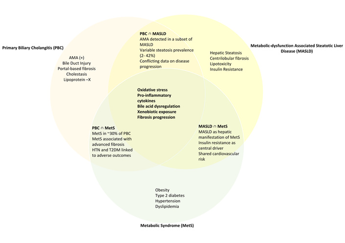
Pathophysiologic mechanisms and overlap between primary biliary cholangitis (PBC), metabolic dysfunction-associated steatotic liver disease (MASLD), and metabolic syndrome (Mets). Each circle represents disease-specific features, while overlapping zones illustrate shared pathophysiologic mechanisms and clinical associations. The central zone highlights mechanisms common to all three conditions. AMA: anti-mitochondrial antibodies; T2DM: type 2 diabetes mellitus; HTN: hypertension.
Among the individual components of metabolic syndrome, the association with PBC is biologically plausible and clinically relevant. Obesity, through leptin-ghrelin dysregulation and adipokine-mediated hepatic inflammation, may accelerate biliary fibrosis. Hypertension carries a synergistic risk with cirrhosis for adverse liver-related outcomes. Hyperlipidemia, paradoxically, may be protective against atherosclerosis in PBC due to the unique properties of Lp-X, yet its inverse association with liver-related mortality reflects the hepatic dysfunction that accompanies advanced disease. Diabetes independently increases the risk of PBC-related cirrhosis, with an odds ratio exceeding 2. Taken together, these findings support the routine metabolic risk factor screening in all patients with PBC, and for hepatologists to address these comorbidities proactively.
The therapeutic landscape offers an additional dimension of overlap. Agents approved or in trials for PBC, including UDCA, OCA, Elafibranor, and fibrates, have each been studied in MASLD/MASH with mixed results. The anti-cholestatic properties of fibrates and the FXR agonism of OCA represent mechanistic bridges between the two diseases. Future clinical trials in patients with confirmed co-existing PBC and MASLD are needed to determine whether treating one condition meaningfully alters the course of the other.
The detection of AMA in patients with MASLD raises unresolved questions about subclinical autoimmunity in the MASLD population. Whether this represents early or latent PBC, a shared immunologic susceptibility, or an epiphenomenon remains unclear. Longitudinal follow-up studies of AMA-positive MASLD patients are needed to determine whether treatment or closer surveillance in this group is warranted.
Further studies are needed to better understand the impact of metabolic risk factors on the prognosis of individuals with PBC. PBC does not exist in metabolic isolation, and clinicians caring for these patients should systematically evaluate for and address metabolic comorbidities. Researchers should prioritize prospective studies designed to clarify causality, define combined disease phenotypes, and test targeted therapeutic strategies in this understudied population.
ALP: alkaline phosphatase
AMA: anti-mitochondrial antibodies
APRI: AST-to-platelet ratio index
BA: bile acid
CAD: coronary artery disease
Fib-4: Fibrosis-4 index
FXR: farnesoid X receptor
GGT: gamma-glutamyl transferase
INR: international normalized ratio
IPTW: inverse probability of treatment weighting
LCAT: lecithin-cholesterol acyltransferase
LDL-C: low-density lipoprotein cholesterol
LFTs: liver function tests
Lp-X: lipoprotein-X
MASH: metabolic dysfunction-associated steatohepatitis
MASLD: metabolic dysfunction-associated steatotic liver disease
NAFLD: non-alcoholic fatty liver disease
NASH: nonalcoholic steatohepatitis
OCA: obeticholic acid
PBC: primary biliary cholangitis
PPAR: peroxisome proliferator-activated receptor
SLD: steatotic liver disease
T2DM: type 2 diabetes mellitus
UDCA: ursodeoxycholic acid
MM: Conceptualization, Investigation, Writing—original draft, Writing—review & editing. MA: Investigation, Writing—original draft, Writing—review & editing. WKS: Validation, Supervision, Writing—review & editing. KQ: Supervision, Validation, Writing—review & editing. All authors read and approved the submitted version.
Kamran Qureshi, MD, has received honoraria from Madrigal Pharmaceuticals, Gilead Sciences, and Salix Pharmaceuticals as a member of the speaker bureau. The other authors declare no conflicts of interest relevant to this work.
Not applicable.
Not applicable.
Not applicable.
Not applicable.
The authors received no specific funding for this work.
© The Author(s) 2026.
Open Exploration maintains a neutral stance on jurisdictional claims in published institutional affiliations and maps. All opinions expressed in this article are the personal views of the author(s) and do not represent the stance of the editorial team or the publisher.
Copyright: © The Author(s) 2026. This is an Open Access article licensed under a Creative Commons Attribution 4.0 International License (https://creativecommons.org/licenses/by/4.0/), which permits unrestricted use, sharing, adaptation, distribution and reproduction in any medium or format, for any purpose, even commercially, as long as you give appropriate credit to the original author(s) and the source, provide a link to the Creative Commons license, and indicate if changes were made.



View: 332
Download: 23
Times Cited: 0


