 Review


Affiliation:
Department of Pharmacology and Pharmaceutical Sciences, Alfred E. Mann School of Pharmacy and Pharmaceutical Sciences, University of Southern California, Los Angeles, CA 90089, USA
ORCID: https://orcid.org/0000-0001-7225-6126

 ,
,
Affiliation:
Department of Pharmacology and Pharmaceutical Sciences, Alfred E. Mann School of Pharmacy and Pharmaceutical Sciences, University of Southern California, Los Angeles, CA 90089, USA
Email: vpapadop@usc.edu
ORCID: https://orcid.org/0000-0002-1183-8568

Explor Dig Dis. 2026;5:1005135 DOI: https://doi.org/10.37349/edd.2026.1005135
Received: March 26, 2026 Accepted: June 23, 2026 Published: August 26, 2026
Academic Editor: Jose C. Fernandez-Checa, Institute of Biomedical Research of Barcelona (IIBB), CSIC, Spain
The article belongs to the special issue Fibrosis and Hepatobiliary Cancer
Metabolic dysfunction-associated steatotic liver disease (MASLD) is strongly associated with obesity, insulin resistance, and increased cardiovascular risk. Male hypogonadism has emerged as a potentially modifiable risk factor, and testosterone replacement therapy (TRT) has been proposed as a potential adjunctive treatment for MASLD and metabolic dysfunction-associated steatohepatitis (MASH) in hypogonadal men. Cross-sectional and longitudinal studies demonstrate a consistent inverse association between serum testosterone and MASLD prevalence and severity. Interventional evidence from randomized controlled trials (RCTs) and observational cohorts suggests TRT is associated with reductions in hepatic steatosis and improvements in liver-related biomarkers. In selected hypogonadal men, particularly those with metabolically active disease, TRT may contribute to MASH resolution and fibrosis improvement, although histological data remain limited. The most consistent response is observed in men with concurrent type 2 diabetes (T2D), obesity, or obstructive sleep apnea (OSA) and significant baseline steatosis. Preclinical data support convergent mechanisms involving the androgen receptor (AR), adenosine monophosphate-activated protein kinase (AMPK), and antifibrotic pathways. While recently approved therapies such as resmetirom and semaglutide represent significant advances in MASH treatment, their distinct mechanisms suggest that complementary roles alongside TRT are biologically plausible, though this remains entirely hypothetical in the absence of combination trial data. Taken together, TRT may represent a promising adjunctive therapy for reducing hepatic steatosis and improving the histopathological features of MASH in selected hypogonadal men with MASLD, particularly those with obesity or T2D and significant baseline steatosis; however, routine clinical use will require large, well-powered Phase 3 RCTs featuring standardized histological endpoints, extended follow-up, and rigorous cardiovascular and oncologic safety data.
Metabolic dysfunction-associated steatotic liver disease (MASLD), formerly known as non-alcoholic fatty liver disease (NAFLD), is characterized by hepatic steatosis involving ≥ 5% of hepatocytes in the absence of significant alcohol intake and in the presence of at least one cardiometabolic risk factor, such as obesity, type 2 diabetes (T2D) mellitus, or dyslipidemia [1, 2]. MASLD exists along a disease spectrum ranging from simple steatosis to metabolic dysfunction-associated steatohepatitis (MASH), advanced fibrosis, cirrhosis, and hepatocellular carcinoma (HCC). Together, these conditions contribute to a substantial and increasing global health burden that affects an estimated 30% of adults worldwide [3, 4]. Until March 2024, no targeted pharmacotherapy for MASH was approved by the U.S. Food and Drug Administration (FDA). That year, resmetirom (Rezdiffra), a selective thyroid hormone receptor-β agonist, received accelerated approval as the first disease-specific treatment for adults with noncirrhotic MASH and moderate-to-advanced fibrosis (stages F2–F3) [5–7]. This was followed in August 2025 by accelerated approval of semaglutide (Wegovy), a glucagon-like peptide-1 (GLP-1) receptor agonist, establishing the first GLP-1-based therapy for MASH [8–10]. Despite these therapeutic advances, lifestyle interventions remain the cornerstone of MASLD management, as a 3–5% weight loss improves hepatic steatosis, while greater and sustained weight loss (> 10%) can improve MASH and fibrosis [11–15]. However, long-term adherence to dietary and exercise modifications is often suboptimal, underscoring the critical role of adjunctive pharmacotherapies, such as GLP-1 receptor agonists and other emerging agents targeting metabolic pathways, in modifying the disease and mitigating progression.
Among the modifiable risk factors implicated in MASLD pathogenesis, endocrine dysregulation, specifically male hypogonadism, has attracted increasing attention. Late-onset hypogonadism, defined as total testosterone (TT) levels below 300 ng/dL on two consecutive morning measurements, engages in a bidirectional relationship with key features of metabolic syndrome, including insulin resistance (IR) and dyslipidemia, which collectively promote ectopic lipid deposition in hepatocytes [16–18]. Hypogonadism contributes to visceral adiposity and systemic inflammation [e.g., via elevated tumor necrosis factor-α (TNF-α) and interleukin-6 (IL-6)], impairing insulin signaling and upregulating hepatic de novo lipogenesis (DNL) through activation of sterol regulatory element-binding protein-1c (SREBP-1c), thereby exacerbating steatosis [17, 19–21]. Reciprocally, IR and compensatory hyperinsulinemia disrupt the hypothalamic-pituitary-gonadal (HPG) axis, further suppressing testosterone production and perpetuating a vicious cycle of metabolic dysfunction [22–24]. Epidemiological studies demonstrate that men with the lowest testosterone levels face a markedly higher risk of MASLD and MASH relative to eugonadal counterparts [25–27]. Testosterone replacement therapy (TRT), administered via intramuscular (IM), transdermal, or oral formulations, restores eugonadal testosterone levels and, in principle, could disrupt this pathological cycle by improving insulin sensitivity, reducing visceral adiposity, and directly modulating hepatic lipid metabolism [19, 28–32].
This narrative review synthesizes the evolving bench-to-bedside evidence on TRT in the management of MASLD over the past decade (2015–2025). We first delineate the molecular mechanisms underpinning androgen-mediated hepatoprotection, integrating preclinical data with epidemiological trends linking testosterone deficiency to disease severity. We then review the epidemiological evidence linking testosterone to MASLD risk and severity before appraising the interventional clinical data, including recent evidence from the EARTH study and the LiFT program. We conclude by proposing a phenotype-driven framework for patient stratification and outlining priorities for future research, including combination strategies with approved MASH therapies.
Male hypogonadism and MASLD are bound together in a self-reinforcing metabolic cycle. IR and hyperinsulinemia suppress the HPG axis at multiple levels: IR directly impairs Leydig cell steroidogenesis, attenuates pituitary luteinizing hormone (LH) output, and, particularly in the context of chronic hyperinsulinemia, may disrupt hypothalamic gonadotropin-releasing hormone (GnRH) pulsatility, collectively impairing testicular testosterone synthesis [22–24]. Visceral adiposity further amplifies this suppression via estrogen-mediated negative feedback and direct pro-inflammatory cytokine (TNF-α, IL-6) signaling at the hypothalamus and pituitary [17, 20, 21]. In turn, hypogonadism promotes adipogenesis, reduces skeletal muscle mass, and exacerbates whole-body IR, creating a vicious cycle that accelerates hepatic lipid accumulation and MASLD progression (Figure 1) [17, 19, 20]. Understanding this reciprocal loop is essential to appreciating why TRT may offer a mechanistically distinct and potentially complementary strategy in MASLD management, alongside conventional pharmacotherapies.
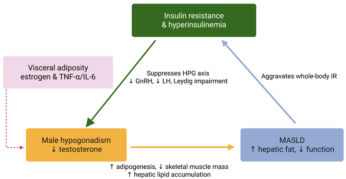
Interplay between insulin resistance (IR), hypogonadism, and metabolic dysfunction-associated steatotic liver disease (MASLD) in a self-reinforcing metabolic cycle. IR and chronic hyperinsulinemia directly impair testicular steroidogenesis at the level of Leydig cells, attenuate pituitary luteinizing hormone (LH) secretion, and disrupt hypothalamic gonadotropin-releasing hormone (GnRH) pulsatility, collectively reducing testosterone production. Visceral adiposity further exacerbates this suppression through increased estrogen-mediated negative feedback and pro-inflammatory cytokine signaling (TNF-α, IL-6) acting on the hypothalamic-pituitary axis. The resulting male hypogonadism promotes adipogenesis, decreases skeletal muscle mass, and worsens systemic IR. This reciprocal relationship establishes a vicious cycle that accelerates hepatic lipid accumulation and contributes to the development and progression of MASLD. Created in BioRender. Li, Y. (2026) https://BioRender.com/05wj7um.
At the hepatic level, TRT regulates lipid handling and steatosis progression. Activation of hepatic androgen receptors (ARs) suppresses DNL by downregulating core lipogenic enzymes, including acetyl-CoA carboxylase (ACC) and fatty acid synthase (FAS), thereby reducing triglyceride (TG) synthesis. In castration models, lipogenic transcriptional programs driven by SREBP-1c are significantly upregulated, promoting endoplasmic reticulum (ER) stress and lipid accumulation; TRT reverses these effects, restoring metabolic balance and alleviating steatosis [18, 33–38] (Figure 2).
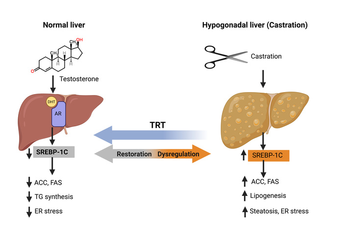
Androgen regulation of lipid metabolism via hepatic SREBP-1c. Testosterone is converted to DHT in the liver, which activates the AR to maintain normal SREBP-1c expression and preserve hepatic lipid homeostasis (left). Castration disrupts AR signaling, resulting in SREBP-1c dysregulation, hepatic lipid accumulation, and fatty liver (right). TRT restoration reverses these metabolic abnormalities. ACC: acetyl-CoA carboxylase; AR: androgen receptor; DHT: dihydrotestosterone; ER: endoplasmic reticulum; FAS: fatty acid synthase; SREBP-1c: sterol regulatory element-binding protein-1c; TRT: testosterone replacement therapy. Testosterone PubChem CID: 6013; PubChem 2D structure URL: https://pubchem.ncbi.nlm.nih.gov/compound/6013#section=2D-Structure; PubChem 3D structure URL: https://pubchem.ncbi.nlm.nih.gov/compound/6013#section=3D-Conformer. Created in BioRender. Li, Y. (2026) https://BioRender.com/e0x221t.
Complementing this AR-dependent control, preclinical hypogonadism models indicate that TRT can activate hepatic energy-sensing pathways: in high-fat diet (HFD)-induced MASLD mice, TRT induces adenosine monophosphate-activated protein kinase α (AMPKα) phosphorylation at Thr172, leading to functional inhibition of ACC, enhanced mitochondrial fatty acid β-oxidation, and suppression of lipid droplet biogenesis through downregulation of perilipin-2 [32, 39]. These coordinated actions result in ~40% improvement in histologic steatosis scores, normalization of circulating TG and cholesterol, and attenuation of inflammatory mediators (decreased TNF-α and IL-6), consistent with earlier rodent studies demonstrating a reduction in inflammation without adverse glycemic perturbation [30, 38–41] (Figure 3). TRT further promotes hepatic lipid clearance by enhancing very low-density lipoprotein (VLDL) secretion, preventing intracellular TG retention, and reinforcing its antisteatotic profile [42]. The clinical outcomes observed in the LiFT trial, including ≥ 30% reductions in liver fat content by magnetic resonance imaging-proton density fat fraction (MRI-PDFF) and steatosis resolution in 53.8% of treated patients, are consistent with, though not mechanistically confirmatory of, the preclinically established AMPK pathway.
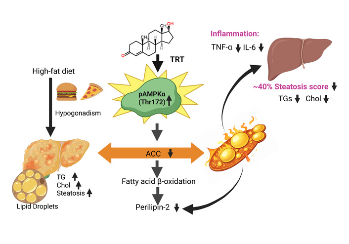
TRT ameliorates hepatic steatosis & dyslipidemia. TRT activates hepatic energy sensing in high-fat diet-induced MASLD mice with hypogonadism by increasing AMPKα phosphorylation (Thr172), leading to ACC inhibition, enhanced mitochondrial β-oxidation, and reduced lipid droplet formation via downregulation of perilipin-2. These effects result in ~40% reduction in steatosis scores, normalization of TG and cholesterol, and decreased inflammatory markers (↓ TNF-α, ↓ IL-6). ACC: acetyl-CoA carboxylase; AMPKα: adenosine monophosphate-activated protein kinase α; MASLD: metabolic dysfunction-associated steatotic liver disease; TG: triglyceride; TRT: testosterone replacement therapy. Testosterone PubChem CID: 6013; PubChem 2D structure URL: https://pubchem.ncbi.nlm.nih.gov/compound/6013#section=2D-Structure; PubChem 3D structure URL: https://pubchem.ncbi.nlm.nih.gov/compound/6013#section=3D-Conformer. Created in BioRender. Li, Y. (2026) https://BioRender.com/8i0oxyr.
Beyond the liver, TRT may exert systemic metabolic effects that indirectly protect hepatic function, although responses vary across populations and trial designs. Some studies report reductions in adiposity-related measures and improvements in IR [36, 43]. Enhanced GLUT4 (glucose transporter type 4, encoded by Slc2a4) translocation in skeletal muscle and adipose tissue caused by TRT contributes to improved glucose disposal and reductions in homeostatic model assessment of IR (HOMA-IR), while AMPK activation may amplify these benefits by sensing ATP depletion in insulin-resistant states and coordinating cellular energy homeostasis [20, 32, 39, 44–46] (Figure 4). The resulting suppression of hyperinsulinemia-driven hepatic lipogenesis provides an additional mode of steatosis control [30, 36, 39, 44]. Meta-analyses corroborate these systemic effects, reporting mean hepatic fat reductions of ~6.66% and, after baseline adjustment, relative liver fat decreases of 38.3% following TRT [36].
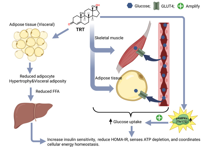
Systemic metabolic effects of TRT on hepatoprotection. TRT attenuates visceral adiposity, thereby reducing adipose tissue-derived FFA flux to the liver and mitigating hepatic lipotoxicity. This reduction in ectopic lipid burden alleviates hepatocellular stress associated with insulin resistance. In parallel, TRT enhances GLUT4 translocation and glucose uptake in skeletal muscle and adipose tissue, improving systemic insulin sensitivity and lowering HOMA-IR. Furthermore, activation of AMPK (pAMPKα Thr172) functions as a central metabolic sensor under insulin-resistant conditions, sensing ATP depletion in insulin-resistant states and coordinating cellular energy homeostasis. AMPK: adenosine monophosphate-activated protein kinase; FFA: free fatty acid; GLUT4: glucose transporter type 4; HOMA-IR: homeostatic model assessment of insulin resistance; TRT: testosterone replacement therapy. Testosterone PubChem CID: 6013; PubChem 2D structure URL: https://pubchem.ncbi.nlm.nih.gov/compound/6013#section=2D-Structure; PubChem 3D structure URL: https://pubchem.ncbi.nlm.nih.gov/compound/6013#section=3D-Conformer. Created in BioRender. Li, Y. (2026) https://BioRender.com/dzvaf4d.
Importantly, TRT also modulates inflammatory and fibrotic pathways central to MASLD progression. AMPK activation attenuates NLRP3 inflammasome signaling and suppresses Kupffer cell-mediated inflammatory responses, thereby reducing hepatocyte ballooning and improving liver biochemistry [i.e., decreased alanine aminotransferase (ALT)/aspartate aminotransferase (AST)] in preclinical HFD models, without evidence of fibrosis exacerbation [39, 47, 48] (Figure 5). These mechanistic insights are reflected clinically in the LiFT trial, where fibrosis improved in 80% of TRT-treated patients compared with 33% in placebo controls [49].
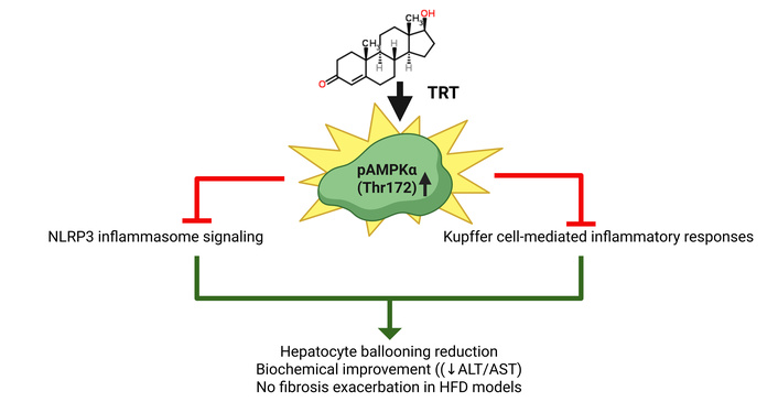
TRT activates hepatic AMPK to attenuate inflammation in MASLD. TRT increases AMPKα phosphorylation at Thr172 (↑ pAMPKα), leading to suppression of NLRP3 inflammasome signaling and Kupffer cell-mediated inflammation. In preclinical HFD models, this is associated with reduced hepatocyte ballooning and improved liver enzymes (↓ ALT/AST) without worsening fibrosis. ALT: alanine aminotransferase; AMPK: adenosine monophosphate-activated protein kinase; AST: aspartate aminotransferase; HFD: high-fat diet; MASLD: metabolic dysfunction-associated steatotic liver disease; TRT: testosterone replacement therapy. Testosterone PubChem CID: 6013; PubChem 2D structure URL: https://pubchem.ncbi.nlm.nih.gov/compound/6013#section=2D-Structure; PubChem 3D structure URL: https://pubchem.ncbi.nlm.nih.gov/compound/6013#section=3D-Conformer. Created in BioRender. Li, Y. (2026) https://BioRender.com/8m3rfr2.
Collectively, these interconnected pathways, now strengthened by AMPK-centered evidence, position TRT as a rational adjunctive therapy for a biologically defined subset of MASLD patients. Further studies are warranted to better delineate its indirect effects on fibrogenesis. Future phase 3 trials should evaluate potential synergistic strategies combining TRT with AMPK-modulating agents (e.g., metformin).
Beyond its metabolic and lipogenic effects, testosterone exerts significant anti-inflammatory actions that are directly relevant to MASLD pathobiology. Chronic low-grade hepatic and systemic inflammation is a defining and self-perpetuating feature of MASLD, contributing to disease progression from simple steatosis toward steatohepatitis, fibrosis, and cirrhosis [50]. Testosterone has been shown to suppress multiple pro-inflammatory mediators, including TNF-α, IL-1β, and IL-6, while promoting an anti-inflammatory cytokine milieu, effects mediated at least in part through AR-dependent modulation of NF-κB signaling [41]. In the context of male hypogonadism, the loss of these testosterone-mediated anti-inflammatory effects may therefore represent an independent, additive mechanism through which androgen deficiency accelerates MASLD progression, distinct from, yet synergistic with, the hyperinsulinemia-driven HPG axis suppression described above. This inflammatory axis further strengthens the biological rationale for TRT as a hepatoprotective intervention in hypogonadal men with MASLD and underscores the need for future trials to incorporate inflammatory biomarkers (e.g., high-sensitivity CRP, IL-6, TNF-α) as secondary endpoints alongside histological outcomes.
Early cross-sectional studies from 2015 established a robust inverse association between testosterone and MASLD. For instance, a Korean cohort study (n = 495 men) found that low serum TT was independently associated with MASLD, with men in the lowest testosterone quintile having a significantly higher odds of MASLD (OR 4.52, 95% CI 2.09–9.80) compared to those in the highest quintile, even after adjusting for visceral adipose tissue and IR [25]. Similarly, an Italian study (n = 55 men with spinal cord injury) found that low TT and free testosterone levels were independently associated with MASLD, with men with testosterone levels < 300 ng/dL showing ~12-fold greater risk of MASLD than those with levels ≥ 300 ng/dL after adjustment for confounders [51]. These findings were supported by a 2017 meta-analysis of 16 observational studies (n = 13,721 men and 5,840 women) that confirmed inverse associations between TT and sex hormone-binding globulin (SHBG) and MASLD [52].
More recently, a 2022 meta-analysis of ten cross-sectional studies (n = 2,995 men) further confirmed that lower TT levels are associated with increased MASLD severity, with a weighted mean difference of −0.35 ng/mL (95% CI −0.50 to −0.20) and an odds ratio of 0.79 (95% CI 0.73–0.86) per unit increase in TT, indicating a higher likelihood of moderate-to-severe disease in men with low testosterone [27]. A longitudinal Korean cohort study (n = 1,944 men) reported that although testosterone levels were lower in men with MASLD in cross-sectional analyses, baseline testosterone did not independently predict incident or regression of MASLD at 4.2-year follow-up after full adjustment for obesity and metabolic parameters, suggesting that metabolic factors mediate this relationship [53]. In a T2D cohort (n = 175 men), low testosterone levels correlated with hepatic TG measured by magnetic resonance spectroscopy but were not associated with histological severity of liver necroinflammation or fibrosis, indicating that the relationship is driven by underlying IR and obesity rather than MASH severity [54]. Overall, hypogonadism is consistently associated with increased MASLD risk, with men in the lowest testosterone quintiles showing 3.5- to 5-fold greater odds of MASH compared to those with higher levels [25, 26].
Evidence from 2015–2025 derived from observational studies, randomized controlled trials (RCTs), prospective cohort studies, and a 2025 meta-analysis of nine interventional studies, supplemented by subgroup analyses, is summarized in Table 1. Early research mainly examined associations, whereas later work shifted toward efficacy-focused trials.
Clinical studies of TRT in men with MASLD and other metabolically at-risk populations (2012–2025).
| Study (Year) | Design/Population | Intervention/Duration | Key hepatic outcomes | Limitations |
|---|---|---|---|---|
| Hoyos et al. (2012) [55] | RCT; n = 67 men with obesity and severe OSA (54 completed) | IM T undecanoate 1,000 mg at 0, 6, 12 weeks vs. placebo; 18 weeks | Liver fat ↓ 0.09 HU attenuation ratio (95% CI 0.009–0.17, p = 0.03); insulin sensitivity improved (HOMA-IR ↓ 1.14 units, 95% CI –2.27 to –0.01, p < 0.05); lean muscle mass ↑ 1.6 kg (95% CI 0.69–2.5, p = 0.0009); arterial stiffness ↓ 3.2% | OSA-specific population; short duration (18 weeks); concurrent weight loss program; no change in total weight, fat mass, or VAF; ALT/AST unchanged |
| Ng Tang Fui et al. (2016) [43] | RCT; n = 100 men with obesity (BMI ≥ 30, T ≤ 12 nmol/L); 82 completed | IM T undecanoate 1,000 mg q10w vs. placebo; 10-week VLED followed by 46-week weight maintenance (56 weeks total) | Fat mass ↓ 2.9 kg (MAD –5.7 to –0.2, p = 0.04); visceral fat ↓ 2,678 mm2 (p = 0.04); lean mass preserved; weight loss almost exclusively from fat in T group | Men with obesity and without diabetes; no liver fat measurements; body composition focus only; concurrent hypocaloric diet in all participants |
| Albhaisi et al. (2020) [56] LiFT Trial | Single-arm study; n = 21, hypogonadal males with MRI-proven MASLD (hepatic fat ≥ 8%) | LPCN 1144 (Oral T undecanoate): 450 mg daily. Duration: 16 weeks | Steatosis: 81% (17/21) showed improvement; mean decrease 4.04% in hepatic fat. MASLD resolution: 46.7% (10/21) achieved < 5% fat on MRI. Enzymes: significant reduction in ALT and AST | No control group; very small sample (n = 21); short duration (16 weeks); MRI only (no biopsy) |
| Khripun et al. (2020) [57] | RCT; 60 men with T2D and hypogonadism (mean age 54 years) | 1% transdermal T gel (AndroGel, 50 mg/day) vs. standard hypoglycemic therapy alone; 6 months | TRT group (n = 30) vs. control group (n = 30): ↓ hepatic fat fraction by 1.7 times; ↓ AST by 31%, ↓ ALT by 21%, ↓ GGT by 15.9% (p < 0.05); improved adipose tissue function (↓ leptin by 1.4×, ↓ resistin by 1.5×, ↑ adiponectin by 1.3×, p < 0.01); ↓ visceral obesity, ↓ hyperinsulinemia by 1.5×, ↓ HOMA-IR by 2.2×; ↓ total cholesterol and TG; improved glycemic control (↓ fasting glucose and HbA1c) | Single-center study; specific population (T2D with hypogonadism only); no long-term safety or efficacy data beyond 6 months; adverse events not specifically reported |
| Yassin et al. (2020) [58] | Prospective controlled registry study; n = 505 hypogonadal men (age 61 ± 10 years, TT ≤ 350 ng/dL); 321 TRT, 184 controls | IM T undecanoate 1,000 mg q12w (following initial 6-week interval); up to 12 years | FLI ↓ from 83.6 ± 12.08 to 66.91 ± 19.38 (vs. ↑ from 69 to 81 in controls); GGT ↓ from 39.31 ± 11.62 to 28.95 ± 7.57 U/L; bilirubin ↓ from 1.64 ± 4.13 to 1.21 ± 1.89 mg/dL; TG ↓ from 252.35 ± 90.99 to 213 ± 65.91 mg/dL; CVD mortality ↓ (7.8% vs. 15.2% in controls) | Non-randomized; observational design; treatment interruption in 147 men for 17 months due to reimbursement issues; surrogate markers (FLI) rather than imaging or biopsy; self-selection bias (controls opted out of treatment) |
| Al-Qudimat et al. (2021) [59] | Prospective controlled registry study; n = 496 hypogonadal men (age 59 ± 9.5 years, TT ≤ 12.1 nmol/L); 312 TRT, 184 controls | IM T undecanoate 1,000 mg q12w (following initial 6-week interval); 8 years | FLI ↓ from 83.70 ± 12.15 to 67.12 ± 19.21 (vs. stable in controls); bilirubin ↓ from 1.69 ± 4.21 to 1.31 ± 1.91 mg/dL; GGT ↓ from 39.45 ± 11.51 to 29.11 ± 7.68 U/L; TG ↓ from 254.87 ± 92.99 to 213.37 ± 66.91 mg/dL; CVD-related mortality improved | Non-randomized; observational design; surrogate markers (FLI) rather than imaging or biopsy; self-selection bias (controls opted out of treatment); no histological outcomes |
| Maseroli et al. (2021) [37] | Prospective observational study; 78 men with severe obesity (bariatric surgery candidates): eugonadal (n = 17), untreated hypogonadal (n = 46), treated hypogonadal (n = 15) | T undecanoate 1,000 mg IM q12w; mean 30 weeks | TTh vs. untreated hypogonadal: ↓ NAS and steatosis scores (p < 0.05); ↓ liver TG to eugonadal levels (p < 0.05); ↓ FLI (p = 0.001); ↓ total cholesterol (p = 0.022); improved hepatic gene expression (insulin signaling, lipid metabolism); enhanced preadipocyte mitochondrial function and insulin sensitivity | Observational design: selection bias (symptomatic patients treated); liver biopsies only at surgery (no baseline); short duration; small TTh group (n = 15) |
| TEREPINS study (2021) [60] LiFT Trial | Single-arm pilot; n = 3. Hypogonadal males with biopsy-proven MASH | LPCN 1144 (Oral T undecanoate): 450 mg daily. Duration: 52 weeks | Steatosis: Fatty liver grade improved from 2 to 1 in all 3 subjects.Fibrosis: Mean change in central fibrosis: 2.75 (improvement in all 3) | Extremely small sample (n = 3); study terminated early; no control group; results only in trial registry (no peer-reviewed publication) |
| Apostolov et al. (2022) [61] Secondary analysis of Gianatti 2014 | Secondary analysis of RCT; n = 39 men with T2D and low T (subset of 88 from parent trial; 20 T, 19 placebo) | IM T undecanoate 1,000 mg at 0, 6, 18, 30 weeks; 40 weeks | Liver fat ↓ 38.3% relative reduction (95% CI 25.4%–49.0%, p < 0.001) by MRI; median absolute reduction 3.5% vs. placebo increase 1.2% (between-group difference 4.7%, p < 0.001); lean mass ↑ 2.1 kg; fat mass ↓ 3.0 kg; no change in ALT, glucose, or HbA1c | Secondary analysis: not all parent trial participants had MRI; MRI IP/OP sequences used (not MRI-PDFF); liver enzymes within normal range (no significant inflammation); T2D-specific population; 40-week duration may be too short for some outcomes |
| LiFT Trial (2021) [49] | Phase 2 RCT; N = 56 randomized (3 arms); NASH Resolution set n = 37 (Treatment A: n = 13, Treatment B: n = 13, Placebo: n = 11). Biopsy-proven MASH (NAS ≥ 4, F1–F3), hypogonadal or eugonadal men | LPCN 1144 (oral T undecanoate): Treatment A: 225 mg BID; Treatment B: 225 mg BID + d-alpha tocopherol; vs. placebo. Duration: 36 weeks | Steatosis: ↓ up to 9.2% absolute and 46.8% relative vs. placebo (12 weeks, MRI-PDFF). MASH Resolution: 53.8% (7/13) vs. 9.1% (1/11) placebo.Fibrosis: No significant benefit over placebo; requires confirmation.Enzymes: ALT ↓ 23.4 U/L; AST ↓ 13.3 U/L vs. placebo | Small sample (n = 37); wide CIs due to small numbers; 36-week duration may be insufficient for long-term fibrosis assessment |
| Lee et al. (2024) [62] | RCT (secondary analysis of T Trials); n = 479 (246 TRT, 233 placebo); CT subgroup n = 140; older hypogonadal men (≥ 65 years, T < 275 ng/dL) | AndroGel 1%, 5 g daily titrated to T 400–800 ng/dL: 12 months | No significant differences in NAFLD scores: LAP (P = 0.98), HSI (P = 0.67), MASLD-MS (P = 0.52); or CT measures: liver HU (P = 0.24), LSR (P = 0.74). MASLD prevalence by HU < 40: 24.6% vs. 24.5% at 12 months (P = 0.21); by LSR < 1: 21.2% vs. 30.4% (P = 0.69). Subgroup analysis (MASLD+): no T benefit | Secondary analysis (not powered for MASLD); small CT subgroup; CT less sensitive than MRI-PDFF; no histology; short duration (12 months); excluded severe obesity (BMI > 37) and uncontrolled DM (HbA1c > 8.5%); no fibrosis assessment |
| Shigehara et al. (2025) [63] EARTH Study | RCT sub-analysis; n = 186 hypogonadal Japanese men (88 TRT, 98 controls); subgroup with FIB-4 ≥ 1.30 at baseline (n = 60; 28 TRT, 32 controls) | IM T enanthate 250 mg q4w; 12 months | Overall cohort: no change in FIB-4; FIB-4 ≥ 1.30 subgroup: FIB-4 ↓ from 1.98 ± 0.52 to 1.87 ± 0.60 in TRT group (p = 0.0277) vs. no change in controls; improvements correlated with ↓ waist circumference, ↓ body fat, ↓ TG | Retrospective sub-analysis; surrogate marker (FIB-4) rather than imaging or biopsy; no direct measurement of liver fat or histology; Japanese population only; modest effect size in FIB-4 reduction |
| Mahmoud et al. (2025) [36] | Systematic review and meta-analysis; 9 studies (3 RCTs, 4 non-randomized studies, 2 single-arm studies); adult men with MASLD/MASH, mostly hypogonadal (LiFT included hypogonadal and eugonadal men) | Varied TT (oral LPCN 1144/T undecanoate, IM/parenteral T undecanoate/Reandron, and transdermal AndroGel/T gel); follow-up ranged from 12 weeks to 8 years | All studies reported evidence of reduced hepatic steatosis with TT. LiFT showed MASH resolution and fibrosis improvement/regression; Albhaisi et al. [56] reported MASLD resolution by MRI. TT was associated with decreased liver enzymes; adverse events were generally comparable in comparative studies | Significant heterogeneity in populations, interventions/formulations, routes, doses, durations, endpoints, and outcome assessments; small samples/imprecision; observational studies had confounding/selection-bias concerns; larger high-quality double-blinded placebo-controlled RCTs are needed |
Yassin et al. (2020) [58] and Al-Qudimat et al. (2021) [59] are derived from the same patient registry and should not be considered independent datasets. ALT: alanine aminotransferase; AST: aspartate aminotransferase; BMI: body mass index; CI: confidence interval; CT: computed tomography; CVD: cardiovascular disease; DM: diabetes mellitus; FIB-4: fibrosis-4 index; FLI: fatty liver index; GGT: gamma-glutamyl transferase; HbA1c: hemoglobin A1c; HOMA-IR: homeostatic model assessment of insulin resistance; HSI: hepatic steatosis index; HU: Hounsfield unit; IM: intramuscular; LAP: lipid accumulation product; LPCN: Lipocine Inc.; LSR: liver-spleen ratio; MAD: mean absolute difference; MASLD: metabolic dysfunction-associated steatotic liver disease; MASLD-MS: MASLD metabolic score; MASH: metabolic dysfunction-associated steatohepatitis; MRI: magnetic resonance imaging; MRI IP/OP: MRI in-phase/opposed-phase; MRI-PDFF: MRI-proton density fat fraction; NAFLD: non-alcoholic fatty liver disease; NAS: NAFLD activity score; OSA: obstructive sleep apnea; q4w: every 4 weeks; q10w: every 10 weeks; q12w: every 12 weeks; RCT: randomized controlled trial; T: testosterone; T2D: type 2 diabetes; TG: triglyceride; TRT: testosterone replacement therapy; TT: total testosterone; TTh: testosterone therapy; VAF: visceral adipose fat; VLED: very low energy diet.
Based on the studies summarized in Table 1, the following analyses focused on the key features relevant to the relationship between TRT and hepatic outcomes in hypogonadal men with MASLD.
TRT, regardless of modality/formulation, consistently reduces hepatic steatosis in men with hypogonadism. Magnetic resonance imaging (MRI)-based assessment showed a 38.3% relative reduction in hepatic fat content over 40 weeks in T2D men treated with injectable testosterone undecanoate [61]. Similarly, in the LiFT program, oral testosterone undecanoate [(Lipocine Inc.) LPCN 1144] resulted in significant radiologic improvements. Notably, the LiFT Phase 2 trial included both hypogonadal and eugonadal men, and subgroup-specific effects were not fully delineated; therefore, the extent to which observed benefits are attributable to correction of hypogonadism versus pharmacologic testosterone exposure remains uncertain. After 16 weeks, MASLD resolved in nearly half of treated men, where resolution was defined as MRI-PDFF < 5%, with mean relative reductions in liver fat of approximately 50–55% across trials [49, 56]. In obese men with severe obstructive sleep apnea (OSA), TRT resulted in smaller but statistically significant reductions in computed tomography (CT)-quantified liver fat over 18 weeks, reflected by modest increases in hepatic attenuation relative to placebo [55]. Additional randomized evidence from a single-center trial in men with T2D and hypogonadism demonstrated a 1.7-fold reduction in hepatic fat fraction following six months of transdermal testosterone gel, accompanied by significant improvements in aminotransferases, GGT, IR, and visceral adiposity [57], reinforcing the consistency of steatosis reduction in metabolically high-risk populations. These trial-level findings are further supported by a 2025 systematic review and meta-analysis of nine interventional studies, which reported directionally consistent reductions in hepatic steatosis across TRT formulations and treatment durations, along with improvements in liver enzymes and, in some trials, MASLD or MASH resolution, albeit with substantial heterogeneity in study design, populations, and endpoints [36].
Two long-term observational reports [58, 59], likely derived from overlapping or related registry datasets, demonstrated sustained improvement with continued TRT exposure. Given similarities in study design, authorship, and patient populations, these reports should not be interpreted as independent cohorts, and their findings are best considered collectively rather than cumulatively. Across these studies, which followed large patient cohorts for 8–12 years, fatty liver index (FLI) declined from the high-risk range (~83–84) to intermediate-risk values (~66–67), accompanied by reductions in GGT, bilirubin, TGs, and cardiovascular mortality. Treatment interruptions were associated with partial reversal of hepatic and metabolic improvements, supporting a treatment-dependent effect, though these findings relied on surrogate indices rather than direct imaging or histological confirmation. A prospective observational study of hypogonadal men with severe obesity undergoing bariatric surgery reported lower steatosis and NAFLD activity scores, reduced hepatic TG content, and normalization of liver lipid profiles relative to untreated hypogonadal controls in TRT-treated individuals [37].
Evidence for modification of fibrosis with TRT remains exploratory. In a 2024 sub-analysis of the EARTH study, hypogonadal men with high baseline fibrosis risk [fibrosis-4 index (FIB-4) ≥ 1.30] demonstrated a modest but statistically significant reduction in FIB-4, from 1.98 ± 0.52 to 1.87 ± 0.60 (p = 0.0277), which was associated with improvements in waist circumference, body fat, and TG. In contrast, there was no significant change in FIB-4 in men with low baseline fibrosis risk [63], suggesting that any potential antifibrotic effect may be confined to individuals with pre-existing hepatic injury and may be mediated indirectly through improvements in adiposity and metabolic dysfunction, rather than direct hepatic remodeling.
The safety profile of TRT in men with MASLD appears to be generally favorable. A systematic review and meta-analysis found that adverse events were comparable between testosterone-treated and placebo groups, with no severe cardiac or hepatic events, including cholestasis and HCC, reported [36]. In the study of Apostolov et al. [61], TRT was well-tolerated with rare, serious adverse events that were not significantly different from placebo. In the EARTH study sub-analysis, TRT administered over 12 months was generally well-tolerated in Japanese hypogonadal men, with no serious adverse events attributable to the treatment reported [63].
Predictable androgenic effects have been documented, including prostate-related changes such as modest PSA elevations, but no study reported prostate cancer in either treatment or placebo groups [36]. PSA increases above 1 μg/L occurred in ~10% of testosterone-treated patients [43]; benign prostatic hyperplasia was not reported as an outcome in that source. In studies that directly reported hematological endpoints, TRT increased hemoglobin and hematocrit [55], with one patient experiencing an increase in hemoglobin to > 180 g/L [43]. Other MASLD-focused sources discuss polycythemia as a potential adverse event or focus on hepatic outcomes without reporting hematological parameters [36]. In the Maseroli observational study of men with severe obesity undergoing bariatric surgery, there were no hepatic concerns in TRT-treated hypogonadal men, though the study duration was relatively short (mean 30 weeks) and safety monitoring was not the primary endpoint [37].
Cardiovascular safety remains the most consequential area of uncertainty surrounding TRT. Concerns about its potential for adverse cardiovascular events have been longstanding and are well-documented in the literature [64–66]; however, short- to medium-term data have not demonstrated a significant increase in major adverse cardiovascular events, offering some reassurance for near-term clinical use [67]. Long-term registry data have further reported favorable outcomes, including significantly reduced overall mortality in TRT-treated patients compared with controls (7.8% vs. 15.2%, p = 0.035 in the Al-Qudimat cohort, with similar trends observed in the Yassin registry) [58, 59]. These findings contrast with meta-analytic evidence. Meta-analyses reported an approximately threefold increased risk of myocardial infarction with TRT [relative risk (RR) 3.12, 95% CI 0.34–29.01]; notably, this estimate was limited by small event numbers and wide CIs crossing unity, precluding definitive conclusions [36]. More recently, a real-world cohort study also demonstrated that long-term TRT was associated with increased cardiovascular risk [68], reinforcing the concern that short- to medium-term reassurance cannot be extrapolated to prolonged exposure. Taken together, these conflicting signals highlight the critical need for large-scale, prospective safety registries with extended follow-up. In parallel, pharmacological strategies aimed at augmenting endogenous testosterone production rather than exogenous replacement may represent an alternative avenue warranting further investigation, potentially offering a more favorable cardiovascular risk profile [69].
Formulation-specific data are reassuring. The LPCN 1144 oral testosterone prodrug study reported no serious adverse events, with only mild to moderate adverse events unrelated to treatment observed [56]. In the Phase 2 LiFT trial, LPCN 1144 demonstrated a favorable safety profile over 36 weeks, with adverse events distributed similarly between treatment and placebo groups [49, 56]. The small biopsy-based pilot study (n = 3) conducted over 52 weeks reported no safety concerns; however, the extremely limited sample size precludes meaningful conclusions regarding safety or efficacy [60]. In the Lee et al. [62] secondary analysis of the T Trials of older hypogonadal men (≥ 65 years), transdermal testosterone gel was generally well-tolerated over 12 months, with safety profiles consistent with the broader T Trials cohort, though this population excluded severe obesity [body mass index (BMI) > 37] and uncontrolled diabetes [hemoglobin A1c (HbA1c) > 8.5%]. Importantly, no studies reported hepatic adenoma or HCC in either treatment or control groups [36].
Taken together, the safety considerations surrounding TRT can be organized into three domains that are directly relevant to patient selection and clinical monitoring. With respect to cardiovascular risk, prior evidence has been inconsistent; however, the TRAVERSE trial provides reassurance in the short- to medium-term, demonstrating that TRT is noninferior to placebo with respect to major adverse cardiovascular events, albeit with a higher incidence of certain adverse events such as atrial fibrillation, pulmonary embolism, and acute kidney injury [67], a more recent real-world cohort study reported increased cardiovascular risk with long-term TRT exposure [68], and earlier registry data yielding apparently favorable mortality outcomes should be interpreted cautiously given the methodological limitations discussed above. Clinicians should therefore formally assess cardiovascular risk prior to initiation and exercise particular caution in men with pre-existing cardiac disease, uncontrolled hypertension, or a recent cardiovascular event—groups that were largely excluded from the trials reviewed here. With respect to hematological safety, erythrocytosis is a predictable and dose-dependent effect of TRT, and polycythemia is noted as a potential adverse event in MASLD-focused evidence [36]. Hemoglobin and hematocrit should be measured at baseline and monitored periodically during treatment, with dose reduction or temporary discontinuation considered if hematocrit exceeds 54%, consistent with established TRT monitoring guidance [16, 43]. With respect to prostate safety, MASLD-focused sources support a need for vigilance by reporting PSA-related signals [43], while baseline PSA assessment, digital rectal examination where clinically indicated, follow-up monitoring, and contraindications in men with known or suspected prostate or breast cancer should be grounded in established TRT guidance [16]. Regarding biochemical thresholds for treatment initiation, AUA guidelines recommend confirming hypogonadism with at least two morning TT measurements below 300 ng/dL in the presence of consistent symptoms, supplemented by calculated free testosterone and SHBG in cases where SHBG abnormality is suspected, a clinically important consideration given that obesity and IR, both prevalent in MASLD, suppress SHBG and may cause TT to underestimate bioavailable androgen [16].
Overall, while TRT demonstrates an acceptable safety profile in hypogonadal men with MASLD, monitoring for hematological changes, prostate parameters, and cardiovascular risk factors remains essential, and longer-term studies are needed to definitively establish safety across diverse patient populations.
TRT appears best suited to hypogonadal men (TT < 300–350 ng/dL) [16] with imaging- or histologically-confirmed MASLD and significant metabolic comorbidities, particularly T2D, obesity, or OSA. These populations show the most consistent hepatic responses across trials: a 38.3% reduction in liver fat in men with T2D [61], MASLD resolution in 48%, with a 55% mean relative liver fat reduction in the LiFT open-label study [56]. Improvements in steatosis have also been demonstrated with transdermal T gel in men with T2D [57] and with IM formulations in men with obesity and severe OSA [43, 55]. Men with an increased risk of fibrosis (FIB-4 ≥ 1.30) represent an additional higher-yield subgroup, where TRT produced a statistically significant FIB-4 reduction over 12 months (EARTH study) [63], while older men with low baseline steatosis or normal testosterone showed no meaningful benefit [62].
However, TT alone is an imperfect biomarker for therapeutic decision-making. TT levels are strongly influenced by SHBG, which is frequently altered in obesity, IR, and MASLD, and may therefore not reflect biologically active testosterone [17, 18, 27, 52]. This can lead to misclassification, with normal TT masking reduced free testosterone and low TT not necessarily indicating clinically meaningful androgen deficiency. In addition, TT does not capture AR sensitivity or tissue-specific androgen activity, both of which may influence treatment response. Accordingly, incorporation of complementary measures, such as calculated free testosterone and SHBG, may improve patient stratification and help explain heterogeneity in clinical outcomes. Consistent with this, some interventional studies, including the LiFT trial, included eugonadal participants, further limiting the specificity of conclusions regarding hypogonadism as the primary therapeutic target.
Prior to initiating TRT, pre-treatment screening should encompass hematological parameters (hemoglobin, hematocrit), prostate assessment (PSA, digital rectal examination), and formal cardiovascular risk evaluation [36]. Absolute contraindications include active prostate or breast cancer and uncontrolled erythrocytosis.
Despite encouraging findings, the current evidence base is constrained by several important methodological limitations. Many studies rely on surrogate indices of hepatic steatosis and fibrosis, such as the FLI and FIB-4, which were originally developed for risk stratification rather than for monitoring treatment-induced changes, thereby limiting inference regarding true histological improvement [58, 59, 63]. While MRI-based techniques, particularly MRI-PDFF, allow accurate quantification of hepatic steatosis [56, 61], they do not capture the defining histological features of MASH, including lobular inflammation, hepatocellular ballooning, and fibrosis stage. In addition, most RCTs are of relatively short duration (generally under one year), which is insufficient to meaningfully assess fibrosis progression or regression, processes that typically evolve over years [55–57, 63]. Conversely, longer-term observational studies reporting sustained or durable improvements lack randomization and are therefore susceptible to selection bias and residual confounding [37, 58, 59]. Furthermore, substantial heterogeneity in study populations, TRT formulations, outcome definitions, biopsy availability, and follow-up duration complicates interpretation, limits generalizability, and constrains pooled inference in meta-analyses [36]. TRT is not included in current American Association for the Study of Liver Diseases (AASLD) and European Association for the Study of the Liver (EASL) guideline recommendations for MASLD [2, 11]; this absence should be interpreted in light of the current evidence landscape rather than as a conclusion explicitly attributed to those guidelines.
Large, well-powered Phase 3 RCTs incorporating standardized, clinically meaningful histological endpoints remain a critical unmet need in this field. Future trial designs should adopt regulatory-grade endpoints, specifically MASH resolution without worsening of fibrosis and ≥ 1-stage fibrosis improvement, alongside sufficient follow-up duration to adequately assess long-term disease modification. In this regard, studies such as the biopsy-based TEREPINS trial represent meaningful advances toward establishing the therapeutic efficacy of TRT in MASLD. Beyond trial design, biomarker-driven patient enrichment strategies will be essential for identifying subgroups most likely to derive benefit. Specifically, candidate enrichment criteria include baseline FIB-4 ≥ 1.30 and SHBG levels, together providing a more targeted and mechanistically informed approach to patient selection [63].
Given the complex and multifactorial pathophysiology of MASLD, combination therapeutic strategies targeting complementary mechanisms are a logical next step. Recently approved therapies, including resmetirom, a thyroid hormone receptor-β agonist [5–7], and semaglutide, a GLP-1 receptor agonist [8–10], primarily target hepatic lipid metabolism and systemic energy balance. TRT operates through AR-mediated transcriptional regulation and AMPK activation—mechanisms that are largely non-overlapping with those of resmetirom or semaglutide, though clinical validation of these pathways specifically in MASLD remains limited. This mechanistic distinctiveness positions TRT as a potentially complementary agent in combination regimens, particularly in metabolically high-risk men with hypogonadism and comorbid T2D or obesity. Of note, the convergence of TRT and metformin on AMPK signaling raises the possibility that co-administration could produce additive or synergistic benefits on hepatic lipid homeostasis and insulin sensitivity, an avenue that warrants dedicated prospective investigation [39, 48].
Looking ahead, integrating TRT with approved agents like resmetirom and semaglutide may offer a mechanism‑based, personalized approach to MASLD management in hypogonadal men. However, prospective clinical evidence is currently lacking, and dedicated combination trials are needed to establish efficacy and safety.
While preclinical studies provide strong mechanistic support for TRT in MASLD, translational validation in human tissue remains a critical gap. Experimental evidence demonstrates that testosterone activates AMPK signaling, suppresses lipogenic transcriptional programs (notably SREBP-1c), enhances mitochondrial β-oxidation, and attenuates both inflammatory and fibrotic signaling cascades [32, 39]. To confirm whether these mechanisms translate to the human liver, future studies should incorporate paired pre- and post-treatment liver biopsies, coupled with integrated multi-omics (transcriptomic, proteomic, and metabolomic) analyses. Such approaches would help verify pathway-level changes and identify potential biomarkers of therapeutic response. Particular attention should be paid to validating the effects of TRT on hepatic stellate cell (HSC) activation and fibrogenesis. This is especially important because clinical evidence in the TRT field currently lacks direct data on liver fibrogenesis.
Current evidence is largely derived from relatively homogeneous populations, limiting generalizability across diverse ethnic, geographic, and metabolic subgroups. Future studies should therefore prioritize inclusion of underrepresented populations to better characterize treatment effects across varying disease phenotypes and cardiometabolic risk profiles.
In addition, cost-effectiveness analyses are currently lacking, despite the rapidly expanding therapeutic landscape for MASLD, including TRT, resmetirom, and GLP-1 receptor agonists. Comparative studies evaluating the economic and clinical value of these therapies, both as monotherapy and in combination, are essential for informing clinical decision-making and healthcare policy development.
Finally, head-to-head comparisons across TRT formulations (oral, IM, and transdermal) in well-defined MASLD populations are needed to identify the optimal treatment strategies and facilitate integration into future clinical guidelines.
Building on these limitations, several priorities should guide the future development of TRT in MASLD. First, adequately powered Phase 3 RCTs with standardized histopathological endpoints and extended follow-up are urgently needed, consistent with contemporary MASLD regulatory and guideline expectations. Ongoing studies, including LPCN 1144 and the paired-biopsy TEREPINS trials, are critical next steps. Second, combination strategies incorporating FDA-approved agents such as resmetirom and semaglutide warrant systematic evaluation, given that TRT’s androgenic and AMPK-mediated mechanisms are largely non-overlapping with thyromimetic and incretin pathways [39]. Third, biomarker-driven stratification using SHBG, baseline FIB-4 (≥ 1.30) [63], and AR genetic variants may enhance therapeutic precision and optimize patient selection. Fourth, translational mechanistic studies incorporating paired pre- and post-treatment liver biopsies are needed to validate, in humans, the AMPK activation, lipogenic suppression, and antifibrotic stellate cell effects observed in preclinical models. Fifth, long-term safety registries should systematically track HCC incidence, cardiovascular events, and hematological complications, because existing meta-analytic and registry data provide incomplete coverage of these outcomes: cardiovascular signals are reported in registry datasets, whereas HCC incidence and hematological complications require more systematic capture [36, 58, 59]. Sixth, future trials should enroll more diverse populations and incorporate formal cost-effectiveness analysis, both of which are currently absent from the evidence base.
Over the past decade, TRT has evolved from an epidemiological association to a mechanistically supported candidate therapy for hypogonadal men with MASLD, with preclinical and clinical evidence consistently pointing toward benefit in men with concurrent T2D, obesity, or significant baseline steatosis. However, the current evidence—largely based on surrogate imaging endpoints, short trial durations, and limited histological data—remains insufficient for routine clinical use, and TRT is best characterized as promising but not yet established. Its absence from current AASLD and EASL guidelines appropriately reflects this evidentiary gap. Rigorous pre-treatment screening and multidisciplinary oversight are essential, and routine use cannot be recommended until large, well-powered Phase 3 RCTs with histological endpoints and long-term safety data are available. Such trials represent the most critical next step in establishing TRT’s role in individualized MASLD management.
AASLD: American Association for the Study of Liver Diseases
ACC: acetyl-CoA carboxylase
ALT: alanine aminotransferase
AMPK: adenosine monophosphate-activated protein kinase
ARs: androgen receptors
AST: aspartate aminotransferase
BMI: body mass index
CT: computed tomography
DNL: de novo lipogenesis
EASL: European Association for the Study of the Liver
ER: endoplasmic reticulum
FAS: fatty acid synthase
FDA: U.S. Food and Drug Administration
FIB-4: fibrosis-4 index
FLI: fatty liver index
GGT: gamma-glutamyl transferase
GLP-1: glucagon-like peptide-1
GLUT4: glucose transporter type 4
GnRH: gonadotropin-releasing hormone
HbA1c: hemoglobin A1c
HCC: hepatocellular carcinoma
HFD: high-fat diet
HOMA-IR: homeostatic model assessment of insulin resistance
HPG: hypothalamic-pituitary-gonadal
HSC: hepatic stellate cell
IL-6: interleukin-6
IM: intramuscular
IR: insulin resistance
LH: luteinizing hormone
LPCN: Lipocine Inc.
MASH: Metabolic dysfunction-associated steatohepatitis
MASLD: metabolic dysfunction-associated steatotic liver disease
MRI: magnetic resonance imaging
MRI-PDFF: magnetic resonance imaging-proton density fat fraction
NAFLD: non-alcoholic fatty liver disease
OSA: obstructive sleep apnea
RCTs: randomized controlled trials
RR: relative risk
SHBG: sex hormone-binding globulin
SREBP-1c: sterol regulatory element-binding protein-1c
T: testosterone
T2D: type 2 diabetes
TG: triglyceride
TNF-α: tumor necrosis factor-α
TRT: testosterone replacement therapy
TT: total testosterone
VLDL: very low-density lipoprotein
The diagrams were generated using BioRender.
YL: Conceptualization, Writing—original draft. VP: Conceptualization, Writing—review & editing, Supervision. Both authors read and approved the submitted version.
Vassilios Papadopoulos, who is the Editorial Board Member of Exploration of Digestive Diseases, had no involvement in the decision-making or the review process of this manuscript. Another author declares no conflicts of interest.
Not applicable.
Not applicable.
Not applicable.
Not applicable.
This work was supported by the USC Alfred E. Mann School of Pharmacy and Pharmaceutical Sciences and the John Stauffer Dean’s Chair in Pharmaceutical Sciences at USC (to VP). The funders had no role in study design, data collection and analysis, decision to publish, or preparation of the manuscript.
© The Author(s) 2026.
Open Exploration maintains a neutral stance on jurisdictional claims in published institutional affiliations and maps. All opinions expressed in this article are the personal views of the author(s) and do not represent the stance of the editorial team or the publisher.
Copyright: © The Author(s) 2026. This is an Open Access article licensed under a Creative Commons Attribution 4.0 International License (https://creativecommons.org/licenses/by/4.0/), which permits unrestricted use, sharing, adaptation, distribution and reproduction in any medium or format, for any purpose, even commercially, as long as you give appropriate credit to the original author(s) and the source, provide a link to the Creative Commons license, and indicate if changes were made.



View: 959
Download: 17
Times Cited: 0
Patrizia Pontisso, Maurizio Parola
Siva Bala Subramaniyan, Balasubramaniyan Vairappan
Anna Fichera, Mirella Fraquelli
Lorenzo Mainardi ... Chiara Raggi


