Systematic Review


Affiliation:
1Department of Computer Science and Engineering, Bennett University, Greater Noida 201310, Uttar Pradesh, India
2Stroke Monitoring and Diagnostic Division, AtheroPoint™, Roseville, CA 95661, USA
ORCID: https://orcid.org/0009-0008-6528-8160

 ,
,
Affiliation:
3Department of Cardiology and Vascular Sciences, Indraprastha APOLLO Hospitals, New Delhi 110001, India
ORCID: https://orcid.org/0000-0003-2957-3825
,
Affiliation:
4Vascular Screening and Diagnostic Centre and University of Nicosia Medical School, 2408 Nicosia, Cyprus
ORCID: https://orcid.org/0000-0003-3912-7394
,
Affiliation:
5Division of Cardiovascular Medicine, Atrium Health Sanger Heart and Vascular Institute, Charlottesville, NC 28204, USA
ORCID: https://orcid.org/0000-0003-1756-2504
,
Affiliation:
2Stroke Monitoring and Diagnostic Division, AtheroPoint™, Roseville, CA 95661, USA
ORCID: https://orcid.org/0009-0003-5631-0550
,
Affiliation:
6Rheumatology Department, Hygeia Hospital, 15123 Athens, Greece
ORCID: https://orcid.org/0000-0002-0828-6176
,
Affiliation:
2Stroke Monitoring and Diagnostic Division, AtheroPoint™, Roseville, CA 95661, USA
ORCID: https://orcid.org/0000-0002-2844-6050
,
Affiliation:
7Department of Radiobiology and Molecular Genetics, National Institute of the Republic of Serbia, University of Belgrade, 110010 Belgrade, Serbia
ORCID: https://orcid.org/0000-0002-0012-2636
,
Affiliation:
8Department of Radiology, Azienda Ospedaliero Universitaria, 40138 Cagliari, Italy
ORCID: https://orcid.org/0000-0003-2870-3771
,
Affiliation:
2Stroke Monitoring and Diagnostic Division, AtheroPoint™, Roseville, CA 95661, USA
9Department of Innovation, Global Biomedical Technologies, Inc., Roseville, CA 95661, USA
10Department of Electrical and Computer Engineering, Idaho State University, Pocatello, ID 83209, USA
11Symbiosis Institute of Technology, Nagpur Campus, Symbiosis International (Deemed University), Pune 440008, Maharashtra, India
Email: jasjit.suri@atheropoint.com
ORCID: https://orcid.org/0000-0001-6499-396X

Explor Cardiol. 2026;4:1012122 DOI: https://doi.org/10.37349/ec.2026.1012122
Received: November 01, 2025 Accepted: July 09, 2026 Published: September 16, 2026
Academic Editor: Eugenio Picano, Italian National Research Council, Italy
Background: The cardiovascular risk and patient management are evaluated on the basis of risk scores that account for the multifactorial risk factors and thus fail in the estimation of an individual’s cardiovascular disease (CVD) risk. Advances in the field of medical imaging, especially cardiac computed tomography angiography (CTA) and magnetic resonance imaging (MRI), have laid the foundation of radiogenomics.
Methods: Research from the past 10 years was collected using the Preferred Reporting Items for Systematic Reviews and Meta-Analyses (PRISMA) methodology from PubMed, Google Scholar, ScienceDirect, and MEDLINE, and some additional sources like websites and arXiv.
Results: A total of 860 records were identified, and after the removal of duplicates, 800 remained for the screening for relevance by the abstracts and titles. After screening, 225 articles were assessed for eligibility, and the records that were either out of scope, had insufficient data, or were neither written nor translated in English were excluded. Finally, 80 records were identified to be included in the current review.
Discussion: We speculated on the application of artificial intelligence (AI) algorithms to identify CVD and its prognosis. Radiomic features, which are extracted by CTA and MRI, have shown great diagnostic accuracy of coronary plaques, and on the other hand, some studies exploited radiogenomics integration, which suggests that further research needs to be done in this field.
Cardiovascular disease (CVD) is a group of conditions that affect the heart or blood arteries. It is typically linked to an accumulation of fatty deposits in the arteries, raising the possibility of blood clots. Nowadays, CVD is the leading cause of death globally [1]. Nearly 17.9 million people die annually because of this. However, there are many ways to reduce the risk of developing these conditions [2, 3]. Although the precise etiology of CVD is unknown, a number of factors can raise your chance of developing it. These are referred to as “risk factors.”
Now, these risk factors can be of different types, like lifestyle, smoking, drinking, family history, environmental factors, and genetic factors [4]. These risk factors vary person to person. There are many CVD risk prediction calculators like the Framingham Risk Score (FRS) which is one of the first and is now outdated, systematic coronary risk evaluation (SCORE) which includes only the fatal CVD events resulting in underestimation of risk and QRISK which is not applicable outside the countries like Germany, Italy, England and Scotland, but these risk calculators do not incorporate the individulal’s genetic profiles, do not identify the low-risk patients and do not evaluate the lifetime CVD risk [5, 6]. Generally, the same treatment is given to all patients with CVD, regardless of their risk profile [7]. However, the concept of personalized medicine (PM) is to manage patients based on their characteristics, such as genetic disposition, which is unique for all patients [8, 9]. Correct treatment should be given to the correct individual at the correct time. An individual’s genetic background can guide the selection of the most effective treatments and preventive strategies for individuals with a specific genotype.
Clinicians are increasingly aiming to align medical tests and treatments with each individual’s unique biological characteristics. This approach utilizes data such as blood chemistry, genetic information, and medical imaging to determine the most effective interventions [10, 11]. This means healthcare is moving away from one-size-fits-all treatments. By understanding these biological factors, healthcare providers can tailor therapies that are more effective and safer for each person [12, 13]. In recent years, AI has emerged as the driving force of personalized cardiovascular medicine by overcoming the limitations of conventional CVD models [14, 15]. It helps in integrating the large, high-dimensional, and non-linear data, like imaging and genetics, without using handcrafted feature selection [6, 16]. This multimodal data integration has led to the development of radiogenomics [17].
Radiogenomics is the combination of two words, Radiomics and Genomics [18, 19]. In radiomics, computational extraction of medical imaging features captures imaging phenotypes that are otherwise not visible to the naked eye [20]. The steps in radiomics include image data acquisition, preprocessing of image data, identifying the region of interest (ROI), segmentation, and then different types of feature extraction, and using these features for the interpretation of risk [21]. Since its initial use in cancer, radiomics has proven useful in classifying tumors according to their histology, stage, and prognosis, as well as in linking imaging phenotypes to genetic changes [22, 23]. This review brings together recent studies on how AI-led radiogenomics advances cardiovascular care in a personalized framework.
The concept of PM is to devise a care plan tailored to each patient’s unique characteristics [24, 25]. Advances in AI have enabled the detailed analysis of patient data, thereby providing a more efficient and individualized approach to medical care [9, 26]. AI algorithms, particularly machine learning (ML) and deep learning (DL), are widely used to improve diagnostic accuracy by learning patterns from past patient records and medical images. Furthermore, DL models such as CNNs have shown excellent results in image-based medical applications, including disease detection and classification [27]. These models help clinicians analyze patient-specific data, which allows them to initiate appropriate treatment early.
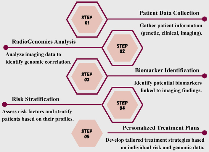
Integrating AI and radiogenomics is very useful as it links the genotypic features with the phenotypic ones [28]. Figure 1 depicts the systematic workflows for radiogenomics. The first step is the patient data collection, which includes the radiological images and molecular data [29]. This is followed by radiogenomic analysis, where the AI extracts the most important features and links them with genomic alterations [30]. These correlations point to clinically significant biomarkers that help categorize patients based on risk, disease severity, or anticipated treatment response [31]. This method makes it possible to more precisely customize treatment plans to the distinct clinical features of each patient.
In addition to diagnosis and treatment planning, AI has become an important component of drug discovery and development [32, 33]. It reduces development time and cost while increasing the success rate. While big data technologies and computing resources are becoming more affordable, it would be easier to develop a customized digital model, such as a “digital-twin,” that will mimic the human body and allow doctors to test all potential interventions and treatments before recommending them to actual patients [34–36]. Moreover, by using explainable AI (XAI), the interpretations of the AI-driven outcomes and decisions are enhanced [37, 38]. It helps the doctors to evaluate the reliability of predictions and support informed decision-making rather than replacing clinical judgement.
AI is also being incorporated into robotic platforms, virtual assistants, and monitoring tools to support healthcare delivery [39, 40]. These technologies assist with remote patient monitoring, patient education, and guided therapeutic interventions, including targeted drug discovery. Overall, the integration of AI and PM enhances evidence-based clinical decisions, improves patient outcomes, and has the potential to reduce the burden on the healthcare systems.
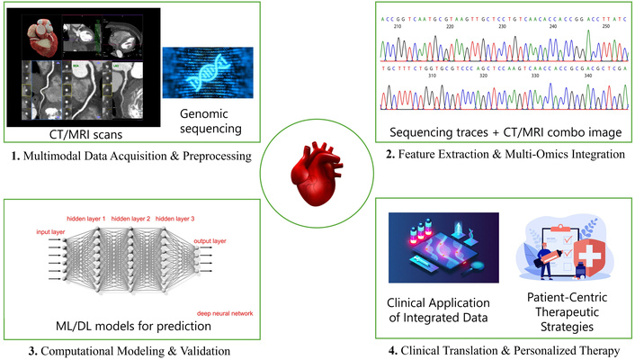
After the genomic revolution in the early 1990’s, oncology researchers have focused on investigating the fundamental causes of diseases at the genetic level to enable personalized treatments. CVDs are one of the most difficult diseases to diagnose because of their multifactorial nature, and also, the symptoms appear late during the disease progression [41, 42]. Conventional ways of genetic analysis rely on invasive biopsies or post-operative pathological tissues to perform the procedure; therefore, they cannot be applied to every patient [21, 43]. As we enter the next era of PM and big data, a large number of research studies are being carried out in the field of “Radiogenomics” [44]. It is the high-throughput extraction of image features from medical imaging. It allows data to be collected and used in a clinical decision support system to increase predictive, prognostic, and diagnostic accuracy [45]. Figure 2 depicts the stages in radiogenomics for CVD. The radiogenomics-CVD-based workflow consists of the following four steps: (i) First is multimodal data acquisition and preprocessing. In this step, multimodality imaging technologies such as CT, MRI, and PET are used to capture different physical and chemical properties of tissues. These radiomics features provide a multidimensional view of the CVDs [31, 46]. (ii) Second, feature extraction and multi-omics integration. In this step, high-dimensional feature data is extracted to quantitatively determine the characteristics of ROI [47]. After this, the data are fused with multiple omics data, such as proteomics, clinical, genomics, and transcriptomics, to accurately model clinical outcomes in patients with CVD [48, 49]. (iii) Third, computational modelling and validation. In this step, a DL model is trained and validated to link radiogenomic features with the symptoms of heart disease. They map gene regulatory networks to uncover the molecular relationships that underlie CVD development [50]. To maximize the performance of these algorithms, various cross-validation techniques like k-fold validation are used [51]. (iv) Last is the clinical translation and personalized therapy. Here, the outputs generated in the above-mentioned steps are incorporated into the clinical decision support systems so that they can assist the doctors during real-world decision-making [52]. This also helps in customized therapy planning of the patient, pharmacogenomics, and generating the polygenic risk scores for the early detection of CVDs [53, 54].
In order to analyze the various CVD methodologies within the AI framework, we adopted the Preferred Reporting Items for Systematic Reviews and Meta-Analyses (PRISMA) strategy [55]. Two authors (MS and JSS) from our team independently conducted the search within the four major databases: PubMed, Google Scholar, ScienceDirect, MEDLINE, and some additional sources like the websites and arXiv.
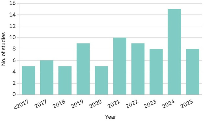
The search was made till November 1, 2025, and only studies published within the last 10 years were considered in this review. Figure 3 demonstrates the distribution of studies per year. The count of research papers has varied gradually with time, showing a sudden increase in 2024, indicating that the amount of research is increasing in this field. The key terms used for the search are: “CVD”, “CVD using DL”, “CVD risk assessment using DL”, “AI”, “Radiogenomics”, “AI and radiogenomics”, “Radiomics”, “Genomics”, “PM in CVD”, “AI in the framework of PM”, “Precision medicine”, “Medical imaging”, and “Bias in AI”. This study employed a combination of search criteria by applying Boolean operators like “AND”, “OR”, and “NOT”. This process yielded 860 research articles, of which 80 were deemed suitable according to the selection criteria established for the current study.
Our study utilized the published research papers written or translated into English. The inclusion and exclusion criteria used in our investigation are shown in Table 1. We omitted the non-research papers, such as magazines, theses, and dissertations. The final pool of publications focused on AI applications for examining CVDs, incorporating radiogenomics and PM.
Inclusion and exclusion criteria for PRISMA.
| S.No. | Attributes | Inclusion criteria | Exclusion criteria |
|---|---|---|---|
| 1 | Language | Written/Translated in English | Not written/Translated in English |
| 2 | Research type | Research articles, book chapters, case studies, websites | Magazines, thesis/dissertation |
| 3 | Research area | CVD, AI, PM, Oncology, and Radiogenomics | Not CVD, AI, PM, Oncology, or Radiogenomics |
| 4 | Location | Worldwide | None |
CVD: cardiovascular disease; PM: personalized medicine.
The initial search across four databases yielded 840 records, and an additional 20 records were identified through other sources, such as websites and arXiv, for a total of 860 records. After duplicate removal (n = 60), 800 unique records remained and were screened at the title and abstract levels. Of these, 200 records were excluded based on the title and 375 based on the abstract. A total of 225 full-text articles were assessed for eligibility. During this phase, 145 studies were excluded for being out of scope (n = 72), having insufficient details (n = 46), or being non-English (n = 27). Ultimately, 80 studies met all inclusion criteria and were included in the qualitative synthesis. The complete study selection process is illustrated in the PRISMA flow diagram (Figure 4).
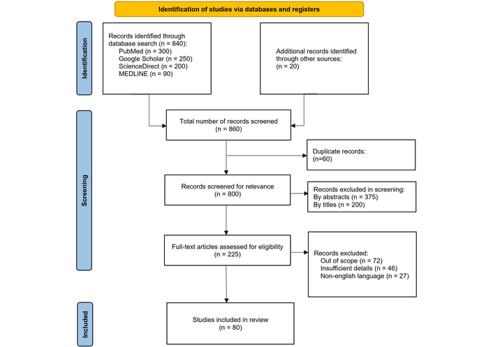
PRISMA flowchart for systematic paper selection and quality assessment. Adapted from ‘PRISMA’ (http://www.prisma-statement.org/). Accessed November 4, 2025. © 2024–2026 The PRISMA Executive. Distributed under a Creative Commons Attribution (CC BY 4.0) license.
The list of references in Tables 2 and 3 comprises 57 articles out of 80, as the remaining 23 focus on the global burden of CVD, the importance of AI, or precision medicine in general. The chosen literature covers a broad spectrum of AI methods, data sources, and study methodologies. In numerous CVD-focused studies, radiomics-based ML and DL models were used for risk stratification, disease detection, disease categorization, and prognostic evaluation, relying on medical imaging data. While wearable device data and electrocardiogram (ECG) signals were used for screening and early detection, electronic health records (EHR) data had an impact on outcome prediction. Oncology research has shown advanced integration of multi-omics and radiogenomics. Molecular subtype classification, treatment response assessment, and survival prediction were all done using AI frameworks. These studies provide insightful information that can guide individualized cardiovascular therapy. The significance of openness, interpretability, and clinical trust in AI-driven healthcare systems was emphasized in several studies that also addressed ethical issues, explainability, and governance of AI.
Comprehensive assessment of studies included in the review.
| Study population | Clinical domain | Data source | AI model type | Outcome | No. of studies | References |
|---|---|---|---|---|---|---|
| Adults with CVDs | CVD | Imaging | Radiomics-based ML | Risk prediction/plaque characterization | 4 | [22, 26, 31, 41] |
| Adults with CVDs | CVD | Imaging | DL | Diagnosis/screening | 5 | [15, 56–59] |
| Adults with CVDs | CVD | Imaging | Traditional ML | Risk prediction | 2 | [6, 60] |
| Adults with CVDs | CVD | Genomics | PRS models | CVD risk prediction | 2 | [4, 54] |
| Adults with CVDs | CVD | Genomics | ML | Genotype-phenotype prediction | 1 | [23] |
| Adults with CVDs | CVD | ECG/wearables | DL | Screening/detection | 3 | [57, 58, 61] |
| Adults with CVDs | CVD | EHR/clinical records | DL | Prognosis/outcome prediction | 1 | [62] |
| Mixed population cohorts | CVD | Multi-omics | DL | Precision medicine | 2 | [49, 50] |
| General CVD populations | CVD | Conceptual | Conceptual/AI frameworks | Precision cardiology | 5 | [7, 10, 11, 34, 35] |
| Cancer patients | Oncology | Imaging | CNN | Diagnosis/molecular subtype prediction | 4 | [14, 63–65] |
| Cancer patients | Oncology | Imaging | Radiomics-based ML | Prognosis/treatment response | 5 | [18–20, 63, 66] |
CVD: cardiovascular disease; CNN: convolutional neural network; DL: deep learning; ECG: electrocardiogram; EHR: electronic health records; ML: machine learning; PRS: polygenic risk score.
Comprehensive assessment of studies included in the review.
| Study population | Clinical domain | Data source | AI model type | Outcome | No. of studies | References |
|---|---|---|---|---|---|---|
| Cancer patients | Oncology | Imaging + genomics | Radiogenomics AI | Precision oncology | 7 | [28–30, 42–44, 52] |
| Cancer patients | Oncology | Multi-omics | DL | Survival prediction | 2 | [48, 67] |
| Oncology populations | Oncology | Conceptual | Conceptual frameworks | Precision oncology | 2 | [53, 68] |
| Mixed populations | Methodological | Imaging | DL | Model benchmarking | 2 | [14, 15] |
| Mixed populations | Methodological | Imaging | Radiomics systems | Feature extraction/validation | 2 | [21, 51] |
| Mixed populations | Methodological | Imaging + genomics | Radiogenomics AI | Method development | 2 | [47, 69] |
| Not population specific | Ethics/Governance | Conceptual | XAI | Ethics, transparency | 6 | [37, 38, 70–73] |
| Not population specific | Digital health | Wearables/EHR | DL | Decision support/digital twin | 5 | [34–36, 39, 40] |
| Drug discovery datasets | Cross-domain | Molecular/computational | DL | Drug discovery | 2 | [32, 33] |
DL: deep learning; EHR: electronic health records; XAI: explainable artificial intelligence.
Cardiovascular risk assessment has traditionally used clinical risk scores, such as the Framingham Risk Score, which estimate outcomes over predetermined time periods based on a small set of clinical and demographic factors, such as age, sex, blood pressure, cholesterol, smoking status, and diabetes history [74]. Even though these tools have been helpful for a long time, it is becoming recognized that they are unable to adequately reflect the complexity and variability of CVDs [75]. However, AI-driven models that integrate multimodal datasets—from genomic and molecular signatures to imaging biomarkers—offer previously unheard-of precision in diagnosis, customized treatment planning, and patient classification. Non-invasive radiogenomics may further reduce patient risk and discomfort by eliminating the need for biopsies [44]. As shown in Table 4, which highlights significant advances over the last five years, radiogenomics and AI have shown numerous applications in oncology. However, research on these approaches in CVD has been scant. Applying lessons from cancer to CVDs could improve treatment outcomes, risk assessment, and early detection.
Applications of radiogenomics in oncology.
| Author | Year | Cancer type | Imaging modality | AI | Results | Aim |
|---|---|---|---|---|---|---|
| Yan et al. [63] | 2021 | Glioma | MRI | Bayesian-regularization neural networks | AUC(IDH): 0.884AUC(1p19q): 0.815AUC(TERT): 0.669 | To predict the molecular groups (IDH, 1p19q, and TERT) in gliomas and assess their prognosis |
| Verduin et al. [76] | 2021 | Glioblastoma | MRI | Multivariable Cox-regression model | AUC(EGFR amplification): 0.707AUC(MGMT methylation): 0.667 | To predict overall survival and key molecular markers in glioblastoma patients |
| Zeng et al. [67] | 2021 | Clear cell renal cell carcinoma | CT | RF | AUC: 0.971 | To predict molecular characteristics and overall survival in ccRCC |
| Lu et al. [64] | 2023 | Colorectal cancer | CT | Yolov7 | AUC: 0.9591 | To determine the colorectal tumor location and predict the stage, and RAS gene mutation |
| Lee et al. [65] | 2023 | Colorectal cancer (stage IV) | 18F-FDGPET | ML algorithms with the best performance achieved by kNN | AUC: 0.791 | To predict tumor mutational burden (TMB) and prognosis in patients with stage IV colorectal cancer |
| Zhou et al. [66] | 2024 | Triple-negative breast cancer | Dynamic contrast enhanced-MRI | LR | AUC: 0.93 | To predict pCR from radiogenomic models |
| Ogbonnaya et al. [77] | 2024 | Prostate cancer | MRI | Pearson’s correlation | AUC: 0.95 | To create a radiogenomics map and predict prostate cancer |
| Buzdugan et al. [28] | 2025 | IDH-wild-type glioblastoma | Multiparametric MRI | RF, XGBoost, LightGBM, DNN | CI: 0.86 | To refine survival prediction in glioblastoma patients |
AUC: area under the curve; CI: concordance index; ccRCC: clear cell renal cell carcinoma; DNN: deep neural network; EGFR: epidermal growth factor receptor; FDGPET: fluorodeoxyglucose positron emission tomography; IDH: isocitrate dehydrogenase; kNN: k-nearest neighbors; MGMT: O6-methylguanine–DNA methyltransferase; pCR: pathological complete response; RAS: rat sarcoma; RF: random forest; TERT: telomerase reverse transcriptase.
To put the findings discussed in the review in context, it is essential to examine how recent research has applied AI across a range of CVD use cases. These studies differ significantly in terms of AI model, study design, and clinical goal. Key AI-driven CVD investigations are compiled in Table 5 to enable an organized comparison of their goals, approaches, and published results. Lekadir et al. [56] developed a CNN to determine the proportion of lipid core, fibrous tissue, and calcified tissue in carotid plaque from ultrasound images. The CNN outperformed SVM-based methods with pixel-level accuracy of 0.75 ± 0.16 and correlation with expert area measurements of 0.87–0.93 across tissue types using about 90,000 picture patches from 53 in vivo examples. These findings show that DL can enhance CVD risk stratification and early detection of plaques at risk of rupture. Attia et al. [57] trained a CNN model using paired 12-lead ECG and echocardiogram data to identify asymptomatic left ventricular dysfunction (ALVD). Patients with normal ventricular function at the start who tested positive were four times more likely to develop ventricular dysfunction later. When ECG is combined with AI, it can serve as a powerful screening tool for detecting ALVD in asymptomatic individuals. They further extended their study to identify cardiac dysfunction using a smartwatch’s single-lead ECG [58]. This approach worked even in non-clinical and real-world setting demonstrating the potential of wearable technology to detect asymptomatic heart problems and enable early intervention. Heo et al. [78] used and compared ML techniques to predict the long-term outcomes in ischemic stroke patients. The model that used a deep neural network (DNN) considerably outperformed the traditional Acute Stroke Registry and Analysis of Lausanne (ASTRAL) score (AUC 0.839; P < 0.001), with an AUC of 0.888, whereas ML algorithms, random forest (RF), and logistic regression (LR) models performed similarly to ASTRAL. McGilvray et al. [62] developed an ensemble DL model using longitudinal EHR data to predict which HF patients are at risk of severe decompensation or non-response to medical therapy. The model achieved an AUC of 0.91, which shows high prediction performance. This would spare the rest of the individuals from the associated risk, expense, and unnecessary high-risk procedures. Wang et al. [59] used CMR images to automate screening and diagnosis of 11 types of CVD across several Chinese facilities. They combined cine and late gadolinium enhancement (LGE) MRI with the AI models that have utilized a two-staged DL approach with video-based Swin Transformer models. In fact, this methodology demonstrated robust generalizability across some institutions and outperformed experienced cardiologists in some diagnoses. This outlines the potential of AI to improve the accuracy of CMR interpretation. Furthermore, in a recent study by Saikumar and Rajesh [60], the RCNN integrated with the Adaptive Random Forest (ARF) model outperformed conventional ML techniques in identifying heart disease from CT/MRI images, achieving 99% accuracy, enabling early detection of overlooked cardiac problems. Moreover, recent advances in DL have enabled high-accuracy prediction of CVD events from ECG images. To forecast four heart diseases, researchers in a study by Hasan et al. [61] developed a lightweight CNN model alongside conventional weighted classifiers, including GNB, XGBoost, SVM, DT, RF, and LR. With an incredible 99.29% accuracy, the ensemble model outperformed baseline techniques, while the CNN model extracted 100 significant features per image.
Examples of AI in CVD research.
| Author | Year | Diseases | Model | Results | Summary |
|---|---|---|---|---|---|
| Lekadir et al. [56] | 2016 | Carotid atherosclerosis | CNN | Acc: 75% | To characterize carotid plaque composition |
| Attia et al. [57] | 2019 | ALVD | CNN | AUC: 0.93 | To analyze smartwatch-recorded ECG signals and predict LV dysfunction |
| Heo et al. [78] | 2019 | Ischemic stroke | DNN, RF, LR | AUC(DNN): 0.888AUC(RF): 0.857AUC(LR): 0.849 | To predict long-term functional outcomes at 3 months in patients with ischemic stroke patients |
| Attia et al. [58] | 2022 | Cardiac dysfunction | AI algorithm (unspecified DL model) | AUC: 0.885 | To identify patients with cardiac dysfunction |
| McGilvray et al. [62] | 2022 | Heart failure (HF) | Ensemble DL model | AUC: 0.91 | To identify HF medical therapy non-responders |
| Wang et al. [59] | 2024 | *11 CVD types | Video Swin Transformer | AUC: 0.988 | To develop a video-based DL approach for automatic screening and diagnosis of CVDs using CMR |
| Saikumar et al. [60] | 2024 | CAD | RCNN | Acc: 99.173% | To detect CAD on radiology datasets |
| Hasan et al. [61] | 2025 | AHB, MI, HMI, NHB | CNN | Acc: 99.29% | To predict four cardiac conditions using ECG images |
*11 CVD types: hypertrophic cardiomyopathy (HCM), dilated cardiomyopathy (DCM), coronary artery disease (CAD), left ventricular noncompaction (LVNC), restrictive cardiomyopathy (RCM), cardiac amyloidosis (CAM), hypertensive heart disease (HHD), myocarditis, arrhythmogenic right ventricular cardiomyopathy (ARVC), pulmonary arterial hypertension (PAH), and Ebstein’s anomaly. Acc: accuracy; AHB: abnormal heartbeat; ALVD: asymptomatic left ventricular dysfunction; AUC: area under the receiver operating characteristic curve; CAD: coronary artery disease; CMR: cardiovascular magnetic resonance; CVD: cardiovascular disease; DL: deep learning; DNN: deep neural network; ECG: electrocardiogram; HMI: history of myocardial infarction; LR: logistic regression; MI: myocardial infarction; NHB: normal heartbeat; RCNN: region-based convolutional neural network; RF: random forest.
While AI offers many benefits for the healthcare sector, it also poses significant challenges. First, when imaging and genomic data are combined, they generate complex, heterogeneous datasets. To trace patterns from such a scarce dataset requires very high-end algorithms, which increase computational cost [69]. Second, an individual’s medical data is highly confidential and must be protected against breaches or cyberattacks. This necessitates robust encryption of patient-specific data. Healthcare institutions must adhere to the legal framework for the ethical use of patient data to protect data integrity from unauthorized use [70]. Third, bias in AI prediction algorithms [71]. It could be at different stages that contribute to health disparities. One of the main reasons is the underrepresentation of a specific population in the training data, which could be due to the inaccessibility of the healthcare facilities [72]. Additionally, AI predictions might yield accurate results on the data on which they were trained, but perform poorly on new, unseen data. It remains unclear to what extent it has understood the general principles [79]. The last is the concept of a “black box,” which results from the decision-making process’s lack of transparency regarding the reasons why AI generated the particular result [73]. For many years, clinical decision support tools have been integrated into EHR systems to assist physicians in identifying patients who are at higher risk, promoting medication safety, and adhering to evidence-based standards [80]. Historically, these systems have been used as advising tools, allowing medical professionals total discretion over whether to accept or reject the advice. The balance of power drastically shifts when AI starts acting on its own rather than just issuing commands. This shift highlights the need for close supervision as autonomy grows since it poses significant challenges to patient safety, accountability, ethical responsibility, and openness.
AI algorithms and radiogenomics have become promising tools for creating individualized therapy for CVD patients. The routine quantitative mapping of cardiac CT scans, which produces a variety of features that can be fed into ML and DL algorithms for CVD diagnosis and risk stratification, has been made possible by the quick development of radiomics. By helping clinicians make the right judgments and relieving them of difficult image processing duties, these innovative technologies have the potential to revolutionize current healthcare. Oncology accounts for the majority of this field’s study, while CVD receives less attention. With more investigation, the lessons learned from oncology-based studies can be applied to CVD.
Also, AI-based CVD imaging is still in its early stages, but it has a lot to offer to both clinicians and patients as it catalyzes the treatment towards more personalized care.
ALVD: asymptomatic left ventricular dysfunction
ASTRAL: Acute Stroke Registry and Analysis of Lausanne
CVD: cardiovascular disease
DL: deep learning
ECG: electrocardiogram
EHR: electronic health records
ML: machine learning
PM: personalized medicine
PRISMA: preferred reporting items for systematic reviews and meta-analyses
RF: random forest
ROI: region of interest
MS: Conceptualization, Data curation, Formal analysis, Investigation, Methodology, Project administration, Supervision, Validation, Visualization, Writing—original draft, Writing—review & editing. NK: Data curation. AN: Data curation. AS: Investigation. SG: Writing—review & editing. GK: Resources. IMS: Resources. EI: Writing—review & editing. LS: Formal analysis. JSS: Conceptualization, Methodology, Project administration, Supervision. All authors read and approved the submitted version.
Jasjit S. Suri, who is the Editorial Board Member of Exploration of Cardiology, had no involvement in the decision-making or the review process of this manuscript. The other authors declare no conflicts of interest.
Not applicable.
Not applicable.
Not applicable.
The primary data for this systematic review were sourced online from databases listed in the methods. Referenced articles are accessible on PubMed, Google Scholar, ScienceDirect, MEDLINE, and additional sources, including websites and arXiv. Additional supporting data are available from the corresponding author upon request.
Not applicable.
© The Author(s) 2026.
Open Exploration maintains a neutral stance on jurisdictional claims in published institutional affiliations and maps. All opinions expressed in this article are the personal views of the author(s) and do not represent the stance of the editorial team or the publisher.
Copyright: © The Author(s) 2026. This is an Open Access article licensed under a Creative Commons Attribution 4.0 International License (https://creativecommons.org/licenses/by/4.0/), which permits unrestricted use, sharing, adaptation, distribution and reproduction in any medium or format, for any purpose, even commercially, as long as you give appropriate credit to the original author(s) and the source, provide a link to the Creative Commons license, and indicate if changes were made.


View: 221
Download: 8
Times Cited: 0