 Perspective


Affiliation:
Diagnostic Centre, Diagnostic Medical Imaging, Unimed Porto Alegre, Porto Alegre 90560-003, Brazil
Email: mtorres.mt10@gmail.com
ORCID: https://orcid.org/0000-0002-5357-9138


Explor Cardiol. 2026;4:1012123 DOI: https://doi.org/10.37349/ec.2026.1012123
Received: May 05, 2026 Accepted: July 15, 2026 Published: September 16, 2026
Academic Editor: Andrea Borghini, Institute of Clinical Physiology - National Research Council (IFC-CNR), Italy
Medical radiation exposure in cardiology represents a significant and often underappreciated component of patient risk, with profound implications for individual and population health. Ionizing radiation is a proven carcinogen even at low doses, yet awareness among prescribers and practitioners remains suboptimal. Cardiologists are responsible for approximately half of all medical radiation exposure, with cumulative doses exceeding 100 mSv in one out of five adult cardiology patients and children with congenital heart disease—equivalent to 5,000 chest X-rays and corresponding to one excess cancer per 100 exposed individuals. Radiation exposure also contributes to non-cancer risks, including accelerated atherosclerosis, through mechanisms involving DNA instability and chronic inflammation. Professional exposure among interventional cardiologists is 4–5 times higher than that of diagnostic radiologists, with documented increases in chromosomal aberrations and vascular disease. Current guidelines emphasize justification, optimization, and education as cornerstones of radiation safety. Technological advances have enabled substantial dose reductions across all imaging modalities, and non-ionizing alternatives such as echocardiography and cardiac magnetic resonance offer effective diagnostic options without carcinogenic risk. This review examines the scope of radiation exposure in cardiology, associated cancer and non-cancer risks, current guideline recommendations, and strategies for achieving radiologic sustainability through responsible imaging practices.
The integration of radiation risk assessment into clinical decision-making represents a fundamental shift in cardiovascular medicine. Until the early 2000s, radioprotection was notably absent from cardiology textbooks, resulting in widespread ignorance of radiation doses and associated risks among both prescribers and practitioners [1]. This knowledge gap persists to varying degrees despite accumulating evidence that ionizing radiation is a proven carcinogen even at low exposure levels. The consequences extend beyond individual patient safety to encompass population health, occupational risks for healthcare workers, and medicolegal considerations. The use and the types of diagnostic techniques, in particular, interventional radiology, are increasing in complexity and versatility and are projected to increase steadily in the near future, expanding the therapeutic potential of the cardiologist, but also exposing the patient to potential risks due to cumulative exposures.
Medical radiation exposure in the US for the radiologic year 2016 reached the equivalent of 129 chest X-rays (CXRs) per person annually, with cardiologists responsible for approximately half of this burden [2]. Interventional cardiologists face professional exposures 4–5 times higher than diagnostic radiologists, placing them at increased risk for both cancer and non-cancer diseases, including premature atherosclerosis [3]. The past decade has witnessed growing recognition that radiation-aware practice constitutes a key quality indicator in cardiology, essential for protecting patients, staff, and practitioners themselves. The essence of radiation safety in cardiology is to adhere to the fundamental principles of radioprotection, stemming from the ALARA (As Low As Reasonably Achievable) principle. All doses should be kept as low as possible, avoiding ionizing radiation whenever a non-ionizing option is feasible.
This perspective examines the current state of radiation exposure in cardiac imaging, quantifies associated risks, evaluates guideline recommendations, and discusses strategies for achieving radiologic sustainability in cardiovascular practice.
Diagnostic and interventional fluoroscopy procedures represent the largest source of radiation exposure in cardiology (Figure 1).
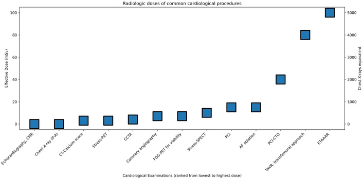
Doses of common cardiology examinations expressed in mSv (left Y-axis) and in equivalents of chest X-ray (right Y-axis). 1 mSv = 50 CXRs; 1 CXR = 0.02 mSv. AF: atrial fibrillation; CMR: cardiovascular magnetic resonance; CCTA: coronary computed tomography angiography; ETAAAR: endovascular thoracoabdominal aortic aneurysm repair; FDG-PET: fluorodeoxyglucose-positron emission tomography; PCI: percutaneous coronary intervention; SPECT: single-photon emission computed tomography.
Reference doses vary widely due to numerous factors, including patient habitus, procedure complexity, operator experience, and equipment characteristics. Diagnostic coronary angiography typically delivers approximately 7 mSv (equivalent to 350 CXRs), while percutaneous coronary interventions (PCIs) average 15 mSv (750 CXRs) [4, 5]. Complex interventions such as chronic total occlusion dilation can reach 81 mSv (4,050 CXRs), and aortic aneurysm repair procedures range from 76–119 mSv (3,800–5,950 CXRs) [6, 7]. The radiation dose is reported as median and interquartile range (Table 1).
| Invasive fluoroscopy | Effective dose (mSv) | CXRs |
|---|---|---|
| Diagnostic coronary angiography | 7 (2–16) | 350 (100–800) |
| PCI | 15 (7–57) | 750 (350–2,850) |
| Thoracic angiography (pulmonary or aorta) | 5 (4–9) | 250 (200–450) |
| Abdominal angiography or aortography | 12 (4–48) | 600 (200–2,400) |
| Pelvic vein embolization | 60 (44–78) | 3,000 (2,200–3,900) |
| TIPS placement | 70 (20–180) | 3,500 (1,000–9,000) |
| Aortic valvuloplasty | 39 (12–100) | 1,950 (600–5,000) |
| Dilation chronic coronary occlusion | 81 (17–194) | 4,050 (850–9,700) |
| Aortic aneurysm repair procedure | 76–119 | 3,800–5,950 |
| Renal angioplasty | 54 | 2,700 |
| Iliac angioplasty | 58 | 2,900 |
| Cardiac electrophysiology | ||
| Diagnostic EP studies | 3.2 (1.3–23.9) | 160 (65–1,195) |
| Ablation procedure | 15.2 (1.6–59.6) | 760 (80–2,980) |
| Regular PM or ICD implant | 4 (1.4–17) | 200 (70–850) |
| CRT implant | 22 (2.2–95) | 1,100 (110–4,750) |
| Computed tomography | ||
| CCTA | 7.4 (3.5–15.5) | 370 (175–775) |
| CACS | 1.2 (0.7–2.2) | 60 (35–110) |
| Nuclear cardiology | ||
| PET N-13 ammonia stress-rest (1,100 MBq) | 2.0 (1.6–2.4) | 100 (80–120) |
| 99mTc -Sestamibi (1,100 MBq, 1 day) stress-rest | 6.5 (3.9–8.6) | 325 (195–430) |
CXRs: chest X-rays; CRT: cardiac resynchronization therapy; PCI: percutaneous coronary intervention; CT: computed tomography; PET: positron emission tomography; PM: pacemaker. CACS: coronary artery calcium scoring; CCTA: coronary computed tomography angiography.
Electrophysiology procedures contribute substantially to both patient and operator exposure. Diagnostic electrophysiology studies average 3.2 mSv (160 CXRs), while ablation procedures—particularly for atrial fibrillation—deliver approximately 16.6 mSv (830 CXRs) [6, 7]. Cardiac resynchronization therapy implantation involves about 22 mSv (1,100 CXRs), reflecting the complexity of these interventions [4, 5]. The radiation dose is reported as median and interquartile range (Table 1).
Coronary computed tomography angiography (CCTA) has emerged as a significant source of medical radiation, with current protocols delivering approximately 7.4 mSv (370 CXRs) and new technology reaching < 3 mSv [4, 5]. Coronary artery calcium scoring (CACS), while lower at 1.2 mSv (60 CXRs), remains significant when considering its frequent use in asymptomatic populations. However, technological advances have enabled dramatic dose reductions, with modern scanners achieving up to 95% reduction compared to reference doses [8]. The radiation dose is reported as median and interquartile range (Table 1).
Nuclear cardiology procedures demonstrate considerable dose variability depending on tracer selection and protocol. Traditional thallium-201 stress-rest single‑photon emission computed tomography (SPECT) studies deliver 40.7 mSv (2,035 CXRs), while contemporary technetium-99m-based protocols range from 3.9 to 8.6 mSv (195–430 CXRs) [4, 5]. Positron emission tomography (PET) offers lower doses, with ammonia-13 stress-rest protocols averaging 2.0 mSv (100 CXRs) [8]. The American Society of Nuclear Cardiology has prioritized dose reduction, establishing as a primary goal that 50% of examinations should achieve < 10 mSv exposure [4, 5]. The radiation dose is reported as median and interquartile range (Table 1).
Children show greater radiosensitivity than adults, making pediatric radiation exposure particularly consequential [6]. The radiation dose is reported as median (Table 2) and has been dramatically reduced in the last 10 years [6]. The updated estimates for common invasive pediatric cardiology examinations show a constant downward trend in all examinations in all institutions, due to technological advances in imaging systems, growing awareness of the long-term effects of radiation exposure in pediatric populations, and adoption of radiation-sparing technology by operators, for instance, with decreasing use of image quantification and reduction of frame rate during fluorography acquisition [6].
The median radiation dose for common pediatric cardiology examinations (modified from [6]).
| Procedure | Effective dose (mSv) | Equivalent CXRs |
|---|---|---|
| Closure of atrial septal defect | 0 | 0 |
| Patent ductus arteriosus closure | 1.3 | 65 |
| Aortic valvuloplasty | 1.3 | 65 |
| Myocardial biopsy | 1.8 | 90 |
| Ventricular septal defect closure | 2.3 | 115 |
| Pulmonary hypertension | 1.3 | 65 |
| Aortic coarctation stenting | 1.9 | 95 |
| Right ventricular outflow tract stent | 2.7 | 135 |
CXRs: chest X-rays.
The 2020 American College of Cardiology/American Heart Association guidelines explicitly acknowledge that radiation exposure remains a critical consideration for congenital heart disease patients requiring serial imaging over a lifetime [7].
The relationship between radiation dose and cancer risk follows a stochastic model: increasing dose increases the probability, not severity, of malignancy. Cumulative exposure in cardiology patients can be substantial, with one in five adults and children with congenital heart disease exceeding 100 mSv lifetime exposure—equivalent to 5,000 CXRs and corresponding to approximately one excess cancer per 100 exposed individuals [9–12]. Recent data have reaffirmed the linear no-threshold model even below the 50 mSv threshold, and the cancer risk tends to be higher than previously assumed, exactly in the low-dose, low-dose rate range, where DNA damage is more frequently sublethal and therefore more often transmissible to subsequent generations of cells [13].
Risk varies significantly by age, sex, and genetic factors. Children face a fourfold higher risk than adults for equivalent doses. Women demonstrate 38% greater vulnerability than men, particularly due to highly radiosensitive organs such as the breast [13]. A 35-year-old woman undergoing a thallium scan faces cancer risk as high as 1 in 300, compared to approximately 1 in 1,000 for a middle-aged man undergoing myocardial perfusion imaging.
Specific malignancies reflect organ-specific dosing. Invasive cardiology procedures predominantly expose bone marrow, lungs, stomach, and thyroid, while nuclear cardiology delivers the highest doses to colon, bone, and bladder [2, 4, 5, 12, 13]. Chromosomal aberrations—reliable intermediate biomarkers for cancer—increase acutely after fluoroscopy procedures and persist decades after exposure in both patients and interventional cardiologists [11, 14].
The population burden of medical radiation-induced cancer is substantial. Approximately 9 million myocardial perfusion imaging scans performed in 2006 in the United States translate into roughly 8,000 additional lifetime cancers [15]. Computed tomography utilization in 2023 was estimated to contribute 5% of all US cancers [16]. Global estimates suggest medical radiation may account for 5–10% of all cancers when using updated exposure assessments [17–19].
The ISCHEMIA trial [20] showed that the risk of malignancy deaths increased linearly with the number of exposures to ionizing testing, such as coronary CT, myocardial perfusion scintigraphy, invasive coronary angiography, and PCI. Compared to patients with 1 radiation exposure, the risk increased 2-fold in patients with 2 radiation exposures and 4-fold in patients with 3 radiation exposures, with a median follow-up time of 3.2 years in a population of patients with chronic coronary syndromes.
Research involving patients with congenital heart disease indicates a greater risk of cancer in children who undergo more than six radiation-based procedures at a young age. The highest radiation exposure to specific organs—along with the associated predicted cancer risk—has been observed in the thyroid gland and female breast tissue [21].
Emerging evidence implicates radiation exposure and cardiovascular disease, with risk magnitude potentially comparable to cancer [22]. This association extends beyond the high-dose radiotherapy setting to include cumulative diagnostic exposures in the low-dose range, below 100 mSv [23]. Shared biological mechanisms include DNA instability, chronic low-grade inflammation, and increased oxidative stress [23].
Occupational studies demonstrate increased subclinical carotid atherosclerosis and premature vascular aging in interventional cardiologists with long-term radiation exposure [3]. These findings align with observations from atomic bomb survivors and radiation workers, suggesting that vascular effects represent a genuine stochastic risk of low-dose exposure rather than a threshold phenomenon [19].
Interventional cardiologists face professional exposures 4–5 times higher than diagnostic radiologists, approaching levels observed in early radiation workers and contemporary astronauts [24]. Documented health effects include increased chromosomal aberrations in circulating lymphocytes, elevated rates of left-sided brain tumors (ipsilateral to higher dose received from typical X-ray tube positioning), and multiple cutaneous malignancies in unprotected skin areas. Population-based studies demonstrate increased cancer risk among cardiologists who frequently perform PCI [24].
Cardiac sonographers face two distinct radiation exposure sources: patient-emitted radiation following nuclear imaging and scatter radiation in interventional laboratories. Patients injected with radiotracers become “hot” sources; at 90 minutes post-technetium-99m administration, chest wall dose equivalents measure 0.37–0.58 mSv. A 24-minute echocardiogram can deliver up to 0.16 mSv to sonographers, with higher exposure during left-handed scanning. Transport workers face approximately 0.02 mSv during 10-minute patient transfers [25].
Protective apparel offers limited benefit against high-energy photons emitted by radioisotopes. Lead aprons block 90% of X-rays but prove ineffective against positron-emitting tracers (fluorine-18, nitrogen-13), modestly effective against technetium-99m, and effective only against low-energy thallium-201 photons. Optimal protection involves temporal separation: 24 hours for PET or technetium studies, one week for Thallium examinations [5, 25–27].
The 2005 American College of Cardiology/American Heart Association clinical competence statement established that “the responsibility of all physicians is to minimize the radiation injury hazard to their patients, to their professional staff, and themselves” [27]. The 2009 American Heart Association position paper identified “Top-10 things to know about ionizing radiation in cardiac imaging,” emphasizing that studies using radiation should be ordered only after thoughtful consideration of potential benefits and consistent with appropriateness criteria [28].
The 2014 American Heart Association scientific statement articulated three cornerstones of radiation safety: education, justification, and optimization [29]. The European Society of Cardiology 2014 position paper added specific recommendations: avoid ionizing testing when non-ionizing alternatives provide comparable accuracy, select the lowest-dose options when ionizing studies are necessary, and document delivered dose in patient records [4]. European law (Euratom directive 2013/59) has mandated dose reporting since February 2018.
The 2018 position paper on radiation safety from the American College of Cardiology highlighted that effective training ought to foster a culture that respects radiation risks and demonstrates a dedication to reducing exposure while optimizing protection [5]. This cultural shift has produced measurable effects: myocardial perfusion imaging declined 50% between 2006 and 2016, largely replaced by functional testing with stress echocardiography or anatomic testing with CCTA [2].
Continuous technological innovation has enabled substantial dose reductions across all imaging modalities. Patient-specific personalized scanning protocols of multidetector coronary computed tomography achieves 60% dose reduction compared to reference standards. Nuclear cardiology has realized 50% reductions through improved cameras and software and a shift from Thallium to Technetium-based radiotracers [8]. Electrophysiology now offers near-zero fluoroscopy techniques for complex ablations. Invasive cardiology protocols achieve 40% reduction through optimized acquisition parameters [4, 5, 29]. Manufacturers increasingly compete on decreasing dose rather than increasing resolution, recognizing that radiologic sustainability provides a marketing advantage. Artificial intelligence also holds promise for dose optimization and iodinated contrast reduction in CCTA and invasive fluoroscopy.
Non-ionizing imaging alternatives should constitute first-line approaches whenever possible. Stress echocardiography effectively replaces myocardial perfusion imaging in most patients, with comparable accuracy and zero carcinogenic risk. Lung ultrasound can substitute for chest computed tomography in monitoring pulmonary edema and infection, offering additional advantages of bedside performance and minimized contagion risk. Right heart hemodynamics can be assessed non-invasively, potentially replacing invasive catheterization in many pulmonary hypertension patients [29].
Prescriber and practitioner awareness remains suboptimal despite decades of evidence. Educational initiatives must target all levels: medical students, residents, fellows, and practicing physicians. Informed consent documents require revision to clearly communicate radiation risks rather than obscure them with technical language [30]. Electronic health records should accumulate lifetime radiation dose, enabling evidence-based decisions about serial imaging.
No doubt ionizing radiation gives an immense benefit to diagnosis and therapy in modern cardiology. Although cardiologists cannot be scared of ionizing radiation, they must be scared of radiation negligence and always consider radiation in the risk side of the risk-benefit assessment of every diagnostic or therapeutic procedure, especially when discretionary or repeated imaging is performed, where alternatives exist.
Radiation exposure in cardiac imaging represents a clinically significant and often modifiable risk factor for cancer and accelerated atherosclerosis. The cumulative burden in cardiology patients frequently reaches levels associated with measurable population harm, while occupational exposures place healthcare workers at increased risk. Guideline recommendations have evolved to emphasize justification, optimization, and education as essential components of quality cardiovascular care. Technological advances enable substantial dose reduction, and non-ionizing alternatives offer effective diagnostic options without carcinogenic risk. A radiation-aware practice protects patients, staff, and practitioners while enhancing the overall sustainability of cardiovascular medicine. Action needs to be taken to reinforce the culture of radiation through appropriate changes in medical school and cardiology fellowship curricula, prepare transparent informed consent forms before the incorporation of dose in the written report, and adopt conservative approaches for human research and scientific publication to always justify the applications of radiation doses.
The basic idea is straightforward: if you can avoid taking a dose entirely, that’s the best option, both for patients and for intensively exposed doctors, like invasive cardiologists and electrophysiologists. No dose is the best dose.
CCTA: coronary computed tomography angiography
CXR: chest X-ray
PCIs: percutaneous coronary interventions
PET: positron emission tomography
MART: Conceptualization, Writing—original draft, Writing—review & editing. The author read and approved the submitted version.
The author declares that there are no conflicts of interest.
Not applicable.
Not applicable.
Not applicable.
Not applicable.
Not applicable.
© The Author(s) 2026.
Open Exploration maintains a neutral stance on jurisdictional claims in published institutional affiliations and maps. All opinions expressed in this article are the personal views of the author(s) and do not represent the stance of the editorial team or the publisher.
Copyright: © The Author(s) 2026. This is an Open Access article licensed under a Creative Commons Attribution 4.0 International License (https://creativecommons.org/licenses/by/4.0/), which permits unrestricted use, sharing, adaptation, distribution and reproduction in any medium or format, for any purpose, even commercially, as long as you give appropriate credit to the original author(s) and the source, provide a link to the Creative Commons license, and indicate if changes were made.



View: 244
Download: 6
Times Cited: 0


