 Original Article


Affiliation:
1Department of Medicine, Albert Szent-Györgyi Medical School, University of Szeged, H-6725 Szeged, Hungary
Email: nemes.attila@med.u-szeged.hu
ORCID: https://orcid.org/0000-0002-7570-6214


 ,
,
Affiliation:
1Department of Medicine, Albert Szent-Györgyi Medical School, University of Szeged, H-6725 Szeged, Hungary
Affiliation:
1Department of Medicine, Albert Szent-Györgyi Medical School, University of Szeged, H-6725 Szeged, Hungary
2Division of Haematology, Department of Medicine, Albert Szent-Györgyi Medical School, University of Szeged, H-6725 Szeged, Hungary
Explor Med. 2026;7:1001427 DOI: https://doi.org/10.37349/emed.2026.1001427
Received: December 09, 2025 Accepted: July 26, 2026 Published: September 13, 2026
Academic Editor: Lindsay A. Farrer, Boston University School of Medicine, USA
The article belongs to the special issue Assessment of Atrial and Ventricular Volumes and Functional Properties: Novel Insights
Aim: Hypocoagulability and associated alterations in myocardial mechanics seen in hemophilia could theoretically impact valvular geometry. Therefore, the purpose of the present study was to characterize mitral and tricuspid annular dimensions and functions in patients with hemophilia via 3D speckle-tracking echocardiography.
Methods: Of the 17 hemophilia patients originally assessed, four were excluded due to inadequate image quality. The remaining cohort consisted of 13 males with hemophilia (mean age: 40.8 ± 16.3 years; 11 with hemophilia A and 2 with hemophilia B). Thirty age- and gender-matched healthy males (mean age: 40.4 ± 9.2 years) served as the control group.
Results: Hemophilia patients presented with significant dilation of end-diastolic and end-systolic mitral annulus (MA) dimensions (diameter, area, and perimeter), leading to a reduction in MA fractional area change (MAFAC) compared with control subjects. In contrast, among the tricuspid annulus (TA) parameters, dilation was confined to end-diastolic values, while end-systolic dimensions were preserved.
Conclusions: Dilated MA, along with its functional deterioration, is present in patients with hemophilia compared to matched controls. TA dilation seems less pronounced.
Hemophilia is a rare, mostly inherited disorder that has two main types: hemophilia A and B, and develops as a consequence of low amounts of coagulation factors VIII and IX, respectively. Based on experience, 85% of patients have hemophilia A [1–3]. The coagulation abnormalities in hemophilia, driven by qualitative blood alterations, could theoretically impair myocardial mechanics and valvular function. Recent studies have demonstrated altered left ventricular (LV) rotational mechanics and segmental strains in these patients, resulting in subsequent changes to left atrial (LA) functional features [4–6]. Consequently, the present study aimed to characterize mitral annulus (MA) and tricuspid annulus (TA) morphology and function via three-dimensional (3D) speckle-tracking echocardiography (STE) in patients with hemophilia, with comparisons made to age- and gender-matched healthy subjects.
The study initially enrolled 17 patients with hemophilia, all of whom were receiving ongoing treatment and regular care at our department’s tertiary hematology center. Due to inadequate image quality for 3D echocardiographic analysis, 4 patients were excluded. Consequently, the final study cohort comprised 13 hemophilia patients (mean age: 40.8 ± 16.3 years, all males), from which 11 proved to be type A and 2 cases had type B. In all subjects, the diagnosis was established during infancy. None of the hemophilia patients and controls were obese, but 2 subjects were smokers. No known cardiovascular disease was present in any subject, 10 hemophilia patients were HCV positive and hemophilic arthropathy was present in 6 patients. Symptoms proved to be mild in 6 patients, and severe in 7 individuals. Factor level was below 1% in 8 patients with hemophilia, 4% in 2 cases, 6% in 1 case, and 9% in the remaining cases, respectively. Eight patients received on-demand therapy and prophylactic therapy was administered in 5 cases. The mean dose of factor VIII or IX was between 2,000–6,000 U/week for each patient. Their results were compared to 30 age- and gender-matched control subjects (mean age: 40.4 ± 9.2 years, all males). The control subjects had no symptoms, did not have any known cardiovascular risk factors or disorders, and were not taking any medications. A complete evaluation using two-dimensional (2D) Doppler echocardiography and 3DSTE was performed in both the hemophilia and control groups. The findings are from the Motion Analysis of the heart and Great vessels bY three-dimensionAl speckle-tRacking echocardiography in Pathological cases (MAGYAR-Path) Study, with the objective of characterizing 3DSTE-derived myocardial and valvular alterations unique to specific disorders, such as hemophilia (‘magyar’ means ‘Hungarian’ in the Hungarian language). The study was approved by the Institutional and Regional Biomedical Research Committee of the University of Szeged (number: 71/2011, latest approval 17th March 2025). The study conformed to the ethical guidelines of the Declaration of Helsinki, and all subjects gave their informed consent.
Standard echocardiography was performed on all hemophilia patients and control subjects to measure LA diameter and the thickness of the interventricular septum and LV posterior wall via the parasternal long-axis view. Furthermore, LV volumes were measured, and LV-EF was determined in accordance with Simpson’s method. A commercially available Toshiba Artida® echocardiographic tool (Toshiba Medical Systems, Tokyo, Japan) was used in all cases. For 2D Doppler examination, it was attached to a 1–5 MHz PST-30BT phased-array transducer. Early (E) and late (A) diastolic velocities of transmitral flow and their ratio (E/A) were determined by pulsed Doppler [7].
Data acquisitions were performed by a Toshiba ArtidaTM cardiac ultrasound machine (Toshiba Medical Systems, Tokyo, Japan) attached to a PST-25SX matrix-array transducer with 3D capability. Following image optimalisations (magnitude, gain, etc.), 6 subvolumes were acquired within six heart cycles and during a single breath-hold. Subsequently, offline image analysis was performed on these full-volume 3D datasets using 3D Wall Motion Tracking software version 2.7 (UltraExtend, Toshiba Medical Systems, Tokyo, Japan) [8–11].
Assessment of MA/TA dimensions followed a recently described method [12–15]. Briefly, MA/TA endpoints were defined on apical two- and four-chamber longaxis views by optimizing image planes, then measurements were performed on C7 cross-sectional short-axis views (Figure 1). Respecting the cardiac cycle, end-diastolic (at the time of peak R wave on ECG) and end-systolic (identified as the initial frame demonstrating aortic valve closure, coinciding with the termination of the T-wave on the ECG) parameters were assessed. The following parameters were determined.
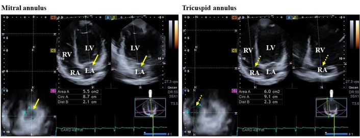
Three-dimensional (3D) speckle-tracking echocardiography-based analysis of mitral and tricuspid valve annuli. (A) Apical four-chamber long-axis view; (B) apical two-chamber long-axis view; and (C7) cross-sectional views of valvular annuli, of which their determination lon-axis views helped to define and optimally select. The yellow arrow represents the mitral annulus, while the yellow dotted arrow represents the tricuspid annulus. Area: mitral/tricuspid annular area; Circ: mitral/tricuspid annular perimeter; Dist: mitral/tricuspid annular diameter. LA: left atrium; LV: left ventricle; RA: right atrium; RV: right ventricle.
MA/TA diameter (MAD/TAD): perpendicular line connecting the peak of MA/TA curvature and the middle of the straight MA/TA border,
MA/TA area (MAA/TAA) measured by planimetry,
MA/TA perimeter (MAP/TAP) measured by planimetry.
MA/TA fractional shortening (MAFS/TAFS) = [end-diastolic MAD/TAD – end-systolic MAD/TAD]/end-diastolic MAD/TAD × 100,
MA/TA fractional area change (MAFAC/TAFAC) = [end-diastolic MAA/TAA – end-systolic MAA/TAA]/end-diastolic MAA/TAA × 100.
Count and percentage format (%) was used for categorical variables, and mean standard deviation (SD) format was used for continuous variables. Statistical tests proved to be two-sided and p < 0.05 was considered to be significant. Levene’s test was performed for homogeneity of variances: parametric data were analyzed using Student’s t-test, whereas non-parametric variables were compared using the Mann–Whitney-Wilcoxon test. Proportions for categorical data were assessed using Fisher’s exact test. To determine intraobserver and interobserver agreements, Bland–Altman analysis and interclass correlations (ICCs) were utilized. Statistical analyses were executed with IBM SPSS Statistics for Windows, version 29.0 (IBM Corp., Armonk, NY, USA)
There was a higher rate of hypertension and hyperlipidaemia in hemophilia patients, other demographic data were similar between patients and the healthy subjects (Table 1). Mean systolic and diastolic blood pressure (123.5 ± 2.8 vs. 121.3 ± 2.3 mmHg, p = ns), heart rate (71 ± 5 vs. 75 ± 6 bpm, p = ns) and cardiac output (5.8 ± 0.5 vs. 5.6 ± 0.4 L/min, p = ns) were comparable between the groups.
Baseline demographic and two-dimensional echocardiographic data of patients with hemophilia and healthy controls.
| Data | Controls(n = 30) | Hemophilia patients(n = 13) | p value |
|---|---|---|---|
| Demography | |||
| Age (years) | 40.4 ± 9.2 | 40.8 ± 16.3 | 0.910 |
| Male gender (%) | 30 (100) | 13 (100) | 1.000 |
| Hypertension (%) | 0 (0) | 4 (31) | 0.001 |
| Diabetes mellitus (%) | 0 (0) | 1 (8) | 0.124 |
| Hyperlipidaemia (%) | 0 (0) | 3 (23) | 0.006 |
| 2D echocardiography | |||
| LA diameter (mm) | 38.5 ± 3.5 | 38.2 ± 3.4 | 0.749 |
| LV end-diastolic diameter (mm) | 48.1 ± 3.0 | 49.6 ± 2.6 | 0.119 |
| LV end-diastolic volume (mL) | 108.2 ± 18.4 | 117.2 ± 14.7 | 0.125 |
| LV end-systolic diameter (mm) | 31.9 ± 2.1 | 31.2 ± 2.5 | 0.310 |
| LV end-systolic volume (mL) | 39.6 ± 10.3 | 38.7 ± 7.8 | 0.776 |
| Interventricular septum (mm) | 9.8 ± 1.3 | 10.1 ± 1.0 | 0.553 |
| LV posterior wall (mm) | 9.7 ± 1.6 | 9.8 ± 1.0 | 0.805 |
| LV ejection fraction (%) | 64.4 ± 2.9 | 67.1 ± 4.2 | 0.048 |
| E (cm/s) | 72.6 ± 14.8 | 71.8 ± 13.2 | 0.877 |
| A (cm/s) | 58.4 ± 17.9 | 67.2 ± 13.9 | 0.125 |
| E/A | 1.3 ± 0.2 | 1.1 ± 0.3 | 0.049 |
2D: two-dimensional; E and A: early and late diastolic transmitral flow velocity; LA: left atrium; LV: left ventricular.
Standard 2D echocardiographic findings remained similar between hemophilia patients and controls, except for elevated LV-EF and decreased E/A ratios in the hemophilia cohort. All other echocardiographic variables showed no significant differences between the groups. Additionally, valvular regurgitation ≥ grade 1 or significant valvular stenosis was entirely absent in both cohorts (Table 1).
Dilated MA diameter, area, and perimeter measured both in end-diastole and end-systole, and consequent decreased MAFAC were present in hemophilia patients compared to those of healthy controls. Among the TA parameters in patients with hemophilia, only end-diastolic area and perimeter showed dilation, while end-systolic TA dimensions were preserved (Tables 2 and 3).
Mitral annular data as assessed by three-dimensional echocardiography between hemophilia patients and controls.
| Data | Controls(n = 30) | Hemophilia patients(n = 13) | p value |
|---|---|---|---|
| Morphological parameters | |||
| End-diastolic MA diameter (MAD-D) (cm) | 2.47 ± 0.35 | 2.79 ± 0.25 | 0.006 |
| End-diastolic MA area (MAA-D) (cm2) | 7.66 ± 1.90 | 10.70 ± 2.55 | < 0.001 |
| End-diastolic MA perimeter (MAP-D) (cm) | 10.46 ± 1.22 | 11.30 ± 3.33 | < 0.001 |
| End-systolic MA diameter (MAD-S) (cm) | 1.83 ± 0.40 | 2.19 ± 0.38 | 0.010 |
| End-systolic MA area (MAA-S) (cm2) | 4.02 ± 1.34 | 6.91 ± 2.31 | < 0.001 |
| End-systolic MA perimeter (MAP-S) (cm) | 7.48 ± 1.28 | 9.99 ± 1.71 | < 0.001 |
| Functional parameters | |||
| MAFAC (%) | 47.17 ± 15.91 | 35.57 ± 16.44 | 0.042 |
| MAFS (%) | 26.18 ± 15.87 | 21.75 ± 10.19 | 0.368 |
MA: mitral annulus; MAFAC: mitral annular fractional area change; MAFS: mitral annular fractional shortening.
Tricuspid annular data evaluated by three-dimensional echocardiography between hemophilia patients and controls.
| Data | Controls(n = 30) | Hemophilia patients(n = 13) | p value |
|---|---|---|---|
| Morphological parameters | |||
| End-diastolic TA diameter (TAD-D) (cm) | 2.41 ± 0.41 | 2.65 ± 0.34 | 0.071 |
| End-diastolic TA area (TAA-D) (cm2) | 7.68 ± 1.94 | 9.06 ± 2.32 | 0.050 |
| End-diastolic TA perimeter (TAP-D) (cm) | 10.68 ± 1.25 | 11.54 ± 1.32 | 0.049 |
| End-systolic TA diameter (TAD-S) (cm) | 1.99 ± 0.37 | 2.08 ± 0.37 | 0.428 |
| End-systolic TA area (TAA-S) (cm2) | 5.92 ± 1.78 | 5.65 ± 1.34 | 0.628 |
| End-systolic TA perimeter (TAP-S) (cm) | 9.34 ± 1.20 | 9.50 ± 1.46 | 0.709 |
| Functional parameters | |||
| TAFAC (%) | 26.57 ± 12.01 | 23.75 ± 9.42 | 0.420 |
| TAFS (%) | 21.56 ± 8.47 | 17.70 ± 6.46 | 0.109 |
TA: tricuspid annulus; TAFAC: tricuspid annular fractional area change; TAFS: tricuspid annular fractional shortening.
Inter- and intraobserver variability for end-diastolic and end-systolic MA/TA parameters (diameter, area, and perimeter) obtained via 3DSTE is summarized in Table 4.
Intra- and interobserver variability for mitral and tricuspid annular dimensions as evaluated by three-dimensional speckle-tracking echocardiography.
| Data | Intraobserver agreement | Interobserver agreement | ||
|---|---|---|---|---|
| Mean ± 2SD difference in values obtained by 2 measurements of the same observer | ICC between measurements of the same observer | Mean ± 2SD difference in values obtained by 2 observers | ICC between independent measurements of 2 observers | |
| Mitral annular dimensions | ||||
| End-diastolic MA diameter | 0.03 ± 0.20 cm | 0.94 (p < 0.0001) | 0.04 ± 0.31 cm | 0.95 (p < 0.0001) |
| End-diastolic MA area | –0.03 ± 0.87 cm2 | 0.95 (p < 0.0001) | 0.03 ± 0.66 cm2 | 0.96 (p < 0.0001) |
| End-diastolic MA perimeter | –0.03 ± 0.71 cm | 0.96 (p < 0.0001) | –0.07 ± 0.71 cm | 0.95 (p < 0.0001) |
| End-systolic MA diameter | –0.03 ± 0.11 cm | 0.97 (p < 0.0001) | 0.03 ± 0.26 cm | 0.96 (p < 0.0001) |
| End-systolic MA area | –0.02 ± 0.32 cm2 | 0.95 (p < 0.0001) | –0.06 ± 0.61 cm2 | 0.98 (p < 0.0001) |
| End-systolic MA perimeter | 0.06 ± 0.81 cm | 0.95 (p < 0.0001) | 0.05 ± 0.55 cm | 0.97 (p < 0.0001) |
| Tricuspid annular dimensions | ||||
| End-diastolic TA diameter | 0.03 ± 0.23 cm | 0.95 (p < 0.0001) | 0.03 ± 0.34 cm | 0.95 (p < 0.0001) |
| End-diastolic TA area | –0.02 ± 1.34 cm2 | 0.95 (p < 0.0001) | 0.04 ± 0.68 cm2 | 0.97 (p < 0.0001) |
| End-diastolic TA perimeter | –0.06 ± 0.81 cm | 0.97 (p < 0.0001) | –0.10 ± 0.66 cm | 0.97 (p < 0.0001) |
| End-systolic TA diameter | –0.03 ± 0.51 cm | 0.97 (p < 0.0001) | 0.04 ± 0.51 cm | 0.97 (p < 0.0001) |
| End-systolic TA area | –0.04 ± 0.50 cm2 | 0.97 (p < 0.0001) | –0.05 ± 0.47 cm2 | 0.97 (p < 0.0001) |
| End-systolic TA perimeter | 0.08 ± 0.51 cm | 0.95 (p < 0.0001) | 0.05 ± 0.81 cm | 0.95 (p < 0.0001) |
MA: mitral annular; TA: tricuspid annular; SD: standard deviation.
Due to suboptimal image quality, 24% of the hemophilia cohort (4 out of 17 patients) had to be excluded from the analysis. Consequently, the overall feasibility rate for MA/TA measurements was 76%.
Based on the literature, the relationship between the mitral/tricuspid valve and hemophilia is about how to manage the patient during a possible surgery [16], how to treat the patient in the presence of a prosthetic valve [17], or what to do in case of a co-existing peripheral [18] or coronary artery disease or atrial fibrillation [19]. Literature data do not support an increased prevalence or severity of mitral/tricuspid regurgitation/stenosis in hemophilia. Nevertheless, early studies have confirmed differences in heart function in hemophilia [20, 21]. In healthy circumstances, a relationship between mitral/tricuspid dimensions and functional properties and myocardial characteristics can be seen [22, 23]. Compromised LV rotational mechanics—specifically reduced twist and apical rotation—have been established as key myocardial abnormalities in recent hemophilia studies [4]. Nevertheless, the prevalence of a near-total absence of LV twist—termed LV ‘rigid body rotation’—remained comparable to that of healthy controls [4]. These abnormalities were associated with the reduction of midventricular and basal LV circumferential strains representing specific segmental LV deformation abnormalities without alterations of global LV strains [5]. Moreover, these LV abnormalities were associated with impaired LA end-systolic reservoir function represented by reduced total LA emptying fraction and peak mean segmental LA circumferential and longitudinal strains as well [6].
The findings of the present study complement this knowledge by demonstrating MA dilation accompanied by functional deterioration. Based on these results, however, cardiac involvement is not limited to the left side of the heart. In patients with hemophilia, the TA also exhibited abnormal dilation, although these alterations were less pronounced. These findings suggest that despite being a hematological disorder, hemophilia, which is related to changes in blood quality, can be associated with significant abnormalities in myocardial mechanics and valvular annular dimensions. Cardiovascular risk factors such as hypertension and hypercholesterolemia were present in this hemophilia population, and their potential confounding effects cannot be ruled out. It is a well-established fact that associations exist between annular alterations and myocardial mechanics, even in healthy circumstances, as previously demonstrated by prior studies [23, 24]. Consequently, further investigations involving a larger cohort of hemophilia patients and utilizing alternative methodologies are warranted to confirm these findings. Additionally, a more detailed evaluation of the right heart using modern non-invasive imaging modalities in relation to the current findings is justified. Furthermore, these findings highlight the potential importance of routine cardiac screenings expanded to include echocardiography within this patient population.
Certain limitations must be considered while analyzing the data:
One of the most important limitations of 3D echocardiography is its image quality. In routine practice, 2D echocardiography-derived image quality is higher than that of 3D echocardiography. However, the feasibility and reproducibility of measurement of MA/TA dimensions seems to be acceptable.
Comparison of 2D echocardiography vs. 3D echocardiography-derived MA/TA dimensions was not proposed.
As previous studies on the same topic confirmed, assessment of dimensions of MA/TA is not based on a real 3D evaluation of these valvular annuli respecting their spatial 3D saddle-shape, but only on the evaluation of their 2D-projected image. This fact should be kept in mind when data are interpreted [12–15].
3DSTE echocardiography is an ideal method for simultaneous chamber quantifications and determination of volumes, volume-based functional properties, strains, rotational features, etc. of certain chambers. This study did not aim to perform such analyses.
In some studies, speckle-tracking echocardiography-derived analysis of MA/TA function was demonstrated. However, this study did not aim to perform such measurements.
Quantitative determination of mitral and tricuspid valve regurgitations was not performed; they were excluded only visually.
These findings raise the question of whether the aortic and pulmonary valves show similar abnormalities to those of the MA and TA. However, no literature data support this hypothesis; therefore, further investigations are required for confirmation.
It could be concluded that dilated MA, along with its functional deterioration, is present in patients with hemophilia compared to matched controls. TA dilation seems less pronounced.
2D: two-dimensional
3D: three-dimensional
MA: mitral annulus
MAA: mitral annular area
MAD: mitral annular diameter
MAFAC: mitral annular fractional area change
MAFS: mitral annular fractional shortening
MAP: mitral annular perimeter
TA: tricuspid annulus
TAA: tricuspid annular area
TAD: tricuspid annular diameter
TAFAC: tricuspid annular fractional area change
TAFS: tricuspid annular fractional shortening
TAP: tricuspid annular perimeter
AN: Conceptualization, Investigation, Visualization, Formal analysis, Writing—original draft, Writing—review & editing. NA: Validation, Writing—review & editing. ZB: Supervision, Resources. All authors read and approved the submitted version.
Attila Nemes, who is the Editorial Board Member and Guest Editor of Exploration of Medicine, had no involvement in the decision-making or the review process of this manuscript. The other authors declare no conflicts of interest.
The study was approved by the Institutional and Regional Biomedical Research Committee of the University of Szeged (number: 71/2011, latest approval 17th March 2025). The study conformed to the ethical guidelines of the Declaration of Helsinki.
All subjects gave their informed consent.
Not applicable.
The datasets for this manuscript are not publicly available due to local restrictions. Requests for accessing the datasets should be directed to the corresponding author.
No funding.
© The Author(s) 2026.
Open Exploration maintains a neutral stance on jurisdictional claims in published institutional affiliations and maps. All opinions expressed in this article are the personal views of the author(s) and do not represent the stance of the editorial team or the publisher.
Copyright: © The Author(s) 2026. This is an Open Access article licensed under a Creative Commons Attribution 4.0 International License (https://creativecommons.org/licenses/by/4.0/), which permits unrestricted use, sharing, adaptation, distribution and reproduction in any medium or format, for any purpose, even commercially, as long as you give appropriate credit to the original author(s) and the source, provide a link to the Creative Commons license, and indicate if changes were made.



View: 281
Download: 16
Times Cited: 0
Elizaveta A. Rogozhkina ... Oksana M. Drapkina


