 Original Article


Affiliation:
1Central Laboratory, Peking University School and Hospital of Stomatology, Beijing 100081, China

Affiliation:
1Central Laboratory, Peking University School and Hospital of Stomatology, Beijing 100081, China
Affiliation:
2Hunan Key Laboratory of Oral Health Research, Central South University, Changsha 410008, Hunan, China
3Xiangya Stomatological Hospital, Central South University, Changsha 410008, Hunan, China
4Xiangya School of Stomatology, Central South University, Changsha 410008, Hunan, China
Email: 569904896@qq.com
 ,
,
Affiliation:
1Central Laboratory, Peking University School and Hospital of Stomatology, Beijing 100081, China
5State Key Laboratory of Innovative Immunotherapy, Shanghai 200240, China
Email: caixinjia1994@163.com
Explor Med. 2026;07:1001426 DOI: https://doi.org/10.37349/emed.2026.1001426
Received: April 03, 2026 Accepted: July 28, 2026 Published: September 03, 2026
Academic Editor: Lindsay A. Farrer, Boston University School of Medicine, USA
Aim: To investigate interobserver variability in programmed cell death ligand 1 (PD-L1) combined positive score (CPS) assessment in head and neck squamous cell carcinoma (HNSCC) and to develop an artificial intelligence (AI)-based model for predicting PD-L1 expression and patient prognosis from hematoxylin and eosin (H&E)-stained slides.
Methods: Fifty HNSCC specimens were independently evaluated for PD-L1 by pathologists with different experience levels. Agreement was assessed using Fleiss’ and Cohen’s κ. Whole-slide images were processed into tiles for deep learning using DenseNet121. Tile-level features were integrated via two machine learning pipelines to construct whole-slide prediction models. Multiple algorithms were tested, with performance evaluated in validation and testing cohorts. Prognostic value was analyzed using AI-derived risk stratification.
Results: Interobserver agreement was low (Fleiss’ κ = 0.34), indicating substantial variability in CPS assessment. DenseNet121 achieved moderate predictive performance (AUC 0.641 in validation, 0.616 in testing). AI models significantly improved prediction accuracy, with logistic regression demonstrating the best performance (AUC 0.900 in validation, 0.851 in testing). AI-derived prediction scores effectively stratified overall survival, with multiple models showing significant prognostic discrimination (P < 0.05).
Conclusions: AI models integrating deep learning and machine learning can accurately predict PD-L1 expression and stratify prognosis in HNSCC, outperforming tile-level deep learning alone.
Head and neck cancer ranks as the seventh most common malignancy worldwide and remains a significant global public health challenge [1, 2]. Among these tumors, head and neck squamous cell carcinoma (HNSCC) accounts for more than 90% of cases [1]. In 2022, approximately 758,000 new cases were reported globally, underscoring the substantial disease burden associated with HNSCC [3]. Despite advances in multimodal treatment strategies, the overall 5-year survival rate remains unsatisfactory and is markedly lower in advanced stages and low-income regions [4–6]. These limitations highlight the urgent need for more effective therapeutic approaches and improved strategies for precise patient stratification [7–9].
In recent years, immunotherapy, particularly immune checkpoint inhibitors (ICIs) targeting the programmed cell death protein 1 (PD-1)/programmed cell death ligand 1 (PD-L1) axis, has emerged as a promising treatment modality for HNSCC [10–12]. Anti-PD-1 agents were approved by major regulatory agencies, including the Food and Drug Administration and the European Commission [13]. However, clinical responses remain limited, with relatively low objective response rates observed for agents such as pembrolizumab [14]. This variability in therapeutic efficacy underscores the critical need for reliable predictive biomarkers to optimize patient selection and improve treatment outcomes. Currently, PD-L1 expression, assessed by immunohistochemistry and quantified using the combined positive score (CPS), serves as the primary biomarker guiding immunotherapy decisions in clinical practice [15]. However, CPS evaluation is associated with several intrinsic limitations. The assessment is semi-quantitative and highly dependent on pathologist expertise, resulting in considerable variability, particularly in borderline cases. Furthermore, intratumoral heterogeneity and sampling bias further compromise the reproducibility and robustness of PD-L1 evaluation [12, 16, 17]. Collectively, these challenges limit the clinical utility of CPS and emphasize the need for more objective, standardized, and scalable assessment methods [18].
Artificial intelligence (AI), particularly deep learning-based computational pathology, has demonstrated considerable potential in extracting high-dimensional features from hematoxylin and eosin (H&E)-stained whole-slide images (WSIs) [19–22]. Recent studies suggest that AI models can infer molecular characteristics and tumor microenvironment features directly from routine histopathological images, offering a convenient and cost-effective alternative to conventional assays [23–25]. However, there remains a lack of clinically adopted AI-based tools for the assessment of PD‑L1 expression.
In this study, we developed an AI-based framework that integrates deep learning and machine learning to predict PD-L1 expression from H&E-stained slides in HNSCC. Tile-level features were extracted using a convolutional neural network and subsequently aggregated into slide-level representations through multi-feature fusion strategies. The model was validated in both internal and external cohorts, and its prognostic value was further assessed. Our results demonstrate that this AI-driven approach achieves robust predictive performance and may serve as a valuable tool to assist pathologists in PD-L1 evaluation, thereby supporting precision immunotherapy in HNSCC.
A total of 457 H&E-stained WSIs of HNSCC collected from 2014 to 2017 were used as the training cohort. An additional 104 WSIs obtained between 2013 and 2014 from the same institution served as the validation cohort, while 182 WSIs from another hospital were used as an independent testing cohort. Baseline clinicopathological characteristics of the three cohorts are summarized in Table S1. This study was approved by the Institutional Review Board of these hospitals.
Formalin-fixed, paraffin-embedded (FFPE) tissue samples were sectioned and mounted on adhesive slides. Immunohistochemical staining for PD-L1 was performed using a fully automated system (BOND, Leica Biosystems) following standard protocols, including deparaffinization, rehydration, antigen retrieval, endogenous peroxidase blocking, incubation with an anti-PD-L1 monoclonal antibody, application of secondary antibodies, and hematoxylin counterstaining. The CPS was calculated as the number of PD-L1-positive cells (including tumor cells, lymphocytes, and macrophages) divided by the total number of viable tumor cells, multiplied by 100. A minimum of 100 viable tumor cells was required for evaluation [11]. PD-L1 staining was independently assessed by two experienced pathologists.
WSIs were segmented into non-overlapping image tiles, and tiles lacking tissue content were excluded. Image intensities were normalized using Z-score standardization across RGB channels. A tile-level deep learning model based on DenseNet121 was trained to predict PD-L1 expression. Transfer learning was employed with pre-trained weights. Following tile-level prediction, each tile was assigned a probability score. Two independent feature aggregation pipelines were developed: PALHI pipeline and BoW pipeline. The features derived from both pipelines were integrated to construct slide-level representations. These features were then input into multiple machine learning classifiers. Model performance was evaluated in the validation and testing cohorts, and the optimal model was selected based on predictive accuracy. The overall workflow is illustrated in Figure 1.
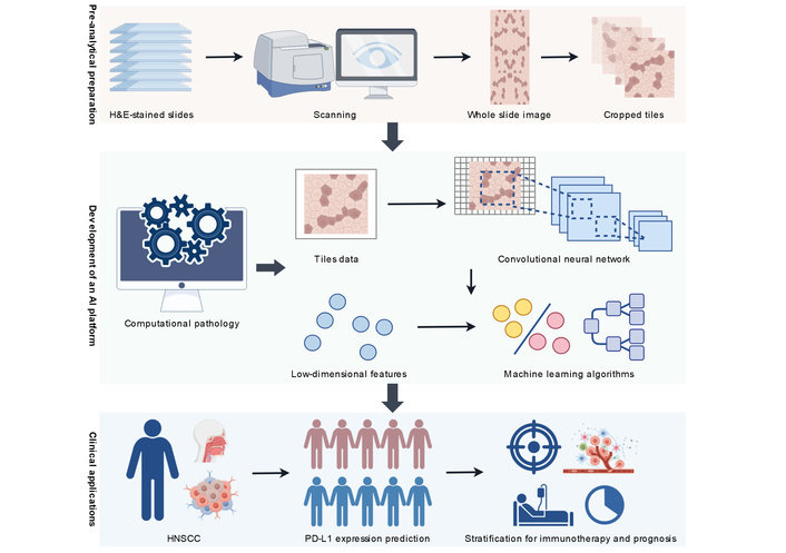
Development of the computational pathology-based AI framework for predicting PD-L1 expression and prognosis in HNSCC. AI: artificial intelligence; HNSCC: head and neck squamous cell carcinoma; PD-L1: programmed cell death ligand 1. Created by figdraw.com.
Receiver operating characteristic (ROC) curves were generated to evaluate model performance by plotting sensitivity against 1-specificity across varying thresholds. The area under the ROC curve (AUC) was used as the primary metric of predictive accuracy, with higher AUC values indicating better performance. All analyses were conducted using Python. Deep learning models were implemented using the PyTorch library, and machine learning algorithms were developed using the scikit-learn package.
Fifty HNSCC specimens were independently evaluated for PD-L1 expression (CPS ≥ 1, Figure 2) by three pathologists with varying levels of experience (junior, mid-career, senior). The overall interobserver agreement was low, with a Fleiss’ κ value of 0.34 (P < 0.001), indicating fair agreement. Pairwise comparisons revealed moderate agreement between the junior and mid-career pathologists (Cohen’s κ = 0.40, P < 0.001), weak agreement between junior and senior pathologists (κ = 0.21, P = 0.015), and substantial agreement between mid-career and senior pathologists (κ = 0.65, P < 0.001). These findings confirm the considerable variability in PD-L1 CPS assessment across observers. Given the higher consistency observed between the latter two pathologists, they were selected for PD-L1 CPS evaluation in the training, validation, and testing cohorts following additional calibration.
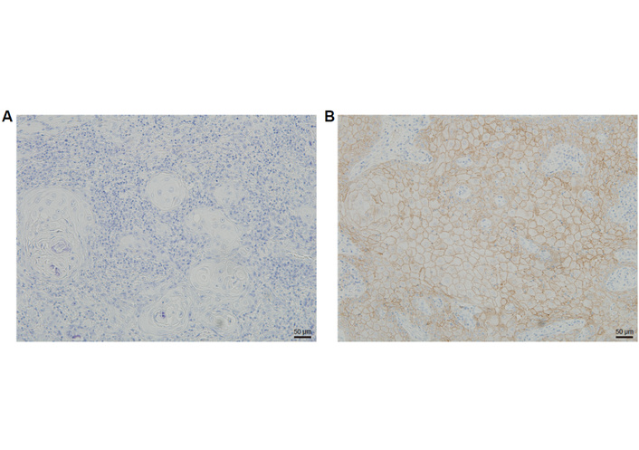
Representative immunohistochemical negative (A) and positive (B) staining of PD-L1 in HNSCC. HNSCC: head and neck squamous cell carcinoma; PD-L1: programmed cell death ligand 1.
To develop predictive models, H&E-stained WSIs were preprocessed by cropping into 512 × 512 pixel tiles and applying data augmentation to address staining variability. A DenseNet121-based deep learning model with transfer learning was trained for tile-level prediction. The tile-level model demonstrated modest performance, with an AUC of 0.641 (95% CI: 0.635–0.647) in the validation cohort and 0.616 (95% CI: 0.611–0.621) in the testing cohort, indicating limited predictive capability at the tile level alone. To improve performance, tile-level features were aggregated into slide-level representations using PALHI and BoW pipelines. These features were subsequently integrated into multiple machine learning classifiers. As summarized in Table S2 and Figure 3, all models achieved improved performance compared with tile-level prediction. Among them, the logistic regression (LR) model achieved the best performance, with an AUC of 0.900 (95% CI: 0.832–0.967) in the validation cohort and 0.851 (95% CI: 0.789–0.913) in the testing cohort. Other models, including support vector machines (SVM), ExtraTrees, and AdaBoost, also demonstrated strong predictive performance, whereas NaiveBayes showed relatively lower accuracy. These results indicate that integrating deep learning-derived features with machine learning significantly enhances the prediction of PD-L1 expression at the whole-slide level.
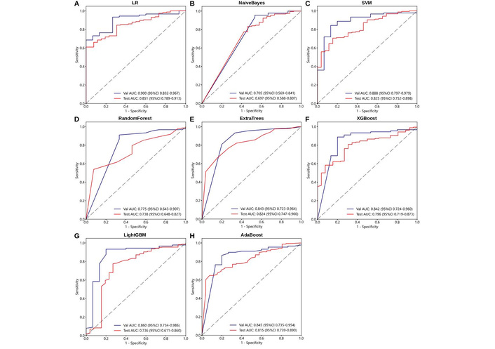
ROC curves of AI models for PD-L1 prediction in the validation (Val) and testing (Test) cohorts. (A) LR; (B) NaiveBayes; (C) SVM; (D) RandomForest; (E) ExtraTrees; (F) XGBoost; (G) LightGBM; (H) AdaBoost. AI: artificial intelligence; LR: logistic regression; PD-L1: programmed cell death ligand 1; ROC: receiver operating characteristic; SVM: support vector machines.
Given the controversial relationship between PD-L1 expression and HNSCC prognosis [26–28], we further evaluated the prognostic relevance of the AI-derived models. Survival data from the 50 HNSCC cases used for interobserver analysis were included. Based on the predicted probabilities of PD-L1 expression derived from the models, patients were stratified into high-risk and low-risk groups. Kaplan-Meier survival analysis demonstrated that several models, including LR (P = 0.005), SVM (P = 0.014), ExtraTrees (P = 0.023), and AdaBoost (P = 0.036), were able to significantly stratify overall survival (Figure 4). In contrast, other models showed limited prognostic discrimination. These findings suggest that AI-predicted PD-L1 expression is associated with clinical outcomes and may serve as a potential prognostic indicator in HNSCC. Moreover, the integration of AI-based models into clinical workflows may facilitate risk stratification and support personalized treatment decision-making.
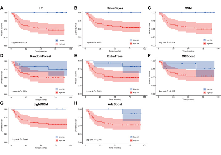
Kaplan-Meier survival curves of HNSCC patients stratified by AI-predicted PD-L1 expression probabilities using different models. (A) LR; (B) NaiveBayes; (C) SVM; (D) RandomForest; (E) ExtraTrees; (F) XGBoost, (G) LightGBM; (H) AdaBoost. HNSCC: head and neck squamous cell carcinoma; LR: logistic regression; PD-L1: programmed cell death ligand 1; SVM: support vector machines.
In the present study, we developed and validated a computational pathology framework that enables prediction of PD-L1 expression directly from routine H&E-stained WSIs in HNSCC. By integrating deep learning-derived morphological representations with machine learning-based feature aggregation, the proposed system achieved robust predictive performance across both internal and external cohorts. Importantly, our findings further demonstrate that AI-inferred PD-L1 status is not only technically feasible but also clinically informative, as evidenced by its ability to stratify patient survival. These results position AI-assisted histopathological analysis as a promising surrogate approach for molecular biomarker assessment in precision oncology, as has been shown in previous studies [24, 29–37].
A key clinical challenge addressed by this study is the limited reproducibility of PD-L1 CPS evaluation. Consistent with prior reports [38, 39], we observed only fair interobserver agreement among pathologists, with substantial variability particularly between less and more experienced observers. This highlights the intrinsic subjectivity and complexity of CPS scoring, which requires simultaneous evaluation of tumor and immune compartments within heterogeneous tissue architecture [11, 13–15, 17, 40]. From a clinical standpoint, such variability may directly impact therapeutic decision-making, especially in borderline cases where CPS thresholds determine eligibility for ICIs [38, 39]. The AI-based approach proposed here offers a standardized and reproducible alternative, potentially reducing observer-dependent bias and improving consistency in PD-L1 assessment across institutions.
Beyond reproducibility, our study contributes to the growing body of evidence that morphological features captured in H&E images encode latent molecular and immunological information [24, 37]. Although PD-L1 expression is conventionally assessed by immunohistochemistry, our results suggest that tumor architecture, stromal composition, and immune cell distribution patterns may collectively reflect underlying immune checkpoint activity. The relatively modest performance of the tile-level deep learning model indicates that local features alone are insufficient to capture this complexity. In contrast, the substantial improvement observed after WSI-level feature integration (AUC up to 0.900 in validation and 0.851 in testing) underscores the importance of global spatial context and multi-feature fusion. This aligns with emerging paradigms in computational pathology emphasizing hierarchical modeling and spatially aware feature aggregation [21, 22, 41].
From a methodological perspective, the dual-pipeline design combined with ensemble machine learning represents a flexible and scalable strategy for translating tile-level predictions into clinically meaningful outputs. Among the evaluated algorithms, LR demonstrated optimal performance, suggesting that, despite the complexity of upstream feature extraction, the final decision boundary may remain linearly separable in high-dimensional feature space. This finding has practical implications, as simpler models may offer advantages in interpretability, computational efficiency, and clinical deployment. Importantly, this study extends the utility of AI beyond biomarker prediction to prognostic stratification. By leveraging predicted PD-L1 probabilities, several models effectively distinguished patients with significantly different overall survival outcomes. This observation reinforces the biological relevance of PD-L1-associated immune states in HNSCC progression and supports the concept that AI-derived surrogate biomarkers may capture prognostically meaningful tumor-immune interactions [23, 29, 42]. Clinically, such models could complement existing staging systems by providing additional risk stratification, thereby informing postoperative surveillance and adjuvant treatment strategies.
From a translational perspective, the proposed framework offers several potential advantages. First, it utilizes routinely available H&E slides, eliminating the need for additional staining, reducing cost, and preserving tissue. Second, it enables retrospective analysis of archived specimens, facilitating large-scale studies and real-world validation. Third, it provides rapid and automated assessment, which may be particularly valuable in resource-limited settings where access to standardized immunohistochemistry and expert pathology review is constrained. Collectively, these features support the integration of AI-based tools into clinical workflows as decision-support systems rather than replacements for pathologists.
Nevertheless, several challenges remain before clinical implementation. The biological interpretability of AI models is still limited, and the specific morphological correlates of PD-L1 expression inferred by the model warrant further investigation using explainable AI techniques. Additionally, while our model demonstrated strong performance across two centers, broader validation in multi-center, multi-platform datasets is essential to ensure generalizability, as variability in tissue processing, staining protocols, and scanner characteristics may affect model robustness; standardization efforts and domain adaptation techniques will be critical for real-world deployment. Furthermore, independent validation by external research groups is essential to confirm the reproducibility, robustness, and clinical applicability of the framework across diverse patient populations and practice settings. Finally, prospective clinical trials are needed to determine whether AI-assisted PD-L1 assessment can improve patient selection for immunotherapy and ultimately translate into better clinical outcomes. Importantly, the current study focused exclusively on predicting PD-L1 positivity using a threshold of CPS ≥ 1, which represents a clinically relevant but relatively inclusive cutoff. Future studies should extend this framework to predict higher thresholds, particularly CPS ≥ 20, which are increasingly used to guide immunotherapy decision-making and may provide additional prognostic and therapeutic value. Incorporating multiple clinically meaningful PD-L1 cutoffs into AI-based prediction models may further enhance their utility for precision oncology and patient stratification.
In summary, we developed a robust AI-based computational pathology framework capable of predicting PD-L1 expression directly from H&E-stained slides in HNSCC. The model demonstrated strong performance across independent cohorts and showed potential in prognostic stratification. This approach provides a convenient, reproducible, and scalable alternative to conventional immunohistochemical assessment and may serve as a valuable adjunct tool for guiding immunotherapy and personalized clinical decision-making in HNSCC.
AI: artificial intelligence
CPS: combined positive score
H&E: hematoxylin and eosin
HNSCC: head and neck squamous cell carcinoma
ICIs: immune checkpoint inhibitors
LR: logistic regression
PD-1: programmed cell death protein 1
PD-L1: programmed cell death ligand 1
ROC: receiver operating characteristic
SVM: support vector machines
WSIs: whole-slide images
The supplementary tables for this article are available at: https://www.explorationpub.com/uploads/Article/file/1001426_sup_1.pdf.
YC: Data curation, Formal analysis, Investigation, Visualization, Writing—original draft. CD: Formal analysis, Investigation, Writing—review & editing. LL: Data curation, Formal analysis, Writing—review & editing. XC: Conceptualization, Formal analysis, Funding acquisition, Writing—review & editing. All authors read and approved the submitted version.
The authors declare that there are no conflicts of interest.
This study was approved by the Institutional Review Board of Peking University Hospital of Stomatology (PKUSSIRB-202497028) and Xiangya Stomatological Hospital (20230024) and complies with the Declaration of Helsinki.
Participants were informed and voluntarily agreed to participate. Personal information was protected throughout the study, with data securely stored and used exclusively for research purposes.
Not applicable.
The datasets that support the findings of this study are not publicly available due to ethical restrictions but are available from the corresponding author upon reasonable request.
This work was supported by Talent development plan for the future in Medical-Engineering Integration by BRA-CDCHE and ZTA (MBRC0012025013), State Key Laboratory of Innovative Immunotherapy. The funders had no role in study design, data collection and analysis, decision to publish, or preparation of the manuscript.
© The Author(s) 2026.
Open Exploration maintains a neutral stance on jurisdictional claims in published institutional affiliations and maps. All opinions expressed in this article are the personal views of the author(s) and do not represent the stance of the editorial team or the publisher.
Copyright: © The Author(s) 2026. This is an Open Access article licensed under a Creative Commons Attribution 4.0 International License (https://creativecommons.org/licenses/by/4.0/), which permits unrestricted use, sharing, adaptation, distribution and reproduction in any medium or format, for any purpose, even commercially, as long as you give appropriate credit to the original author(s) and the source, provide a link to the Creative Commons license, and indicate if changes were made.



View: 683
Download: 24
Times Cited: 0


