Review


Affiliation:
1Department of Chemistry, American University, Washington, DC 20016, USA
Email: mkonak@american.edu
ORCID: https://orcid.org/0000-0002-1068-8326


 ,
,
Affiliation:
2Department of Microbiology and Immunology, Midwestern University, Downers Grove, IL 60515, USA
ORCID: https://orcid.org/0000-0002-0564-3975
Explor Drug Sci. 2026;4:1008178 DOI: https://doi.org/10.37349/eds.2026.1008178
Received: March 12, 2026 Accepted: July 16, 2026 Published: August 27, 2026
Academic Editor: Francisco Javier Luque Garriga, University of Barcelona, Spain
The article belongs to the special issue The Rise of Targeted Covalent Inhibitors in Drug Discovery
Plant metabolites are an invaluable source of bioactive molecules, and a high percentage of them can react covalently with their targets. Lipid-derived α,β-unsaturated systems (Michael acceptors), which are present in all plants, regulate signaling pathways in cells. In addition, they potentially represent novel molecular targets and mechanisms of action in drug development. The irreversible covalent binding of the majority of these electrophilic molecules to their corresponding molecular targets, combined with, in certain cases, unfavorable pharmacokinetic properties, i.e., absorption, distribution, metabolism, and excretion (ADME), has shifted their use predominantly to that of molecular probes for target identification. In this review, we present examples of structural modification of the original naturally occurring Michael acceptor-containing compounds, as well as examples of incorporating naturally occurring functionalities in the design of reversible covalent probes and drug candidates in order to improve ADME and increase target selectivity.
Electrophilic drugs are electron-deficient compounds that form covalent bonds with nucleophilic sites to increase pharmacological efficacy or generate covalent inhibition, being effective at low doses, but the drawback is that they can be nonspecific and toxic at higher doses [1]. An electrophilic Michael acceptor (MA) moiety is present in many natural products. Molecules containing it interfere with numerous plant physiological processes. The most important of these processes is stimulation of cell survival gene expression. Genes most commonly affected are those upregulated in plants during environmental stress and pathogenesis. Molecules containing MAs can be synthesized by plants, either via enzyme catalysis or by non-enzymatic processes, e.g., oxidation by reactive oxygen species (ROS). The α,β-unsaturated systems typically found in natural products are: exo-methylene lactones, enones, enals, and β-cyclopropyl-substituted MAs [1, 2]. There are strong parallels in electrophile-stimulated gene expression in both plants and mammals, especially those involved with sulfhydryl-containing nucleophiles/cysteine-selective protein modification. Evolution in the kingdom Plantae resulted in the development of electrophilic moieties as part of plant growth defense systems, with one of the most successful ones being the MA warheads. The phytoprostanes (Plantae) and isoprostanes (Animalia) have a similar evolutionary origin, and their metabolites [plant hormones and mammalian prostaglandins (PGs)] play critical roles in plant injuries and mammalian wound and inflammatory responses, respectively. The structural similarities of the plant hormones and mammalian PGs indicate cross-kingdom adoption of lipid-derived signals against tissue injuries in both plants and animals, which makes the plant defense systems an invaluable source of ideas for drug development.
Eukaryotes possess an endogenous defense system made of a series of signaling cascades whose function is to protect the organism against different stressors and maintain cellular redox homeostasis. The response of an organism to ROS is activation of the nuclear factor erythroid 2-related factor 2 (Nrf2)-driven antioxidant response element (ARE), which leads to the induction of a multitude of cytoprotective phase II enzymes [3, 4]. The best characterized mechanism of ARE activation is the Kelch-like ECH-associated protein 1 (Keap1)-dependent regulation of Nrf2. Under normal conditions, Keap1 binds to Nrf2 through Cul3-based E3 ubiquitin ligase complex and promotes the Nrf2 degradation by the ubiquitin proteasome pathway. Keap1 is a cysteine-rich protein that, upon detecting stressors, undergoes modifications leading to the discontinuation of Nrf2 ubiquitination [5–7]. The latter leads to the accumulation of Nrf2, its nuclear translocation, and the subsequent binding to the ARE, thus, promoting the expression of Nrf2 target genes, such as NAD(P)H:quinone oxidoreductase 1 (NQO1), heme oxygenase 1 (HMOX1), glutamate cysteine ligase (GCL), and glutathione S-transferase (GST) [8]. Modulators of Nrf2 containing the MA acceptor warhead from plant origin have been identified and utilized in drug discovery, mainly, but not limited to, anti-cancer drugs.
In this review, we focus on the function of the plant kingdom’s toolbox of MAs for plant survival and development, from those with very high electrophilicity, thus low selectivity (usually used as chemical weapons by plants), to those that achieve target selectivity through different synthetic chemistry mechanisms. Some MAs exist in latent forms (masked functionality) in plant cells, which are released as chemical signals in response to biotic/abiotic stressors. We discuss the potential utilization of plant-produced MAs as leads for drug discovery using illustrative examples from the structural modifications made by plants and humans to achieve target selectivity (in addition to fast stress response), since the potential of the natural products containing the α,β-unsaturated systems as human drugs has already been extensively reviewed [9–11].
A selective approach was used for this review to identify applicable relevant peer-reviewed publications. The focus for this examination was on the innate challenge in the design of selective enzyme modulators having MA as the electrophilic warhead. Specifically, we discuss the structural overlap of cyclic enones used by plants and man for plants’ defense against abiotic and biotic insults, and in drug development. We give several examples of strategies to use prodrugs (having masked enone functionality) to achieve more favorable physicochemical properties of potential drug candidates and improve target selectivity. Only English language publications were considered for inclusion. All major academic databases and search engines, including SciFinder, Reaxys, Google Scholar, and PubMed were searched. Both foundational and contemporary studies included in this review span from 1978 to 2026. Of the 155 included references, 85 articles (55%) were published within the last decade (2015–2026), demonstrating the continued interest in using Nature’s toolbox in tuning MAs as warheads in covalent binding to molecular targets of interest to human health. Search terms such as “Michael acceptors”, “enzyme specificity”, “enzyme promiscuity”, and “covalent inhibitors” were used to identify relevant publications. Abstracts were evaluated, followed by full-text reviews of those with relevance to the topic of this review.
The chemical weapon of plants against predators involves the production of chemical defense electrophilic compounds with good selective toxicity against target organism(s). Energetically, the synthesis of these defense compounds is at a low cost for the plant. This is ensured by the rapid oxidative cleavage of long-chain polyunsaturated fatty acids (PUFAs) to give a variety of low molecular weight (MW) α,β-unsaturated aldehydes, as well as α,β,γ,δ-unsaturated aldehydes.
Known enzymatically synthesized MA-containing molecules include volatile unsaturated alkyl aldehydes, such as 2,4-heptadienal, 2,4-octadienal, and 2,4-decadienal [12–14], produced by algae, as well as 2-(E)-hexenal and (E)-4-hydroxy-2-nonenal (HNE) found in higher plants [15–22].
One of the most studied releases of polyunsaturated aldehydes as a response to wounding is that produced by algae, including marine diatoms. The latter are pelagic, bloom-forming algae, which release α,β,γ-unsaturated aldehydes upon cell wounding [23, 24]. These compounds stall mitosis [25] and negatively impact the hatching success of predators, specifically these algae’s most important predator, the pelagic copepods [25–27]. There are excellent reviews on the effect of polyunsaturated aldehydes as chemical defense at the population level of the benthic diatoms to which the reader is directed [28–30].
This mechanism is also found in macrophyte defense reactions [31–34]. The brown algae Laminaria digitata (kelp) releases aldehydes in response to different stressors [14, 30, 32]. When oligoguluronate is used to mimic pathogenic microbe attack, it induces an oxidative burst in the alga, leading to the release of different chain length unsaturated aldehydes. Environmental stress is believed to be the trigger for aldehyde production by L. digitata, resulting in their detection in the tidal pools occupied by this alga. These stressors include exposure to ozone, ultraviolet (UV) light, as well as desiccation, changes in temperature and salinity [32]. It has been suggested that this aldehyde production leads to the synthesis of oxylipins, thus acting as inducers of the alga’s metabolic responses [30, 32]. Therefore, it appears that the released aldehydes act as both external and internal chemical signals in L. digitata that can be produced in response to both biotic and abiotic stressors. That assumption was recently further examined, leading to the determination of a specific metabolic response by the alga to different aldehydes [32, 33].
Both C6 and C9 unsaturated aldehydes have been found to affect plant mitochondria (Figure 1): (E)-2-hexenal specifically changes the redox status of the mitochondria, while HNE acts as a potent inhibitor of land plants’ mitochondrial terminal oxidases [33, 34]. In animals, HNE (Figure 1) is generated from the peroxidation of PUFAs as a major, stable end product of oxidative stress, which is a toxic, bioactive marker in various human diseases such as cancer, diabetes, and Alzheimer’s [34–38]. While attempts to include the α,β-unsaturated alkyl aldehydes in the arsenal of human drugs, more specifically as anticancer reagents, have so far been unsuccessful due to the high reactivity of these aldehydes as electrophiles. The potential adverse effects of aldehydes, especially α,β-unsaturated aldehydes, on various mammalian cells are well-established [39–41].
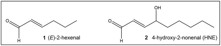
Structures of unsaturated alkyl aldehydes used by plants as chemical weapons and signaling molecules. 1 (E)-2-hexenal and 2 4-hydroxy-2-nonenal (HNE).
Research on marine algae such as Phaeodactylum tricornutum indicates that fucoxanthin synthesis can be induced by signaling molecules such as methyl jasmonate (MeJA), a known stress-response signal in plants (see Cycloalkenones as MAs), suggesting its role in defense mechanisms [42, 43]. Fucoxanthin is an antioxidant and protects cell components from ROS. Enhanced concentration of ROS has been implicated in many pathological conditions, including cancer. The major machinery in the cells of Animalia that neutralizes excess ROS functions through the activation of the ARE.
Fucoxanthin (3, Figure 2), a major carotenoid in brown algae and diatoms, functions primarily as a light-harvesting pigment and photoprotective agent. In addition, it potentially plays a role as a signaling molecule. Under excess light, fucoxanthin is part of an adaptive, photoprotective signaling system. It plays a role in the xanthophyll cycle, where intermediate pigments like diadinoxanthin are converted to diatoxanthin to dissipate excess energy and prevent ROS-induced damage [44, 45]. The latter controls the activation of many phase II detoxification enzymes. The transcription factor that recognizes the ARE, Nrf2, can be activated by a variety of small molecules, most of which contain an α,β-unsaturated carbonyl system. The MA acceptor functionality enables it to interact with proteins and, in animal models, trigger antioxidant signaling pathways, specifically by activating the Nrf2/ARE system [46–50]. While extensive literature exists on fucoxanthin’s signaling role in animals (e.g., MAPK, NF-κB pathways), in plants and algae, it is primarily defined as a crucial pigment for photosynthetic light-harvesting, structural stabilization of complexes, and light-stress signaling. The importance of fucoxanthin for human health has been extensively reviewed [46–49].
The MA motif has also been found in derivatives of monounsaturated C18 and C16 fatty acids isolated from the green macroalga Ulva lactuca (Figure 3), C18 fatty acid (4a, Figure 3), C16 fatty acid (4b, Figure 3), and an amide derivative (4c, Figure 3) of the C18 acid [51]. These compounds’ activity to ARE is attributed to the presence of the unsaturated keto-moiety between C7 and C9 (Figure 3). The most active compound, the C18 acid 4a (Figure 3) induces the expression of ARE-regulated cytoprotective genes, including NQO1, heme oxygenase 1, thioredoxin reductase 1, both subunits of the glutamate-cysteine ligase (catalytic subunit and modifier subunit), and the cystine/glutamate exchange transporter, in IMR-32 human neuroblastoma cells [51].
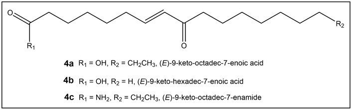
Keto-enoic acids isolated from the edible green alga Ulva lactuca (Florida coast) activate the transcription factor that recognizes the ARE, Nrf2. ARE: antioxidant response element; Nrf2: nuclear factor erythroid 2-related factor 2.
In addition to acyclic α,β-unsaturated aldehydes and ketones, another product of oxylipin metabolism is the jasmonic acid (JA) precursor, 12-oxo-phytodienoic acid (12-OPDA), found in algae to higher plants [51–54]. Biologically synthesized 12-OPDA is cis-(+)-12-OPDA, 9, Figure 4, (referred herein as 12-OPDA for the 8-[(1S,5S)-4-oxo-5-[(Z)-pent-2-enyl]cyclopent-2-en-1-yl]octanoic acid, usually found in plants). Allene oxide cyclase is the enzyme responsible for the production of enantiomerically pure cis-(+)-12-OPDA [55, 56]. In Arabidopsis thaliana, bioconjugation of JA, 10, Figure 4 with isoleucine results in the synthesis of the plant hormone (+)-7-iso-Jasmonoyl-L-isoleucine (JA-Ile, 18, Figure 5). JA-Ile regulates growth, reproduction, and defense responses against pathogens and chewing insects by binding to its receptor COI1-JAZ [57–59]. The initiation of the plants’ response to environmental stress via binding of JA-Ile to COI1-JAZ is referred to as the canonical pathway. Several reports have demonstrated that 12-OPDA activates stress responses in A. thaliana, tomato, and maize via a JA-Ile-independent pathway, i.e., a noncanonical pathway (Figure 5) [55, 60].
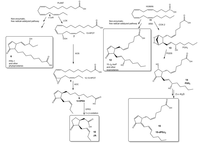
Structural similarity between mammalian cyclopentenone-based prostaglandins (e.g., 15) and 12-OPDA (9). Adapted with permission from [61]. Accessed Jul 11, 2026. © The Author(s). 12-OPDA: 12-oxo-phytodienoic acid; 15-dPGJ2: 15-deoxy-Δ12,14prostaglandin J2; JA: jasmonic acid.
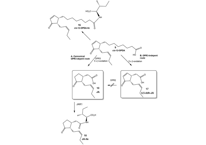
The noncanonical biosynthetic pathway of JA-Ile in A. thaliana [69]. 12-OPDA: 12-oxo-phytodienoic acid; 4,5-ddh-JA: 7-iso-4,5-didehydro-jasmonic acid; JA: jasmonic acid.
In the kingdom Plantae, both alkyl α,β-unsaturated enones and 12-OPDA (9, Figure 4) are ubiquitous chemical signaling compounds with overlapping triggers, i.e., environmental stress, including wounding, and pathogenesis [52, 53, 55, 61–64]. Exogenous 12-OPDA affects the expression of approximately 200 genes in A. thaliana. In algae and other representatives of the land plants, the expression of 12-OPDA-specific response genes has been confirmed [53, 54]. The essential compound moiety for this signal appears to be the presence of the α,β-unsaturated carbonyl group [52, 53].
12-OPDA (9, Figure 4) is structurally similar to the mammalian cyclopentenone PGs (cyPGs, e.g., 15, Figure 4), with the cyclopentenone being the electrophilic (MA) moiety [61]. Both cyPGs and 12-OPDA are important regulators of reproductive systems. For example, while cyPGs play a role in labor contractions, 12-OPDA is involved in regulating seed dormancy, germination, and embryogenesis [65–67]. The covalent binding of 12-OPDA to thiol groups by Michael addition, termed OPDAylation, affects the activity of its target proteins, such as cyclophilin 20-3 (EC:5.2.1.8) and thioredoxins, that are essential for the cellular redox system in plants [55, 68]. Recently, the intricate interplay between the 12-OPDAylation of protein thiols and the binding of 12-OPDA to glutathione (GSH) has been shown to be under kinetic vs. thermodynamic control, respectively. This control of 12-OPDA (9, Figure 4) concentrations permits rapid modification of the target protein due to the higher nucleophilicity of its cysteine as compared to the thiol group of cysteine in GSH. This ensures rapid induction of OPDA signaling followed by detachment from the protein (de-OPDAylation) in response to the increasing levels of GSH [68].
The downstream metabolites of 12-OPDA (9, Figure 4), specifically tetranor-cis-OPDA (tn-cis-OPDA) and 7-iso-4,5-didehydro-JA (4,5-ddh-JA) (17, Figure 5), upregulate the expression of 12-OPDA marker genes, such as ZAT10 and ERF5. Similar to 12-OPDA, its downstream metabolites, tn-cis-OPDA and 4,5-ddh-JA, function independently of the JA-Ile-COI1-JAZ-MYCs canonical jasmonate signaling module, with their electrophilic MA-acceptor functionalities being essential for their bioactivity [69]. This finding suggests that both 12-OPDA (9, Figure 4) and its derivative 4,5-ddh-JA (17, Figure 5), which contains the α,β-unsaturated moiety, function as endogenous chemical signals in A. thaliana. The cyclopentenone functionality embedded in 12-OPDA compounds functions as a chemical response to stress in marine and terrestrial algae as well as vascular plants [69–76]. It has been demonstrated that dn-OPDA is the evolutionary precursor of JA-Ile. It is suggested that the low hydrophilicity of dn-OPDA exerted evolutionary pressure for the formation of the polar JA-Ile hormone, as it permits easier distribution through a plant vasculature [77].
Abscisic acid (ABA, 22, Figure 6) is another MA-containing compound that shares striking similarities with 12-OPDA (9, Figure 4) in both chemical structure and biological function [52, 77–85]. ABA is a 15-carbon sesquiterpenoid (22, Figure 6), synthesized by oxidation from a product of the carotene metabolism, neoxanthin (19, Figure 6), whose structure shares great similarity with that of fucoxanthin (3, Figure 2).
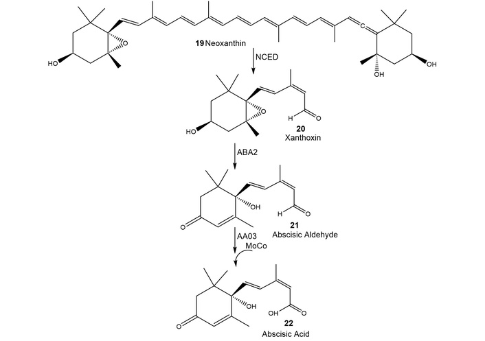
Abscisic acid (ABA) biosynthesis pathway. ABA2: hydrogenase; AAO3: abscisic aldehyde oxidase; MoCo: molybdenum cofactor. By activating AO, MoCo directly allows plants to produce sufficient levels of ABA [88].
Similarly to 12-OPDA (9, Figure 4), ABA (22, Figure 6) is a chemical signal in the defense responses of plants. In addition, both compounds share the same electrophilic unsaturated moiety, which binds to sulfhydryl groups, such as cysteine in GSH and proteins [77–85]. Furthermore, ABA affects 12-OPDA concentration [86, 87].
ABA (22, Figure 6) has activity across taxonomic kingdoms affecting prokaryotes, fungi, and animals, including humans [88]. The pathway for ABA synthesis in fungi is dissimilar from the oxylipin pathway in plants. In fungi, ABA is synthesized via the mevalonic acid (MVA) pathway, utilizing a unique cyclase (BcStc5) to convert farnesyl diphosphate to α-ionylideneethane [89, 90]. Further oxidative modifications of the cyclized product (α-ionylideneethane) to afford 1ʹ,4ʹ-trans-dihydroxy-α-ionylideneacetic acid, which undergoes alcohol oxidation to furnish ABA. Currently, in many naturopathic products and drug development programs, chiral cyclohexenones, e.g., ABA, are the reactive functionality present [91–98]. Different synthetic approaches have been utilized to achieve regio- and stereoselective C–C and C–X bond formation for cyclohexenone synthesis [98, 99]. Human proteomics research recently showed that there are proteins that specifically bind ABA, some of which may play a role in cancer and diabetes [100]. It has been proposed that ABA (22, Figure 6) synthesis in humans, as in fungi, proceeds via farnesyl pyrophosphate cyclization [100]. It appears that mammals contain a latent, fungal-like ABA synthesis pathway that is inducible under metabolic or xenobiotic stress. If confirmed, this will allow for a rational ABA-based drug design for the treatment of chronic pathologies such as inflammation, diabetes, and cancer. [100].
Another example of a parallel metabolic pathway in mammals and plants is that of the PGs. Until recently, PGs metabolism and functions were mainly associated with mammals. However, recently it has been recognized that PGs are present in plant species, such as onions, poplar trees, and birch (Betula alba L.) pollen [101, 102]. The synthesis of PGs in both macro- and micro-algal marine species occurs when the algae are under stress conditions, but their exact role in mediation of stress responses in marine algae is still under investigation [103]. Recently, studies of the PG biosynthetic pathway during different growth phases of the centric diatom Thalassiosira rotula show that PGs are released primarily during the stationary and senescent growth phases, suggesting a possible signaling function for these compounds [104]. Both PGs and plant hormones are the end result of pathways involving bioactive lipids.
In general, bioactive lipids in both plants and animals have been reported to govern both cellular homeostasis and pathogenic inflammatory processes. The endogenously produced electrophilic α,β-unsaturated ketones and their derivatives (i.e., MA) from hydroxylated PUFAs are chemically reactive signaling mediators that induce tissue-protective events. The mechanism of action of these MAs is post-translational alkylation of nucleophilic cysteines in key transcriptional regulatory proteins and enzymes that govern cellular metabolic and inflammatory homeostasis. The cycloenone motif has been proven by Nature to be a very successful electrophilic warhead that can be produced rapidly to alleviate oxidative stress. Therefore, it is not surprising that MAs evolutionary development for plant cellular homeostasis can be used similarly by mammals.
For OPDA induced growth inhibition in breast cancer cells, 12-OPDA (9, Figure 4) degradation of cyclin D1 protein is a key event [105]. Cancer cells’ treatment with 12-OPDA exhibit a progressive decline in cyclin D1 expression, which is tightly associated with the accumulation of hypophosphorylated form of the retinoblastoma protein (Rb) and G1 arrest. 12-OPDA induces Nrf2-dependent antioxidative response, which in turn may have contributed to the observed decrease in H2O2-induced ROS levels in human neuroblastoma SH-SY5Y cells and breast cancer [106–108]. It is interesting to note that in the last several years, the anticancer activity of JA (10, Figure 4) and its derivatives has also been reported [109–111].
The use of MAs as target modulators is currently an active area of drug development. These target modulators bind non-covalently or covalently to their molecular targets. The chronological order of MA-containing compounds in drug development ranges from natural products (irreversible) to current trends (irreversible/reversible) based on nature-inspired warheads. The covalent binding of the enzyme modulators offers significant advantages over non-covalent ones, since the covalent warhead could target a single amino acid residue. However, typically, enzyme modulators rarely act on a single molecular target, and with those being irreversible, the off-target effects can lead to undesired effects, e.g., toxicity. Therefore, strategies to minimize off-target effects both by nature and man include the preparation of compounds with high specificity toward a molecular target and/or those that bind reversibly. The reversible covalent modulation ensures high potency of binding (covalent binding with the molecular target), and the potential for tuning the residence time on target through structural modification around the electrophilic warhead, thus securing the binding through non-covalent interactions in the binding site.
Nature has used plant hormones containing either cyclopentenone or cyclohexenone as electrophilic moieties that are able to bind covalently with sulfhydryl groups, which range from H2S to those found in proteins. These hormones (12-OPDA and ABA) are not limited to plants but can be found in all living organisms on our planet, including humans. Cyclopentenone and cyclohexenone are present in many of the evolutionarily ancient signaling molecules [76]. The endocyclic double bond of the α,β-unsaturated system of the electrophilic warheads appears to be advantageous in specific binding to target proteins, as compared to open-chain compounds, which in turn should lead to fewer off-target effects. For example, binding of 12-OPDA (9, Figure 4) to proteins containing cysteine, e.g., thioredoxin, is favored over OPDA adduct formation with GSH. This is despite the fact that, under physiological conditions, the concentration of free thiols, for example GSH, exceeds that of 12-OPDA by approximately 102- to 103-fold [112]. This rationale from nature has been recently demonstrated to be effective via an increased specificity in using cyclohexenone as the MA functionality in inhibitors of c-Jun kinase, involved in human carcinogenesis [113].
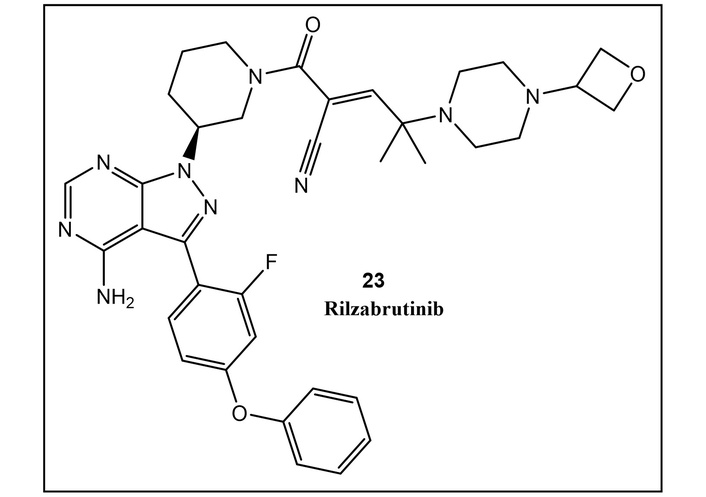
Cyanoacrylamide is often the electrophilic warhead of the currently used reversible synthetic covalent enzyme modulators, such as, but not limited to, those used as kinase inhibitors, including the recently FDA-approved rilzabrutinib (23, Figure 7, formerly known as PRN1008, in 2025) [114].
Cyanoacrylamide moiety is attacked by one reachable nucleophile in the enzyme binding site (most often, but not limited to, a non-catalytic cysteine). However, the potential for binding of the reversible acyclic warheads (i.e., cyanoacrylamide) to other molecular targets, including those in the redox system in cells (e.g., GSH), still exists. Introducing a cyclic MA acceptor moiety in compound (24, Figure 8) to reduce the aforementioned potential interactions with off-target cysteines due to its less accessible 3D shape (found in naturally occurring MAs) has hindered its binding to GSH [113, 115]. This strategy, utilizing cyclohexenone-based MA (25, Figure 8), led to the preparation of inhibitors of c-Jun Terminal Kinase, where, in addition to the cyclic moiety, the chirality of the γ-carbon in 1aR-IN-8 (25, Figure 8) plays a significant role in guiding the inhibitor to the “correct” nucleophile and secures binding specificity equivalent to that of its irreversible counterpart JNK-IN-8 [113, 115].
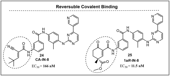
The placement of the cyano group at the α-position of the α,β-conjugated system leads to highly reactive cyanoenone functionality, which preferentially binds covalently to cysteines in proteins [116–118]. An example of this is the replacement of the original secondary alcohol of the glycyrrhetinic acid scaffold with a 2-cyano-substituted cyclohexenone ring (Figure 9), resulting in a pentacyclic triterpenoid derivative equipped with a potentially reversible MA [116].
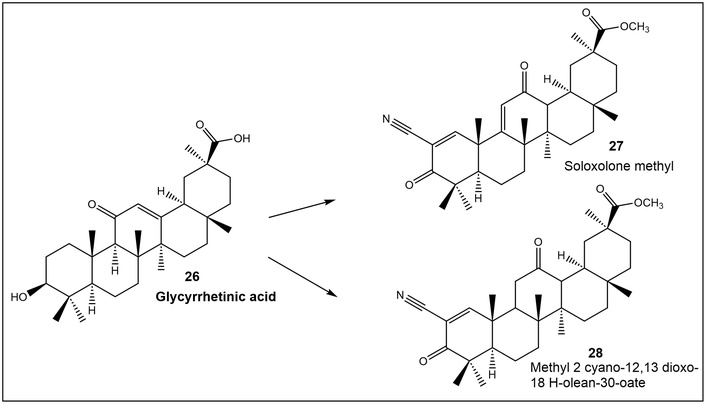
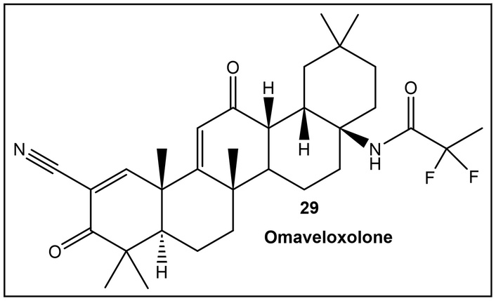
Compound 28, a cyanoenone derivative of glycyrrhetinic acid, has been demonstrated to function, with high selectivity, as an inhibitor of cancer cell growth and NO production in LPS-activated J-774 cells [116]. Furthermore, omaveloxolone (29, Figure 10) with structural similarity to Soloxone methyl (27, Figure 9) is the first FDA-approved drug for Friedreich ataxia treatment (2018) [117].
Orally active omaveloxolone (29, Figure 10) binds primarily to Cys151, of the cysteine-based sensor protein Keap1, which leads to the accumulation of transcription factor Nrf2. In Friedreich’s ataxia, an autosomal recessive degenerative disease of the nervous system, the nuclear factor Nrf2 pathway is suppressed, causing oxidative stress and mitochondrial dysfunction. This stress leads to central and peripheral neuron cell damage. The Nrf2 pathway may be activated by omaveloxolone as it blocks the ubiquitination and degradation of Nrf2, which in turn inhibits transcription of proinflammatory genes. An additive effect of omaveloxolone may be its binding to cysteines in proteins involved in inflammatory cascades (e.g., IKKβ), resulting in inhibition of inflammation [117, 118].
The cyclohexenone motif has been used in other small molecules biosynthesized by plants. Zeylenone (30, Figure 11), a naturally occurring cyclohexene oxide, was first isolated from the extract of Uvaria grandiflora. This naturally occurring compound has demonstrated activity against cervical carcinoma, gastric cancer, and prostate cancer, with limited cytotoxicity against normal cell lines [119–121]. While a detailed mechanism of action/role in plants of zeylenone is currently lacking in the literature, it is predicted to act as a defense compound against different stressors due to its MA moiety, and having cytotoxic activity suggests a role in defending the plant against external threats.
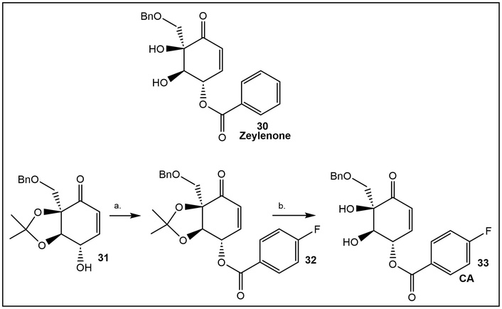
Studies in vivo show that zeylenone (30, Figure 11) is susceptible to esterase hydrolysis with accompanying loss of biological activity. Thus, structural modifications to address hydrolytic instability and resultant activity of the prepared zeylenone derivatives have been carried out using glioblastoma (GBM) cancer lines as the target cell [122]. (+)-Zeylenone derivative CA (33, Figure 11, (1R, 2R, 3S)-3-p-fluorobenzoyl-zeylenone) demonstrated the lowest IC50 value in GBM cells. IC50 values of CA were notably low in GBM cell lines, particularly in U251 (5.161 µM) and A172 (6.440 µM). The molecular mechanism by which CA exerts its anticancer activity is by attenuating the downregulation of cyclin-dependent kinase inhibitors p27 and p16 by the polycomb repressive complex 2 (PRC2). This has been confirmed by in vivo studies. Furthermore, compound CA has the potential to synergistically potentiate the anti-tumor effects of EZH2 inhibitors.
EZH2, a histone methyltransferase enzyme, is the catalytic subunit of the PRC2. PRC2 primarily targets developmental genes, particularly transcription factors, to keep them in a repressed state in specific cell types (e.g., HoX gene family) by methylating histone H3 on lysine 27. It plays a crucial role in cell differentiation, development, and, when mutated or overexpressed, promotes cancer progression, making it a key therapeutic target [122].
Structures found in natural and synthetic sesquiterpene lactones (STLs), e.g., cyclopentenone, have also been explored as potential enzyme inhibitors in the treatment of cancers [123, 124]. Cyclopentenone has been established as a structural motif in anticancer drug development that, when present in compounds, increases the likelihood of specific binding [124]. The simplest representative, 2-cyclopenten-1-one, inhibits the activity of cyclin A promoters in breast cancer cells and cyclin D1, which, when overexpressed, acts as a proto-oncogene in cancers like breast, lung, and lymphoma [123]. Pentacyclic lactones with an exocyclic double bond (α-exo-methylene-γ-butyrolactones) found in terpenes such as andrographolide are also among the highly explored MA-containing compounds as anti-inflammatory agents and as potential treatments for neurological disease, e.g., Alzheimer’s disease [125–127].
The cyclopentenone-based PGs and their cyclopentenone-based synthetic mimics have been demonstrated to be potent inhibitors of NF-κB activation by inflammatory cytokines, mitogens, and viral infection [128]. In addition, these compounds inhibit NF-κB-dependent anticancer activity by directly binding and modifying the β subunit of the IκB kinase complex (IKK) [128]. Specifically, the natural cyclopentenone 15-deoxy-Δ12,14PG J2 (15-dPGJ2) (15, Figure 4) is a potent inhibitor of constitutive IκB kinase and NF-κB activities in chemotherapy-resistant ER-negative breast cancer cells [128]. Development of various approaches to modify the cyclopentenone ring to explore its potential in drug development followed [129, 130]. Illustrative examples of strategies using masked MA functionality by utilizing the presence of suitable substituents at either α- or β-position of the α,β-unsaturated systems are discussed below [131, 132].
Conversely, highly reactive MAs are of concern since their promiscuity can lead to high toxicity [133–135]. Since exocyclic enones, such as parthenin 1 (34, Figure 12), are highly reactive MAs, different approaches to lower their toxicity have been employed, such as blocking the exocyclic MA acceptor group, which lowers their MA reactivity.
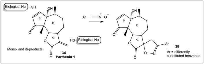
Differently substituted parthenin 1 derivatives 35, with low mammalian cytotoxicity.
STLs are 15-carbon secondary metabolites, primarily found in the Asteraceae family, that act as crucial chemical defenses against other plants, insects, and microbes. They commonly contain a cyclopentenone moiety as the electrophilic center. Their different roles in benefiting the plant producer vary depending on the plant and the compound type, being able to function against both environmental stress and pathogens [136, 137]. STLs are currently under investigation by numerous research groups due to their ability to bind to targets relevant to the treatment of cancer, inflammation, and infectious diseases [138].
The STL, parthenin 1 (34, Figure 12), is the main allelopathic compound in the invasive Parthenium hysterophorus (famine weed). It serves as a natural defensive mechanism, suppressing the germination and growth of competing plants. It enables P. hysterophorus to dominate ecosystems by hindering the growth of the surrounding flora. Spiro derivatives of parthenin 1 (35, Figure 12) reduce the natural product’s active MAs functionalities from two to one. After determining that the cyclopentenone functionality is essential for the bioactivity, derivatives were synthesized and then tested against three cancer cell lines as inhibitors of NF-κB. [131]. The in vivo screening showed improved activity with low mammalian toxicity of the spiro parthenin 1 derivatives (35, Figure 12) as compared to parthenin 1 [131]. For additional modification of this double MA, the reader is directed to the recent review [139].
The cyclopentenone derivatives shown below (Figure 13) with different substituents on the α-carbon of the conjugated system have been prepared. These derivatives have activity against cancer lines and low cytotoxicity to normal human cells [132]. From the derivative series, the most active compound was compound HCP33 (Figure 13), of the α-hydroxy-substituted cyclopentenones (37, Figure 13) having a p-chloro-substituted phenylthio group at C4 of the cyclopentenone. Derivative HCP33 is nontoxic to healthy cell lines, while showing significant activity in the breast cancer cell lines [132].
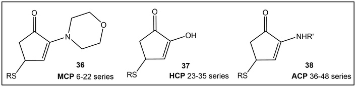
Differently substituted cyclopentenone derivatives at the α-carbon. α-Hydroxy-substituted cyclopentenone with p-Cl-substituted phenylthio group proved to be nontoxic to healthy cell lines, while demonstrating good activity against breast cancer cell lines [132].
The pro-drug approach, masking of the MA warhead present in natural products, was used in the synthesis of drugs to secure cysteine-selective protein modification and low cytotoxicity. Electrophiles that need to be activated by protein binding prior to becoming reactive (latent electrophiles) are expected to have high selectivity towards protein nucleophiles. Thus, the latent electrophiles are more likely to have environment-dependent protein reactivity towards a single protein or a very limited number of proteins. The inertness of the latent electrophiles (pro-drugs) towards the majority of the cellular proteome provides another option to minimize possible off-target activity/toxicity [140].
As noted above, the cycloenone is a common structural feature in many natural products [141–145]. One example is 2-oxyalkyl-cyclohex-2-enone found in antheminones, shown to display notable toxicity towards a range of different cancer cell lines [146]. An alternative approach to lowering cycloenone’s electrophilicity is one that utilizes cycloenone having a leaving group at the α-position of the α,β-unsaturated system moiety. This strategy generates more reactive species with an exocyclic double bond at the targeted site, thereby alleviating multidrug resistance (MDR) in cancer cell lines. The strategy is based on converting the cycloenone to its exocyclic derivative by human GST P1-1 (hGSTP1-1). hGSTP1-1 is known to play a role in detoxifying antitumor drugs by catalyzing their conjugation to the ubiquitous cofactor GSH. The antitumor 2-crotonyloxymethyl-2-cycloalkenones (COMCs, 39, Figure 14) were envisioned as substrates of hGSTP1-1. The exocyclic enone (41, Figure 14) is capable of either reacting with another equivalent of GSH to give the GSMC derivative or alkylating biomacromolecules, such as DNA [143–146].
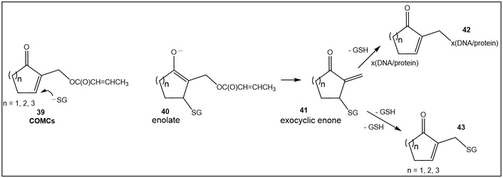
COMCs: generating exocyclic system 41 from endocyclic α,β-unsaturated 39, at the targeted site as a means to alleviate drug resistance of cancer cells.
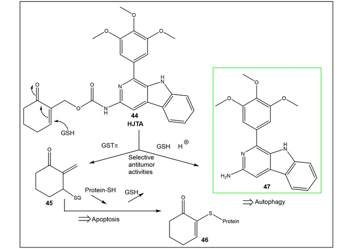
Another example of cancer-targeted therapy based on the cycloenone’s activation by tumor-targeting GSH/GSTπ is the synthesis of the theranostic agents HJTA and HJTB [147]. These cyclohexenone derivatives have antitumor activity and possess a pH-responsive fluorescent tag, leading to GSH-dual-responsive fluorescence in tumor cells but not in normal cells. HJTA (44, Figure 15) illuminates orthotopic colonic tumors through the blood circulation system for 7 hours in intraoperative mice and has potent and selective antiproliferative activities and colonic tumor inhibition in mice. Furthermore, HJTA induces enhanced cancer cell apoptosis and autophagy by regulating the expression of apoptotic and autophagic proteins [147].
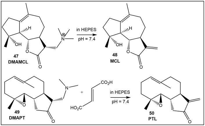
Several synthetic strategies, such as incorporating sulfoxides at the β-carbon of the α,β-unsaturated ester in brefeldin A, a lactone, produced a prodrug of brefeldin A [148]. Derivatizing the MA acceptor with amines is another prodrug strategy, exemplified in the figure below [149]. Dimethylamine has been used for the guaianolide, a STL, isolated from Tanacetum parthenium (Feverfew) by synthesizing the dimethylamino Michael adduct of the exocyclic double bond [149]. In parthenolide (PTL, 50, Figure 16) and guaianolides (MCL, 48, Figure 16), the sole MA functionality, responsible for their anticancer properties, is that of the lactone; thus, the prodrugs’ preparation involved the exocyclic double bond. The data indicated that guaianolide prodrug (DMAMCL, 47, Figure 16) has an advantage over that of the PTL (50, Figure 16), due to its superior in vivo kinetic properties [149].
An interesting example of masked MA functionality was found in the isolated cymopol (51, Figure 17) and related aromatic diols, synthesized by the marine green alga Cymopolia barbata [150]. These brominated aromatic alcohols proved to be activators of transcription factor Nrf2-mediated antioxidant response, increasing cellular antioxidant status [150]. The hypothesis that they might function as an MA that could alkylate the Keap1 cysteines, similar to the known mechanism for the tert-butylhydroquinone (tBHQ)/tert-butylquinone (tBQ) redox cycling pair, was elegantly confirmed [150, 151]. Cymopol (51, Figure 17) proved to be the most active compound, since its oxidation to quinone would be more prone to redox cycling due to the presence of a para-OH substitution. Cymopol was chemically oxidized to produce quinone 52 (Figure 17), which, upon incubation with Keap1 protein, led to the formation of the covalent adducts 53–56 (Figure 17). Cymopol quinone alkylates various cysteine residues of Nrf2’s cytoplasmic repressor protein Keap1, due to the presence of multiple electrophilic centers in its structure, and at the amino acid level of the cysteine-rich target protein Keap1 [150].
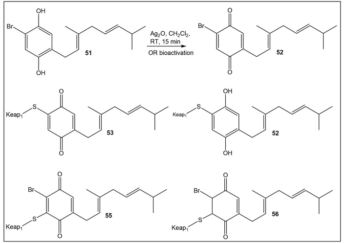
Cymopol, 51 as a masked MA functionality of quinone 52 [150]. Keap1: Kelch-like ECH-associated protein 1
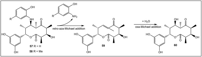
Retro-aza-Michael reaction has also been used by nature and man to unmask the α,β-unsaturated functionality of MA [151, 152]. STLs, such as 57, Figure 18, are phenol-substituted macrolides, which have been isolated from the rare actinomycete Saccharothrix sp. A1506 [153].
STL-B (57, Figure 18) has demonstrated the most potent cytotoxicity against human fibrosarcoma HT1080 cells. Mechanistic studies indicated that the unmasked MA (compound 59), obtained by the release of the o-aminophenol group of 57 via a retro-aza-Michael reaction, was responsible for the cytotoxicity [152]. Further investigation of the unmasking of the MA utilizing methyl-substituted o-aminophenol (58, Figure 18), introduced via co-incubation with Sacharothrix sp. A1506 has demonstrated that electron-donating groups (e.g., as in p-methyl-substituted o-aminophenol) have a positive effect on the reaction [151, 152]. The ease of the retro-aza-Michael reaction with ring-activating groups vs. unsubstituted o-aminophenol allows for fine-tuning of the reaction in addition to confirming the unmasking mechanism [152].
In drug development, the current interest in covalent inhibition (year 2025 marked the approval of the 100th covalent drug by the FDA) brings renewed attention to natural products as leads and as sources of ideas for structural modification that would lead to better target selectivity [154]. Progress in chemical proteomics aids our understanding of the behavior of covalent modifiers, from their molecular target promiscuity and toxicity to tightly controlled target selectivity. The latter depends on the protein’s sensitivity towards the covalent modifier, as well as on the pH and the concentration of excess thiol groups in the cell. The cycloenones, on their own and as the pharmacophore of the STL family, proved to be highly successful covalent modifiers used by plants and humans. Despite utilizing the same cycloenone/α-exo-methylene-γ-butyrolactone moiety as the electrophilic warheads, nature achieves diverse target selectivity by diversifying the rest of the structure.
Having this great structural diversity, uncovering more covalent modifiers with different targets and binding modes from plants and other organisms is just a matter of time. The use of masked functionality by nature inspires the preparation of prodrugs possessing mildly electrophilic moieties, which increases their molecular target specificity, as well as, in some cases, their reversibility. Developing pH/GSH-dual-responsive fluorescent probes using the cycloenone moiety in cancer-targeting therapeutic activity provides new tools for precise diagnosis and tumor treatment. In addition, the recent discoveries of new pro-electrophilic moieties in natural products enrich the warhead chemical space beyond traditional MAs.
12-OPDA: 12-oxo-phytodienoic acid
4,5-ddh-JA: 7-iso-4,5-didehydro-jasmonic acid
ABA: abscisic acid
ARE: antioxidant response element
cyPGs: cyclopentenone prostaglandins
GSH: glutathione
GST: glutathione S-transferase
hGSTP1-1: human glutathione S-transferase P1-1
HNE: 4-hydroxy-2-nonenal
IKK: IκB kinase complex
JA: jasmonic acid
Keap1: Kelch-like ECH-associated protein 1
MA: Michael acceptor
NQO1: NAD(P)H:quinone oxidoreductase 1
Nrf2: nuclear factor erythroid 2-related factor 2
PGs: prostaglandins
PRC2: polycomb repressive complex 2
PUFAs: polyunsaturated fatty acids
ROS: reactive oxygen species
STLs: sesquiterpene lactones
The authors would like to thank the American University and the Midwestern University Offices of Research and Sponsored Programs, as well as the Midwestern University College of Graduate Studies, for their support.
MIK: Conceptualization, Writing—original draft. BJP: Writing—review & editing. Both authors read and approved the submitted version.
The authors declare no conflicts of interest.
Not applicable.
Not applicable.
Not applicable.
Not applicable.
Not applicable.
© The Author(s) 2026.
Open Exploration maintains a neutral stance on jurisdictional claims in published institutional affiliations and maps. All opinions expressed in this article are the personal views of the author(s) and do not represent the stance of the editorial team or the publisher.
Copyright: © The Author(s) 2026. This is an Open Access article licensed under a Creative Commons Attribution 4.0 International License (https://creativecommons.org/licenses/by/4.0/), which permits unrestricted use, sharing, adaptation, distribution and reproduction in any medium or format, for any purpose, even commercially, as long as you give appropriate credit to the original author(s) and the source, provide a link to the Creative Commons license, and indicate if changes were made.



View: 480
Download: 14
Times Cited: 0