 Review


Affiliation:
Department of Internal Medicine, Mountain Area Health Education Center, Asheville, NC 28803, USA
Email: jas.virk@mahec.net
ORCID: https://orcid.org/0000-0001-9938-6991


Explor Cardiol. 2026;4:1012117 DOI: https://doi.org/10.37349/ec.2026.1012117
Received: April 18, 2026 Accepted: June 15, 2026 Published: August 11, 2026
Academic Editor: Eugenio Picano, Italian National Research Council, Italy
Pembrolizumab, a programmed cell death protein 1 inhibitor, has had a substantial impact on cancer treatment across multiple malignancies, but is associated with immune-related cardiovascular toxicities that pose significant clinical challenges. Complications such as myocarditis, arrhythmias, and cardiomyopathy are emerging as significant entities with high morbidity and mortality. This article is a narrative review and was not designed or conducted as a systematic review. No formal systematic review protocol was followed and attempts to identify or include all published studies on this topic were not undertaken. The goal is to provide a clinically oriented synthesis of the current literature on pembrolizumab-associated cardiotoxicity. A literature search was conducted using PubMed, Medline, and Google Scholar databases for articles published between January 2014 and March 2025. Search terms included ‘pembrolizumab,’ ‘PD-1 inhibitor,’ ‘immune checkpoint inhibitor,’ ‘cardiotoxicity,’ ‘myocarditis,’ ‘pericarditis,’ ‘arrhythmia,’ and ‘cardiac adverse events,’ used individually and in combination. Inclusion criteria encompassed English-language clinical trials, observational studies, systematic reviews, meta-analyses, pharmacovigilance analyses, clinical practice guidelines, and case reports involving human subjects. Non-English publications, preclinical studies without clinical correlates, and editorials without original data were excluded. Reference lists of identified articles were manually reviewed to identify additional relevant publications. Study selection was performed by a single author. Pembrolizumab-induced myocarditis, although rare, carries a high mortality rate and typically presents within the first few weeks of treatment, including a relative risk of myocarditis ~4.5 with combination immune checkpoint inhibitor therapy. Proposed mechanisms of this cardiotoxicity, though not settled, include shared antigenic targets between tumor and cardiac tissue and impaired immune tolerance. Current management relies on prompt recognition, immunosuppression with high-dose IV methylprednisolone as first-line therapy, and additional immunomodulatory agents for refractory cases. Emerging evidence from case reports and small cohort studies suggests potential benefit from abatacept with ruxolitinib in steroid-refractory cases; however, prospective validation is needed. Baseline cardiac screening and serial monitoring of cardiac biomarkers including high-sensitivity troponin and NT-proBNP, and a multidisciplinary cardio-oncology approach are essential for early detection and optimal outcomes.
Pembrolizumab, a humanized monoclonal antibody targeting programmed cell death protein 1 (PD-1), has substantially changed treatment options since its approval by the U.S. Food and Drug Administration in 2014. By blocking the PD-1/programmed death ligand 1 (PD-L1) immune checkpoint pathway, pembrolizumab restores T-cell activity against tumor cells, enabling the immune system to recognize and destroy cancer cells. Pembrolizumab has demonstrated efficacy across a diverse range of cancers, including melanoma, non-small cell lung cancer, head and neck squamous cell carcinoma, and renal cell carcinoma [1]. Despite its therapeutic benefits, pembrolizumab and other immune checkpoint inhibitors (ICIs) are associated with a unique spectrum of immune-related adverse events (irAEs), including potentially life-threatening cardiotoxicity.
Cardiotoxicity associated with pembrolizumab, though relatively rare compared to other irAEs, carries significant morbidity and mortality. A pharmacovigilance study of 101 patients with ICI-associated myocarditis found that 33 patients had timing of myocarditis onset in relation to ICI initiation, and of these 33 patients, the median onset of myocarditis development was 27 days (IQR 5–155 days) [2].
Cardiac irAEs can manifest in diverse forms, including myocarditis, pericarditis, arrhythmias, and heart failure. Understanding the mechanisms, clinical presentations, risk factors, and management strategies for pembrolizumab-associated cardiotoxicity is essential for oncologists, cardiologists, and other clinicians caring for patients receiving ICI therapy.
The pathophysiology of pembrolizumab-induced cardiotoxicity is complex and not completely understood, but it fundamentally relates to the disruption of immune homeostasis. Under normal physiological conditions, the PD-1/PD-L1 pathway serves as a critical immune checkpoint that prevents excessive T-cell activation and maintains self-tolerance [3, 4]. Cardiac endothelial cells and cardiomyocytes upregulate PD-L1 expression under inflammatory conditions, particularly in response to interferon-gamma (IFN-γ) secreted by T cells; this upregulation serves as a cardioprotective mechanism that limits T-cell-mediated myocardial injury [5, 6]. Genetic deletion of PD-L1 in mice models transforms transient myocarditis into lethal disease, reinforcing the pivotal role of this pathway in cardiac immune tolerance [5]. Separately, PD-1 receptor-deficient BALB/c mice develop autoimmune dilated cardiomyopathy mediated by IgG autoantibodies against cardiac troponin I, providing evidence that PD-1 is critical for preventing autoimmune cardiac disease [7, 8]. However, human ICI-associated myocarditis appears to be primarily T-cell mediated rather than autoantibody-driven, as investigations have shown no evidence of B cells or antibody-antigen deposits in affected human myocardial tissues [9, 10]. T-cell receptor (TCR) sequencing has revealed identical or highly similar TCR clonotypes in both tumor tissues and inflamed myocardium from the same patients, suggesting cross-reactivity between tumor antigens and cardiac proteins [11]. Histopathological studies demonstrate an inflammatory infiltrate predominantly composed of CD8+ T cells interspersed with CD4+ T cells and macrophages, with upregulation and positive staining of PD-L1 in myocardial tissue [12]. These activated T cells recognize cardiac antigens, either through molecular mimicry between tumor antigens and cardiac proteins such as α-myosin heavy chain or through loss of tolerance to native cardiac antigens [13]. Beyond direct T-cell-mediated injury, depletion or functional impairment of regulatory T cells following PD-1 blockade may reduce immune tolerance to cardiac self-antigens [14].
Additionally, cytokine release may play a substantial role in cardiotoxicity. The activation of T cells following PD-1 blockade can trigger the release of pro-inflammatory cytokines such as IFN-γ, tumor necrosis factor-α (TNF-α), and interleukin-6 (IL-6), which can directly damage cardiomyocytes and promote further inflammatory responses [15].
The timing of cardiotoxicity onset provides additional insights into mechanisms. The median time to onset of ICI-associated myocarditis was 34 days, with 81% of patients presenting within 3 months of initiating ICI therapy, though late presentations of up to 454 days have been reported [16]. This early occurrence suggests that the immune system rapidly recognizes and attacks cardiac antigens once the PD-1 checkpoint is released. However, delayed cases indicate that immune activation can have persistent effects even after treatment discontinuation [17].
Pembrolizumab-induced cardiotoxicity presents with a wide spectrum of clinical manifestations, ranging from asymptomatic biomarker elevation to myocarditis with cardiogenic shock and death. ICI-associated myocarditis has a reported incidence of 0.04% to 1.14%, but exhibits a significantly higher mortality rate ranging from 25% to 50% compared to other irAEs [18]. The wide range of reported mortality rates reflects differences in data sources, time periods, case ascertainment methods, and evolving management practices. Notably, the International Cardio-Oncology Society (IC-OS) position statement documents a declining mortality trend from 45.5% before 2016 to approximately 23% in 2021–2022, likely reflecting improved awareness, earlier detection, and more aggressive immunosuppressive treatment [19].
The clinical presentation can be highly variable and nonspecific, making early recognition challenging. In a systematic review of 106 ICI-induced myocarditis cases, more than half of the cases were life-threatening or severe [20]. However, some patients remain asymptomatic despite significant cardiac involvement, and the condition may only be detected through routine laboratory or imaging surveillance. The nonspecific nature of symptoms means that clinicians must maintain a high index of suspicion, particularly during the first few months of pembrolizumab therapy.
Cardiac biomarkers play an important role in detecting and monitoring cardiotoxicity. Troponin elevation, particularly high-sensitivity troponin I or T, is the most sensitive marker for myocardial injury and may be detected before symptom onset. Even modest troponin elevations should prompt further cardiac evaluation in patients receiving pembrolizumab. B-type natriuretic peptide (BNP) or N-terminal pro-BNP (NT-proBNP) elevation suggests myocardial stress and often accompanies troponin elevation in cases of myocarditis [21]. However, standardized surveillance protocols remain under investigation.
Electrocardiographic (ECG) abnormalities that should raise suspicion for myocarditis in patients receiving ICI include new PR interval prolongation, atrioventricular block, ventricular arrhythmias, or T-wave changes [22]. Conduction abnormalities are especially concerning and may include first-, second-, or third-degree heart block. Atrial arrhythmias are possible, with one early case series of 8 patients reporting atrial fibrillation in up to 30% of cases [23]; however, this estimate should be interpreted with caution given the small sample size, and larger registries are needed to establish the true prevalence of atrial fibrillation in ICI-associated myocarditis. In a multicenter registry study of 140 ICI myocarditis cases and 179 controls, QRS duration was significantly prolonged at the time of myocarditis diagnosis (110 ± 22 ms vs. 93 ± 19 ms in controls, p < 0.001), and each 10 ms increase in QRS duration conferred a 1.3-fold increase in odds of major adverse cardiac events (MACE) (95% CI: 1.07–1.61, p = 0.011), whereas QTc interval was not associated with adverse outcomes [24]. These findings suggest that QRS prolongation, rather than QTc prolongation, may serve as a more clinically meaningful prognostic marker in ICI-associated myocarditis.
Echocardiography is essential for assessing cardiac structure and function. Findings in ICI-related myocarditis may include reduced left ventricular ejection fraction (LVEF), regional wall motion abnormalities, or pericardial effusion [25]. However, echocardiographic findings can be subtle or absent even in cases of biopsy-proven myocarditis, particularly early in the disease course. Global longitudinal strain (GLS) has emerged as a more sensitive marker of subclinical myocardial dysfunction than conventional LVEF. In a multicenter registry of 101 patients with ICI-associated myocarditis, GLS was reduced in patients presenting with both preserved and reduced ejection fraction. Among patients with preserved LVEF (≥ 50%), every 1% absolute decline in GLS magnitude was associated with a 4.4-fold increase in MACE, demonstrating that GLS can identify high-risk patients who would otherwise be missed by LVEF assessment alone [26]. The NCCN guidelines now recommend echocardiography “if possible with LV strain measurement” in the workup of suspected ICI cardiotoxicity [27].
Cardiac magnetic resonance imaging (CMR) has emerged as a powerful tool for diagnosing ICI-related myocarditis. CMR is the reference imaging test for the diagnosis of myocarditis, with the approach evolving since publication of the initial Lake Louise criteria to incorporate quantitative approaches including T1/T2 mapping [28]. According to the updated Lake Louise criteria, the two main criteria for diagnosing myocarditis are myocardial edema based on T2 maps and nonischemic myocardial injury based on T1 maps [29].
In a study of patients with checkpoint inhibitor-associated myocarditis, 95% met the nonischemic myocardial injury criteria, 53% met the myocardial edema criteria, and 48% met both criteria [28]. Native T1 values were significantly elevated in 78% of patients and T2 values in 43%, with T1 values showing greater sensitivity than T2 mapping. CMR findings may include myocardial edema indicated by elevated T2 signal, myocardial hyperemia shown by early gadolinium enhancement, and myocardial injury or fibrosis demonstrated by late gadolinium enhancement (LGE), often in a non-ischemic pattern [28].
Endomyocardial biopsy represents the gold standard for definitive diagnosis of myocarditis but is not routinely performed due to its invasive nature and procedural risks [30]. Biopsy is typically reserved for cases where the diagnosis is uncertain or in severe cases not responding to initial treatment. Histopathologic features include prominent interstitial inflammatory changes consisting of histiocytes and lymphocytes, with immunohistochemistry stains usually positive for CD163, CD3, CD8, Granzyme B, PD-1, and PD-L1 [31].
Other cardiac manifestations of pembrolizumab therapy include pericarditis, stress cardiomyopathy, acute coronary syndrome, and vasculitis affecting coronary or other vessels [9]. The overlap of myocarditis with other irAEs, particularly myositis and myasthenia gravis (the myositis-myasthenia-myocarditis overlap syndrome), carries high morbidity and mortality rates and requires aggressive immunosuppression [32]. Table 1 further compares the various diagnostic modalities in assessing ICI-associated myocarditis.
Comparison of diagnostic modalities for ICI-associated myocarditis.
| Modality | Key findings | Sensitivity | Limitations | Role | References |
|---|---|---|---|---|---|
| High-sensitivity troponin (I or T) | Elevated in myocardial injury; may precede symptoms | High | Not specific to myocarditis; may be elevated in other conditions | Screening and monitoring; every 1% troponin rise associated with worse outcomes | [33–35] |
| NT-proBNP/BNP | Elevated with myocardial stress and heart failure | Moderate | Nonspecific; elevated in many cardiac and non-cardiac conditions | Adjunctive biomarker | [33, 36] |
| ECG | Conduction abnormalities (prolonged QRS, bundle branch block, AV block), atrial/ventricular ectopy, ST-T wave changes; QRS prolongation independently predicts MACE | Moderate | Nonspecific; may be normal early | Baseline and serial monitoring | [19, 24] |
| Echocardiography with GLS | Reduced LVEF, wall motion abnormalities, pericardial effusion; GLS detects subclinical dysfunction | Moderate for LVEF; higher for GLS | LVEF may be preserved in early/mild myocarditis | Initial imaging; GLS superior to LVEF for early detection | [33, 36, 37] |
| Cardiac MRI | T1/T2 mapping, LGE in non-ischemic pattern; 95% meet non-ischemic injury criteria | High | Limited availability; contraindicated with some devices; may miss early disease | Confirmatory imaging | [33, 36, 38] |
| Endomyocardial biopsy | CD8+ T-cell infiltrate, CD163+, PD-L1+ staining | Gold standard | Invasive; sampling error; procedural risk | Reserved for uncertain diagnosis or refractory cases | [19, 31, 33, 39, 40] |
ICI: immune checkpoint inhibitor; NT-proBNP/BNP: N-terminal pro-B-type natriuretic peptide/B-type natriuretic peptide; ECG: electrocardiogram; AV: atrioventricular; MACE: major adverse cardiac events; GLS: global longitudinal strain; LVEF: left ventricular ejection fraction; LGE: late gadolinium enhancement; PD-L1: programmed death ligand 1.
Combination ICI therapy is a more frequent culprit of cardiotoxicity compared to ICI monotherapy, with the relative risk of myocarditis in those who received ipilimumab and nivolumab combination compared to nivolumab alone being 4.5, likely reflecting enhanced immune activation when multiple checkpoint pathways are blocked simultaneously [41]. In dual ICI treatment groups, the incidence of myocarditis increased from 0.06% to 0.27%, with increased severity and earlier onset compared to monotherapy [10].
In a case-control study, myocarditis cases had a higher prevalence of diabetes mellitus and sleep apnea compared to controls [42]. Most commonly reported risk factors in comparison with controls appear to be diabetes mellitus, hypertension, and smoking [43]. However, the presence of pre-existing cardiac disease does not represent an absolute contraindication to pembrolizumab therapy.
The three most common primary tumors associated with ICI myocarditis were lung cancer (26.7%), melanoma (19.0%), and esophageal/gastric cancer (6.9%) [20]. Some evidence suggests higher rates of cardiac irAEs in patients with lung cancer, possibly related to mediastinal tumor location or shared antigens between lung tissue and myocardium.
Genetic factors likely play a role in determining susceptibility to ICI-related cardiotoxicity. Human leukocyte antigen (HLA) genotypes may influence the likelihood of developing autoimmune reactions against cardiac tissue [44]. Certain HLA haplotypes have been associated with increased risk of myocarditis in small studies, but genetic screening is not yet part of routine clinical practice. Concurrent or prior use of other cardiotoxic therapies, including anthracyclines, trastuzumab, or radiation therapy to the chest, may increase vulnerability to pembrolizumab-induced cardiac injury, though prior exposure does not represent an absolute contraindication to ICI therapy [45] (Table 2).
Risk factors for ICI-associated cardiotoxicity.
| Category | Risk factor | References |
|---|---|---|
| Patient-level | Diabetes mellitus | [42] |
| Hypertension | [43] | |
| Sleep apnea | [42] | |
| Smoking | [43] | |
| Age > 85 years | [19, 46] | |
| Pre-existing cardiac disease | [19, 33] | |
| Treatment-level | Combination ICI therapy | [46–48] |
| Prior cardiotoxic chemotherapy | [9] | |
| Prior chest radiation | [9, 49] | |
| Tumor-level | Thymic epithelial tumors | [19, 46] |
| Melanoma | [46] | |
| Lung cancer | [20] | |
| Genetic | HLA genotype (under investigation) | [50] |
ICI: immune checkpoint inhibitor; HLA: human leukocyte antigen.
The management of pembrolizumab-induced cardiotoxicity requires prompt recognition, immediate discontinuation of ICI therapy in most cases, and immunosuppressive treatment, particularly for myocarditis. A multidisciplinary approach involving oncologists and cardiologists is essential for optimal outcomes.
Upon suspicion or diagnosis of significant cardiotoxicity, pembrolizumab should be discontinued immediately. While mild cases may allow for treatment resumption after resolution, moderate to severe cardiotoxicity generally represents a contraindication to further pembrolizumab therapy [51]. The administration of an ICI following an irAE is referred to as a rechallenge. The decision to rechallenge with pembrolizumab after cardiac irAE resolution must be made carefully, weighing the cancer treatment benefits against the substantial risk of recurrent and potentially more severe cardiotoxicity. In this scenario, other ICIs should be considered in the treatment algorithm. An observational cohort study found a 28.8% recurrence rate of the same irAE associated with the discontinuation of ICI therapy after a rechallenge with the same ICI [52].
If a patient is re-challenged and toxicity returns, this warrants permanent discontinuation of immunotherapy. However, irAEs that respond to immunosuppressive therapies may pose a lower risk for rechallenge. Considerations for immunotherapy rechallenge after a hold vary based on organ system. For cardiac symptoms, permanent discontinuation is warranted in the setting of grade 2–4 myocarditis [27]. An absolute contraindication to restarting ICI therapy is life-threatening toxicity, particularly cardiac, pulmonary, or neurologic toxicity [53]. The IC-OS statement reported that among 16 patients with nonfatal myocarditis who underwent ICI rechallenge, 38% experienced recurrence of cardiac toxicity [19]. If rechallenge is considered—such as in patients with complete resolution of grade 1 myocarditis, a compelling oncologic indication, and no alternative therapy—it should only be attempted when the myocarditis has fully resolved clinically and biochemically.
The Common Terminology Criteria for Adverse Events (CTCAE) grading system provides a framework for guiding treatment intensity: Grade 1 (asymptomatic), Grade 2 (moderate symptoms limiting activities of daily living), Grade 3 (severe symptoms requiring hospitalization), Grade 4 (life-threatening symptoms), and Grade 5 (death) [33] (Table 3). Escalation of immunosuppressive intensity should be based on severity grade and clinical symptoms.
Management of ICI-associated myocarditis by CTCAE severity grade.
| CTCAE grade | Clinical features | ICI management | Immunosuppression | Cardiac support | References |
|---|---|---|---|---|---|
| Grade 1 (Asymptomatic) | Abnormal biomarkers or imaging only; no symptoms | Hold ICI; consider permanent discontinuation | Prednisone 1 mg/kg/day with taper | Serial monitoring of troponin, ECG, echo | [19, 33] |
| Grade 2 (Moderate) | Mild symptoms limiting instrumental ADLs | Permanently discontinue ICI | Methylprednisolone 1–2 mg/kg/day IV; consider escalation if no improvement in 24–48 hours | Cardiology consultation; standard HF therapy if needed | [19, 27, 48] |
| Grade 3 (Severe) | Severe symptoms requiring hospitalization | Permanently discontinue ICI | Pulse methylprednisolone 500–1,000 mg/day IV × 3–5 days, then taper; consider abatacept ± ruxolitinib if steroid-refractory | ICU monitoring; antiarrhythmics; temporary pacing if needed | [19, 27, 33, 48] |
| Grade 4 (Life-threatening) | Hemodynamic compromise, cardiogenic shock, malignant arrhythmias | Permanently discontinue ICI | Pulse methylprednisolone 1,000 mg/day IV; consider addition of abatacept + ruxolitinib; consider ATG, IVIG, or plasmapheresis | Mechanical circulatory support (IABP, ECMO, VAD); advanced HF/transplant consultation | [27, 33] |
Infliximab should be avoided or used with extreme caution in patients with reduced LVEF due to risk of exacerbating heart failure [27, 33]. ICI: immune checkpoint inhibitor; CTCAE: Common Terminology Criteria for Adverse Events; ECG: electrocardiogram; ADLs: activities of daily living; IV: intravenous; HF: heart failure; ATG: antithymocyte globulin; IVIG: intravenous immunoglobulin; IABP: intra-aortic balloon pump; ECMO: extracorporeal membrane oxygenation; VAD: ventricular assist device.
High-dose corticosteroids represent first-line therapy for ICI-related myocarditis. Initial use of high-dose methylprednisolone (501–1,000 mg/day) was associated with fewer cases of MACE compared to intermediate-dose (60–500 mg/day) and low-dose (< 60 mg/day) methylprednisolone (22%, 55%, and 62% respectively) [54]. Notably, these dosing data derive from a non-randomized retrospective study and are subject to selection bias, including the possibility that patients receiving higher-dose steroids had more aggressive initial management overall. Prospective randomized trials comparing steroid dosing strategies are lacking. For patients with fulminant myocarditis, cardiogenic shock, or refractory disease, pulse-dose methylprednisolone may be administered [55].
Following initial intravenous therapy and clinical stabilization, patients typically transition to oral prednisone with a prolonged taper over several weeks to months to prevent relapse [56]. In a retrospective case series from China, 16 of 24 patients (67%) evolved toward corticosteroid resistance despite prompt use of high-dose corticosteroids, with deterioration often occurring during steroid tapering [57]. The optimal duration and tapering strategy have not been established and should be individualized based on clinical response.
For patients who fail to respond adequately to corticosteroids or who have severe presentations, additional immunosuppressive agents may be considered early. It should be noted that the evidence base for all second-line agents in ICI-associated myocarditis remains limited, consisting primarily of case reports, case series, and observational cohorts. Therapies that have been tested include abatacept and Janus kinase (JAK) inhibitors such as tofacitinib and ruxolitinib [58–60]. Options also include intravenous immunoglobulin (IVIG), mycophenolate mofetil, tacrolimus, and infliximab (anti-TNF-α therapy) [61]. IVIG has been used successfully in some cases and may provide additional immune modulation without further T-cell suppression [62].
Abatacept is a CTLA-4 immunoglobulin fusion protein binding CD80/CD86 on antigen-presenting cells and leads to global T-cell anergy by specifically reversing pathways activated by ICI [63]. The rationale for abatacept use has been supported by preclinical mouse models of ICI myocarditis [64]. JAK inhibitors impair T-cell activation via blockade of proinflammatory cytokines, with ruxolitinib having the additional theoretical benefit of decreasing CD86 expression on macrophages, potentially synergizing with CD86 blockade by abatacept [63]. In a small case series, tofacitinib (5 mg twice daily) was used in 11 corticosteroid-resistant patients, with seven patients recovering, showing a promising therapeutic effect [65]. While these results are encouraging, the small sample size and absence of a control group preclude definitive conclusions. Infliximab has been used in some cases, though with variable results [66]. Antithymocyte globulin (ATG) is an option for severe, refractory cases, though its use carries risks of increased immunosuppression and infection [67].
Supportive cardiac care is essential in managing ICI-related cardiotoxicity. Patients with heart failure require standard heart failure medical management including diuretics, angiotensin-converting enzyme inhibitors or angiotensin receptor blockers, and beta-blockers. Patients with significant arrhythmias may require antiarrhythmic medications, with amiodarone being commonly used for ventricular arrhythmias [56, 68].
Conduction abnormalities require careful monitoring and may necessitate temporary or permanent pacemaker placement [69]. High-grade atrioventricular block associated with ICI myocarditis may not resolve despite immunosuppression, requiring permanent pacing. Patients with hemodynamically unstable arrhythmias may need temporary pacing or implantable cardioverter-defibrillator placement [70].
In cases of fulminant myocarditis with cardiogenic shock, mechanical circulatory support may be lifesaving. Options include intra-aortic balloon pump, extracorporeal membrane oxygenation, or ventricular assist devices. Early consultation with advanced heart failure and transplant cardiology teams is essential for patients with severe hemodynamic compromise, as some patients may require consideration for cardiac transplantation as a refractory treatment option [71–73]. However, cardiac transplantation after ICI-induced myocarditis remains controversial. Concerns exist regarding the risk of recurrent immune-mediated injury in the transplanted heart and the theoretical risk of allograft rejection in patients with prior immune checkpoint-blockade induced immune activation. The long-term immunosuppressive therapy regimen necessary after transplantation may also complicate future cancer management. These considerations should be discussed with advanced heart failure and transplant teams on a case-by-case basis.
Cardiac monitoring during and after treatment is imperative. Serial cardiac biomarkers, ECGs, and echocardiography should be performed regularly to assess treatment response and detect relapse [33]. The frequency of monitoring should be individualized based on disease severity and clinical course. Long-term follow-up is necessary as some patients may develop chronic cardiac dysfunction or late complications even after initial resolution [74] (Figure 1).
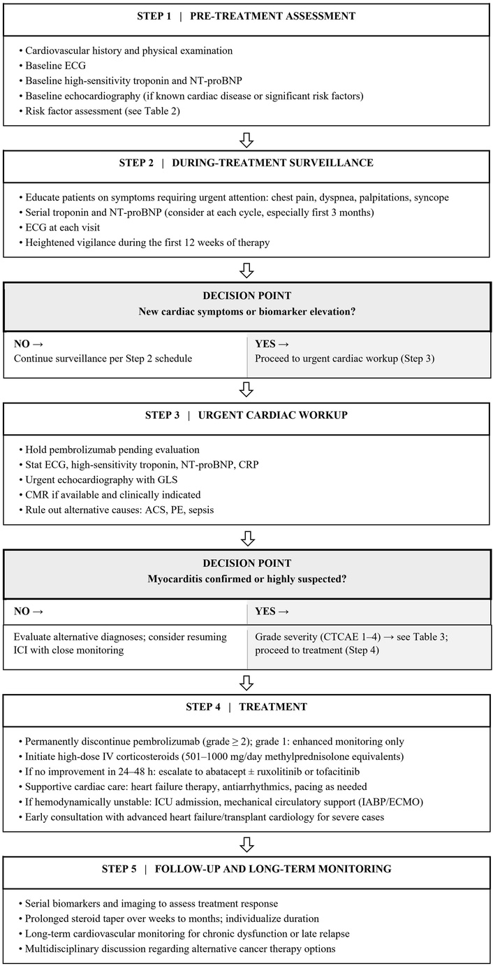
Clinical algorithm for screening, diagnosis, and management of pembrolizumab-induced cardiotoxicity. ECG: electrocardiogram; NT-proBNP: N-terminal pro-B-type natriuretic peptide; CRP: C-reactive protein; GLS: global longitudinal strain; CMR: cardiac magnetic resonance imaging; ACS: acute coronary syndrome; PE: pulmonary embolism; ICI: immune checkpoint inhibitor; CTCAE: Common Terminology Criteria for Adverse Events; IV: intravenous; IABP: intra-aortic balloon pump; ECMO: extracorporeal membrane oxygenation.
The prognosis of pembrolizumab-induced cardiotoxicity varies considerably depending on the severity of presentation and timing of intervention. In a systematic analysis of 116 case reports, the mortality rate was 47.4%, with only 26 cases of recovery (22.4%) and 35 cases of improvement (30.2%) [20]. Deaths typically resulted from cardiogenic shock, ventricular arrhythmias, or complete heart block [20]. The concurrent presence of myositis, myasthenia gravis, or respiratory muscle involvement significantly worsens prognosis [75, 76].
Patients who survive the acute phase may experience variable degrees of cardiac recovery. Some patients achieve complete normalization of cardiac function and biomarkers, while others develop persistent left ventricular dysfunction, chronic heart failure, or persistent conduction abnormalities requiring permanent pacing. Factors associated with worse outcomes include delayed recognition and treatment, severe initial presentation with hemodynamic compromise, elevated troponin levels, and requirement for mechanical circulatory support [77, 78].
Long-term cardiovascular monitoring is essential for all patients who develop ICI-related cardiotoxicity, as late relapses or complications can occur. The impact of cardiotoxicity on cancer outcomes also requires consideration, as treatment interruption or discontinuation may affect oncologic efficacy. However, continuing pembrolizumab in the setting of significant cardiotoxicity carries unacceptable risk, and alternative cancer treatment strategies must be pursued when possible [33].
Strategies for early detection and prevention of pembrolizumab-induced cardiotoxicity are critical. While no approach can eliminate risk, several practices may facilitate earlier recognition and potentially improve outcomes. The 2022 ESC Cardio-Oncology Guidelines recommend a structured cardiovascular risk assessment before initiating ICI therapy, incorporating baseline cardiovascular risk factors, prior cardiac history, and planned treatment regimen to stratify patients into low, moderate, and high cardiovascular risk categories. Higher-risk patients warrant more intensive monitoring, including serial biomarkers at each treatment cycle and periodic echocardiography [79].
Baseline cardiac assessment before pembrolizumab initiation should include a thorough cardiovascular history, physical examination, and ECG. Baseline echocardiography may be considered for patients with known cardiac disease or significant cardiovascular risk factors. Baseline cardiac biomarkers, particularly troponin and pro-BNP, may provide reference values for comparison if symptoms develop, though routine baseline testing remains debated [80].
During pembrolizumab therapy, clinicians should maintain vigilance for cardiac symptoms, particularly during the first few months of treatment. Patients should be educated about symptoms requiring immediate medical attention, including chest pain, dyspnea, palpitations, or syncope. These symptoms should trigger immediate cardiac assessment including ECG, cardiac biomarkers, and echocardiography. Even modest troponin elevations warrant careful evaluation, as small increases may herald more significant injury if treatment continues. Some experts advocate for periodic cardiac biomarker monitoring during treatment, though the optimal frequency and cost-effectiveness of routine surveillance remains uncertain.
Prophylactic immunosuppression to prevent cardiotoxicity is not recommended, as it would likely diminish the anticancer efficacy of pembrolizumab. However, for patients with multiple risk factors or concerning baseline findings, heightened surveillance is warranted.
Improved risk prediction tools incorporating clinical factors, biomarkers, and potentially genetic information could enable more personalized risk assessment and surveillance strategies. A novel ICI myocarditis risk score incorporating troponin elevation, thymoma histology, low QRS voltage, LVEF < 50%, and cardiomuscular symptoms has been developed and validated, with 30-day primary outcomes incidence ranging from 4% (score = 0) to 81% (score ≥ 4), offering a practical tool for early risk stratification [81]. Clinical trials examining the addition of abatacept to high-dose corticosteroids as initial therapy for ICI-induced myocarditis are ongoing and may establish evidence-based second-line treatment protocols [33]. Novel biomarkers including microRNAs (e.g., miR-155 and exosomal miR-34a-5p) and cardiac-specific autoantibodies are under investigation for early detection and risk stratification [82–84]. Advanced imaging techniques, including strain imaging, parametric mapping on CMR, and nuclear imaging approaches, may detect subclinical cardiac involvement before overt clinical manifestations develop [85].
This narrative review has several limitations. As a narrative rather than systematic review, the literature search and syntheses were not conducted according to a formal systematic review protocol, and the findings are subject to the inherent limitations of this methodology, including the potential for incomplete capture of all relevant studies. Article selection was performed by a single author, which introduces the possibility of selection bias in the identification and inclusion of studies. Additionally, much of the available evidence on ICI-associated cardiotoxicity is derived from case reports, case series, pharmacovigilance databases, and retrospective studies, which are themselves subject to bias, confounding, and limited generalizability.
Pembrolizumab-associated cardiotoxicity, though uncommon, carries substantial morbidity and mortality and demands a proactive cardio-oncology approach. Clinicians prescribing pembrolizumab should obtain baseline cardiac biomarkers and ECG, maintain heightened clinical vigilance during and after treatment completion, and pursue further evaluation for new cardiac symptoms that may develop. When myocarditis is suspected, immediate ICI discontinuation with high-dose corticosteroid treatment remains the cornerstone of management, with consideration of immunosuppressive agents for steroid-refractory cases. A multidisciplinary team including oncology, cardiology, and heart failure specialists is essential for optimizing both cardiac and oncologic outcomes. As ICI use expands, integrating structured cardiovascular surveillance into oncology practice will be critical for reducing cardiac complications of pembrolizumab therapy.
BNP: B-type natriuretic peptide
CMR: cardiac magnetic resonance imaging
ECG: electrocardiogram/electrocardiographic
GLS: global longitudinal strain
HLA: human leukocyte antigen
ICIs: immune checkpoint inhibitors
IC-OS: International Cardio-Oncology Society
irAE: immune-related adverse event
IVIG: intravenous immunoglobulin
JAK: Janus kinase
LVEF: left ventricular ejection fraction
MACE: major adverse cardiac events
PD-1: programmed cell death protein 1
PD-L1: programmed death ligand 1
TCR: T-cell receptor
TNF-α: tumor necrosis factor-α
IFN-γ: interferon-gamma
Artificial intelligence–based tools (including large language models) were used to assist with drafting, language refinement, and organization of this manuscript. Open Evidence was used for literature search assistance while Claude (Anthropic) AI was used for writing support. All scientific content, interpretations, and conclusions were generated, verified, and approved by the author, who assumes full responsibility for the work.
JV: Conceptualization, Investigation, Writing—original draft, Writing—review & editing. The author has read and approved the submitted version.
The author declares that he has no conflicts of interest.
Not applicable.
Not applicable.
Not applicable.
Not applicable.
Not applicable.
© The Author(s) 2026.
Open Exploration maintains a neutral stance on jurisdictional claims in published institutional affiliations and maps. All opinions expressed in this article are the personal views of the author(s) and do not represent the stance of the editorial team or the publisher.
Copyright: © The Author(s) 2026. This is an Open Access article licensed under a Creative Commons Attribution 4.0 International License (https://creativecommons.org/licenses/by/4.0/), which permits unrestricted use, sharing, adaptation, distribution and reproduction in any medium or format, for any purpose, even commercially, as long as you give appropriate credit to the original author(s) and the source, provide a link to the Creative Commons license, and indicate if changes were made.



View: 974
Download: 17
Times Cited: 0


