 Review


Affiliation:
1Department of Cardiac Surgery, St. Petersburg State University, 199034 Saint Petersburg, Russian Federation
Email: mryadinskiy17101998@mail.ru
ORCID: https://orcid.org/0000-0002-1773-0160


 ,
,
Affiliation:
1Department of Cardiac Surgery, St. Petersburg State University, 199034 Saint Petersburg, Russian Federation
ORCID: https://orcid.org/0000-0002-1296-8161
,
Affiliation:
1Department of Cardiac Surgery, St. Petersburg State University, 199034 Saint Petersburg, Russian Federation
ORCID: https://orcid.org/0000-0003-2267-2580
,
Affiliation:
2Cardiology Department, Research Cardiology Center “Medika”, 197110 Saint Petersburg, Russian Federation
ORCID: https://orcid.org/0000-0002-4007-3322
,
Affiliation:
3Department of Advanced Surgery, Military Medical Academy of S.M. Kirov, 194044 Saint Petersburg, Russian Federation
ORCID: https://orcid.org/0000-0002-4916-2911
,
Affiliation:
1Department of Cardiac Surgery, St. Petersburg State University, 199034 Saint Petersburg, Russian Federation
ORCID: https://orcid.org/0000-0002-9085-4872
Explor Cardiol. 2026;4:1012116 DOI: https://doi.org/10.37349/ec.2026.1012116
Received: March 25, 2026 Accepted: May 18, 2026 Published: July 29, 2026
Academic Editor: Alexandre Abizaid, Institute Dante Pazzanese de Cardiologia, Brazil
Moderate ischemic mitral regurgitation (IMR) remains a ‘grey area’ for clinicians, with evidence-based decision-making for patients referred for coronary artery bypass grafting (CABG) being particularly challenging. IMR results from left ventricular remodeling and subvalvular deformation, not primary leaflet pathology. Even moderate regurgitation (effective regurgitant orifice area 20–39 mm2) is associated with a 1.5–2-fold increased risk of death. Among diagnostic methods, stress echocardiography is valuable but underutilized. An exercise-induced increase in the effective regurgitant orifice area of ≥ 13 mm2 or an absolute value ≥ 30 mm2 identifies patients with prognostically significant dynamic regurgitation requiring valve intervention, despite moderate resting values. Most randomized trials, particularly the Cardiothoracic Surgical Trials Network study, show that adding mitral valve annuloplasty to CABG reduces regurgitation severity but does not improve two-year survival or heart failure hospitalization rates, resulting in only a class IIb recommendation for the combined procedure. Modern imaging techniques, including three-dimensional echocardiography and cardiac magnetic resonance, provide added value in assessing IMR, annular geometry, and myocardial viability. Natriuretic peptides offer additional prognostic information. Transcatheter percutaneous mitral valve repair (‘edge-to-edge’) shows promise in high-risk patients and opens new prospects for hybrid revascularization. Optimal medical therapy, including Sacubitril/Valsartan and SGLT2 inhibitors, may slow IMR progression through enhanced reverse remodeling. In the absence of convincing evidence for any single approach, the current standard remains an individualized strategy implemented by the Heart Team, simultaneously considering clinical status, stress echocardiography data, anatomical characteristics, and surgical risks. Future research priorities include standardization of stress echocardiography protocols, validation of prognostic models, and long-term assessment of transcatheter approach outcomes in this complex patient category.
Coronary artery disease (CAD) remains the leading cause of morbidity and mortality worldwide. However, a significant proportion of patients referred for coronary artery bypass grafting (CABG) have comorbid valve pathologies, among which ischemic mitral regurgitation (IMR) is particularly prominent. This condition is a functional (secondary) mitral regurgitation caused not by structural damage to the mitral valve leaflets, but by remodeling and distortion of the subvalvular apparatus due to an ischemic myocardial injury.
Epidemiological data indicate a high prevalence of mitral regurgitation among patients with CAD. According to large registry studies, mitral regurgitation of varying severity is detected in 40–50% of patients in the acute phase of myocardial infarction and persists in the chronic phase in a significant proportion of patients [1, 2]. According to Bursi et al. [3], a moderate or severe mitral regurgitation occurs in at least 12% of patients who survived a myocardial infarction. Among patients referred for an isolated CABG, the incidence of a concomitant moderate mitral regurgitation, according to various estimates, ranges from 12 to 30%, making this problem highly relevant for the cardiac surgical practice [4, 5]. True prevalence of this condition may be even higher if we consider the dynamic nature of the IMR and its dependence on loading conditions during examination [6].
Clinical significance of the mitral regurgitation is determined by its proven negative impact on patient prognosis. Numerous studies have convincingly demonstrated that the presence of even a moderate mitral regurgitation of ischemic origin is associated with a significant deterioration in long-term outcomes. In 2001, Grigioni et al. [7] demonstrated that mitral regurgitation is an independent predictor of mortality after myocardial infarction, with the risk of death increasing proportionally to the degree of regurgitation. It is known that even a moderate mitral regurgitation increases the risk of death by 1.5–2 times compared to patients without mitral regurgitation. Moreover, an uncorrected moderate mitral regurgitation during the isolated CABG is accompanied by an increased frequency of hospitalizations due to decompensated chronic heart failure or deterioration of functional class and reduces the quality of life in the postoperative period [8]. Several studies also indicate that some patients experience the progression of the mitral regurgitation after isolated revascularization, which further worsens the long-term prognosis [9].
Despite its obvious clinical significance, a moderate/borderline mitral regurgitation presents the greatest therapeutic dilemma and resides in the so-called ‘gray area’ of decision-making. While the management strategy for patients with severe mitral regurgitation is generally well-defined (most clinical guidelines support mitral valve correction during a CABG, and in cases of mild regurgitation, the additional valve intervention is usually not indicated), the approach to patients with moderate mitral regurgitation remains a topic of active discussion [10, 11]. In this regard, the decision-making complexity is due to several factors. First, the dynamic nature of mitral regurgitation makes it difficult to accurately and reproducibly assess the degree of regurgitation, its severity can vary significantly depending on the hemodynamic conditions, the level of physical activity, and the drug therapy being administered [12]. Secondly, for some patients, the myocardial revascularization alone results in a considerable reduction in the degree of mitral regurgitation due to the restoration of blood supply to ischemic segments and improvement of papillary muscle function, whereas for other patients, the regurgitation persists or progresses, indicating irreversibility remodeling of the left ventricle (LV) [13]. Thirdly, the addition of the mitral valve repair to the CABG procedure increases the time of cardiopulmonary bypass and aortic cross-clamping, which can potentially increase perioperative risks, especially for patients with comorbidities [14].
Current clinical guidelines indicate that there is uncertainty on this issue. The American Heart Association/American College of Cardiology (AHA/ACC, 2020) guidelines classify the simultaneous correction of moderate IMR during the CABG as class IIb with level of evidence B, stipulating that this intervention may be considered without providing a definitive recommendation [11]. European guidelines (ESC/EACTS, 2025) also do not provide a clear solution for this category of patients, leaving the decision to the multidisciplinary cardiac team [10]. Large meta-analyses and randomized clinical trials addressing this issue are few and show conflicting results.
RIME study demonstrated the superiority of the combined approach (CABG + annuloplasty) in terms of functional parameters, the left ventricular reverse remodeling, MR reduction, and BNP levels [15], while a larger study conducted by the Cardiothoracic Surgical Trials Network found no significant differences in survival or adverse cardiovascular events between a CABG alone and the CABG combined with the mitral valve repair groups at 2-year follow-up [9]. These discrepancies may be explained by heterogeneity of the study populations, differences in methodology for assessing the degree of regurgitation, and the duration of follow-up.
Thus, the problem of choosing the optimal management strategy for patients with moderate IMR referred for the CABG remains unresolved and requires the available data systematization. This review analyzes current understanding of the pathophysiology, diagnosis, and surgical treatment of the moderate IMR, as well as a critical analysis of the evidence base underlying the existing clinical guidelines, with the aim of identifying the most appropriate approach to manage this complex category of patients.
IMR is a common complication of global or regional pathological remodeling of the LV due to chronic ischemic heart disease. IMR is defined as mitral regurgitation caused by chronic changes in the LV structure and function due to ischemic heart disease. It is not a valve disease, but rather a valvular consequence of increased leaflet tethering and weakened leaflet closing forces [16].
IMR pathogenesis is based on an imbalance between two opposing forces acting on the leaflets during systole: increased tethering from the subvalvular structures and weakened closing forces from the myocardium [17]. The triggering mechanism is ischemic injury and subsequent remodeling of the LV zones to which the papillary muscles are attached (mainly in inferior and lateral infarctions) [18].
Geometric remodeling of the ventricle leads to displacement of the papillary muscles in the apical, posterior, and lateral directions. Since the papillary muscles are connected to the leaflets via inextensible chordae, their dislocation alters the valve configuration: the coaptation zone is displaced into the LV cavity. This gives the anterior leaflet a characteristic bend, known in the literature as the ‘seagull sign’ [16]. In parallel, the dilation of the fibrous ring usually develops, most pronounced in the septolateral direction [19]. Simultaneously with the increase in leaflet traction, a decrease in closing forces is observed, caused by a decrease in global and regional myocardial contractility, weakening of the systolic contraction of the ring, as well as the dyssynchronous contraction of the papillary muscles and the development of the intraventricular dyssynchrony (especially in the basal segments). Regurgitation resulting from these processes triggers a positive feedback mechanism: the volume overload of the LV aggravates its dilation, which, in turn, increases the displacement of the papillary muscles, further dilation of the annulus, which contributes to the steady progression of the valve insufficiency [18–20]. An example of morphological changes and the dynamic nature of the IMR is shown in Figure 1.
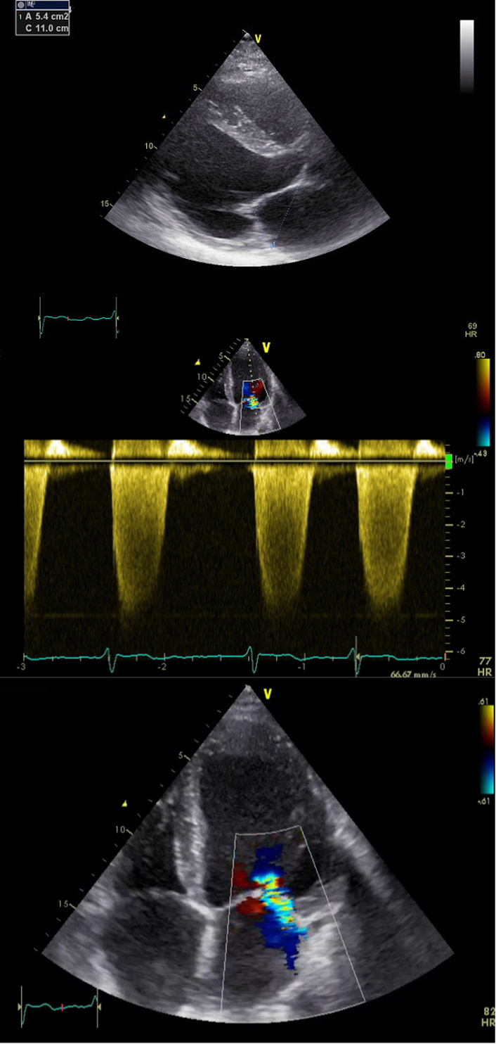
Example of the morphological changes and dynamic nature of IMR. The drawing is taken from clinical practice; patient consent was obtained.
A distinctive feature of the IMR is its pronounced dynamic nature, which is most fully assessed during stress tests, especially stress echocardiography [21, 22]. The degree of the regurgitation recorded at rest does not correlate with the exercise-induced changes in either the increase in the effective regurgitant orifice area (EROA) or the regurgitant volume [23]. Direction and magnitude of these changes are determined by the ability of the LV to undergo further remodeling and the exacerbation of valve deformation under load, as well as the ability of the LV walls and the papillary muscles to contract synchronously. Another manifestation of the dynamic nature of the IMR is the possibility of a decrease in the regurgitant volume against the background of successful drug therapy inducing the reverse (positive) LV remodeling [22]. In this regard, the ESC/EACTS clinical guidelines (2025) clearly emphasize the need for stress echocardiography in patients with inconclusive/uncertain IMR parameters at rest [10].
According to the Carpentier classification, the most common variant of the IMR is type IIIb, the pathogenetic basis of which is limited leaflet movement during systole. In cases where the insufficiency develops with preserved leaflet mobility but is caused by the dilation of the fibrous ring (annuloectasia), the IMR is classified as type I. A separate case represents the IMR type II, which is based on excessive leaflet excursion (prolapse). This dysfunction results from ischemic myocardial injury: acute (e.g., papillary muscle rupture) or chronic, accompanied by fibrotic changes and pathological elongation of the papillary muscles [24].
Recognition of the papillary muscle dysfunction as a distinct nosological entity is associated with the work of Burch et al. [25, 26], who in the 1960s described the ‘papillary muscle dysfunction syndrome’, laying the clinical and hemodynamic foundations to study this condition.
In subsequent publications, the authors systematized the concepts of the pathophysiology of papillary muscle dysfunction in relation to CAD and demonstrated that ischemic damage and the infarction of the papillary muscles lead to a hemodynamically significant mitral regurgitation associated with an unfavorable prognosis [27]. These works laid the conceptual basis for further study of the IMR mechanisms.
Classification proposed by Carpentier (1983) [24] and based on the analysis of the movement of the mitral valve leaflets became a key milestone in understanding of the mitral regurgitation pathogenesis. The identification of the type IIIb MR provided a clear differentiation of the IMR from the primary organic valve pathology and established the universal language for describing the regurgitation mechanisms.
In parallel, approaches to assess the severity of the mitral regurgitation have been developed. First systematic grading was the angiographic classification of Sellers et al. (1964) [28], based on the degree of left atrial opacification during left ventriculography, which provided a four-point system for assessing the severity of the MR. Despite its subjectivity and invasiveness, Sellers’ classification remained the primary tool for stratifying the severity of MR for a long time.
Introduction in the 1980s and 1990s of the Doppler color imaging method marked the transition to noninvasive diagnostics. Initial echocardiographic approaches relied on visual assessment of the area of the regurgitant jet in the left atrium: a jet-to-left atrial area ratio of less than 20% was considered as mild MR, 20–40% as moderate, and greater than 40% as severe. The simplicity of the Doppler’s method has ensured its widespread use in clinical practice. However, the accumulated experience has revealed the significant limitations of this approach: the visualized jet size is highly dependent on instrument settings (gain, pulse repetition rate, Nyquist limit), hemodynamic conditions (blood pressure, volume status), and a systematic underestimation of the regurgitation severity in eccentric jets adjacent to the wall (the Coanda effect). A significant interobserver variability further limited the reproducibility of the results.
Awareness of these shortcomings stimulated the development of quantitative methods. The proximal isovolumetric surface area (PISA) method, based on proximal flow convergence zone analysis, allowed for the calculation of the EROA and regurgitant volume, providing an objective quantitative characterization of the MR severity [29, 30].
At the same time, a method for measuring the width of the vena contracta (the narrowest cross-section of the regurgitant jet directly at the distal orifice) was validated, demonstrating relative independence from hemodynamic conditions and instrument settings [31]. A volumetric method based on a comparison of stroke volumes obtained using pulsed-wave Doppler imaging through the mitral and aortic valves complemented the arsenal of quantitative assessments.
These approaches were systematized in 2003 by the American Society of Echocardiography (ASE) guidelines [32]. The 2006 and 2008 ACC/AHA guidelines for the management of patients with the valvular heart disease established uniform quantitative thresholds for the severe MR regardless of etiology: EROA ≥ 40 mm2, regurgitant volume ≥ 60 mL, and vena contracta width ≥ 7 mm [33, 34]. This unified approach reflected the prevailing view at the time that the regurgitation severity could be assessed using the same criteria for both primary and secondary MR.
However, some clinical studies have questioned the universality of these threshold values for secondary mitral regurgitation. Of fundamental importance was the work of Grigioni et al. (2001) [7], which demonstrated that in patients with ischemic MR, an EROA ≥ 20 mm2 is associated with a significant increase in mortality, whereas in primary MR, a comparable worsening of prognosis is observed only with an EROA ≥ 40 mm2. Enriquez-Sarano et al. [35] confirmed that quantitative determinants of adverse outcomes vary significantly depending on the mechanism of regurgitation: in degenerative MR, the critical threshold is EROA ≥ 40 mm2, whereas in functional MR, values that are half as large are associated with progression of heart failure and increased mortality. These data laid the foundation for the subsequent revision of the threshold criteria for severe secondary MR in European guidelines and identified a fundamental discrepancy between the European and American approaches to classification.
Accumulating evidence on the different prognostic value of quantitative regurgitation parameters in primary and secondary MR has led to a fundamental divergence between the European and American classification approaches, the full extent of which is presented in Table 1. Although the formal definition of severe MR has been revised upward to be more consistent with the ACC/AHA position of EROA ≥ 40 mm2 and regurgitant volume ≥ 60 mL, the document simultaneously introduces the concept of ‘clinical significance’, defining a lower threshold of EROA ≥ 30 mm2 and regurgitant volume ≥ 45 mL, below which MR is formally classified as moderate, but above which it has a significant adverse impact on prognosis [10]. This dual threshold approach, indicated by an asterisk in Table 1, represents a pragmatic reconciliation of a long-standing European-American discrepancy, recognizing that the binary classification of MR as ‘moderate’ or ‘severe’ is insufficient to reflect the prognostic complexity of ischemic regurgitation.
Comparative table of ischemic mitral regurgitation severity thresholds across key Guidelines.
| Parameter | ACC/AHA2006 [33] | ACC/AHA2014 [36] | ACC/AHA2020 [11] | ESC/EACTS2012 [37] | ESC/EACTS2017 [38] | ESC/EACTS2021 [39] | ESC/EACTS2025 [10] |
|---|---|---|---|---|---|---|---|
| EROA, mm2 | ≥ 40 | ≥ 40 | ≥ 40 | ≥ 20 | ≥ 20 | ≥ 20 | ≥ 40 (≥ 30*) |
| RVol, mL | ≥ 60 | ≥ 60 | ≥ 60 | ≥ 30 | ≥ 30 | ≥ 30 | ≥ 60 (≥ 45*) |
| RF, % | ≥ 50 | ≥ 50 | ≥ 50 | ≥ 50 | ≥ 50 | ≥ 50 | ≥ 50 |
| VC, mm | ≥ 7 | ≥ 7 | ≥ 7 | ≥ 7 | ≥ 7 | ≥ 7 | ≥ 7 |
EROA: effective regurgitant orifice area; RVol: regurgitant volume; RF: regurgitant fraction; VC: vena contracta. *: Clinically significant threshold according to ESC/EACTS 2025.
The 2014 American ACC/AHA guidelines, in contrast, retained uniform quantitative criteria for severe MR (EROA ≥ 40 mm2, regurgitant volume ≥ 60 mL) regardless of the regurgitant mechanism [36]. According to their authors’ opinion, the arguments in favor of a unified approach included an insufficient evidence base for lowering the threshold values, methodological limitations of the PISA method in functional MR (elliptical shape of the regurgitant orifice, dynamic nature of the EROA during systole), as well as concerns about overdiagnosis of severe MR with subsequent unjustified expansion of indications for intervention. The 2020 ACC/AHA guideline update formally maintains the previous position. However, it contains an important caveat, the meaning of which is that in secondary MR the poor prognosis may be associated with lower quantitative parameters, and at the same time it is recommended that a comprehensive, integrated assessment considering the clinical context and the degree of LV dilation and dysfunction [11]. Although this statement is not reflected yet in a formal revision of the thresholds, it nevertheless indicates the presence of a gradual convergence of the two main guidelines, a trajectory that Table 1 makes visually evident when read chronologically from left to right.
In this regard, the case of moderate IMR, which falls into the ‘gray area’ of clinical recommendations, is currently of considerable interest (the so-called ‘borderline’ IMR, especially with an EROA of 30–39 mm2, where regurgitation is formally moderate according to traditional criteria, but may already be clinically significant. The concept of ‘disproportionate’ MR proposed by Grayburn et al. (2019) [40] further emphasizes that in the setting of significant LV dilation and dysfunction, even moderate regurgitation in quantitative terms can have a disproportionately significant hemodynamic impact, which should be considered while determining the indications for intervention. As it is discussed in subsequent sections, the data presented in Table 1 and taken together highlight that no single numerical threshold can be universally applied and that optimal decision-making in the borderline IMR requires the integration of quantitative echocardiographic parameters with clinical context, myocardial viability data, and the dynamic behavior of regurgitation under stress.
The evolution of concepts of mitral regurgitation has led to significant differences between the main international recommendations. The 2020 AHA/ACC guidelines use a binary classification system that distinguishes between primary MR (caused by structural valve abnormalities) and secondary MR (caused by left ventricular remodeling), with severity thresholds based on resting echocardiographic measurements: EROA ≥ 0.40 cm2 and regurgitant volume (≥ 60 mL) [11]. The 2025 ESC/EACTS guidelines propose a different approach, introducing a three-category classification that includes ‘disproportionate MR’ as a separate entity, defined as secondary MR with severity parameters (EROA ≥ 0.30 cm2, regurgitant volume ≥ 45 mL) that exceed those expected based on the degree of left ventricular dysfunction [10]. This European classification recognizes that secondary MR exists on a spectrum between purely functional regurgitation and more severe organic manifestations, while emphasizing the dynamic nature of the condition and the inadequacy of assessing the state at rest alone. Importantly, the ESC/EACTS guidelines emphasize the use of exercise or pharmacological stress echocardiography to assess risk, particularly in patients with moderate MR at rest who may experience significant deterioration during physical stress. The AHA/ACC guidelines, while recognizing the value of exercise testing, rely more heavily on resting measurements to grade severity and guide surgical treatment decisions. These fundamental differences provide the necessary context for understanding the comparative diagnostic strategies and treatment recommendations presented in this review.
IMR is a dynamic condition whose severity can vary significantly depending on stress conditions [41]. During physical exertion, worsening regurgitation is due to several interrelated mechanisms: exercise-induced ischemia leads to deterioration of regional contractility and increased leaflet tethering; increased afterload increases the transmitral pressure gradient; impaired coordination of papillary muscle contraction and decreased mitral valve closing forces further aggravate leaflet coaptation failure [42, 43]. The fundamental work of Lancellotti et al. demonstrated that exercise-induced increases in IMR are an independent predictor of adverse cardiovascular events, including hospitalizations for heart failure and cardiovascular mortality [44]. However, in a subgroup of patients, a paradoxical decrease in MR is observed during physical exercise, which is associated with the presence of myocardial contractile reserve, which consists of improved contractility of viable segments, increased closure forces, and a decrease in the LV volume. Preserved contractile reserve is associated with a more favorable prognosis [45].
Key quantitative parameters in stress echocardiography are the EROA and regurgitant volume compared to baseline values at rest. Lancellotti et al. [44] found that an EROA of ≥ 20 mm2 during exercise and an increase in the EROA by ≥ 13 mm2 from rest are significant predictors of poor prognosis in relation to patients with functional MR. However, these data are not supported by the more recent EACVI statement (2022), which indicates that an EROA ≥ 30 mm2 and a peak regurgitant volume ≥ 45 mL have a significant impact on outcomes in patients with IMR [46]. These data are also reflected in the ESC/EACTS clinical guidelines (2025) [10]. Therefore, it is logical to conclude that an EROA ≥ 30 mm2, an increase in the EROA by ≥ 13 mm2 from rest, and a peak regurgitant volume ≥ 45 mL are significant predictors of poor prognosis. Pulmonary artery systolic pressure during exercise ≥ 60 mmHg serves as an additional marker of the hemodynamic significance of regurgitation and is associated with an increased risk of cardiac events [47]. Evaluation of contractile reserve (increase in stroke volume, improvement in regional contractility) and deformation analysis (global longitudinal deformation and its dynamics during exercise) expand the diagnostic capabilities of stress echocardiography, allowing for more accurate prediction of the reversibility of LV dysfunction after revascularization.
Stress echocardiography results allow stratification of patients with moderate mitral regurgitation into groups with different prognoses. Patients with moderate mitral regurgitation at rest and a significant exercise-induced increase in regurgitation severity (EROA ≥ 30 mm2, increase ≥ 13 mm2) are characterized by an increased risk of cardiovascular events, which may serve as an argument in favor of simultaneous mitral valve correction during planned revascularization [10, 48]. Conversely, the absence of significant MR dynamics during exercise is associated with a more favorable prognosis and supports the performance of isolated CABG. The detection of ‘occult’ severe IMR cases in which mild regurgitation at rest increases to moderate or severe regurgitation during exercise is of particular clinical importance. These cases may remain undetected by standard transthoracic echocardiography. Integration of stress echocardiography results into a decision-making algorithm allows for individualized management strategies. However, it should be noted that this approach has not yet been formally endorsed as a mandatory component of preoperative evaluation in the current ACC/AHA and ESC/EACTS guidelines [10, 11].
Despite the lack of mandatory status in current guidelines, the practical implementation of stress echocardiography in routine clinical practice should be strictly selective. Systematic performance of this study is most appropriate in patients with moderate IMR at rest (EROA 20–29 mm2) who exhibit a mismatch between the severity of clinical symptoms (exertional dyspnea, episodes of pulmonary edema) and resting echocardiographic findings.
Implementing this tool into the clinical algorithm requires careful consideration of the patient’s physical capabilities and potential contraindications. In patients unable to perform an exercise test on a treadmill or a semi-supine bicycle ergometer, the utility of SE may be limited. Furthermore, pharmacological stress (e.g., dobutamine) is generally less preferred for assessing MR dynamics, as it does not fully replicate the physiological changes in afterload and transmitral pressure gradients characteristic of physical exertion.
Consequently, candidates for systematic stress echocardiography are those who meet the following criteria:
Presence of CAD requiring revascularization.
Moderate IMR (or mild IMR with suspected ‘occult’ severity) according to resting TTE.
Sufficient physical capacity to perform exercise.
Clinical necessity to refine the surgical strategy (isolated CABG vs. concomitant mitral valve repair).
For patients who do not meet these criteria due to physical frailty or severe comorbidities, clinical decisions should be based on a multiparametric resting assessment and overall clinical status. This underscores the vital role of the ‘Heart Team’ in personalizing management strategies when standardized guideline mandates are absent.
The scheme for integrating stress echocardiography into the preoperative assessment protocol for patients with moderate IMR is presented in Figure 2.
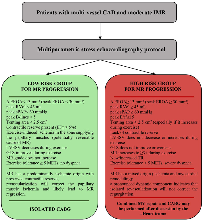
The role of stress echocardiography in decision-making regarding surgical strategy in patients with moderate ischemic mitral regurgitation.
Natriuretic peptides (brain-derived natriuretic peptide, BNP) and their N-terminal fragment (NT-proBNP) are recognized as markers of myocardial stress and neurohormonal activation, reflecting the degree of volume overload and LV dysfunction. In mitral regurgitation, natriuretic peptide levels correlate with the severity of valvular disease, the degree of cardiac chamber remodeling, and the functional class of heart failure [49, 50]. In the case of moderate MR, the BNP/NT-proBNP determination method is of particular importance as a tool for additional risk stratification in the ‘gray area’, where isolated echocardiographic data do not allow for a clear determination of treatment strategy. Studies have shown that elevated BNP levels in patients with functional MR are an independent predictor of adverse clinical outcomes, including mortality and hospitalization for decompensated heart failure, and their prognostic value is maintained after adjustment for key clinical and echocardiographic parameters [51, 52]. Dynamic monitoring of natriuretic peptide levels allows for the assessment of the progression of hemodynamic deterioration and the effectiveness of therapy. The 2025 ESC/EACTS guidelines recommend measuring natriuretic peptides as part of a comprehensive assessment of patients with valvular heart disease, including identifying subclinical decompensation and determining the optimal timing for intervention [10].
Three-dimensional echocardiography (3D echocardiography) has significantly expanded the capabilities of assessing IMR, overcoming several limitations of two-dimensional methods. It allows for the accurate assessment of the annular geometry (area, perimeter, and saddle shape), leaflet tent area, and tent volume, which are key parameters for determining the mechanism and severity of secondary MR [53]. A fundamental advantage of 3D echocardiography is the ability to perform direct planimetry of the vena contracta area, which is particularly important in IMR characterized by an elliptical shape of the regurgitant orifice, which is systematically underestimated by single-planar measurements [54]. Multiplanar reconstruction of three-dimensional data sets provides a more accurate calculation of the VC area and improves the reproducibility of quantitative assessment [55].
Cardiac magnetic resonance imaging (CMR) with delayed contrast is the reference method for assessing LV volumes and function, allowing accurate calculation of regurgitant volume using a volumetric approach independent of Doppler assumptions [56]. This approach is particularly valuable when echocardiographic findings are inconsistent. In addition to quantitative characterization of regurgitation, MRI provides unique information on myocardial viability: late gadolinium enhancement allows assessment of the location and transmurality of scar tissue, as well as the presence and extent of fibrosis of the papillary muscles and adjacent segments [57]. The volume and nature of fibrosis are independent predictors of the reversibility of the LV remodeling after revascularization and the likelihood of a reduction in IMR, which is of direct importance for the choice of surgical strategy [13].
It is important to note that both 3D echocardiography and MRI are performed exclusively at rest, which represents a significant limitation in the context of IMR. Despite their apparent accuracy and excellent spatial resolution, resting assessments may not reflect the true hemodynamic burden of regurgitation because of the inherent dynamic nature of IMR. The severity of IMR can vary significantly depending on changes in loading conditions, heart rate, and exercise-induced myocardial ischemia. Consequently, even the most sophisticated resting imaging techniques can underestimate or, less commonly, overestimate the functional significance of regurgitation, potentially leading to suboptimal therapeutic decisions. This highlights the additional role of stress echocardiography, which remains the only widely available imaging modality capable of capturing the dynamic behavior of MR under physiological conditions.
These diagnostic approaches, taken together, not only provide a more complete understanding of the mechanisms and severity of IMR but also a basis for translating imaging findings into clinical management. This is particularly relevant for moderate and borderline IMR, where a resting assessment alone is often insufficient to determine the optimal strategy. The combination of stress echocardiography, advanced structural imaging, and markers of myocardial dysfunction helps identify patients whose mitral regurgitation is likely to regress after optimization of medical therapy or revascularization alone, as well as those whose persistent or progressive regurgitation is likely to require additional intervention. Accordingly, the discussion of diagnostic methods naturally leads to the question of treatment choice, where the difficulty lies in balancing the potential benefits of medical, surgical, and transcatheter approaches against the risks associated with each treatment option.
Medical therapy in accordance with clinical guidelines (guideline-directed medical therapy, GDMT) is an integral component of the management of patients with IMR, regardless of the chosen strategy, whether it is conservative or surgical. The pathogenetic rationale for pharmacotherapy is determined by its effect on the key mechanisms of IMR progression: neurohormonal activation, LV remodeling, increased afterload, and volume overload. Traditional components of heart failure therapy, such as angiotensin-converting enzyme inhibitors (ACE inhibitors), angiotensin II receptor blockers (ARBs), beta-blockers, and mineralocorticoid receptor antagonists (MRAs), have demonstrated the ability to reduce the LV volume, decrease the degree of mitral regurgitation, and improve prognosis for this category of patients [58].
Impact of new classes of drugs on the course of functional MR is of particular interest. Sacubitril/Valsartan as a neprilysin inhibitor in combination with an ARB demonstrated superiority over enalapril in reducing mortality and hospitalizations in patients with heart failure with reduced ejection fraction in the PARADIGM-HF trial [59]. Subsequent studies have shown that Sacubitril/Valsartan therapy is associated with more pronounced LV reverse remodeling, reduction in chamber volumes and leaflet tethering area, accompanied by a significant reduction in the degree of functional MR compared with traditional inhibitors [60, 61]. The mechanism of the additional effect of Sacubitril/Valsartan on mitral regurgitation is associated with potentiation of the natriuretic system, enhanced vasodilation, antifibrotic effects, and a reduction in both preload and afterload.
Sodium-glucose cotransporter 2 inhibitors (SGLT2 inhibitors, dapagliflozin and empagliflozin) represent another promising class of drugs. The DAPA-HF and EMPEROR-reduced trials convincingly confirmed their efficacy in reducing cardiovascular mortality and heart failure decompensation [62, 63]. Several subanalyses and observational studies suggest a beneficial effect of SGLT2 inhibitors on the degree of functional MR, attributed to preload reduction, anti-inflammatory and antifibrotic effects, and improved cardiomyocyte energy metabolism [64]. However, direct evidence on the effect of SGLT2 inhibitors on the severity of MR and associated outcomes remains limited, and this issue requires further study in dedicated prospective studies.
Thus, modern GDMT, including Sacubitril/Valsartan and SGLT2 inhibitors along with traditional neurohormonal modulators, can alter the course of moderate/borderline IMR by reducing the severity of regurgitation and slowing remodeling. This highlights the need to optimize medical therapy before deciding on surgical or transcatheter intervention. Figure 3 presents a comprehensive decision-making algorithm illustrating the Heart Team’s multidisciplinary approach and providing practical value for clinicians using this complex scheme. This flowchart clearly outlines the diagnostic and therapeutic cascade after GDMT optimization. Specifically, it identifies clinical triggers, such as persistent symptoms during exercise out of proportion to resting echocardiographic findings, conflicting severity assessments, or the need for myocardial viability assessment, that require a shift from standard resting assessment methods to advanced imaging modalities such as stress echocardiography or cardiac MRI. By integrating clinical status, dynamic imaging data, and GDMT response, this algorithm guides the subsequent selection of the most appropriate surgical or transcatheter strategy [10, 11].
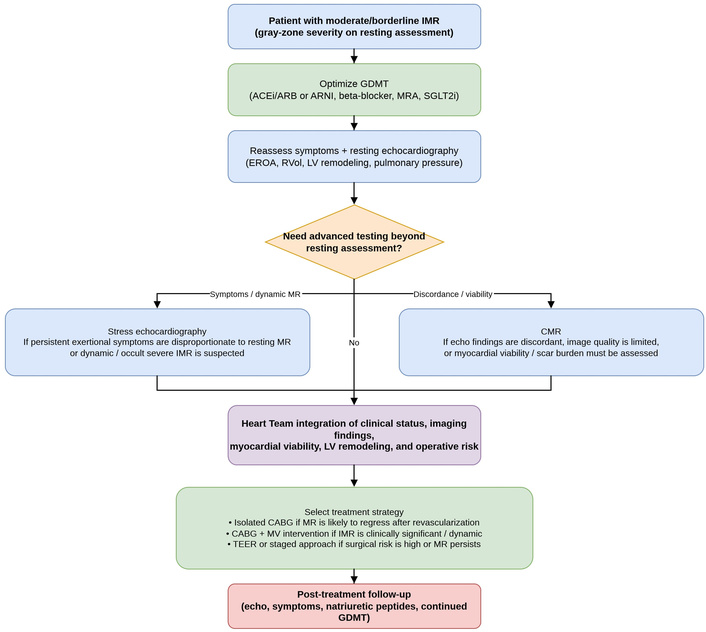
Simplified multidisciplinary Heart Team decision-making algorithm for the diagnostic evaluation and management of patients with moderate/borderline ischemic mitral regurgitation.
For moderate/borderline IMR, two main strategies are used: isolated CABG and CABG combined with mitral valve intervention. In patients with adequate distal coronary flow, viable myocardium, and a high probability of complete myocardial revascularization, isolated CABG is appropriate in most patients with moderate IMR to avoid the risks associated with additional valve intervention (increased cardiopulmonary bypass time, aortic cross-clamping time, opening of cardiac chambers, increased risk of thromboembolic complications, etc.).
In the presence of significant dilation of the fibrous ring, elongation of the papillary muscles, or excess posterior leaflet tissue, valve-preserving reconstructive surgeries are preferable (targeting the leaflets, fibrous ring, or chordopapillary apparatus). Replacement of the mitral valve is indicated in cases of severe limitation of leaflet motion due to extensive scarring of the ventricular walls, significant LV dilation, or when valve-preserving correction is not feasible [10, 11].
There are numerous methods for valve and subvalvular repair (approximation of papillary muscles, chordal resection or neochordal implantation, and mitral valve leaflet resection). However, annuloplasty ring implantation remains the primary method for correcting IMR. Isolated annular dilation (Carpentier type I) utilizes rigid rings with a diameter significantly smaller than the dilated ring, providing effective correction of IMR. In case of symmetrical displacement of the papillary muscles with central regurgitation (symmetrical type IIIb), saddle rings are used, which reduce the anteroposterior size of the ring and bring the P2 segment closer to the anterior leaflet without the need for excessive reduction of the ring size [65, 66]. In case of asymmetrical displacement of the papillary muscles, rings with an asymmetrical deepening in the P2–P3 segment are recommended, improving coaptation [67].
A serious problem is the development of functional mitral stenosis after annuloplasty, which may result from a reduction in the size of the mitral annulus by one or more sizes [68, 69]. However, in several patients who have undergone surgery for IMR, persistence or recurrence of MR is observed, occurring both after isolated CABG and after correct implantation of an annuloplasty ring [70, 71].
The advisability of simultaneous mitral valve correction during CABG in patients with moderate IMR remains one of the most controversial issues in cardiac surgery. The key evidence-based study was the randomized multicenter CTSN trial, in which 301 patients with moderate IMR were randomized to isolated CABG or CABG combined with mitral valve annuloplasty [9]. At 12-month follow-up, the combined intervention resulted in a significantly greater reduction in MR; however, it was not accompanied by improvement in LV reverse remodeling and did not reduce the rate of major adverse cardiovascular events, while increasing cardiopulmonary bypass time and perioperative risks [9]. Results of 2-year follow-up confirmed no difference in survival or readmission rates between the groups, although persistent moderate or severe MR was more common in the isolated CABG group [9].
Similar findings have been obtained in a few observational studies and meta-analyses, demonstrating that the addition of mitral valve annuloplasty to CABG reduces the degree of MR but does not improve mid-term survival [72, 73]. These data formed the basis for the ACC/AHA (2020) and ESC/EACTS (2025) recommendations, which allow simultaneous mitral valve correction in moderate IMR with evidence class IIb, emphasizing the need for an individual approach by a multidisciplinary cardiac team considering the clinical context, anatomical features, and surgical risk [10, 11].
Transcatheter ‘edge-to-edge’ repair (TEER) has become a key minimally invasive option for patients with secondary MR. The seminal COAPT and MITRA-FR trials, both of which included a significant proportion of patients with ischemic etiology, provided critical, yet seemingly contradictory, data on the effectiveness of the transcatheter approach. The COAPT trial demonstrated that in patients with chronic heart failure, reduced LVEF and severe secondary MR refractory to optimal medical therapy, TEER significantly reduces the incidence of hospitalization for heart failure and improves survival and quality of life [46]. However, the MITRA-FR trial did not demonstrate similar benefits [74].
This discrepancy is largely explained by the conceptual basis of proportional and disproportionate MR. Patients in the MITRA-FR study had more significantly dilated LVs with relatively less severe MR (proportionate MR), meaning that their poor prognosis was primarily determined by severe underlying myocardial disease rather than valve defect. Conversely, patients in the COAPT study had less pronounced LV dilation but more severe, hemodynamically significant regurgitation (disproportionate MR), where MR itself was the primary driver of heart failure progression. Thus, valve correction provided significant benefit only in the latter group.
As emphasized in the 2025 ESC/EACTS guidelines, the discrepancy in the results of these studies underscores the critical importance of patient selection, and TEER is indicated for patients with a specific clinical and echocardiographic profile (a “COAPT-like” profile). For patients who do not meet these strict criteria—particularly those with severe ischemic cardiomyopathy and extreme LV remodeling—a strictly individualized approach is required. In such cases, the decision should be made by a multidisciplinary cardiac team on an individual basis, carefully weighing technical feasibility against the risk of clinical futility [10].
Recurrent MR after CABG is a significant clinical challenge given the high risk of reoperation. The 2025 ESC/EACTS guidelines recommend TEER (Class I) in symptomatic patients with severe secondary MR who are not suitable for surgical treatment to reduce hospitalization rates and improve quality of life [10]. Although there are no direct recommendations for the routine use of TEER specifically for recurrent MR after CABG, general principles for the management of high-risk patients with severe secondary MR support this strategy [10, 40].
Since MR improves after isolated revascularization in only a third of patients, a staged approach is becoming increasingly important. The 2025 ESC/EACTS guidelines allow for PCI followed by reassessment of MR severity and possible TEER. This strategy avoids unnecessary valve intervention when MR regresses and offers a minimally invasive solution when it persists, minimizing the risks of extensive single-stage surgery [10].
Isolated CABG is the least invasive option, providing revascularization of viable myocardium with the potential for reverse LV remodeling and spontaneous reduction in the degree of IMR [75]. Advantages include shorter cardiopulmonary bypass time, lower perioperative mortality, and the absence of risks associated with valve intervention. However, according to several observational studies, IMR progression occurs in 30–40% of patients postoperatively, which is associated with a worse long-term prognosis [8, 76].
CABG combined with mitral valve annuloplasty theoretically addresses both the cause (ischemia) and the consequence (regurgitation). A randomized trial conducted by CTSN demonstrated that the addition of restrictive annuloplasty to CABG for moderate mitral regurgitation significantly reduced the degree of regurgitation compared with CABG alone. However, at 2-year follow-up, no differences were found in the primary endpoint (LV end-systolic volume index), mortality, or serious adverse event rates, and a trend toward a higher number of neurological complications was noted in the annuloplasty group [9, 77]. An important limitation remains the high rate of recurrence of IMR in the combined intervention group (up to 11.3% after 2 years), especially with pronounced sailing of the valves [9].
CABG with mitral valve replacement provides radical elimination of regurgitation; however, it is associated with higher operative mortality, the need for anticoagulant therapy (with mechanical prostheses), the risk of prosthesis-related complications, and deterioration of LV function due to disruption of the subvalvular apparatus. This approach is not supported by current guidelines for moderate mitral regurgitation and is used only in cases where repair is technically infeasible [10, 11].
CABG with staged TEER represents a promising staged strategy: revascularization is performed as a first step, then the dynamics of IMR during the reverse remodeling process is assessed, and TEER is performed only in cases of persistent significant regurgitation. This approach avoids unnecessary valve intervention in patients with the potential for spontaneous improvement; however, it carries the risk of MR progression during the waiting period and the need for a repeat procedure. This strategy was formally mentioned for the first time in the updated 2025 ESC/EACTS guidelines [10].
PCI followed by TEER is a minimally invasive strategy potentially suitable for patients at high surgical risk and uncomplicated CAD. The 2025 ESC/EACTS guidelines indicate that PCI followed by TEER after MR reassessment may be considered in symptomatic patients with chronic severe secondary MR (class IIb) [10]. Advantages include avoidance of median sternotomy and cardiopulmonary bypass; however, data from large randomized trials on this combined strategy in moderate MR are currently insufficient.
Thus, no existing approach has demonstrated definitive benefit in moderate/borderline IMR. The decision should be made by a multidisciplinary cardiac team, considering the degree of ischemia, myocardial viability, the severity of LV remodeling, valvular anatomy, and the patient’s comorbidity profile. Clinical guideline positions regarding diagnosis, guideline-based medical therapy, and surgical strategy for moderate/borderline IMR are presented in Table 2.
Comparison of key guidelines for management of moderate/borderline ischemic mitral regurgitation (ESC/EACTS 2025 vs ACC/AHA 2020).
| Aspect | ESC/EACTS 2025 | ACC/AHA 2020 |
|---|---|---|
| Diagnosis | ||
| TTE | IMR assessment should be performed after optimization of medical therapy and in a euvolemic and normotensive state | In patients with chronic secondary MR (stages B to D), TTE is useful to establish the etiology and to assess the extent of regional and global LV remodeling and systolic dysfunction, severity of MR, and magnitude of pulmonary hypertension |
| - | Class I, Level B-NR | |
| Other diagnostic methods | Cardiac magnetic resonance is used to confirm IMR severity and assess cardiac chamber function and dimensions, and to determine the extent of myocardial fibrosis. Owing to the dynamic nature of IMR, stress echocardiography may help to identify patients with severe IMR when values at rest are inconclusive | In patients with chronic secondary MR (stages B to D), noninvasive imaging (stress nuclear PET, CMR, or stress echocardiography), coronary CT angiography, or coronary arteriography is useful to establish etiology of MR and to assess myocardial viability |
| - | Class I, level C-EO | |
| Surgical correction | ||
| Moderate IMR during CABG | May be considered | May be reasonable |
| Class IIb, Level B | Class IIb, level B-R | |
| Preferred technique | Restrictive annuloplasty | Annuloplasty (chordal-sparing) |
| Class I, Level B | Class I, Level B-R | |
| Criteria for intervention | EROA ≥ 40 mm2, RVol ≥ 60 mL (acknowledges significance of EROA ≥ 30 mm2) + clinical/ECHO symptoms or LVESD ≤ 70 mm, LVEF 20–50% | EROA ≥ 40 mm2, RVol ≥ 60 mL, RF ≥ 50% |
| Transcatheter ‘edge-to-edge’ repair (TEER) | ||
| Indications for moderate/borderline IMR | Not recommended for isolated moderate IMR | No specific indications for moderate IMR |
| - | - | |
| Indications for severe secondary MR | Symptomatic severe secondary MR with impaired LVEF (< 50%), and persistent severe ventricular SMR, despite optimized GDMT and CRT, inoperable/high risk, COAPT criteria | Symptomatic severe secondary MR, high surgical risk, COAPT criteria |
| Class I, Level A | Class IIa, Level B-R | |
| Selection criteria | EROA ≥ 30 mm2, LVEF 20–50%, LVESD < 70 mm, optimal GDMT | EROA ≥ 40 mm2, LVEF 20–50%, LVESD < 70 mm, sPAP ≤ 70mmHg, COAPT eligibility |
| Medical therapy | ||
| Guideline-directed medical therapy (GDMT) for HF | Mandatory for all patients with secondary MR | Mandatory for all patients with secondary MR |
| Class I, Level A | Class I, Level A | |
| Components of therapy | ACEi/ARNI, BB, MRA, SGLT2i, diuretics | ACEi/ARNI, BB, MRA, SGLT2i, diuretics |
| Class I, Level A | Class I, Level A | |
| CRT if indicated | Recommended if criteria met (LBBB, QRS ≥ 150 ms, LVEF ≤ 35%) | Recommended if criteria met |
| Class I, Level A | Class I, Level A | |
| Role of medical therapy in moderate IMR | First-line treatment; may be sufficient if LV reverse remodeling occurs | First-line treatment; reassessment of MR before intervention |
| - | - | |
| Follow-up | ||
| Frequency | Echo every 3–6–12 months (according to the HF stage) or with symptom worsening | Echo every 6–12 months |
| - | - | |
IMR: ischemic mitral regurgitation; CABG: coronary artery bypass grafting; EROA: effective regurgitant orifice area; RVol: regurgitant volume; LV: left ventricle; LVEF: left ventricular ejection fraction; LVESD: left ventricular end-systolic diameter; GDMT: guideline-directed medical therapy; ACEi: ACE inhibitors; ARNI: angiotensin receptor-neprilysin inhibitor; BB: beta-blockers; MRA: mineralocorticoid receptor antagonists; SGLT2i: SGLT2 inhibitors; CRT: cardiac resynchronization therapy; LBBB: left bundle branch block; HF: heart failure.
Moderate and borderline IMR is a clinically significant but diagnostically and therapeutically challenging problem. Accumulated data convincingly demonstrate that even moderate regurgitation of ischemic origin is associated with a significantly worse long-term prognosis, which can be explained by the compromised condition of the myocardium and its increased sensitivity to volume overload. The concept of “disproportionate” MR most accurately reflects this pathophysiology. Among diagnostic methods for IMR, stress echocardiography represents a particularly promising, yet currently underutilized, tool for risk stratification and individualized management strategies for patients with moderate IMR. Its ability to detect hemodynamically significant regurgitation, assess contractile reserve, and predict reverse remodeling after revascularization opens the possibility of more accurate candidate selection for combined interventions. However, the widespread adoption of this method is hampered by the lack of standardized stress echocardiography protocols for assessing dynamic IMR, including standardized examination protocols, assessment criteria, and threshold values for prognostically significant parameters. The development and validation of such protocols is a priority for the near future. Data from randomized clinical trials do not provide a definitive answer to the question of the benefit of adding mitral valve intervention to CABG for moderate IMR in terms of the most important endpoints—survival and cardiovascular event rate. The lack of proven benefit, coupled with the increased duration of cardiopulmonary bypass and the potential risks of additional interventions, creates an area of therapeutic uncertainty. This necessitates a thorough discussion of the optimal type and technique of mitral valve intervention. In the current situation, characterized by an incomplete evidence base and heterogeneity of clinical manifestations, an individualized approach based on multimodal imaging results and discussion by a multidisciplinary Heart Team represents the optimal strategy at this time. The decision regarding combined correction of moderate IMR in a patient undergoing CABG should be based on a comprehensive assessment of the clinical status, echocardiographic parameters, stress echocardiographic data, valvular anatomical features, and surgical risk. There is a clear need for further research to resolve key uncertainties.
ARBs: angiotensin II receptor blockers
CABG: coronary artery bypass grafting
CAD: coronary artery disease
EROA: effective regurgitant orifice area
GDMT: guideline-directed medical therapy
IMR: ischemic mitral regurgitation
LV: left ventricle
PISA: proximal isovolumetric surface area
TEER: transcatheter ‘edge-to-edge’ repair
MR: Conceptualization, Writing—original draft. AZ: Conceptualization, Writing—review & editing. MK: Writing—original draft. EK: Writing—original draft. NL: Writing—original draft. DS: Writing—review & editing, Supervision. All authors read and approved the submitted version.
The authors declare that they have no conflicts of interest.
Ethics approval was waived by the Institutional Review Board of St. Petersburg State University, Saint Petersburg, Russian Federation, as this study is a literature review involving no experimental procedures on patients.
Figure 1 is taken from clinical practice; patient consent was obtained.
The unpublished clinical images included in this article were obtained from patients treated at St. Petersburg State University, Saint Petersburg, Russian Federation. Written informed consent for the publication of these images was obtained from all patients. All images were anonymized prior to publication.
Not applicable.
Not applicable.
© The Author(s) 2026.
Open Exploration maintains a neutral stance on jurisdictional claims in published institutional affiliations and maps. All opinions expressed in this article are the personal views of the author(s) and do not represent the stance of the editorial team or the publisher.
Copyright: © The Author(s) 2026. This is an Open Access article licensed under a Creative Commons Attribution 4.0 International License (https://creativecommons.org/licenses/by/4.0/), which permits unrestricted use, sharing, adaptation, distribution and reproduction in any medium or format, for any purpose, even commercially, as long as you give appropriate credit to the original author(s) and the source, provide a link to the Creative Commons license, and indicate if changes were made.



View: 971
Download: 12
Times Cited: 0


