Review


Affiliation:
1Departmental Unit of Allergology & Respiratory Diseases, Severe Asthma Center, Fondazione Poliambulanza Istituto Ospedaliero, 25122 Brescia, Italy
2Italian Society of Allergy, Asthma and Clinical Immunology (SIAAIC), Italy
Email: lombardicarlo@libero.it
ORCID: https://orcid.org/0000-0002-8040-0324


 ,
Affiliation:
3Allergy and Pneumology Outpatient Clinic, 24121 Bergamo, Italy
ORCID: https://orcid.org/0000-0003-2767-6767
,
Affiliation:
4Pulmonology Unit, S. Valentino Hospital, AULSS2 Marca Trevigiana, 31044 Montebelluna, Italy
ORCID: https://orcid.org/0000-0003-3950-5789
Explor Asthma Allergy. 2026;4:1009138 DOI: https://doi.org/10.37349/eaa.2026.1009138
Received: December 30, 2025 Accepted: September 01, 2026 Published: November 02, 2026
Academic Editor: Garry M. Walsh, University of Aberdeen, U.K.
The article belongs to the special issue The Complex Interactions Between Lifestyles and Asthma
The asthma-obstructive sleep apnea (OSA) overlap is a clinically important phenotype, especially in patients with severe or difficult-to-control asthma. Rather than a simple coexistence of independent conditions, this overlap represents a distinct clinical entity modulated by shared pathobiological pathways. This review integrates epidemiologic data, critically balancing association and causation across shared risk factors (obesity, GERD, upper airway disease). We construct an integrated mechanistic framework encompassing mechanical/neuroreflex interactions, intermittent hypoxia–driven inflammation/oxidative stress, and metabolic dysfunction. The overlap is associated with poorer asthma control, increased nocturnal symptoms, reduced quality of life, and higher healthcare utilization. Current evidence is largely observational; randomized controlled trials with respiratory endpoints and prospective cohorts with repeated polysomnography and biomarker panels are needed to establish strict bidirectional causality and identify responders to targeted therapies. A multidisciplinary, treatable-traits approach addressing OSA, obesity, rhinitis, GERD, and inhaler optimization is recommended while awaiting mechanistic and interventional data on CPAP, weight-loss therapies, and biologics.
Asthma is a heterogeneous chronic airway disorder affecting over 300 million people worldwide, characterized by airway inflammation, variable airflow limitation, and symptoms that often worsen at night [1]. Obstructive sleep apnea (OSA) is a sleep-related breathing disorder defined by recurrent upper-airway collapse during sleep, leading to intermittent hypoxia, sleep fragmentation, and excessive daytime sleepiness [2]. The “asthma–OSA overlap” has emerged as a clinically relevant phenotype, particularly among patients with severe or difficult-to-control asthma [3]. To move beyond mere descriptive epidemiology, this review establishes an original conceptual framework that bridges the gap between raw statistical correlation and definitive causation. By synthesizing epidemiologic associations, plausible mechanistic links, clinical consequences, and management strategies, we explicitly grade the current literature (preclinical vs. observational vs. interventional) to expose gaps in bidirectional evidence and prevent clinical over-extrapolation.
Observational studies and meta-analyses report an increased risk of OSA among people with asthma, with a prevalence ranging from 20% to > 60%, depending heavily on asthma severity, diagnostic criteria, and study setting [3–9] (Table 1). However, a critical methodological caveat must be raised: much of the existing large-scale data relies on screening questionnaires (e.g., STOP-Bang) [10] or retrospective international classification of diseases (ICD) administrative billing codes. These methods carry inherent selection and misclassification biases that inherently obscure causal directionality.
Selected key studies evaluating the association between asthma and OSA.
| Author (Year) (Ref.) | Study design | Population & sample size (N) | OSA diagnostic method | Main visualized & clinical outcomes | Quality & methodological limitations |
|---|---|---|---|---|---|
| Julien et al. (2009) [5] | Case-control | 78 adult patients | Full in-laboratory PSG | Higher prevalence and severity of OSA in severe asthma vs. controls. | High diagnostic accuracy; small sample size; single-center setting. |
| Teodorescu et al. (2010) [7] | Cross-sectional | 472 adult asthma patients | Validated risk questionnaires | OSA risk significantly associated with poor asthma control scores. | High clinical utility; subject to recall bias; lacks objective PSG confirmation. |
| Teodorescu et al. (2015) [4] | Population-based cohort | 547 adult patients (longitudinal) | Serial full PSG over time | Baseline asthma significantly predicts incident OSA development. | Strong longitudinal design; well-controlled for major baseline confounders. |
| Kauppi et al. (2016) [6] | Longitudinal follow-up | 197 asthma patients | Clinical tracking and PSG | Baseline asthma severity correlates with accelerated OSA development. | Excellent real-world data; limited by varying follow-up intervals. |
| Kong et al. (2017) [3] | Systematic review & meta-analysis | 7,675 adult patients | Mixed (PSG and questionnaires) | Confirmed that asthma is associated with a significantly increased risk of OSA. | High statistical power; significant heterogeneity across pooled studies. |
| Lafond et al. (2007) [55] | Interventional trial | 20 comorbid patients | PSG pre- and post-treatment | CPAP therapy significantly improves asthma symptoms and quality of life. | Objective proof of concept; unblinded; lacks a placebo/sham-CPAP control arm. |
OSA: obstructive sleep apnea; PSG: polysomnography; CPAP: continuous positive airway pressure.
While longitudinal cohort data—such as the Wisconsin Sleep Cohort—demonstrate that asthma significantly predicts incident OSA over time [4], the reverse timeline (OSA driving incident asthma) remains epidemiologically less robust and relies heavily on unadjusted confounding factors like progressive weight gain [9]. Robust prospective cohorts utilizing baseline and serial gold-standard polysomnography (PSG) combined with standardized, objective asthma phenotyping are fundamentally required to transform these observed associations into confirmed chronological vectors [11, 12].
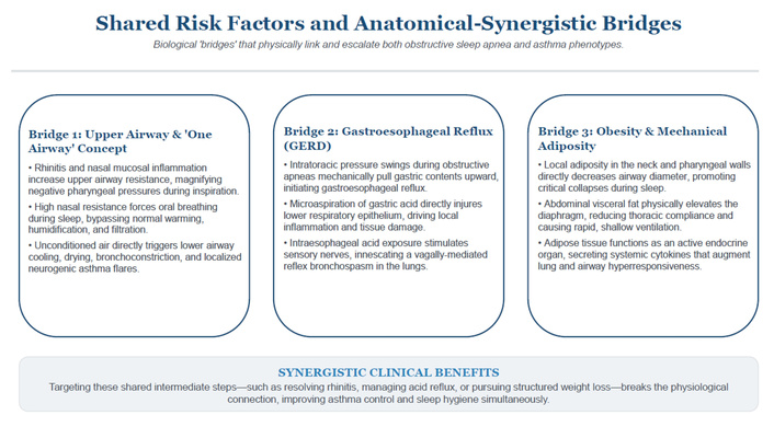
Shared modifiable risk factors and comorbidities linking asthma and OSA are summarised in Table 2, with the mechanistic bridges illustrated in Figure 1.
Asthma-OSA overlap: shared risk factors and comorbidities.
| Modifiable treatable trait | Objective clinical assessment & clues | Recommended multidisciplinary actions |
|---|---|---|
| Obesity & metabolic dysfunction | BMI ≥ 30 kg/m2, elevated waist/neck circumference, dysregulated fasting metabolic panel. | Implement structured lifestyle and dietary modifications; refer to specialized weight-management programs; consider GLP-1/dual agonists based on obesity guidelines; serially re-evaluate AHI and asthma control scores following weight reduction. |
| Obstructive sleep apnea (OSA) | STOP-Bang score ≥ 3, elevated Epworth Sleepiness Scale (ESS ≥ 10), frequent witnessed apneas, loud snoring. | Perform diagnostic Home Sleep Testing (HST) or gold-standard laboratory PSG; initiate targeted therapy (CPAP titration, or customized oral appliances for mild/moderate cases); reassess objective asthma control post-initiation. |
| Upper airway disease & rhinitis | Persistent nasal congestion, rhinorrhea, post-nasal drip, structural ENT examination. | Treat underlying allergic/non-allergic rhinitis aggressively using intranasal corticosteroids and non-sedating antihistamines; refer to ENT for corrective structural evaluation if anatomical obstruction is present. |
| Gastroesophageal reflux disease (GERD) | Frequent nocturnal heartburn, acid regurgitation, chronic laryngopharyngeal symptoms. | Optimize behavioral anti-reflux habits (head-of-bed elevation, early dinners); initiate targeted proton pump inhibitor (PPI) therapy where clinically indicated; consider gastroenterology referral for refractory cases. |
| Poor asthma control & non-adherence | Depressed ACT/ACQ scores, poor inhaler technique observation, irregular prescription refill data. | Formally optimize inhaled pharmacological therapy; comprehensively evaluate and correct inhaler technique and medication adherence; consider step-up therapy or targeted biologics if indicated. |
| Corticosteroid over-exposure | Documentation of frequent systemic rescue bursts or chronic maintenance oral steroid use. | Implement strict steroid-sparing clinical pathways; optimize advanced non-steroidal maintenance or biologic therapies; closely monitor for progressive steroid-induced weight gain and pharyngeal muscle myopathy. |
ACT: Asthma Control Test; ACQ: Asthma Control Questionnaire; BMI: body mass index; AHI: Apnea-Hypopnea Index.
Obesity represents the most dominant shared modifier [13]. Mechanically, excess adiposity reduces lung volumes (functional residual capacity) [14] and increases pharyngeal collapsibility by narrowing the upper airway lumen [15]. Metabolically, adipose tissue acts as an active endocrine organ, releasing systemic pro-inflammatory cytokines (such as TNF-α, IL-6, and leptin) [16] that can worsen underlying eosinophilic or neutrophilic bronchial inflammation and directly impair airway steroid responsiveness [13]. Crucially, obesity often acts as a major confounding variable in epidemiologic studies; its presence alone can artificially inflate the statistical association between asthma severity and OSA risk without a direct causal link between the two airway pathologies themselves.
GERD commonly coexists within this overlap [17]. During an obstructive apnea, the patient generates massive negative intrathoracic pressures to overcome the collapsed upper airway [18]. This mechanical pressure gradient acts as a vacuum, physically drawing gastric acid into the esophagus [19]. Once localized in the upper airway, microaspiration and vagally-mediated neuroreflexes can provoke immediate bronchoconstriction and increase airway edema, establishing a cyclical worsening of nocturnal asthma symptoms [20]. Nevertheless, large randomized trials of aggressive anti-reflux therapy (e.g., proton pump inhibitors) demonstrate highly heterogeneous effects on asthma outcomes, with clinical benefit strictly confined to patients reporting frequent, overt nighttime regurgitation [17], reinforcing that GERD is a potent symptom amplifier rather than a universal root cause.
Allergic rhinitis and chronic rhinosinusitis with or without nasal polyps increase upper airway resistance, forcing a shift to mouth breathing during sleep [21], which destabilizes the pharyngeal airway and predisposes to collapse [22]. Under the “one airway, one disease” unified concept, chronic upper airway inflammation directly communicates with the lower bronchial tree via systemic inflammatory signaling and nasobronchial neuroreflexes [23]. This shared tissue vulnerability means that uncontrolled nasal inflammation simultaneously increases pharyngeal collapsibility and worsens bronchial hyperresponsiveness, acting as a bidirectional clinical modifier.
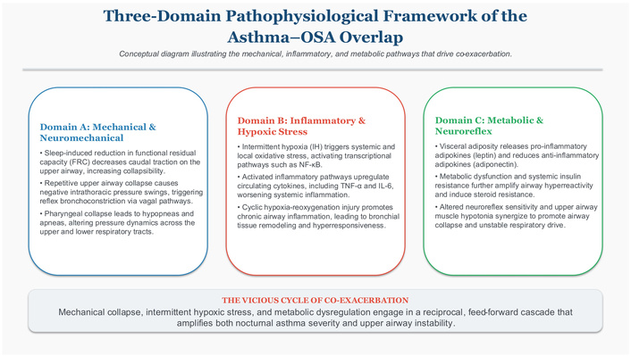
To address previous fragmentation in the literature, we propose a three-domain integrated model where mechanical, inflammatory/immunologic, and metabolic/neuroreflex pathways do not operate in isolation, but rather form an interlocking, self-amplifying pathological loop (Figure 2).
Recurrent upper-airway collapse during sleep produces large negative intrathoracic pressure swings [18]. These mechanical forces stimulate localized mechanoreceptors in the upper airway, triggering a systemic vagal neuroreflex that acutely increases lower airway bronchomotor tone and induces acute nocturnal bronchoconstriction [24]. Simultaneously, sleep-related reductions in lung volumes—markedly pronounced when asthma is complicated by obesity [14]—diminish the caudal longitudinal traction forces normally exerted on the trachea [25]. This loss of physical tension directly increases the anatomical collapsibility of the pharyngeal walls [26]. While these acute mechanical dynamics successfully explain why an asthmatic patient might suffer a sudden bronchospasm immediately following a prolonged apnea episode during sleep, they represent transient physiological interactions and are insufficient on their own to prove a permanent, population-level structural disease causality.
The defining feature of OSA—intermittent hypoxia with rapid reoxygenation cycles—acts as a potent trigger for systemic oxidative stress [27, 28]. Preclinical animal models demonstrate that chronic intermittent hypoxia selectively upregulates the inflammatory transcription factor NF-κB, leading to downstream systemic overproduction of classic pro-inflammatory cytokines, specifically TNF-α, IL-6, and vascular endothelial growth factor (VEGF) [29, 30].
In patients with the overlap phenotype, these circulating cytokines cross into the pulmonary vascular bed, where they actively synergize with local allergic Th2/Th17 tissue inflammation [31]. This process results in a shift toward a mixed neutrophilic airway profile, which is highly characteristic of severe, steroid-resistant asthma [32, 33]. Furthermore, repeated localized hypoxia-reoxygenation injuries within the upper airway induce structural pharyngeal remodeling and upper airway myopathy, rendering the tissues highly vulnerable to further collapse [34]. Over time, this chronic low-grade tissue injury promotes systemic endothelial dysfunction and accelerated structural airway remodeling, leading to fixed, irreversible airflow limitation [35]. It must be heavily emphasized, however, that current human clinical evidence supporting this inflammatory crossover remains almost entirely cross-sectional; high-quality interventional trials evaluating whether local bronchial tissue inflammation or bronchial hyperresponsiveness decreases following targeted OSA therapy remain strictly limited and inconclusive [36].
Metabolic dysfunction linked to obesity (insulin resistance, dysregulated adipokines like leptin and adiponectin) augments both systemic and local tissue-level inflammation, increasing susceptibility to severe asthma and high-grade OSA [37, 38]. Neuroreflex pathways connecting upper airway sensory stimulation and lower airway tone may be modulated or hyperexcited by these metabolic and inflammatory states [24]. Weight-loss pharmacotherapies, such as glucagon-like peptide-1 (GLP-1) receptor agonists and dual GIP/GLP-1 receptor agonists, produce significant weight reduction and have been associated with improved OSA indices (e.g., reductions in the Apnea-Hypopnea Index, AHI) in large clinical populations [39]. However, evidence demonstrating a consistent, direct improvement in underlying asthma pathophysiological markers independent of physical weight loss remains highly preliminary and unconfirmed [40].
The asthma-OSA overlap syndrome produces a severe clinical burden, characterized by significantly poorer asthma control, higher rates of frequent asthma exacerbations, heightened nocturnal awakenings, worse health-related quality of life, and substantially elevated rates of emergency department visits and hospitalizations [41–44]. A critical challenge in daily clinical practice is the profound symptomatic overlap between the two disorders, which frequently leads to the underrecognition and underdiagnosis of OSA in asthmatic cohorts. Symptoms such as profound morning fatigue, unrefreshing sleep, daytime sleepiness, and impaired cognitive concentration are frequently misattributed by both clinicians and patients to poorly controlled asthma or the side effects of high-dose respiratory medications [45]. Conversely, frequent nocturnal asthma awakenings can mask underlying micro-arousals caused by repetitive upper airway collapses [46]. To systematically address this diagnostic blind spot, clinicians should maintain a highly vigilant, low threshold for comprehensive sleep evaluation. Validated screening tools, such as the STOP-Bang questionnaire [10], should be routinely integrated into the standard care workflow for any patient presenting with difficult-to-treat or severe asthma, particularly those with prominent nocturnal symptoms or persistent daytime impairment.
When managing the overlap phenotype, clinicians must carefully consider how standard asthma medications interact with underlying upper airway stability. Prolonged exposure to systemic corticosteroids and high-dose inhaled corticosteroids (ICS) is associated with an elevated risk and increased severity of OSA [47, 48]. The primary proposed mechanisms driving this risk include steroid-induced localized fat deposition in the pharyngeal tissues, localized myopathy of the upper airway dilator muscles, and altered pharyngeal sensory function [47]. However, a major confounding by indication exists: patients requiring high-dose steroids inherently possess more severe, poorly controlled asthma, which independently impairs sleep architecture and upper airway patency [49]. Biologic therapies targeting Type 2 (T2) high inflammation (e.g., anti-IgE, anti-IL5/R, anti-IL4/Rα) have transformed the management of severe asthma, significantly reducing exacerbation rates and systemic steroid requirements [50]. While these agents, such as dupilumab, frequently improve subjective sleep quality by eliminating nocturnal asthma symptoms [51], robust clinical evidence proving a direct, independent reduction in objective OSA severity parameters (such as AHI) remains limited [52].
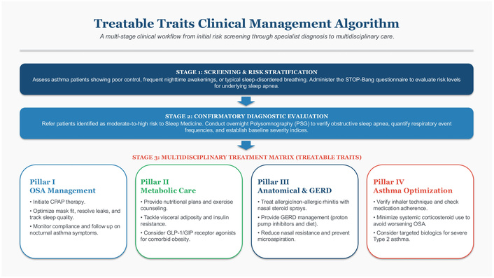
Optimizing outcomes in the asthma-OSA overlap requires a coordinated, dual treatment approach that simultaneously targets the unique requirements of both underlying respiratory diseases while aggressively managing shared treatable traits (Table 3; Figure 3).
OSA: a pragmatic clinical algorithm.
| Clinical phase | Key indicators & validated tools | Recommended clinical action | Measurable endpoints & outcomes |
|---|---|---|---|
| 1. Screening | STOP-Bang, Epworth Sleepiness Scale (ESS), clinical history of snoring/apneas. | Refer the patient to diagnostic sleep testing if STOP-Bang ≥ 3, ESS ≥ 10, or if high clinical suspicion exists. | Successful completion and reporting of diagnostic sleep testing (HST or PSG). |
| 2. Diagnosis | Objective Home Sleep Test (HST) or in-laboratory polysomnography (PSG). | Categorize objective OSA severity based on the Apnea-Hypopnea Index (AHI); record oxygen desaturation index (ODI) and nadir SpO2. | Confirmed baseline values for AHI, ODI, and nadir SpO2. |
| 3. Treatment | Baseline OSA severity index, symptom burden, and documented comorbidities. | Initiate auto-adjusting or fixed continuous positive airway pressure (CPAP) therapy; treat concurrent rhinitis/ENT issues; optimize anti-reflux measures; activate a structured weight-loss pathway. | Verified objective CPAP adherence metrics; documented changes in ACT/ACQ scores; percentage of total body weight lost; reduction in AHI. |
| 4. Follow-up | Clinical re-evaluation at 3–6 months post-treatment initiation. | Formally reassess objective asthma control metrics (ACT/ACQ); consider repeating HST if a significant clinical response or major weight shift occurs. | Stabilization or improvement in asthma control scores; objective resolution of sleep-disordered breathing. |
ACT: Asthma Control Test; ACQ: Asthma Control Questionnaire; OSA: obstructive sleep apnea.
CPAP remains the gold-standard cornerstone therapy for moderate-to-severe OSA [53]. Small interventional trials and observational studies report that consistent CPAP adherence yields significant improvements in subjective asthma control, decreases nocturnal bronchoconstriction, and enhances overall quality of life [54, 55]. Physiologically, CPAP works by pneumatic splinting of the upper airway, which immediately abolishes the large negative intrathoracic pressure swings and effectively halts the downstream vagally-mediated reflex yesbronchoconstriction [54]. Concurrently, standard pharmacological asthma therapy must be systematically optimized. This includes rigorous objective verification of proper inhaler technique, strict electronic or pharmacy-refill tracking of medication adherence, and appropriate step-up titration of inhaled therapies or timely initiation of targeted T2 biologics when indicated to minimize the need for systemic rescue corticosteroids [1, 50].
Given the powerful role of obesity as a shared inflammatory and mechanical driver, structured lifestyle modifications and weight-management programs are central to long-term care [56]. Large-scale clinical evaluations of metabolic therapies, such as the GLP-1 receptor agonist semaglutide and the dual GIP/GLP-1 receptor agonist tirzepatide, demonstrate highly sustained weight reduction alongside clinically significant decreases in AHI and improved systemic metabolic parameters in obese cohorts [39, 57]. The observed respiratory benefits of these metabolic agents predominantly reflect profound physical weight loss, the depletion of compressive mechanical neck adiposity, and a secondary reduction in generalized systemic adipose tissue inflammation [56, 57]. Therefore, clinicians must strictly avoid overstating these findings as a direct “cure” or direct anti-inflammatory treatment for intrinsic bronchial asthma disease itself until dedicated, multi-center randomized controlled trials with primary objective respiratory endpoints (e.g., airway hyperresponsiveness, lung function, exacerbation rates) are completed [40].
To transition the field from association to definitive precision medicine, the following five research priorities are established:
Mechanistically-informed prospective cohorts: Longitudinal studies tracking patients with severe asthma using baseline and serial objective PSG, coupled with deep molecular phenotyping and inflammatory biomarker panels (e.g., systemic and induced sputum cytokines) to chart the exact temporal and causal relationships governing the overlap phenotype over time.
Powered interventional RCTs: Multi-center randomized controlled trials evaluating the long-term efficacy of optimized CPAP therapy vs. sham-CPAP, as well as structured bariatric/lifestyle weight-loss interventions, with primary endpoints focused on objective asthma outcomes, including validated exacerbation rates, spirometric lung function metrics (FEV1), and direct measurements of airway tissue inflammation.
Targeted metabolic RCTs: Dedicated clinical trials investigating the effects of GLP-1 receptor agonists and dual GIP/GLP-1 receptor agonists in obese asthmatic cohorts, utilizing pre-specified, rigorous respiratory endpoints to isolate metabolic drug actions from simple physical weight-loss effects [40].
Endotype and biomarker validation: Discovery and validation of specific serum, sputum, or volatile organic compound biomarkers capable of reliably identifying the precise sub-phenotypes within the asthma–OSA overlap that are highly likely to achieve complete clinical response to targeted upper airway therapies [31].
Advanced digital integration: Development and clinical validation of unobtrusive, home-based sleep monitoring technologies integrated with machine-learning algorithms to facilitate early, large-scale screening and automated phenotypic classification of sleep-disordered breathing directly within respiratory clinics [58].
The asthma-OSA overlap constitutes a clinically important, multifactorial phenotype with clear consequences for disease control, exacerbation risk, and health-care utilization [3, 59]. Current evidence supports a model in which mechanical (upper-airway collapse and intrathoracic pressure swings), inflammatory (intermittent hypoxia–driven systemic and airway inflammation), and metabolic/neuroreflex (obesity-related adipokine and insulin-resistance pathways) mechanisms interact to amplify both nocturnal respiratory instability and lower-airway disease activity. Furthermore, a recent study unveils shared genetic mechanisms associated with OSA and asthma risks, highlighting complex interactions between pathways governing immune response and inflammation [60]. However, the preponderance of cross-sectional and observational data, often derived from imperfect screening tools or administrative sources, precludes definitive causal inferences at the population level. Clinically, the overlap should be approached as a treatable, multidimensional syndrome. Routine, low-threshold screening for sleep-disordered breathing in patients with difficult-to-control or severe asthma is warranted (e.g., STOP-Bang, ESS), followed by objective diagnostic testing (PSG) when indicated. Management must be integrated and two-pronged: (1) targeted therapy for OSA (optimised CPAP or alternative device therapy, ENT management where appropriate) to remove the mechanical trigger of nocturnal bronchospasm and intermittent hypoxia; and (2) rigorous optimization of asthma care (inhaler technique and adherence, steroidsparing strategies including biologics where appropriate, and management of comorbidities). Parallel attention to shared, modifiable drivers—most notably obesity and GERD—should be standard practice, and metabolic interventions (including structured weight-loss programmes and GLP-1-based therapies) are promising adjuncts that require respiratory-focused randomized evaluation. Research priorities must shift from association mapping to mechanism-driven trials and longitudinal phenotyping. Highquality prospective cohorts that combine serial gold-standard sleep testing with deep molecular airway profiling, and adequately powered interventional RCTs (including CPAP vs. sham, bariatric/metabolic therapy trials, and mechanistic studies of biologics on objective sleep and airway endpoints) are essential to establish causality and to identify the subgroups most likely to benefit from specific interventions. Finally, integration of novel home-based sleep monitoring and digital phenotyping will be pivotal to scale screening and personalise care in real-world settings.
In sum, acknowledging and actively managing the asthma-OSA overlap offers a tangible opportunity to improve outcomes through coordinated, mechanism-aware care. Translating this promise into practice will require multidisciplinary clinical pathways, stratified interventional trials with objective respiratory endpoints, and routine incorporation of sleep assessment into severe asthma workflows.
AHI: Apnea-Hypopnea Index
CPAP: continuous positive airway pressure
GERD: gastroesophageal reflux disease
GLP-1: glucagon-like peptide-1
OSA: obstructive sleep apnea
PSG: polysomnography
During the preparation of this work, the authors used an advanced AI text-generation assistant for structural organization, English language editing, and formatting alignment in response to peer reviewers’ feedback. After utilizing the service, the authors thoroughly reviewed, critically edited, and validated all scientific content, definitions, and medical claims, and take full academic and scientific responsibility for the entire finalized content of the publication.
CL: Conceptualization, Resources, Data curation, Supervision, Validation, Methodology, Writing—original draft, Writing—review & editing. MC: Conceptualization, Resources, Data curation, Methodology, Writing—review & editing. FM: Conceptualization, Resources, Data curation, Methodology, Writing—review & editing. All authors critically reviewed, edited, and approved the final revised version of the manuscript.
Carlo Lombardi, who is the Associate Editor of Exploration of Asthma & Allergy, had no involvement in the decision-making or the review process of this manuscript. The other authors declare no conflicts of interest.
Not applicable. This manuscript is a review article based entirely on previously published literature and public aggregated data. It does not involve any primary research, new clinical trials, or direct experimentation on human participants or animal subjects.
Not applicable. This study does not report on primary data collection derived from individual human subjects, and no primary clinical interventions were performed.
Not applicable. This review contains no individual patient clinical details, identifying case descriptions, or personal media/images requiring specific individual publication consent.
All data generated or analyzed during this review are included within this published article and its accompanying tables. The sources cited and synthesized are fully traceable through the provided bibliography list.
The authors received no specific grant or financial funding from any public, commercial, or non-profit sector sponsor for the research, authorship, or publication of this review article.
© The Author(s) 2026.
Open Exploration maintains a neutral stance on jurisdictional claims in published institutional affiliations and maps. All opinions expressed in this article are the personal views of the author(s) and do not represent the stance of the editorial team or the publisher.
Copyright: © The Author(s) 2026. This is an Open Access article licensed under a Creative Commons Attribution 4.0 International License (https://creativecommons.org/licenses/by/4.0/), which permits unrestricted use, sharing, adaptation, distribution and reproduction in any medium or format, for any purpose, even commercially, as long as you give appropriate credit to the original author(s) and the source, provide a link to the Creative Commons license, and indicate if changes were made.



View: 46
Download: 3
Times Cited: 0
Amanda Stanton, Stephen K. Field
Elif Fatma Özkan Pehlivanoğlu ... Uğur Bilge