Original Article


Affiliation:
1Department of Synbiotics, Institute for Genetic Medicine, Hokkaido University, Sapporo, Hokkaido 060-0815, Japan

Affiliation:
2LAB Biotech Co., Ltd., Hokudai Business Spring Bidg, Sapporo, Hokkaido 001-0021, Japan
ORCID: https://orcid.org/0009-0005-5929-2049
 ,
Affiliation:
1Department of Synbiotics, Institute for Genetic Medicine, Hokkaido University, Sapporo, Hokkaido 060-0815, Japan
Email: miyazaki@pop.med.hokudai.ac.jp
ORCID: https://orcid.org/0000-0002-7925-3654

Explor Asthma Allergy. 2026;4:1009137 DOI: https://doi.org/10.37349/eaa.2026.1009137
Received: April 08, 2026 Accepted: August 17, 2026 Published: October 19, 2026
Academic Editor: Giorgio Walter Canonica, Humanitas University, Humanitas Research Hospital, Italy
Aim: Lactic acid bacteria are widely recognized for their health-promoting effects, including allergy prevention and immune activation through gut microbiota modulation. Red perilla leaves possess anti-inflammatory properties and are rich in polyphenols and dietary fibers, exerting prebiotic effects. However, the combined effects of lactic acid bacteria and red perilla leaf extract (RPE) on Japanese cedar pollinosis (JCP) have not been fully elucidated. Therefore, this study investigated the effects of the combined of Pediococcus sp. KB1 (KB1) and RPE, and their anti-inflammatory effects associated with gut microbiota modulation in a JCP mouse model.
Methods: A JCP mouse model was established using 5-week-old BALB/c mice sensitized with cedar pollen extract 1 (Cry j1) and aluminum hydroxide. Mice were divided into five groups: Sham, Control, KB1, RPE, and KB1 with RPE. Sneezing and nasal rubbing were evaluated on the final day. Serum IgE and cytokines were measured by ELISA, and splenic mRNA expression was analyzed by RT-qPCR. Gut microbiota was analyzed using 16S rRNA gene sequencing with QIIME 2.
Results: Combined KB1 and RPE significantly suppressed sneezing and nasal rubbing in JCP mice. The combination reduced serum IgE. Compared with single treatments, the KB1 with RPE group showed increased IL-10 and IL-27 levels in serum and splenic mRNA expression. In addition, gut microbiota composition was altered, with increased butyrate-producing bacteria such as Lachnospiraceae and Oscillospirales and decreased inflammation-associated bacteria including Enterorhabdus, Bacteroides, and Gastranaerophilales. Fecal pH was also reduced in the combination group.
Conclusions: Combined administration of KB1 and RPE was suggested to alleviate Cry j1-induced pollinosis symptoms and was associated with increased serum IL-10 levels and improved gut microbiota composition, including increased butyrate-producing bacteria. These findings suggest that lactic acid bacteria combined with RPE may serve as a complementary approach for cedar pollinosis management.
Over the past several decades, the prevalence of allergic rhinitis (AR) has increased worldwide [1]. Specifically in Japan, the prevalence of AR was 49.2% in 2019, of which cedar pollinosis accounted for 38.8% of the total population [2]. Japanese cedar pollinosis (JCP) is now a national disease in Japan. AR represents a social health care problem that greatly affects not only the quality of life but also work productivity and motivation to learn [3]. In addition, prescribing medicines for numerous patients with AR raises concerns about high costs and side effects, such as interference with daily life [4]. In recent years, the application of bioproducts and sublingual immunotherapy has attracted attention and achieved success [5, 6]. In this modern situation, we became interested in the potential of food-derived compounds that exhibit biological regulatory functions to prevent the development and alleviate the symptoms of pollinosis. The major antigen contributing to JCP is cedar pollen extract 1 (Cry j1), which was first reported in 1983 [7]. These mice exhibit pathophysiological features similar to those observed in patients with JCP, including sneezing and elevated serum IgE levels [6]. Pediococcus sp. KB1 (KB1) was isolated from “Suguki zuke”, a traditional fermented turnip pickle habitually consumed in the Kyoto region, and related strains have been reported to exhibit potential health-promoting properties [8]. Red perilla leaves are important herbal plants with hundreds of bioactive substances, among which perillaldehyde, limonene, and rosmarinic acid are the major bioactive compounds, and they contain 29.4% dietary fiber per 100 g of dried leaves [9]. Red perilla leaves are rich in anthocyanins, which are not found in green perilla leaves [10]. Red perilla has attracted researchers’ interest because of its pharmacological properties. Therefore, red perilla leaf extract (RPE), which possesses the dual characteristic of being rich in both polyphenols and dietary fiber, may act in cooperation with KB1 and contribute to the regulation of immune responses and gut microbiota. Recent studies have reported that resveratrol [11], phlorotannins [12], and Lactococcus lactis [13] are effective in the treatment of AR, including JCP. Although many experimental studies investigating allergic diseases have primarily used ovalbumin (OVA)-induced allergy models, relatively few studies have employed Cry j1-induced models that closely reflect the major allergen involved in JCP. Furthermore, although the individual effects of probiotics and dietary fiber- and polyphenol-rich natural compounds on allergic and immune responses have been reported, studies investigating their combined effects in Cry j1-induced JCP models remain limited. In addition, the relationship between gut microbiota alterations and systemic immune responses under such combined interventions remains largely unclear. For example, in an OVA-induced AR model, resveratrol has been reported to ameliorate allergic symptoms by reducing antigen-specific IgE levels and the secretion of inflammatory cytokines [11]. The anti-allergic properties of phlorotannins have been shown to strongly decrease levels of immunoglobulin E and Th2-type cytokines [12]. For example, based on clinical trials for JCP, Lactobacillus (L.) plantarum-fermented citrus juice has been reported to improve the symptoms of itchy eyes, itchy skin, and QOL loss due to JCP [14]. In this study, we used a Cry j1-sensitized murine model of JCP to investigate the potential of a combination of lactic acid bacteria and RPE as a complementary approach for cedar pollinosis. In this study, symptom improvement was defined as the primary endpoint, serum anti-inflammatory cytokines were evaluated as exploratory outcomes, and changes in the gut microbiota were assessed as preliminary outcomes.
Heat-killed KB1 was obtained from LAB Biotech Co., Ltd. (Hokkaido, Japan). RPE was purchased from Nichino Kagaku Kogyo Co., Ltd. (Osaka, Japan). Cry j1 was purchased from Funakoshi Co., Ltd. (Tokyo, Japan). Aluminum hydroxide was purchased from FUJIFILM Wako Pure Chemical Corporation (Osaka, Japan). KB1 was dissolved in distilled water at a concentration of 10 mg/mouse/day. The RPE was dissolved in distilled water at a concentration of 500 mg/kg/day.
Specific pathogen-free (SPF) male BALB/c mice (5 weeks old) were purchased from CLEA Japan (Tokyo, Japan). The mice were kept in a pathogen-free environment maintained under a controlled 12-h light/dark cycle and acclimatized for 5 days before the experiments. A standard rodent diet, CE-2 (CLEA Japan Inc., Tokyo, Japan) and water were provided ad libitum. All experiments were approved under the animal experimental protocol number (23-0077) and were conducted in accordance with the Guidelines for the Care and Use of Laboratory Animals of Hokkaido University and the Guide for the Care and Use of Laboratory Animals.
After a five-day acclimation period, the mice were assigned to five experimental groups (seven mice per group) while ensuring comparable mean body weights among the groups. Outcome assessment was not performed under blinded conditions. Male mice were used to minimize variability associated with sex hormone-related fluctuations. (1) Sham group; treated with distilled water (2) Control group; Cry j1-induced pollinosis model (3) KB1 group; Cry j1-induced mice orally administered KB1 (10 mg/mouse/day) (4) RPE group; Cry j1-induced mice orally administered RPE (500 mg/kg/day) (5) KB1 with RPE group; Cry j1-induced mice orally administered with both KB1 (10 mg/mouse/day) and RPE (500 mg/kg/day). An experimental murine model of Cry j1-induced pollinosis was developed as previously described, with minor modifications. The doses of KB1 (10 mg/mouse/day) and RPE (500 mg/kg body weight) were selected based on previous studies investigating the immunomodulatory effects of heat-killed lactic acid bacteria and the anti-inflammatory effects of perilla extracts in murine models [8, 15]. As the blood concentrations of polyphenols generally reach peak levels within approximately 1.5 h following ingestion [16], the test substance was administered 1 h prior to Cry j1 sensitization to ensure maximal efficacy during subsequent sample collection. Mice (7 mice per group) were sensitized via intraperitoneal injection of 1 μg Cry j1 and 2 mg aluminum hydroxide on days 5, 12, and 19 during the 31-day test period. Starting on day 22, mice received intranasal administration of 1 μg Cry j1 dissolved in 10 μL PBS ten times. Throughout the 31-day experimental period, mice in the KB1 and/or RPE groups were orally administered KB1 (10 mg/mouse/day) and RPE (500 mg/kg/day), respectively, six times per week. The Sham and Control groups were orally administered distilled water under similar conditions. All outcome measurements were performed at the endpoint to evaluate the cumulative effects of the intervention. During the experimental period, mice were monitored daily for general health condition, activity, food and water intake, and coat condition. Humane endpoints were applied in accordance with institutional guidelines; however, no mice showed severe deterioration or abnormal conditions requiring endpoint intervention. At the end of the experiment, mice were euthanized by exsanguination under isoflurane anesthesia in accordance with institutional guidelines.
On the final day of intranasal sensitization, the Cry j1 solution was injected intranasally again, and the frequency of sneezing and scratching was observed in the mice for 5 min thereafter.
All mice were sacrificed on day 31, and the levels of serum Cry j1-IgE were measured using an LBIS Mouse IgE ELISA kit (FUJIFILM Wako Pure Chemical Corporation). Serum concentrations of IL-10 and IL-27 were detected using a Mouse IL-10 sandwich ELISA kit (Proteintech Group, Inc., USA) and Mouse IL-27 sandwich ELISA kit (Proteintech Group, Inc.), respectively. The cytokine concentrations were determined by measuring the absorbance at 450 nm using a microplate reader.
Leukocytes were stained by adding 10 μL of Turk’s staining solution to 90 μL of blood sample (FUJIFILM Wako Pure Chemical Corporation). Total leukocytes were counted using a hemocytometer under a light microscope.
Total RNA was extracted from 50 mg of spleen tissue using TRIzol reagent (FUJIFILM Wako Pure Chemical Corporation, Osaka, Japan) according to the manufacturer’s instructions. The quality and quantity of RNA were determined using a NanoDrop 1000 spectrophotometer (Thermo Fisher Scientific, USA). Reverse transcription was performed using a cDNA synthesis kit (Takara Bio Inc., Shiga, Japan). Quantitative real-time PCR was performed using TB Green® Premix Ex Taq™ II (Takara Bio Inc, Shiga, Japan) on an Applied BiosystemsTM StepOnePlusTM Real-Time PCR System (Thermo Fisher Scientific, USA). The basic conditions were as follows: initial denaturation at 95°C for 30 s, followed by 45 cycles of denaturation at 95°C for 5 s, and annealing/extension at 60°C for 30 s. GAPDH was used for normalization of gene expression. The mRNA expression levels were assessed using the 2−ΔΔCt method. All reactions were performed in triplicate.
Fresh fecal samples were collected on day 31 of the study and immediately stored at −80°C until analysis. The sample size for each group in the gut microbiota analysis was limited (3–4 per group), and the experiment was performed as a preliminary study within the constraints of the available budget. The gut microbiota composition was analyzed by Cykinso Inc. (Tokyo, Japan). DNA was extracted from the fecal samples using an automated DNA extraction machine (GENE PREP STAR PI-1200A, Kurabo Industries Ltd., Osaka, Japan). The V1–V2 region of the 16S rRNA gene was amplified using 16S_27Fmod and 16S_338R primers. Sequencing was performed on an Illumina MiSeq platform with paired-end reads. Paired-end reads derived from partial 16S rRNA gene sequences were processed and analyzed using QIIME 2 (version 2020.8). Taxonomic assignment was performed using DADA2 and the SILVA database (version 138), and microbial community composition was analyzed at the genus level. The processed microbiota data used in this study were provided by Cykinso Inc. as standardized taxonomic profiles. Detailed information on read depth and normalization procedures was not disclosed by the provider; therefore, downstream analyses were conducted using these processed datasets. In the gut microbiota analysis, given the small sample size, the Games-Howell post hoc test, which does not assume homogeneity of variance, was applied to account for heteroscedasticity.
Fecal samples were collected from the mice and stored at −80°C until analysis. Fecal samples were suspended in phosphate-buffered saline (PBS; pH 7.4) at a 1:5 (w/v) dilution ratio, followed by thorough homogenization to obtain a uniform suspension. The pH of the resulting fecal slurry was measured using a compact pH meter (LAQUAtwin B-212, HORIBA Ltd., Kyoto, Japan) according to the manufacturer’s instructions. Approximately 200 µL of the fecal suspension was dropped onto the sensor surface, and measurements were recorded after the readings stabilized. All measurements were performed in triplicate, and the mean values were used for analysis.
All data are presented as mean ± standard error of the mean (SEM). Statistical analyses were performed using IBM SPSS Statistics (version 29). Differences among groups were evaluated using one-way ANOVA followed by Tukey’s honest significant difference (HSD) test or Games-Howell post hoc tests, as appropriate, based on homogeneity of variance assessed using Levene’s test. These post hoc procedures were applied to control for multiple pairwise comparisons between groups. Statistical significance was set at p < 0.05. For both the primary endpoints and exploratory outcomes, effect sizes were calculated using Cohen’s d for the principal pairwise comparisons, and the precision of the estimated effects was assessed using 95% confidence intervals.
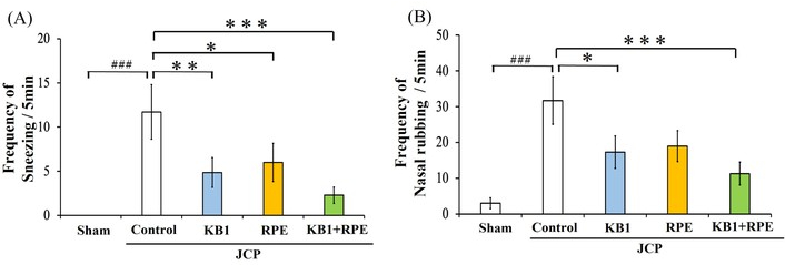
To explore the potential of co-administering KB1 with RPE in attenuating JCP symptoms, we established a mouse model of JCP via intraperitoneal and intranasal sensitization with Cry j1 (Figure 1). These mice exhibited physiological features similar to those of patients with JCP, characterized by increased sneezing and nasal hyperresponsiveness. While BALB/c mice immunized with Cry j1 developed marked nasal inflammatory responses upon intranasal allergen challenge, treatment with either KB1 or RPE alone suppressed these symptoms. Notably, co-administration of KB1 and RPE resulted in a more significant reduction in the frequency of sneezing and nasal scratching than single treatments. These results suggest that this combination may more effectively attenuate JCP symptoms (Figure 2). Consistent with these findings, large effect sizes were observed for the principal pairwise comparisons. For sneezing frequency, Cohen’s d values were 2.73 for Control vs. KB1, 2.48 for Control vs. RPE, 4.79 for Control vs. KB1 plus RPE, 1.69 for KB1 vs. KB1 plus RPE, and 2.25 for RPE vs. KB1 plus RPE. For nasal scratching frequency, the corresponding Cohen’s d values were 2.38, 1.96, 3.45, 0.78, and 1.69, respectively.
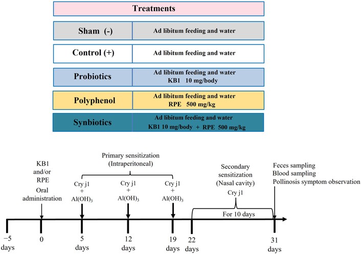
Experimental schedule for the mouse model of JCP with the administration of KB1 and/or RPE. Mice were immunized by intraperitoneal injection of Cry j1 plus alum and challenged with intranasal Cry j1 injection. During the 31-day test period, mice in the KB1 and/or RPE groups were administered KB1 (10 mg/mouse/day) and/or RPE (500 mg/kg/day), dissolved in 0.2 mL of distilled water, by oral administration six times per week. The Sham and Control groups received distilled water under similar conditions. Blood and fecal sampling were performed on the final day. Cry j1-induced JCP was evaluated by counting the frequency of sneezes and nasal scratching immediately after the final Cry j1 challenge. The detailed experimental procedure is described in the Materials and methods section. Cry j1-induced mice were challenged with an oral administration of KB1 and RPE, either with or without the oral administration of KB1 and RPE (Control group). Non-Cry j1-induced mice (Sham group) and Cry j1-induced mice (Control group) were used as negative and positive controls, respectively. JCP symptoms were assessed by counting the frequency of sneezing and nose-rubbing behaviour after the final challenge. Data are expressed as the mean ± standard error of the mean (SEM) for seven mice in each group. Differences between groups were evaluated using one-way ANOVA, followed by Tukey’s HSD test.
Since serum IgE levels are a crucial indicator of the severity of JCP, we measured IgE concentrations in mice orally administered KB1 and/or RPE. As shown in Figure 3, the Cry j1-challenged groups exhibited a significant increase in serum IgE levels compared to the Sham group. In contrast, KB1 and RPE administration suppressed this increase. Furthermore, co-administration of KB1 and RPE resulted in a more pronounced reduction in serum IgE levels than that with either treatment alone (Figure 3).
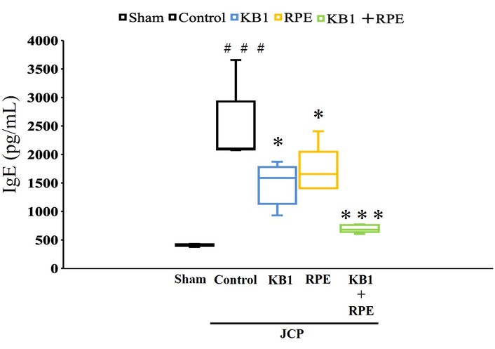
Effect of KB1 and/or RPE on the serum IgE response. JCP mice were challenged with an intranasal Cry j1 injection and KB1 and RPE, or with an oral administration of KB1 and RPE (Control). Non-Cry j1-induced (Sham) mice served as negative controls. Serum IgE levels were measured on day 31. Data are expressed as the mean ± SEM for six to seven mice per group. Differences between groups were evaluated using a one-way ANOVA followed by a Tukey’s HSD test. ### p < 0.001 compared to the Sham group. * p < 0.05, *** p < 0.001 compared to the Control group.
To further characterize systemic inflammation in JCP, we measured the weights of spleen and liver tissues and quantified leukocytes (Table 1). Spleen weight was significantly elevated in the challenged group (Control group) compared with that in the Sham group. However, spleen weight was significantly decreased in both the KB1 and KB1 with RPE groups. No significant differences were observed in liver weights or leukocyte counts (Table 1).
Effect of KB1 and/or RPE on tissue weight and number of leukocytes.
| Parameter | Sham | Control | KB1 | RPE | KB1 + RPE |
|---|---|---|---|---|---|
| Spleen weight (mg) | 137.9 ± 8.0 | 164.7 ± 5.8# | 147.6 ± 9.2* | 150.1 ± 8.6 | 148.6 ± 7.2* |
| Liver weight (g) | 1.55 ± 0.09 | 1.56 ± 0.10 | 1.56 ± 0.10 | 1.66 ± 0.05 | 1.61 ± 0.08 |
| Leukocyte (× 104/mL) | 641.2 ± 57.4 | 561.2 ± 84.6 | 574.9 ± 50.9 | 630.9 ± 142.2 | 529.1 ± 59.6 |
The differences between the groups were evaluated using a one-way ANOVA, followed by a Tukey’s HSD test (seven per group). # p < 0.05 compared to the Sham group. * p < 0.05 compared to the Control group.
To evaluate these effects, serum levels of the anti-inflammatory cytokines IL-10 and IL-27 were quantified in mice administered KB1 and/or RPE (Figure 4). IL-10 and IL-27 levels were significantly higher in the KB1 and RPE co-administration group than in the single-administration groups (Figure 4). Consistent with these findings, large effect sizes were observed for the principal pairwise comparisons. For IL-10, Cohen’s d values were 4.28 for Control vs. KB1, 6.59 for Control vs. RPE, 6.89 for Control vs. KB1 plus RPE, 4.48 for KB1 vs. KB1 plus RPE, and 2.97 for RPE vs. KB1 plus RPE. For IL-27, the corresponding Cohen’s d values were 4.46, 3.38, 9.29, 5.50, and 6.28, respectively.
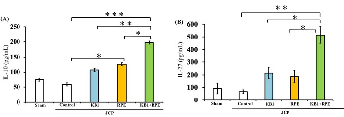
Effects of KB1 and RPE on serum levels of IL-10 (A) and IL-27 (B). JCP mice were challenged via intranasal Cry j1 injection with KB1 and/or RPE, either with or without the oral administration of KB1 and RPE (Control group). Non-Cry j1-induced (Sham) mice served as negative controls. Serum IL-10 and IL-27 levels were measured on day 31. Data are expressed as the mean ± standard error of the mean (SEM) for six to seven mice per group. Differences among groups were evaluated using a one-way ANOVA followed by a Tukey’s HSD test. * p < 0.05, ** p < 0.01, *** p < 0.001 compared to the Control group.
We then quantified the splenic mRNA expression of anti-inflammatory factors and suppressors of excessive immune responses, such as IL-10, IL-27, IFN-γ, and Foxp3 (Figure 5). The primer sequences are shown in Table 2. Similar to serum levels, the splenic mRNA levels of the key anti-inflammatory cytokines IL-10 and IL-27 were significantly higher in the co-administration of KB1 with RPE group than in the single administration group. Furthermore, the expression of IFN-γ and Foxp3 mRNA was significantly higher in the KB1 and RPE co-administration group than in the single-administration group.
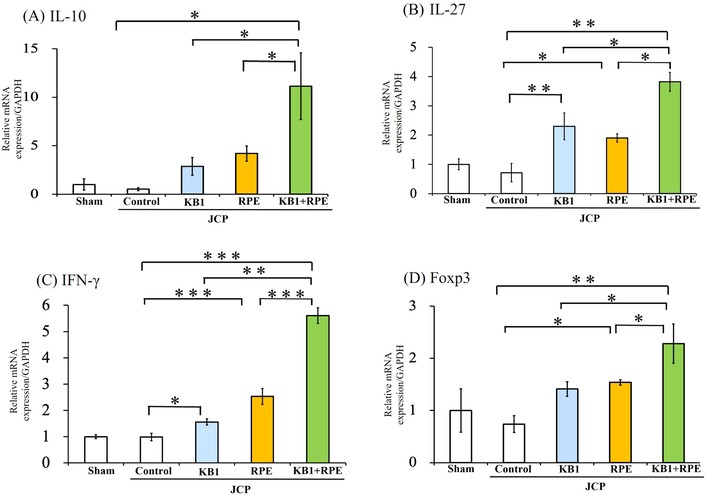
Effects of KB1 and/or RPE on mRNA expression levels in spleen tissue. (A) IL-10; (B) IL-27; (C) IFN-γ and (D) Foxp3. JCP mice were challenged via intranasal Cry j1 injection with KB1 and RPE, or with oral administration of KB1 and RPE (Control). Non-Cry j1-induced (Sham) mice served as negative controls. On day 31, the mRNA expression levels of IL-10, IL-27, IFN-γ, and Foxp3 in spleen tissue were measured. Data are expressed as the mean ± standard error of the mean (SEM) for six to seven mice per group. Differences between groups were evaluated using a one-way ANOVA followed by a Tukey’s HSD test. * p < 0.05, ** p < 0.01, *** p < 0.001 compared to the Control group.
Primer sequences for quantitative real-time PCR.
| Gene | Forward primer (5’→3’) | Reverse primer (5’→3’) |
|---|---|---|
| IL-10 | CGGGAAGACAATAACTGCACCC | CGGTTAGCAGTATGTTGTCCAGC |
| IL-27 | TCTCGATTGCCAGGAGTGAACC | AGTGTGGTAGCGAGGAAGCAGA |
| IFN-γ | ACACCTGATTACTACCTTCTTC | CTTCCTGAGGCTGGATTC |
| Foxp3 | CCCATCCCCAGGAGTCTTG | ACCATGACTAGGGGCACTGTA |
| GAPDH | TTCACCACCATGGAGAAGGC | GGCATGGACTGTGGTCATGA |
The taxa shown in Figure 6 were selected because they represented the predominant bacterial groups detected across the samples and/or exhibited significant differences among the experimental groups. This study evaluated changes in fecal bacterial composition by analyzing microbiota composition relative to the Cry j1-induced Control group (Figure 6). Co-administration of KB1 and RPE resulted in significantly higher abundances of Lachnospiraceae and Oscillospirales, as well as lower levels of Enterorhabdus, compared with the single-administration groups. In contrast, L. reuteri exhibited an increasing trend, although this difference was not statistically significant. Immunization and challenge with Cry j1 significantly decreased the abundance of Clostridia UCG-014, while significantly increasing the abundances of Gastranaerophilales and Bacteroides. Conversely, the KB1 and/or RPE administration groups exhibited higher abundances of Clostridia UCG-014 and lower abundances of Gastranaerophilales and Bacteroides. Additionally, both the KB1 group and the KB1 and RPE co-administration group showed an increased relative abundance of the family Muribaculaceae. Roseburia was significantly more abundant in the RPE group than in the other groups. However, no significant differences were observed in Lactobacillales levels (Figure 6).
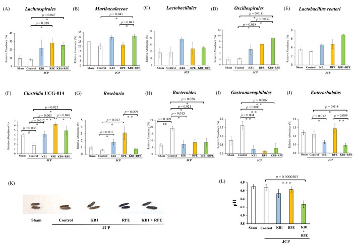
Gut microbiota composition at multiple taxonomic levels (order, family, genus and species), representing the major bacterial taxa detected across the experimental groups. These accounted for approximately 100% of the total relative abundance (A–J), fecal morphological changes (K) and pH (L). The data represent the relative abundance of bacterial taxa that differed significantly between two or more groups. For this analysis of the gut microbiota, feces collected from mice on day 31 were used (A–J). Data are presented as mean ± SEM (n = 3–4). The photograph shows fecal morphology on day 31. Fecal pH was measured using fecal samples suspended in PBS (L). Data are presented as mean ± SEM (seven per group). Differences among groups were analyzed using one-way ANOVA followed by the Games-Howell post hoc test. Fecal pH was analyzed using Tukey’s HSD test. # p < 0.05, ## p < 0.01 compared to the Sham group. * p < 0.05, ** p < 0.01, *** p < 0.001 compared to the Control group.
Fecal pH was measured on day 31 in all experimental groups (Sham, Control, KB1, RPE, and KB1 with RPE). The Control group had a pH of 6.67, whereas the KB1 with RPE co-administration group had a significantly lower pH of 6.27. No significant differences were observed in the single-administration groups compared with the Control group.
Co-administering KB1 and RPE increased the levels of Lachnospiraceae, Oscillospiraceae, and Clostridia UCG-014, while decreasing the levels of Enterorhabdus, Bacteroides, and Gastranaerophilales. Furthermore, co-administration reduced fecal pH. These changes may be linked to elevated serum levels of IL-10 and IL-27, and increased mRNA expression levels of IL-10, IL-27, IFN-γ and Foxp3 in the spleen (Figure 7).
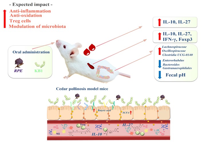
Overview diagram illustrating the effects of KB1 and RPE treatment on metabolism in a Cry j1-induced murine cedar pollinosis model associated with modulation of the intestinal microbiota.
Probiotic-prebiotic and probiotic-polyphenol-based interventions have both been investigated separately in the context of AR [17, 18]. However, the relationships between systemic immune responses, including circulating anti-inflammatory cytokines such as IL-10, and alterations in gut microbiota composition remain insufficiently elucidated for these combined intervention approaches. Based on the premise that the immunological effects of individual nutritional components may differ substantially from those of their mixtures, this study examines KB1 and RPE, which is rich in polyphenols and dietary fibres, in a Cry j1-induced JCP model. This model utilises a major allergen of Japanese cedar pollen and more closely reflects clinically relevant disease pathology than conventional OVA-induced allergy models. Therefore, this model enables a more disease-specific evaluation of immune responses. The present study investigated the effects of KB1 and RPE administration alone and in combination on pollinosis symptoms in this model. We demonstrated that, compared to the administration of KB1 or RPE alone, the co-administration of KB1 and RPE was associated with amelioration of cedar pollinosis symptoms, including reduced IgE production, suggesting that IgE reduction may be related to the improvement of cedar pollinosis. Type 1 allergy, including Cry j1-induced pollinosis, is characterised by allergen-specific IgE as the allergy marker. Therefore, effectively reducing total serum IgE production is thought to suppress mast cell degranulation and improve allergic symptoms. Recent studies have shown that drinking citrus juice fermented with L. plantarum, which contains lactic acid bacteria, for eight weeks can improve allergic symptoms, suppress serum IgE levels and alleviate mast cell-dependent AR in adult patients [18]. Similarly, a 45-day administration of a standard rodent diet containing lactic acid bacteria has been reported to suppress serum IgE levels and mast cell-dependent nasal responses in an AR mouse model [13]. Additionally, quercetin has been reported to exhibit anti-allergic effects by inhibiting the production of histamine and pro-inflammatory mediators, as well as reducing the release of antigen-specific IgE from B cells [19]. In clinical trials, cedar pollinosis patients who were administered 500 mg of apple polyphenols for 12 weeks experienced a significant decrease in sneezing scores [20]. Additionally, although splenic immune activation was evident, as reflected by increased spleen weight and systemic haematological changes (as assessed by abdominal aortic blood leukocyte counts), hepatic changes were limited. This pattern suggests that the immune response was predominantly localised to lymphoid organs, rather than indicating a systemic inflammatory condition. To further confirm the systemic anti-inflammatory properties of KB1 and/or RPE, we analysed the levels of anti-inflammatory mediators in the serum and the mRNA expression of immune tolerance factors in the spleen. IL-10 is an anti-inflammatory cytokine produced by many different types of cells, with the primary source varying depending on the cell type [21]. L. johnsonii has been reported to induce IL-10 production in IL-10-deficient mice and activate native macrophages to become CD206 macrophages (M2), releasing IL-10 to relieve symptoms of colitis [22]. Furthermore, it has been demonstrated that lactic acid bacteria can alleviate cedar pollinosis symptoms by inducing Th1 cells and IL-10-producing regulatory T cells (Tregs) [18]. IL-27 exhibits a wide variety of pro- and anti-inflammatory properties, as well as the capacity to strongly induce IL-10 [23]. One study has shown that Foxp3⁺ Treg cells may modulate dendritic cells to produce IL-27, which then promotes the generation of IL-10-producing T cells [24]. In vitro studies have shown that IL-27 and TGF-β may contribute to IL-10-related immune responses involving Treg cells [25]. Lactic acid bacteria have been reported to promote Treg induction via dendritic cells, increase TGF-β and Foxp3 expression, and suppress Th17 responses [26]. Lactic acid bacterium-fermented citrus juice containing approximately 6.0 × 10¹⁰ heat-killed lactic acid bacterial cells per 100 mL has been reported to alleviate pollinosis symptoms by expanding Treg cells [14]. Previous studies have reported that polyphenols promote the growth of immunologically relevant bacteria and may promote TGF-β production and enhance Treg differentiation [27]. In contrast, resveratrol has been reported to attenuate allergic asthma by increasing Foxp3 expression in lung tissue [28]. Furthermore, administration of resveratrol has been shown to improve autoimmune neuroinflammation by increasing serum IFN-γ production in a mouse model of multiple sclerosis [29]. These results are consistent with those observed in the single-administration groups and suggest that the combined administration of KB1 and RPE may exert an enhanced immunomodulatory effect involving Th1 polarization and Treg induction. In addition, even heat-killed forms of lactic acid bacteria, such as those used in our study, have been reported to retain certain functional properties and have a beneficial effect on intestinal microbiota [30]. Recently, postbiotics have attracted attention for their potential to alleviate inflammatory diseases by promoting immune tolerance and enhancing the bioactivities of probiotics [31]. Postbiotics include short-chain fatty acids (SCFAs) such as lactic acid, butyric acid and propionic acid, which are produced during the fermentation of probiotic bacteria, as well as inactivated bacterial components [30, 31]. Furthermore, the influence of the administration of KB1 and/or RPE on several fecal bacteria was evaluated by analysing changes in microbiota composition compared to the Sham group. In addition, previous studies have reported that Perilla-derived components suppress inflammatory responses by reducing pro-inflammatory cytokines, such as TNF-α and IL-6, and by modulating IgE-mediated allergic reactions [32]. Therefore, the immunological changes observed in the present study may not be solely attributable to alterations in the composition of the gut microbiota, but may also reflect the intrinsic anti-inflammatory properties of RPE itself. It is important to note that the microbiota analysis was performed with a limited sample size, which may constrain the statistical power. Therefore, the observed changes should be interpreted as associative rather than causal. Compared to administration alone, oral administration of KB1 with RPE may be associated with alterations in the composition of the gut microbiota, including an increased abundance of bacteria associated with SCFA production, such as Lachnospiraceae and Oscillospirales, and a decreased abundance of potentially pro-inflammatory bacteria, such as Enterorhabdus and Gastranaerophilales. Lachnospira is a spore-forming, butyrate-producing bacterium with many beneficial effects on the intestinal environment, such as inhibiting colon cancer and improving immune function in the intestinal mucosa [33]. Oscillospirales are recognised as being capable of producing SCFAs, such as butyrate, which have beneficial effects on obesity and chronic inflammation [34]. There was no significant difference for L. reuteri, but the co-administration group showed an increasing trend. Several beneficial effects of L. reuteri have been reported, including the production of the antimicrobial molecule reuterin, strengthening of the intestinal barrier, promotion of anti-inflammatory cytokine IL-10 production in the blood and production of vitamin B12 and folate [35]. Following oral administration of resveratrol, significantly increased levels of the anti-inflammatory cytokine IL-10 have been reported in an acute respiratory distress syndrome model in mice in which L. reuteri microbiota transplantation was performed [36]. The exopolysaccharide produced by L. reuteri has also been demonstrated to induce Treg cells in the spleen [37]. Interestingly, the relative abundance of Clostridia UCG-014 and Roseburia increased the most in the RPE group, whereas Roseburia levels returned to normal with the administration of KB1 with RPE. Clostridia UCG-014 is a butyrate-producing bacterium that is positively correlated with improvements in intestinal morphology and the antioxidant capacity of the ileum and increased SCFAs content [38]. Roseburia is an anaerobic polysaccharide-utilising bacterium derived from the human colon, characterised as a butyric acid producer from glucose [39]. In patients with metabolic syndrome, the consumption of red wine polyphenols has been reported to reduce inflammation, improve insulin resistance and decrease pathogenic bacteria. It has also been shown to significantly increase the abundance of Roseburia [40]. These results suggest that the co-administration of KB1 and RPE may be associated with Treg induction, potentially in relation to increased SCFAs, including various butyrate-producing bacteria, in the gut microbiota. Enterorhabdus has been isolated from a mouse model of spontaneous colitis and has been associated with inflammatory conditions [41]. The relative abundance of Bacteroides was significantly higher in the Control group than in the Sham group, demonstrating that the administration of KB1 and/or RPE shows a tendency to effectively suppress the expansion of Bacteroides. Several reports suggest that an increase in Bacteroides fragilis in the intestinal flora may be a risk factor for pollinosis [42]. Although Bacteroides fragilis constitutes only around 0.5% of the human microbiota [43], its abundance has been reported to be significantly higher in individuals with JCP than in healthy individuals and to be positively correlated with pollinosis symptom scores and Japanese cedar pollen-specific IgE levels [44]. These observations highlight the complexity of the specific regulation of the gut microbiota induced by the combined effects of the cell walls of lactic acid bacteria and the dietary fibres and polyphenols contained in KB1 and RPE. Furthermore, the dietary fibres in RPE are considered prebiotics, which are selectively fermented by the gut microbiota. Their metabolites promote the production of SCFAs and butyrate-producing bacteria, and have been reported to induce specific changes in the composition and functional activity of the gut microbiota [45]. Additionally, fecal pH was assessed as an additional marker of changes to the intestinal environment (Figure 6), with a reduction observed in the combined treatment group. Fecal pH has been associated with intestinal fermentation and SCFA production [46]. Therefore, the reduction in fecal pH observed in the present study may reflect an increase in butyrate-producing bacteria and enhanced SCFA production. However, fecal pH is also known to be influenced by multiple factors, including intestinal transit time, fecal water content, and protein fermentation [47]. Therefore, these findings should be interpreted with caution. In summary, this study provides preliminary evidence that the oral co-administration of KB1 and RPE was associated with greater alleviation of cedar pollinosis symptoms than either treatment alone. This effect was associated with alterations in the gut microbiota, including an increased abundance of putative SCFA-producing bacteria, together with elevated serum IL-10 levels (Figure 7). However, the present study does not establish a causal relationship linking KB1/RPE administration, gut microbiota alterations, immune modulation, and symptom improvement. Further mechanistic studies are required to clarify whether modulation of the gut microbiota directly contributes to the observed anti-allergic effects of KB1/RPE. Thus, the present findings do not demonstrate a causal synbiotic mechanism but rather suggest an association between KB1/RPE administration, immune-related changes, and gut microbiota alterations. These results suggest that KB1 and RPE may have potential as food-derived candidates for further investigation as adjunctive approaches for JCP management. However, additional studies, including human clinical validation, are required before their potential translational application in allergy management can be determined. Given the small sample size in each group, the microbiome analysis in this study is preliminary. Therefore, the observed changes in microbial composition and their potential effects on immune modulation and SCFA-related functions should be interpreted with caution.
AR: allergic rhinitis
Cry j1: cedar pollen extract 1
JCP: Japanese cedar pollinosis
KB1: Pediococcus sp. KB1
L.: Lactobacillus
RPE: Red perilla leaf extract
SCFAs: short-chain fatty acids
AO: Funding acquisition, Investigation, Project administration, Validation, Visualization, Writing—original draft, Data curation, Writing—review & editing. Takao M and Tadaaki M: Supervision, Validation, Data curation, Writing—review & editing. All authors read and approved the submitted version.
Takao Mori is an employee of LAB Biotech Co., Ltd., which provided funding for this study. He contributed to supervision and validation. The other authors declare that they have no conflicts of interest.
All animal experiments were approved by the Institutional Animal Care and Use Committee (IACUC) of Hokkaido University (protocol No. 23-0077) and were conducted in accordance with relevant institutional guidelines and regulations.
Not applicable.
Not applicable.
The sequencing data and all data supporting the findings of this study are available from the corresponding author upon reasonable request.
This work was supported by JSPS KAKENHI Grant Number JP22K07885 and by LAB biotech Co., Ltd. The funders had no role in study design, data collection, analysis, and preparation of the manuscript.
© The Author(s) 2026.
Open Exploration maintains a neutral stance on jurisdictional claims in published institutional affiliations and maps. All opinions expressed in this article are the personal views of the author(s) and do not represent the stance of the editorial team or the publisher.
Copyright: © The Author(s) 2026. This is an Open Access article licensed under a Creative Commons Attribution 4.0 International License (https://creativecommons.org/licenses/by/4.0/), which permits unrestricted use, sharing, adaptation, distribution and reproduction in any medium or format, for any purpose, even commercially, as long as you give appropriate credit to the original author(s) and the source, provide a link to the Creative Commons license, and indicate if changes were made.



View: 113
Download: 3
Times Cited: 0