Original Article


Affiliation:
1Department of Conservative Dental Sciences, College of Dentistry, Prince Sattam bin Abdulaziz University, Al-Kharj 11942, Saudi Arabia
Email: ma.mustafa@psau.edu.sa
ORCID: https://orcid.org/0000-0002-7957-3463


 ,
Affiliation:
2Department of Periodontology and Implantology, HKE’S S. Nijalingappa Institute of Dental Sciences and Research, Kalaburagi 585105, India
ORCID: https://orcid.org/0000-0001-6289-6445
,
Affiliation:
2Department of Periodontology and Implantology, HKE’S S. Nijalingappa Institute of Dental Sciences and Research, Kalaburagi 585105, India
Affiliation:
1Department of Conservative Dental Sciences, College of Dentistry, Prince Sattam bin Abdulaziz University, Al-Kharj 11942, Saudi Arabia
ORCID: https://orcid.org/0000-0002-1388-0890
,
Affiliation:
3Department of Oral and Maxillofacial Surgery and Diagnostic Sciences, College of Dentistry, Prince Sattam bin Abdulaziz University, Al-Kharj 11942, Saudi Arabia
ORCID: https://orcid.org/0000-0002-1589-6685
,
Affiliation:
4Department of Preventive Dental Sciences, College of Dentistry, Prince Sattam bin Abdulaziz University, Al-Kharj 11942, Saudi Arabia
ORCID: https://orcid.org/0000-0003-0474-7067
,
Affiliation:
5Department of Prosthetic Dental Sciences, College of Dentistry, Prince Sattam bin Abdulaziz University, Al-Kharj 11942, Saudi Arabia
ORCID: https://orcid.org/0000-0003-0017-3799
,
Affiliation:
5Department of Prosthetic Dental Sciences, College of Dentistry, Prince Sattam bin Abdulaziz University, Al-Kharj 11942, Saudi Arabia
ORCID: https://orcid.org/0000-0002-5753-6725
Explor Med. 2026;7:1001434 DOI: https://doi.org/10.37349/emed.2026.1001434
Received: May 04, 2026 Accepted: September 20, 2026 Published: November 03, 2026
Academic Editor: Gaetano Isola, University of Catania, Italy
Aim: To compare centre and peripheral tapping techniques during lateral window sinus lift preparation with respect to residual peripheral bone rim thickness and the frequency of Schneiderian membrane perforation in an ex vivo goat maxilla model.
Methods: Twenty goat maxillary specimens were evaluated, with both contralateral sides used, yielding 40 lateral-window preparations arranged as 20 matched pairs. One side of each specimen underwent centre tapping, and the contralateral side underwent peripheral tapping. Residual peripheral bone rim thickness was measured at six predefined locations following window mobilization, and Schneiderian membrane perforation was recorded as present or absent. Residual bone-rim thickness was summarized as mean ± standard deviation (SD) from the available measurements, while membrane perforation was evaluated descriptively.
Results: Peripheral tapping demonstrated greater mean residual peripheral bone rim thickness than centre tapping at all six measurement sites. Based on recalculation from the available group-level summary data, significant differences were observed at the superio-anterior (0.26 ± 0.02 mm vs. 0.14 ± 0.01 mm; p < 0.001), superior (0.27 ± 0.01 mm vs. 0.24 ± 0.01 mm; p < 0.001), superio-posterior (0.12 ± 0.02 mm vs. 0.10 ± 0.02 mm; p = 0.007), inferior (0.13 ± 0.02 mm vs. 0.09 ± 0.02 mm; p < 0.001), and inferio-anterior (0.19 ± 0.02 mm vs. 0.09 ± 0.02 mm; p < 0.001) regions. The inferio-posterior difference was not statistically significant (0.13 ± 0.01 mm vs. 0.12 ± 0.02 mm; p = 0.165). Schneiderian membrane perforation was observed in 8/20 (40.0%) centre-tapping preparations and 4/20 (20.0%) peripheral-tapping preparations.
Conclusions: Peripheral tapping was associated with greater residual peripheral bone-rim thickness at most evaluated sites and a numerically lower observed frequency of Schneiderian membrane perforation than centre tapping. The findings support peripheral tapping as a promising favourable technical profile that merits further paired experimental evaluation.
Rehabilitation of the atrophic posterior maxilla with dental implants remains challenging because tooth loss is followed by progressive alveolar ridge resorption and pneumatization of the maxillary sinus, which may substantially reduce the vertical bone available for implant placement. Maxillary sinus floor augmentation was developed to overcome this anatomical limitation and has subsequently become an established reconstructive procedure for implant rehabilitation of the posterior maxilla [1–3]. Systematic reviews have demonstrated high implant survival following sinus augmentation, supporting the predictability of this approach when appropriate case selection and surgical principles are followed [1, 2].
Sinus floor elevation can be performed through transcrestal or lateral approaches, with selection influenced by residual bone height, sinus anatomy, required augmentation volume, and the planned implant protocol [3, 4]. The lateral-window approach provides direct access to the sinus and remains particularly useful when substantial vertical augmentation is required. However, the procedure requires preparation of a bony window in the lateral sinus wall, followed by controlled mobilization of the bony component and elevation of the Schneiderian membrane [3–5]. Because of the close anatomical relationship between the lateral wall and the underlying membrane, window preparation and mobilization represent critical stages during which membrane injury may occur.
Schneiderian membrane perforation is the most frequently reported intraoperative complication of lateral-window sinus floor elevation [5–7]. A systematic review of prospective controlled studies reported an overall membrane perforation occurrence of approximately 15.7% [8], while a meta-analysis by Al-Dajani [7] reported 388 perforations among 1,652 sinus-lift procedures, corresponding to a weighted incidence of 23.5%. More recent systematic evidence continues to identify membrane perforation as a clinically relevant procedural complication [9, 10]. Although perforation does not invariably result in implant failure when appropriately recognized and repaired, it may complicate membrane elevation and graft containment and can require additional intraoperative management [9, 11].
The susceptibility of the Schneiderian membrane to perforation is multifactorial. Membrane thickness, the presence of sinus septa, residual alveolar bone height, lateral wall morphology and thickness, sinus contour, smoking, and operative variables have all been investigated as potential determinants [7, 10, 12–14]. In a systematic review and meta-regression, Monje et al. [12] reported substantial variation in Schneiderian membrane thickness and emphasized the complexity of relating radiographic membrane dimensions to surgical complications. Subsequent clinical investigations have identified smoking, thin membranes, unfavourable sinus contours, and septa as factors associated with increased perforation risk [13, 15]. A recent systematic review and meta-analysis similarly identified membrane thickness, septa, and smoking among the most relevant factors associated with perforation during lateral-window sinus elevation [10].
Maxillary sinus septa are particularly important during lateral-window preparation because they can interfere with osteotomy design and membrane elevation. A systematic review and meta-analysis involving more than 22,000 sinuses estimated a sinus-level septa prevalence of approximately 33%, highlighting their frequent occurrence [16]. Preoperative three-dimensional assessment is therefore important for identifying septa, lateral wall characteristics, sinus width, membrane morphology, and vascular structures that may influence surgical access [4, 14, 16]. CBCT-based anatomical studies have further demonstrated considerable variability in lateral wall thickness, membrane thickness, sinus dimensions, and the position of relevant vascular structures [14].
Technical modifications have consequently been proposed to improve control during lateral-window preparation. Conventional rotary instruments, piezoelectric devices, manual instruments, different window geometries, and bone-lid approaches have all been investigated [3, 5, 6]. Romanos [17] described a simplified technique in which, after preparation of a small osteotomy, a mallet and mirror holder were used to fracture and mobilize the lateral bony wall. This approach provides a direct conceptual basis for controlled tapping of the prepared lateral window.
Nevertheless, the mechanical effect of the location at which tapping force is applied has received little comparative investigation. In centre tapping, force is concentrated toward the central portion of the prepared bony window, whereas peripheral tapping applies the mobilizing force closer to its margins. These different points of force application could theoretically produce different fracture patterns and consequently influence the amount of residual peripheral bone remaining after mobilization. A residual peripheral bony rim might, in turn, alter the mechanical interaction between the mobilized bony window and the underlying Schneiderian membrane. This proposed mechanism remains hypothetical, however, and should not be interpreted as demonstrating that a thicker residual rim necessarily protects against membrane perforation.
Accordingly, the present ex vivo study was designed to compare centre and peripheral tapping using contralateral sides of goat maxillary specimens. The study specifically evaluated residual peripheral bone-rim thickness at predefined anatomical locations and the observed frequency of Schneiderian membrane perforation. The null hypothesis was that there would be no difference between centre and peripheral tapping in residual peripheral bone rim thickness or membrane perforation frequency.
This exploratory ex vivo comparative study was conducted using 20 freshly obtained goat maxillary specimens devoid of overlying skin and muscles. Both contralateral sides of each maxillary specimen were used, yielding a total of 40 lateral-window preparations arranged as 20 matched pairs. For each specimen, one side was subjected to centre tapping and the contralateral side to peripheral tapping, resulting in 20 preparations for each technique. The specimens were obtained from a slaughterhouse where animals had been routinely slaughtered for food consumption. No animal was sacrificed specifically for the purposes of the present investigation. Baseline cortical bone thickness was not assessed using CBCT or another three-dimensional imaging modality before window preparation. Although the contralateral design reduced between-specimen anatomical variability, potential site-specific differences in lateral wall morphology and cortical thickness could not be completely standardized.
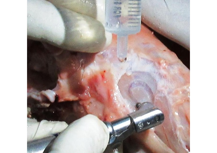
The maxillary sinus was localized using lateral cephalographic imaging (Figure 1). An oval-shaped lateral window was prepared in the lateral maxillary wall using a No. 8 round carbide bur under saline irrigation (Figure 2). Bone removal was continued until a thin residual layer of bone remained over the underlying Schneiderian membrane. The 40 lateral-window preparations were evaluated using two tapping approaches: centre tapping (n = 20) and peripheral tapping (n = 20).
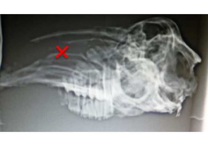
Lateral cephalogram to locate maxillary sinus and a cross mark indicating the location of the sinus.
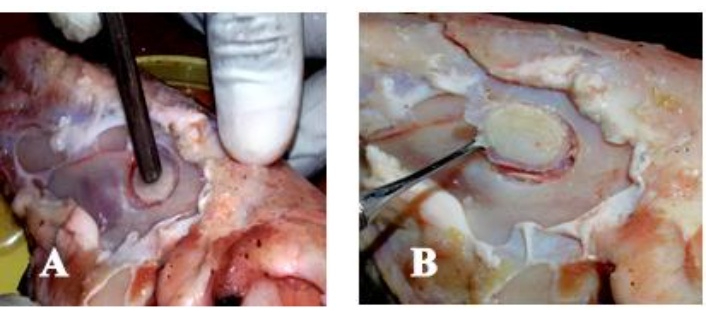
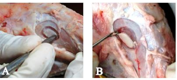
For the centre-tapping technique, the handle of a dental mouth mirror was positioned at the centre of the prepared lateral bony window and tapped with a mallet perpendicular to the lateral bony wall (Figure 3). For the peripheral-tapping technique, the handle of the dental mouth mirror was positioned at the peripheral margin of the prepared lateral bony window, and tapping was similarly performed with a mallet perpendicular to the lateral bony wall (Figure 4). Following mobilization, the lateral bony window was carefully separated from the underlying Schneiderian membrane using the tip of a sinus curette.
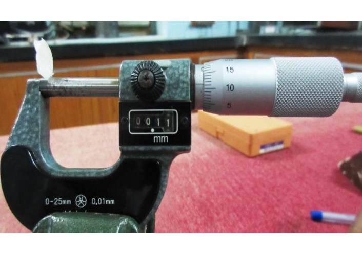
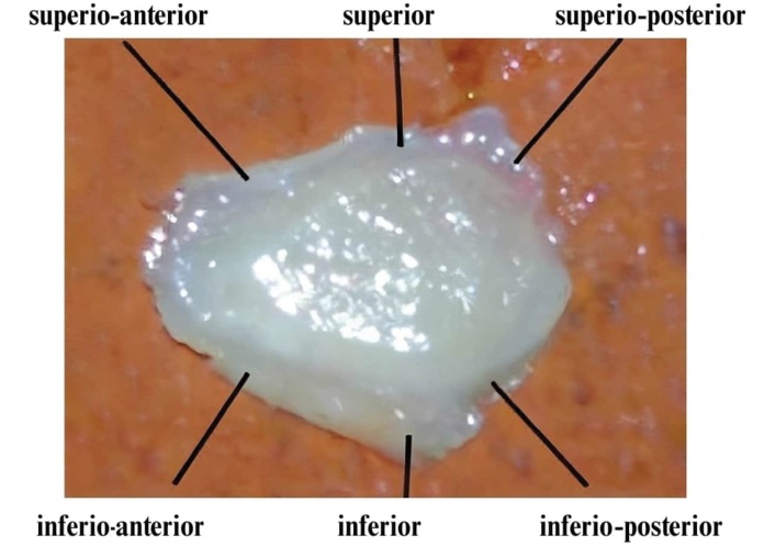
Following mobilization of the lateral bony window, the residual peripheral bone rim thickness was measured using a micrometer (Figure 5). Measurements were obtained at six predefined locations around each lateral window: superio-anterior, superior, superio-posterior, inferio-posterior, inferior, and inferio-anterior regions (Figure 6). All measurements were recorded in millimetres and were subsequently compared between the centre- and peripheral-tapping groups.
Following mobilization and removal of the lateral bony window, the underlying Schneiderian membrane was directly examined for loss of membrane integrity. Membrane perforation was recorded as a binary outcome (present or absent) for each lateral-window preparation. The number and proportion of perforations were subsequently compared between the two tapping techniques.
The study evaluated two principal outcomes: (1) residual peripheral bone rim thickness, measured in millimetres at six predefined locations—superio-anterior, superior, superio-posterior, inferio-posterior, inferior, and inferio-anterior—and (2) Schneiderian membrane perforation, recorded as present or absent following mobilization of the lateral bony window.
Data were entered into Microsoft Excel and analysed using IBM SPSS Statistics, version 21.0. Continuous variables were summarized as mean ± standard deviation (SD), and categorical variables as frequency and percentage. Normality of residual bone-rim thickness measurements was assessed with the Shapiro–Wilk test.
The experimental design consisted of 20 matched specimens, with centre and peripheral tapping performed on contralateral sides of the same specimen. However, the original specimen-level paired measurements were unavailable at the time of the present reanalysis. Consequently, within-specimen correlation could not be reconstructed retrospectively. The available residual bone rim thickness data were therefore evaluated from the reported group-level summary statistics. Between-technique comparisons of residual bone rim thickness were recalculated from the available means, SDs, and sample sizes using two-sided independent-samples t-tests. These analyses should be interpreted as secondary summary-level comparisons rather than paired analyses of the original specimen-level observations. A two-sided p value < 0.05 was considered statistically significant.
Schneiderian membrane perforation was summarized descriptively as frequency and percentage for each tapping technique. Because perforation represented a paired binary outcome and the original specimen-level concordant and discordant pair information was unavailable, an appropriate paired categorical test such as McNemar’s test could not be reconstructed reliably. Therefore, no inferential p value, odds ratio, or confidence interval was used to establish superiority for the membrane-perforation outcome, and the observed difference between techniques was interpreted descriptively.
The original specimen-level paired dataset was not available for retrospective reanalysis. Accordingly, the paired structure of the experiment could not be incorporated into the recalculation of the continuous outcomes, and paired categorical analysis of membrane perforation could not be performed. No individual specimen-level observations were reconstructed or imputed for the purposes of the present analysis.
The primary outcome measures were residual peripheral bone rim thickness at predefined locations and the occurrence of Schneiderian membrane perforation during mobilization of the lateral bony window.
Residual peripheral bone rim thickness was greater with peripheral tapping than with centre tapping at all six predefined measurement locations (Table 1). The largest absolute differences were observed at the superio-anterior and inferio-anterior regions.
Comparison of residual peripheral bone rim thickness between centre and peripheral tapping techniques.
| Measurement site | Centre tapping (n = 20)mean ± SD (mm) | Peripheral tapping (n = 20)mean ± SD (mm) | Mean difference (mm)* | p-value† |
|---|---|---|---|---|
| Superio-anterior | 0.14 ± 0.01 | 0.26 ± 0.02 | 0.12 | < 0.001 |
| Superior | 0.24 ± 0.01 | 0.27 ± 0.01 | 0.03 | < 0.001 |
| Superio-posterior | 0.10 ± 0.02 | 0.12 ± 0.02 | 0.02 | 0.007 |
| Inferio-posterior | 0.12 ± 0.02 | 0.13 ± 0.01 | 0.01 | 0.165 |
| Inferior | 0.09 ± 0.02 | 0.13 ± 0.02 | 0.04 | < 0.001 |
| Inferio-anterior | 0.09 ± 0.02 | 0.19 ± 0.02 | 0.10 | < 0.001 |
* Mean difference calculated as peripheral tapping minus centre tapping. Values are mean ± standard deviation (SD). † Two-sided p-values recalculated from the available group-level means, SDs, and sample sizes using independent-samples t-tests.
At the superio-anterior region, mean residual bone rim thickness was 0.14 ± 0.01 mm with centre tapping and 0.26 ± 0.02 mm with peripheral tapping (p < 0.001). At the superior region, the corresponding values were 0.24 ± 0.01 mm and 0.27 ± 0.01 mm, respectively (p < 0.001).
At the superio-posterior region, mean thickness was 0.10 ± 0.02 mm with centre tapping and 0.12 ± 0.02 mm with peripheral tapping (p = 0.007). At the inferio-posterior region, mean thickness was 0.12 ± 0.02 mm and 0.13 ± 0.01 mm, respectively; this difference was not statistically significant (p = 0.165).
Mean residual rim thickness at the inferior region was 0.09 ± 0.02 mm with centre tapping compared with 0.13 ± 0.02 mm with peripheral tapping (p < 0.001). At the inferio-anterior region, the corresponding values were 0.09 ± 0.02 mm and 0.19 ± 0.02 mm, respectively (p < 0.001). Thus, five of the six evaluated locations demonstrated statistically significant differences in the summary-level comparisons, whereas the inferio-posterior difference was not statistically significant.
Schneiderian membrane perforation was observed in 8 of 20 (40.0%) centre-tapping preparations compared with 4 of 20 (20.0%) peripheral-tapping preparations (Table 2). Correspondingly, membrane integrity was preserved in 12/20 (60.0%) centre-tapping preparations and 16/20 (80.0%) peripheral-tapping preparations. Thus, the observed frequency of Schneiderian membrane perforation was numerically lower with peripheral tapping than with centre tapping. However, because the two preparations represented contralateral sides of the same specimens and the original specimen-level concordant and discordant perforation records were unavailable for retrospective paired categorical analysis, no inferential comparison was performed for this outcome. Consequently, the observed 20-percentage-point difference should be interpreted descriptively and does not establish a statistically significant reduction in membrane perforation with peripheral tapping.
Schneiderian membrane perforation according to tapping technique.
| Tapping technique | Perforation absent, n (%) | Perforation present, n (%) | Total, n |
|---|---|---|---|
| Centre tapping | 12 (60.0) | 8 (40.0) | 20 |
| Peripheral tapping | 16 (80.0) | 4 (20.0) | 20 |
| Total | 28 (70.0) | 12 (30.0) | 40 |
Note: Centre and peripheral tapping were performed on contralateral sides of the same 20 specimens.
The present ex vivo study investigated whether the location of tapping during mobilization of a prepared lateral bony window influenced residual peripheral bone rim thickness and Schneiderian membrane integrity. Peripheral tapping was associated with greater mean residual bone rim thickness at all six evaluated locations, with summary-level comparisons reaching statistical significance at five sites. The second observation was a numerically lower frequency of Schneiderian membrane perforation with peripheral tapping: perforation occurred in 4/20 (20.0%) peripheral-tapping preparations compared with 8/20 (40.0%) centre-tapping preparations. Importantly, because the original specimen-level concordant and discordant perforation records were unavailable, this paired binary outcome could not be subjected to an appropriate retrospective paired analysis. The 20-percentage-point difference must therefore be regarded as descriptive and hypothesis-generating rather than evidence that peripheral tapping significantly reduces membrane perforation.
The importance of preserving Schneiderian membrane integrity during lateral sinus floor elevation is well established. Membrane perforation represents the most common intraoperative complication of the lateral approach [6–8]. Reported frequencies vary considerably according to study design, operator experience, anatomical characteristics, instrumentation, and definitions of perforation. Al-Dajani [7] reported a weighted incidence of 23.5%, whereas Stacchi et al. [8] identified membrane perforation and vascular injury as the principal intraoperative complications reported in controlled studies. This variation is important when interpreting the 40% and 20% frequencies observed in the present experiment, because rates obtained from an ex vivo animal model should not be directly compared with clinical incidence estimates.
The rationale for evaluating tapping location derives from the simplified lateral-window technique described by Romanos [17], in which a mallet and mirror holder were used to mobilize the prepared buccal sinus wall. The present investigation extends this mechanical concept by distinguishing between force applied centrally and force applied near the peripheral margins. The finding that peripheral tapping left a greater residual bony rim at most measurement sites suggests that the location of force application may influence the pattern by which the thin residual lateral wall fractures during window mobilization. However, fracture propagation, applied impact force, window displacement, and stress distribution were not measured in the present experiment. A specific biomechanical mechanism therefore cannot be inferred from residual thickness measurements alone.
Preservation of the lateral bony window itself has received increasing attention in sinus augmentation. Techniques involving replacement or preservation of a bone lid have been investigated because the lateral window participates in healing of the augmented compartment. Schiavon et al. [18], in a systematic review and meta-analysis of the bone-lid technique, found evidence from preclinical studies that repositioning the bony lid may favour new bone formation, although clinical evidence remains limited. These observations demonstrate that the lateral bony window is biologically and mechanically relevant rather than merely an access opening. Nevertheless, the residual peripheral rim evaluated in the present experiment differs from a repositioned bone lid, and the findings of bone-lid studies should therefore not be directly extrapolated to the tapping techniques investigated here.
The relationship between residual bone preservation and membrane perforation is likely to be complex. Anatomical studies demonstrate substantial variability in the lateral sinus wall, Schneiderian membrane, sinus width, septa, and vascular anatomy [14, 19]. Kawakami et al. [14], using CBCT assessment of patients scheduled for lateral sinus floor elevation, reported variation in mucosal thickness, lateral wall thickness, sinus width, and septal anatomy. Zijderveld et al. [19] similarly showed that anatomical or surgical findings frequently required modification of the standard lateral-window procedure. These observations are relevant to the present study because baseline CBCT characterization was not performed. Consequently, contralateral sides cannot be assumed to have had identical cortical thickness or sinus morphology before osteotomy.
Sinus septa constitute one of the best-established anatomical difficulties during lateral sinus augmentation. Earlier systematic evidence reported septa in approximately 13–35% of maxillary sinuses [20], while a more recent meta-analysis estimated a prevalence of 33.2% per sinus and 41.0% per patient [16]. Septa can necessitate modification of lateral-window geometry and complicate membrane elevation [16, 20]. Clinical studies have also associated septa with an increased likelihood of membrane perforation [13, 15]. These findings reinforce the importance of controlling anatomical variables in future studies examining subtle differences between window-mobilization techniques.
Schneiderian membrane thickness represents another potentially relevant factor. Monje et al. [12] reported a pooled mean membrane thickness of approximately 1 mm but emphasized substantial methodological and biological heterogeneity. More recent evidence has continued to examine whether thin membranes are particularly vulnerable. A 2024 systematic review and meta-analysis found that membranes were thinner in perforated cases than in non-perforated cases, although analyses using predefined thickness cut-offs did not consistently demonstrate a significant difference in perforation risk [21]. The most recent systematic evidence available before the present revision nevertheless identifies thin membrane phenotype among the factors warranting consideration during lateral-window surgery [10].
Patient-related and surgical variables may further modify perforation risk. Schwarz et al. [15], in an analysis of 407 sinus augmentation procedures, identified sinus septa, reduced residual bone height, and smoking as relevant risk factors. A later retrospective investigation involving 278 lateral sinus floor elevations similarly associated smoking, thin membrane, unfavourable sinus contour, and septa with perforation [13]. A systematic review specifically evaluating smoking also examined its association with Schneiderian membrane perforation [22]. Thus, perforation cannot reasonably be attributed to a single technical variable, and the numerically lower perforation frequency observed with peripheral tapping in the present study should not be interpreted as demonstrating an isolated causal effect of tapping location.
Instrumentation used for lateral-window osteotomy has also been extensively studied. Conventional rotary instrumentation remains widely used, while piezoelectric surgery has been proposed to provide more selective bone cutting and potentially reduce soft-tissue injury. Jordi et al. [23] reported weighted membrane perforation rates of approximately 24% with conventional rotary instruments and 8% with piezoelectric devices in their meta-analysis. However, comparative evidence has been less definitive. Atieh et al. [24] found no statistically significant reduction in membrane perforation with piezoelectric devices compared with rotary instruments and Stacchi et al. [25] subsequently concluded that although membrane tearing occurred less frequently with piezoelectric surgery, the available evidence was too weak to establish a definitive benefit.
This broader evidence is directly relevant to interpretation of the current findings. A numerical difference—even one that appears clinically substantial—should not automatically be equated with statistical or causal superiority. In the present study, the original analysis produced conflicting p-values and confidence intervals because the paired nature of the experimental design was not adequately incorporated. The revised analysis therefore deliberately avoids an inferential claim for membrane perforation. Since centre and peripheral tapping were performed on contralateral sides of the same 20 specimens, McNemar-type paired analysis would require the original concordant and discordant pair information. These data were unavailable during reanalysis, and reconstructing them would introduce untraceable information. Consequently, 8/20 versus 4/20 is reported transparently as a descriptive observation.
The consequences of Schneiderian membrane perforation also warrant balanced interpretation. Historically, perforations have been regarded as potentially compromising graft containment and sinus augmentation outcomes. Proussaefs et al. [26], using a split-mouth design, reported less new bone formation at perforated sites repaired with collagen membranes than at non-perforated sites. Various repair strategies have subsequently been described, including collagen barrier membranes and techniques adapted to perforation size [11, 27]. Testori et al. [27], for example, described stabilization of collagen membranes for management of large perforations.
However, contemporary evidence indicates that appropriately managed perforation does not necessarily preclude successful implant rehabilitation. Díaz-Olivares et al. [9] reported implant survival of 97.68% beneath previously perforated and repaired membranes compared with 98.88% beneath intact membranes and found no significant difference in implant survival. A broader systematic review of repair approaches similarly concluded that repaired perforations can achieve favorable implant outcomes [28]. More recently, a systematic review and meta-analysis reported implant survival of approximately 97.1% in perforated sinuses and 97.7% in non-perforated sinuses, without a statistically significant difference [29]. These findings indicate that preventing perforation remains desirable, but membrane perforation should not automatically be equated with treatment failure.
The present study has several strengths. The two techniques were evaluated on contralateral sides of the same 20 goat maxillary specimens, thereby reducing some between-specimen biological variability. Residual bone thickness was evaluated at six predefined anatomical sites rather than by a single global measurement, permitting assessment of regional differences in the fracture pattern. In addition, membrane integrity was directly observed after mobilization of the lateral window. Finally, the revised statistical interpretation separates observations supported by available summary statistics from the membrane-perforation outcome for which appropriate paired inferential analysis could not be reconstructed.
Several limitations must nevertheless be acknowledged. First, this was an ex vivo animal experiment, and goat maxillary anatomy and Schneiderian membrane characteristics cannot be assumed to reproduce those of humans. Second, baseline CBCT assessment was not performed; therefore, pre-existing differences in lateral wall thickness, membrane thickness, septal anatomy, and sinus morphology were not quantified. Third, the magnitude and number of mallet impacts were not objectively standardized or recorded, and biomechanical parameters such as fracture propagation, force distribution, and window displacement were not measured. Fourth, the sample comprised only 20 matched specimens and should be considered exploratory.
Most importantly, the original specimen-level paired dataset was unavailable during retrospective reanalysis. This prevented incorporation of within-specimen correlation into recalculation of the continuous outcomes and prevented appropriate paired categorical analysis of membrane perforation. The summary-level comparisons of residual bone rim thickness and descriptive perforation frequencies should therefore be interpreted with these limitations in mind. No specimen-level observations were reconstructed or imputed.
Future studies should prospectively retain complete paired datasets, perform preoperative CBCT characterization of both sides, quantify Schneiderian membrane and lateral wall thickness, document septal anatomy, standardize window dimensions and tapping force, and use predefined paired statistical analyses. Experimental assessment of fracture propagation and force distribution could clarify whether peripheral force application genuinely produces a different mechanical failure pattern. Ultimately, adequately powered prospective clinical studies would be required before peripheral tapping could be recommended as superior to centre tapping in human lateral-window sinus augmentation.
Within the limitations of this ex vivo comparative study, it can be concluded that centre tapping was associated with less peripheral bone-rim thickness than the peripheral tapping method. Peripheral tapping was associated with 4/20 (20.0%) perforation of Schneiderian membrane compared to 8/20 (40.0%) of centre tapping preparation. This difference should be interpreted descriptively and further studies are suggested to make use of paired specimen-level perforation data to confirm the pattern.
SD: standard deviation
MM: Conceptualization, Methodology, Investigation, Writing—original draft, Writing—review & editing. SRD: Conceptualization, Methodology, Investigation, Data curation, Writing—original draft, Writing—review & editing. KDK: Data curation, Formal analysis, Visualization Writing—review & editing. ARA: Project administration, Supervision, Writing—original draft, Writing—review & editing. AAA: Project administration, Supervision, Writing—review & editing. ASA: Project administration, Resources, Writing—review & editing. FMA: Project administration, Resources, Writing—review & editing. KMA: Project administration, Supervision, Writing—review & editing. All authors read and approved the final submitted version.
The authors declare that they have no conflicts of interest.
Ethical approval was not required for this study because it used goat maxillae, and no animals were sacrificed solely for this research. Instead, the specimens were procured from a slaughterhouse, where goats are routinely processed for food consumption.
Not applicable.
Not applicable.
The data presented in this study will be available from the corresponding author upon reasonable request.
The authors extend their appreciation to Prince Sattam bin Abdulaziz University for funding this research work through the project number PSAU/2025/03/33904. The funders had no role in study design, data collection and analysis, decision to publish, or preparation of the manuscript.
© The Author(s) 2026.
Open Exploration maintains a neutral stance on jurisdictional claims in published institutional affiliations and maps. All opinions expressed in this article are the personal views of the author(s) and do not represent the stance of the editorial team or the publisher.
Copyright: © The Author(s) 2026. This is an Open Access article licensed under a Creative Commons Attribution 4.0 International License (https://creativecommons.org/licenses/by/4.0/), which permits unrestricted use, sharing, adaptation, distribution and reproduction in any medium or format, for any purpose, even commercially, as long as you give appropriate credit to the original author(s) and the source, provide a link to the Creative Commons license, and indicate if changes were made.



View: 54
Download: 2
Times Cited: 0