 Review


Affiliation:
1Department of Pathology, Institute of Infectious Diseases and Molecular Medicine, University of Cape Town, Cape Town 7925, WC, South Africa
ORCID: https://orcid.org/0000-0002-2583-4760

 ,
,
Affiliation:
1Department of Pathology, Institute of Infectious Diseases and Molecular Medicine, University of Cape Town, Cape Town 7925, WC, South Africa
2Wellcome Center for Infectious Diseases Research in Africa, University of Cape Town, Cape Town 7925, WC, South Africa
ORCID: https://orcid.org/0000-0002-7792-7977
,
Affiliation:
3Department of Chemistry, Drug Discovery and Development Center and South African Medical Research Council Drug Discovery and Development Research Unit, University of Cape Town, Cape Town 7925, WC, South Africa
ORCID: https://orcid.org/0000-0003-3455-8857
,
Affiliation:
1Department of Pathology, Institute of Infectious Diseases and Molecular Medicine, University of Cape Town, Cape Town 7925, WC, South Africa
ORCID: https://orcid.org/0000-0002-7718-9839
,
Affiliation:
1Department of Pathology, Institute of Infectious Diseases and Molecular Medicine, University of Cape Town, Cape Town 7925, WC, South Africa
4National Health Laboratory Service, Johannesburg 2131, GP, South Africa
Email: muazzam.jacobs@uct.ac.za
ORCID: https://orcid.org/0000-0002-7403-4604

Explor Immunol. 2026;6:1003259 DOI: https://doi.org/10.37349/ei.2026.1003259
Received: January 26, 2026 Accepted: May 19, 2026 Published: July 16, 2026
Academic Editor: Sunil K. Arora, Postgraduate Institute of Medical Education & Research, India
Tuberculosis (TB) accounts for the most deaths amongst humans from an infectious agent. Although the approved vaccines are effective in preventing infant meningitis, they provide inadequate protection for adolescents and adults. There is a need for an improved understanding of immunological determinants of protection from disease as well as the drivers of pathology. Tertiary lymphoid structures (TLS), inducible bronchial-associated lymphoid tissue (iBALT), are an organized accumulation of cells that mount a protective immune response against Mycobacterium tuberculosis (Mtb) in the lung. A comprehensive search of literature was performed in public databases for articles discussing iBALT in TB disease, yielding findings mainly from animal models of pulmonary TB and observational human data. The search revealed a protective role of iBALT characterized by efficient T-cell priming and macrophage activation that restricts Mtb spread. Conversely, dysregulated or chronic TLS formation is associated with excessive cytokine production, myofibroblast activation, autoimmunity, and the progression of post-TB lung disease (PTBLD). Future research must leverage omics technologies to delineate the stromal and immune subsets that govern the protective or pathological iBALT mechanisms.
Tuberculosis (TB) disease remains a significant global health challenge [1]. The infection arises when an individual inhales Mycobacterium tuberculosis (Mtb)-containing droplets. The majority of people are able to prevent the bacteria from causing disease, with only 10% progressing to active TB [2]. Active TB is associated with an infiltration of proinflammatory immune cells followed by progressive lung remodeling, resulting in cavitation, fibrosis, bronchiectasis, pleural disease, and dystrophic calcification [3–9]. This pathology has been observed after individuals were successfully treated with anti-TB standard of care therapy [10]. There is therefore an urgent need for novel clinical interventions capable of preventing infection and minimizing the disease pathology.
Vaccination remains the most effective clinical intervention, reducing transmission and mortality [11]. An ideal TB vaccine prevents infection, reduces disease recurrence, and prevents lung damage [12–14]. Bacillus Calmette-Guérin (BCG), a live-attenuated strain of Mycobacterium bovis, is the only approved TB vaccine to date [15]. Whilst BCG is protective against TB meningitis (TBM) and disseminated disease in children, its efficacy in adolescents and adults is inconsistent [16–19]. Several TB vaccine candidates are currently under varying phases of active clinical trials, as shown in Table 1 [20, 21], with the intention of boosting the protection offered by BCG [22]. Trials with a “no active trials” clinical status at the time of writing this publication are not shown in Table 1.
These tuberculosis vaccine candidates are in active clinical trials.
| Phase 1 | Phase 2a | Phase 2b | Phase 3 |
|---|---|---|---|
| H107e/CAF10bProtein Subunit/AdjuvantROA: IntramuscularSponsor: SSICTID: NCT06050356 | BNT164a1mRNAROA: IntramuscularSponsor: BioNTech SECTID: NCT05547464 | RUTIInactivated Mtb FragmentsROA: SubcutaneousSponsor: Archivel FarmaCTID: NCT04919239 | BCG (Travel Vaccine)Whole Cell BCGROA: IntradermallySponsor: HJFCTID: NCT04453293 |
| Ad5-105KViral VectorROA: AerosolSponsor: CanSino Biologics, Inc.CTID: NCT06732583 | BNT164b1mRNAROA: IntramuscularSponsor: BioNTech, Gates FoundationCTID: NCT05547464 | GamTBvacProtein/AdjuvantROA: IntradermallySponsor: GAM MOH RFCTID: NCT04975737 | |
| ID93 + GLA-SE (QTP101)Protein/AdjuvantROA: IntramuscularSponsor: Quartis, NIHCTID: NCT06714513 | M72/AS01EProtein/AdjuvantROA: IntramuscularSponsor: Gates MRI, GSKCTID: NCT06062238 | ||
| MTBVACLive Attenuated MtbROA: IntradermalSponsor: BiofabriCTID: NCT04975178 |
BCG: Bacillus Calmette-Guérin; CTID: ClinicalTrials.gov ID; HJF: Henry M. Jackson Foundation for the Advancement of Military Medicine; GAM MOH RF: N.F. Gamaleya Federal Research Centre for Epidemiology and Microbiology, Ministry of Health of the Russian Federation; GSK: GlaxoSmithKline; MRI: Medical Research Institute; NIH: National Institute of Health; ROA: Route of Administration; SSI: Statens Serum Institute. Based on the Stop TB Partnership Working Group on New TB Vaccines Resources [23].
Tertiary lymphoid structures (TLS) are immune cell aggregates that form in response to chronic inflammatory stimuli, representing a potential strategy for tailoring lung immune responses to eliminate Mtb [24]. The presence of TLS has been associated with favorable clinical outcomes in TB disease [25]. In this article, peer-reviewed literature was sourced from PubMed and Semantic Scholar, using search terms related to “Tuberculosis”, “Tertiary Lymphoid Structures”, and “iBALT”. This yielded 244 articles from 1975 to 2026, which were stratified based on relevance and by their characterization of inducible bronchial-associated lymphoid tissue (iBALT) as either protective or pathogenic. While all relevant experimental models were considered, findings from human TB lung tissue were prioritized for detailed review. Non-English articles, conference abstracts, and preprints were excluded.
Lung immune responses to infection are mounted in secondary lymph organs, namely bronchial, hilar, and mediastinal lymph nodes [26], allowing antigen-presenting cells (APCs) to display cognate antigens to naïve lymphocytes [27]. Secondary lymphoid organs are divided into B cell zones [i.e., germinal centres (GC)] and inner T cell zones. These zones are interconnected by a complex network of follicular dendritic cells (FDCs), high endothelial venules (HEV), and fibroblastic reticular cells (FRCs), responsible for priming naïve B cells with antigens, lymphocyte migration, and the scaffolding necessary for anchoring cells while presenting antigens to naïve T cells, respectively [28–30]. A type of lung-associated secondary lymphoid organ, known as the bronchus-associated lymphoid tissue (BALT), is present in rats and rabbits [31, 32]. However, humans, non-human primates, and mice lack persistent BALT [33, 34], instead forming transient iBALT in response to chronic inflammation [35]. iBALTs are observed in pathological conditions such as infectious diseases, allograft rejections, malignancies, and autoimmune disorders [36–38]. In TB-infected human lungs, a variation of the iBALT, referred to as granuloma-associated lymphoid tissue (GrALT), has been observed adjacent to necrotic TB lesions [39].
iBALT formation is preceded by a local infection event, in which antigens are thought to be presented to CD4 T cells by lymphoid tissue inducers such as group 3 innate lymphoid cells (ILC3s) [40], resulting in the production of proinflammatory cytokines such as interleukin 6 (IL-6), IL-17, and IL-22 (Figure 1) [41]. These cytokines activate immune, epithelial, endothelial, and stromal cells to express CCL19, CCL21, and CXCL13, resulting in the migration of B cells from the circulatory system to the site of iBALT formation. Additionally, these tissue resident cells also upregulate the expression of adhesion factors such as ICAM-1 and VCAM-1, committing them to a lymphoid tissue organizer or an immunofibroblast cellular fate [25]. As the iBALT continues to develop, more specialized cell types, such as CD21+ FDCs, lymphatic endothelial cells (LECs), and HEVs are induced in response to continued production of chemokines such as CXCL13, CXCL12, and CCL19. At this stage, distinct B-cell and T-cell zones begin to emerge, which are thought to be supported by B-cell FRCs (BRC) and T-cell zone FRCs (TRC), respectively [25, 42].
The cellular composition of iBALT varies, but B and T lymphocytes make up the majority [43]. iBALT contains a B cell zone with a germinal center, where naïve B cells are exposed to antigens by CD21+ FDCs [44]. This interaction leads to somatic hypermutation and affinity selection, resulting in the production of antigen-specific memory B cells and plasma cells [45–47].
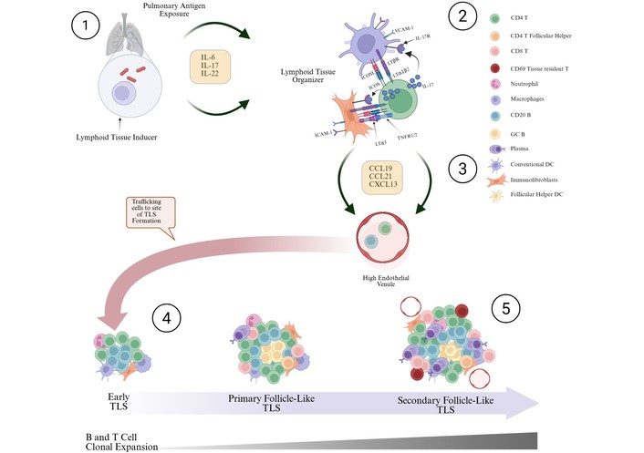
In the presence of chronic inflammation, immunofibroblasts and dendritic cells interact through the ICOS-ICOSL axis with T cells, leading to the production of TLS-inducing chemokines, including CCL19, CCL21, and CXCL13. Based on [48–52]. ICOS: inducible costimulatory; TLS: tertiary lymphoid structures. Created in BioRender. Jacobs, M. (2026) https://BioRender.com/3xmx8fq.
GC B cells express C-X-C motif receptor 5 (CXCR5) to guide the migration of CXCL13+ FDCs or CXCR4, establishing chemokine migration gradients for CXCL13+ FDCs or C-X-C motif ligand 12 (CXCL12)+ FRCs, respectively [53, 54]. Surrounding the GC, CD3 T cells form a cuff known as the T cell zone, where antigen-specific B cells prime CD4+ T cells [41, 55, 56]. CD4+ T cell subpopulations found in iBALTs include follicular helper T cell (Tfh), type 2 helper T cell (Th2), Th17, Forkhead box P3 (FOXP3) regulatory T cell (Treg), and γδ T cells [53, 54, 57–59]. Antigen-specific CD8+ T cells are also present in the T cell zone [60], together with CD83+ lysosomal-associated membrane protein (LAMP) dendritic cells (DCs) that present antigens to T cells [61]. The T cell zone also contains neutrophils, eosinophils, and plasma cells [41]. Other cellular constituents of iBALT include peripheral node addressin (PNAd+)-HEVs and IL-7+ LECs [62, 63]. HEVs express CCL19/CCL21 and CXCL13, establishing migration gradients for CCR7+ T and CCR5+ B cells toward the developing iBALT [64, 65]. Afferent LECs might also play a role in antigen presentation [25]. The maturity of iBALT is determined by its cellular constituents, ascending from early or immature, primary follicle-like, and secondary follicle-like or mature TLS, as illustrated in Figure 1 [66]. Early TLS are unorganized aggregates of B and T cells, whereas primary follicle-like TLS are lymphocyte aggregates containing CD21+ FDCs. Secondary follicle-like TLS are lymphocyte aggregates that include an organized GC, together with PNAd+ HEVs. Secondary follicle-like TLS formation correlates with the expansion of tissue-resident T cells (Trm) [67], which are thought to mediate responses to antigens when TLS are resolved due to the dissipation of the chronic stimuli or with age [68].
Several host immunostimulatory factors are implicated in iBALT formation (Table 2). Zhao et al. [50] conducted an extensive review of the molecular determinants of TLS formation, describing a role for type 1, type 17, and type 2 helper immunostimulatory molecules in iBALT neogenesis. Soldevilla et al. [69] intimated that for effective control of TB, mixed responses might be necessary to counter immune evasion strategies implemented by the pathogen, but this is yet to be demonstrated. A recent study showed the importance of tumor necrosis factor alpha (TNF-α) and TNF superfamily member 14 (TNFSF14), type 1 immunostimulatory molecules, in iBALT formation after intranasal administration of (polyinosinic: polycytidylic acid (I:C)) and ovalbumin in C57BL/6J mice [66]. Zhao et al. [50] reported a role for IL-17, IL-22, and IL-23 in the formation of iBALT in mice. Furthermore, IL-7, IL-13, and oncostatin M, type 2 immunostimulatory molecules, have also been associated with the iBALT formation [63, 70, 71]. Host growth factors such as transforming growth factor beta (TGFβ) and vascular endothelial growth factor have been implicated in TLS formation [50], with the former inducing Tfh to produce TNFSF14 and the latter driving angiogenesis at the site of TLS formation [72–74].
Host immunostimulatory factors associated with TLS formation.
| Immunostimulatory factor | Species | Function | Reference |
|---|---|---|---|
| TNF-α | Mouse | Stromal cell activation and iBALT maintenance | [75] |
| LTα and LTβ | Mouse | iBALT maintenance | [66] |
| CXCL12 | Mouse | Lymphocyte recruitment to developing iBALT | [76] |
| CXCL13 | Mouse | Lymphocyte recruitment to developing iBALT | [77] |
| CCL19 and CCL21 | Mouse | Lymphocyte recruitment to developing iBALT | [77] |
| IL-17 | Mouse | Induces expression of CXCL13, CCL19 and CXCL13 | [78] |
| IL-22 | Mouse | Activation of fibroblasts | [79] |
| IL-23 | Mouse | Action of T cells and ILCs | [78] |
| IL-6 | Mouse | Th2 and Th17 activation | [80] |
| OSM | Mouse | B cell activation | [70] |
ILCs: innate lymphoid cells; OSM: oncostatin M; IL: interleukin; TLS: tertiary lymphoid structures.
TLS orchestrates complex immune responses in the lung, conferring protection in the early stages of TB infection [53, 54]. Mtb infection results in the formation of granulomas that are either restrictive or permissive of bacterial growth [55], enriched in T helper 1/17 cells, and mast and plasma cells, respectively. GrALT, a type of TLS, observed adjacent to TB granuloma, is associated with latent TB infection (LTBI) [39]. Colocalization of B cells, CXCR5+ T cells, DCs, and macrophages has been observed in this tissue associated with the restriction of bacterial growth in adjacent granuloma [56]. It is not clear whether granulomas drive the formation of this tissue or if the tissue supplies the molecular or cellular determinants for granuloma formation. A study in non-human primates coinfected with Mtb and simian immunodeficiency virus (SIV), a model of human immunodeficiency virus (HIV) infection, yielded key insights [57]. The animals were initially infected with a low dose of Mtb, followed by SIV, whilst receiving antiretroviral therapy (ART). LTBI animals showed a significantly lower bacterial burden compared to both ART-treated and ART-naïve groups. iBALT from the coinfected animals showed a significant reduction in CD4+ T cells with SIV infection [57]. Interestingly, there were no significant differences in the bacterial burden between the ART-treated and ART-naïve groups, suggesting that the protective immunity of iBALT is mediated by B cells. A recent study utilized outbred mice to mimic the diverse TB outcomes observed in humans following a low-dose aerosol infection [58]. Using deep learning algorithms to analyse lung tissue showed an enrichment of the iBALT signature in asymptomatic and latently infected animals compared to those with active TB. Griffiths et al. [59] demonstrated the importance of Mtb-primed DCs in the early accumulation of TB-specific CD4+ T cells, associated with B cell follicles, IL-17/IFN-γ secretion, and a 3-log reduction in bacterial burden relative to unvaccinated mice. This protective efficacy was recapitulated in BCG-primed, TB-infected mice by the intratracheal administration of Ag85B, amphiphilic-CpG (toll-like receptor 9 agonist), and FGK4.5 (a CD40 antibody that activates DCs). Studies have reported CXCR5+ Tfh, Th1, and Th17 cells and their association with TB-specific B cells as a critical axis for controlling the infection [53, 56, 60, 61]. Other studies have delivered Mtb antigens via viral vectors and fusion proteins, correlating protection with IL-17-dependent iBALT formation [62, 63]. Intranasal vaccination of rhesus macaques with an attenuated Mtb∆SigH strain elicited robust, iBALT-associated CD4+ and CD8+ T cell-mediated immunogenicity [64], with significantly reduced bacterial burden, lung pathology, and survival relative to BCG-vaccinated animals.
A study in cynomolgus macaques showed that animals vaccinated with Mtb∆SigH generated iBALT, which mounted antigen-specific protective responses against subsequent Mtb infection [65]. Sigma H (SigH) is a bacterial factor that protects the pathogen from the harsh host physiological environment, such as oxidative/nitrosative stress [81, 82], low oxygen partial pressure [83], low pH [84], disruption of cell wall integrity [85], and phagocytosis [73]. Dunlap et al. [86] reported that knocking out mycobacterial membrane protein large 7 (MmpL7) resulted in the overexpression of anti-inflammatory diacylglycerol trehaloses and the formation of protective GrALT.
There is no evidence demonstrating that the presence of iBALTs worsens patient outcomes in TB disease. However, comparison of the immune correlates of pathogenic iBALTs from other pulmonary disorders with TB might provide clues [87–91]. Air pollution is associated with severe TB lung disease [92]. A recent study reported a positive correlation between exposure to cigarette smoke, the severity of TB disease, iBALT formation, and CXCL13 levels in humans [93]. In a mouse model of cigarette smoke exposure, CD4+ T cells acquired a Th1 response with poor effector function [94]. Pathogenic iBALTs are thought to be formed due to chronic inflammation, leading to suboptimal, persistently activated innate and adaptive immune responses [41].
Emerging human single cell sequencing and spatial transcriptomic studies of TB-infected lung tissue are yielding key insights into a potential role of pathogenic iBALT. Krause et al. [39] profiled B cell phenotypes from lung resection surgeries from participants with prior TB episodes, uncovering statistically significant enrichment of tissue resident and antigen secreting cells. Further classification of the antibodies from this study revealed an abundance of Mtb-specific immunoglobulin M (IgM) memory B cells, which have been associated with ectopic lymphoid tissue [95]. A recent study from the same cohort identified a colocalization of MMP1+ CXCL5+ myofibroblasts and SPP1+ macrophages as highly abundant in granulomatous and iBALT-rich TB-infected tissue [96]. This association has been reported in idiopathic pulmonary fibrosis as the primary driver of pathology [97]. It was interesting to note that the myofibroblast signature was enriched in iBALT from HIV+ samples relative to granuloma from the same participants. Furthermore, lung tissue from this cohort showed a low bacterial burden, as most participants received therapy. It remains unclear whether TB pathology at this stage is driven by the persistence of Mtb antigens or by an aberrant, self-perpetuating host immune response. Nayar et al. [49] reported on the role of myofibroblasts in the formation of iBALT via the inducible costimulatory (ICOS)-ICOSL axis [49]. Detection of inflammatory stimuli by immunofibroblasts and APCs resulted in the expression of ICOSL, which binds to ICOS on T cells, which then express lymphotoxin alpha 3 (LTα3) (Figure 1). LTα3 binds to TNFR1/2 on the immunofibroblasts, establishing a positive feedback loop to the production of CCL19, CCL21, and CXCL13. We speculate that this mechanism represents an axis that is dysregulated in PTLD, resulting in tissue damage despite low bacterial burden.
Pathogenic iBALTs are a source of destructive autoantibodies in COPD [98]. In a clinical study, autoantibodies were elevated in active TB patients, reducing upon commencement of anti-TB therapy [99]. TB-COPD comorbidities have been reported in endemic regions [100]. A study reported enrichment of autoimmune IgM in TB-COPD patients compared to the COPD-only group [101]. The authors did not report differences in iBALT abundance. The formation of pathogenic iBALT is also mediated by damage-associated molecular patterns (DAMPs) [41]. DAMPs are host-derived factors that are released by cells during necrosis [102]. IL-1, an example of a DAMP, was associated with pathogenic Th2 immunostimulatory factors after instillation of aluminium salts and silica [35]. IL-1 is protective against Mtb in the acute phase of infection, but its presence in the chronic phase is associated with neutrophil accumulation and lung destruction [103]. PTLD is driven by aberrant Th2 host immunostimulatory molecules such as TGFβ, IL-4, IL-5, and IL-13, activating fibroblasts to produce molecular drivers of excessive extracellular matrix deposition and collagen fibril digestion [104–106]. Ardain et al. [107] reported upregulation of IL-6 and oncostatin M in iBALT-enriched human lung tissue, suggesting a potential link. More research is therefore imperative in human tissue, together with animal models, to determine the context in which iBALT might be pathogenic across the TB disease spectrum.
A vaccine that induces iBALT formation against Mtb has the potential to establish lung-specific protection. Zhao et al. [50] comprehensively reviewed different methods for inducing iBALT, including DCs exposed to antigens or transduced with a viral vector overexpressing iBALT-agnostic host factors, recombinant cytokines, stimulant-loaded hydrogels, toll-like receptors, lymphotoxin β receptors, and stimulator of interferon genes agonists. Nagatake et al. [62] intranasally delivered an Mtb antigen, Ag85B, using a human parainfluenza type 2 viral vector, resulting in the establishment of TB-specific, IL-17-dependent iBALTs. This corresponded to Ag85B-specific antibodies in the serum and bronchoalveolar fluid. However, they did not report any protection data from subsequent Mtb infection. A cancer study demonstrated the protective efficacy of tumor-specific TLS induced using lymphotoxin and the human papilloma virus type 16 E7-antigen [108]. Similar approaches should be explored in TB research for the induction of iBALTs, as this will allow antigen expression to be linked to directed immune activation. Currently, the most promising approach for inducing protective iBALTs is the use attenuated Mtb∆SigH strain [65]. Further evaluation of the safety profile of this approach is necessary before use in humans. This will be achieved by assaying strains incorporating more attenuations (double or triple knockouts) together with SigH, particularly in the severe combined immunodeficiency mouse models [109].
iBALT induction has been shown to sensitize the immune system to other airway pathogens [110], an essential consideration as post lung TB disease patients often develop respiratory comorbidities. TLS might also be an avenue for treatment of central nervous system TB (CNS-TB), a rare form of TB with high morbidity and mortality rates [111, 112]. CNS-TB presents either as TBM, intracranial tuberculoma, or spinal TB [113]. TLS has been observed in the leptomeninges of CNS-TB patients, but its role in disease progression remains unknown [114]. Ramachandran et al. [115] reported on the induction of TLS in a glioblastoma mouse model using an adenovirus vector encoding for TNFSF14, which resulted in the generation of robust anti-tumor T cell responses and prolonged survival.
A critical consideration for approaches that induce TLS is to ensure that they do not result in deleterious toxicities or syndromes, as reported in cancer patients concurrently receiving immune checkpoint inhibitors [116]. Overexpression of both foreign and host immunostimulatory molecules might result in an acute phase response, local and systemic cytokine storms, cell or organ damage, elevated vascular permeability, coagulation disruption, and autoimmune disease [117]. It is likely that the development of immunostimulatory overexpression vectors for the induction of TLS might require the administration of anti-inflammatory molecules in cases where patients show adverse reactions.
Another strategy to consider for the treatment of active TB and post-TB lung disease is to concurrently induce TB-specific iBALT and inhibit host-mediated lung damage. The comorbidity of fibrotic disease with TB presents an opportunity to inhibit the drivers of fibrosis, such as TGFβ and IL-10, while simultaneously inducing iBALT to suppress bacteria that persist in the granuloma. Huang et al. [118] reported a strategy for treating pancreatic ductal adenocarcinoma in a murine model in which they combined an antifibrotic molecule (α-mangostin) and a plasmid vector overexpressing TNFSF14. This strategy reduced the degree of fibrosis whilst inducing tissue remodeling, resulting in a significant reduction in the tumor size and improved survival of the animals. Caution should be exercised when modulating host responses to treat TB disease. Antifibrotic treatments such as pirfenidone and nintedanib reportedly worsen TB pathology [119, 120].
If future research demonstrates a mechanistic role for iBALT in human TB pathology, molecules that are currently used in other diseases to disrupt the formation of iBALT should be explored as adjunct therapy. Monoclonal antibodies targeting IL-17 and podoplanin have been shown to reduce TLS formation [121]. Disrupting the activity of lymphotoxin β receptors using modified ligands inhibited the formation of TLS in the salivary glands of mice in a model of Sjogren’s disease [122]. Clotrimazole inhibited TLS formation by disrupting the metabolism of oxysterol, a key molecule in the positioning of B cells in iBALT [123]. Multi-omics approaches, which allow for the simultaneous characterization of the epigenome, genome, and transcriptome (single cell and spatial), together with high-resolution imaging, will likely provide a clearer picture of protective or detrimental determinants of TLS immunity, establishing a basis for rational design of therapeutic interventions.
APCs: antigen-presenting cells
ART: antiretroviral therapy
BALT: bronchus-associated lymphoid tissue
BCG: Bacillus Calmette-Guérin
CNS-TB: central nervous system tuberculosis
CXCR5: C-X-C motif receptor 5
DAMPs: damage-associated molecular patterns
DCs: dendritic cells
FDCs: follicular dendritic cells
FRCs: fibroblastic reticular cells
GC: germinal centres
GrALT: granuloma-associated lymphoid tissue
HEV: high endothelial venules
HIV: human immunodeficiency virus
iBALT: inducible bronchial-associated lymphoid tissue
ICOS: inducible costimulatory
IgM: immunoglobulin M
IL-6: interleukin 6
LAMP: lysosomal-associated membrane protein
LECs: lymphatic endothelial cells
LTBI: latent tuberculosis infection
LTα3: lymphotoxin alpha 3
Mtb: Mycobacterium tuberculosis
SigH: Sigma H
SIV: simian immunodeficiency virus
TB: tuberculosis
TBM: tuberculosis meningitis
Tfh: follicular helper T cell
TGFβ: transforming growth factor beta
Th2: type 2 helper T cell
TLS: tertiary lymphoid structures
TNFSF14: TNF superfamily member 14
The authors acknowledge Professor Alasdair Leslie, Professor Mohlopheni Jackson Marakalala, and Dr Robert Krause for kindly reviewing the manuscript.
IMM: Conceptualization, Writing—original draft, Writing—review & editing. RMM: Writing—review & editing. RVS: Writing—review & editing. NJH: Supervision. MJ: Conceptualization, Supervision, Funding acquisition. All authors read and approved the submitted version.
The authors declare that they have no conflicts of interest.
Not applicable.
Not applicable.
Not applicable.
Not applicable.
This article was supported by a University of Cape Town University Research Council Postdoctoral Fellowship Award (CC01-2025). The funder had no role in study design, data collection, analysis, decision to publish, or preparation of the manuscript.
© The Author(s) 2026.
Open Exploration maintains a neutral stance on jurisdictional claims in published institutional affiliations and maps. All opinions expressed in this article are the personal views of the author(s) and do not represent the stance of the editorial team or the publisher.
Copyright: © The Author(s) 2026. This is an Open Access article licensed under a Creative Commons Attribution 4.0 International License (https://creativecommons.org/licenses/by/4.0/), which permits unrestricted use, sharing, adaptation, distribution and reproduction in any medium or format, for any purpose, even commercially, as long as you give appropriate credit to the original author(s) and the source, provide a link to the Creative Commons license, and indicate if changes were made.



View: 1549
Download: 47
Times Cited: 0


