 Original Article

Affiliation:
1Department of Medical Microbiology and Immunology, Faculty of Medicine, Assiut University, Assiut 71511, Egypt
ORCID: https://orcid.org/0000-0001-7510-5489
Affiliation:
1Department of Medical Microbiology and Immunology, Faculty of Medicine, Assiut University, Assiut 71511, Egypt
ORCID: https://orcid.org/0000-0003-2097-1106
Affiliation:
2Assiut Chest Hospital, Ministry of Health, Assiut 71517, Egypt
Affiliation:
3Faculty of Pharmacy, Assiut University, Assiut 71526, Egypt
Email: yousyezzeldeen@gmail.com
ORCID: https://orcid.org/0009-0009-0490-4916
Explor Immunol. 2026;6:1003256 DOI: https://doi.org/10.37349/ei.2026.1003256
Received: October 30, 2025 Accepted: May 05, 2026 Published: June 16, 2026
Academic Editor: Danilo Di Bona, University of Foggia, Italy
Aim: This study aimed to evaluate the expression of ectonucleotidases cluster of differentiation 39 (ecto-adenosine triphosphate diphosphohydrolase) (CD39) and cluster of differentiation 73 (ecto-5’-nucleotidase) (CD73) and determine the serum levels of their soluble forms (sCD39 and sCD73) in patients with bronchial asthma compared to healthy controls.
Methods: A case-control study was conducted, including asthmatic patients and age-matched healthy controls. The expression levels of CD39 and CD73 were assessed using quantitative real-time PCR (qRT-PCR), while the serum levels of their soluble forms were measured using enzyme-linked immunosorbent assay (ELISA). The diagnostic performance of these biomarkers was evaluated using receiver operating characteristic (ROC) curve analysis.
Results: Compared to controls, asthmatic patients showed significantly lower levels of CD39 expression and its sCD39, whereas CD73 expression and its sCD73 levels were significantly higher. All four biomarkers demonstrated good diagnostic performance in ROC curve with area under the curve (AUC) values greater than 0.8.
Conclusions: The dysregulation of CD39 and CD73 expressions and their soluble forms is associated with bronchial asthma. These biomarkers may serve as potential diagnostic indicators, with sCD39 showing the best overall diagnostic performance due to its high sensitivity and specificity.
The physiology, comorbidities, clinical presentation, immunopathological pathways, biomarkers of allergic inflammation, and response to treatment of asthma vary, making it a complicated chronic airway illness [1]. It is one of the most common long-term respiratory conditions in the world, affecting over 300 million people, many of whom live in underdeveloped nations [2].
The function of purinergic signaling, specifically the enzymes cluster of differentiation 39 (ecto-adenosine triphosphate diphosphohydrolase) (CD39) and cluster of differentiation 73 (ecto-5’-nucleotidase) (CD73), in controlling inflammation and immunological responses in the airways has been the subject of more recent studies [3]. The cell surface expresses CD39, the archetypal member of the ectonucleoside triphosphate diphosphohydrolase (E-NTPDase) family. Together with CD73, this enzymatic cascade modulates purinergic signaling through P2 receptors and P1 receptors, respectively, by converting adenosine triphosphate (ATP) into adenosine [4].
By attaching to adenosine receptors on different immune cells and having strong anti-inflammatory and immunosuppressive effects, the produced adenosine is essential for preserving tissue homeostasis, especially in the inflamed airway environment of asthma. Dysregulation of CD39 and CD73 expression or activity, however, could tip the scales in favor of inflammation. Reduced CD39 and CD73 mRNA expression in CD4 T cells and peripheral blood mononuclear cells, as well as decreased proportions of CD39 regulatory T cells (Tregs) in asthmatic patients, have been documented, indicating that altered purinergic signaling plays a role in airway inflammation and disease pathogenesis [5, 6].
Monocytes, neutrophils, dendritic cells, myeloid-derived suppressor cells, B lymphocytes, and particular T cell subsets are among the immune cells that express CD39 and CD73 [7]. These enzymes exist in the bloodstream in both soluble and membrane-bound forms. but their exact biological functions are still unknown, soluble versions of membrane-bound CD39 and CD73 have been found in circulation, but they mainly control extracellular nucleotide levels at the cell surface [8, 9].
Therefore, this study aimed to evaluate the expression of cell bound CD39 and CD73, as well as to determine the serum levels of their soluble forms in patients with bronchial asthma compared to healthy controls. These biomarkers were selected due to their known roles in purinergic signaling and immune regulation, which are implicated in airway inflammation. In addition, the diagnostic performance and clinical utility of these four biomarkers were assessed to explore their potential as indicators of disease severity and as therapeutic targets.
This case-control study included 30 patients with chronic bronchial asthma attending Chest Hospital during the period from September 2022 to August 2023. Thirty apparently healthy individuals were considered as a control group.
Using α = 0.05 and power = 0.8, the sample size was determined by looking for a significant difference in biomarker levels between asthmatic patients and healthy controls. The projected necessary sample size, based on EPITOOLS (http://epitools.ausvet.com.au) and assuming a large effect size (Cohen’s d = 0.8), was roughly thirty participants per group. Thus, 30 asthmatic patients and 30 healthy controls were included in the study.
Exclusion criteria included patients with upper and lower respiratory tract diseases, chronic heart or lung disease, systemic corticosteroid use, tuberculosis, malignant disease, or other allergic conditions such as allergic rhinitis (AR), atopic dermatitis, and food allergy.
According to the Global Initiative for Asthma (GINA) 2025 guidelines [10], asthmatic patients were diagnosed based on their history, clinical examination, and the presence of reversible airway obstruction, which is defined as a ≥ 12% rise in forced expiratory volume in 1 second (FEV1) after bronchodilator administration.
The study was conducted in accordance with the ethical standards of the Declaration of Helsinki and was approved by the local Ethical Committee of Chest Hospital, Assiut, Egypt (08-2022-24).
Peripheral venous blood (5 mL) was collected from each participant. The sample was divided into two parts: the first, 3 mL of blood with EDTA was used for RNA extraction; the second part was used for serum preparation by allowing the tubes to clot at room temperature, followed by centrifugation at 1,000 × g for 10 min. The serum samples were stored at −80°C until analysis.
Molecular laboratory tests were performed at the Molecular Biology Unit, Assiut University. Total RNA was extracted using the Qiagen RNeasy Mini Kit (Qiagen, Germantown, MD, USA) according to the manufacturer’s protocol (Qiagen, 2021). The purity and concentration of the RNA were assessed using a Biotech Nanodrop (Thermo Fisher Scientific, Waltham, MA, USA), as described by Wilfinger et al. [11].
Synthesis of cDNA was performed using the Applied Biosystems™ High-Capacity cDNA Reverse Transcription Kit (Applied Biosystems, Foster City, CA, USA) following the manufacturer’s instructions (Applied Biosystems, 2020). The resulting cDNA was diluted appropriately for qRT-PCR analysis.
qRT-PCR was conducted using TaqMan probes on a 7500 Fast Real-Time PCR System (Applied Biosystems, Foster City, CA, USA). Each 20 µL reaction contained 10 µL of TaqMan Universal PCR Master Mix, 1 µL of the specific TaqMan Gene Expression Assay, 5 µL of cDNA template, and 4 µL of nuclease-free water. All reactions were performed in triplicate. Cycling conditions consisted of a hot start at 95°C for 7 min, initial denaturation at 95°C for 20 s, followed by 40 cycles of annealing/extension at 59°C for 60 s [12]. Beta-actin was used as the internal control, and TaqMan probes Hs00969559_m1 and Hs00159686_m1 were used for CD39 and CD73 amplification, respectively (Thermo Fisher Scientific, 2022). Relative mRNA expression levels were calculated using the 2^−ΔΔCt method [13].
Serum concentrations of soluble CD39 (sCD39) and soluble CD73 (sCD73) were determined using sandwich ELISA kits (Wuhan Fine Biotech Co., Ltd, Catalogue No. EH0468 and EH2435, respectively) according to the manufacturer’s instructions. The concentrations were calculated by comparing the optical density of the samples to a standard curve.
Data were analyzed using SPSS software (Statistical Package for the Social Sciences, version 25). Differences between means of the study parameters were evaluated by the independent-sample t-test. Non-parametric quantitative variables were compared using the Mann-Whitney U test. A p-value < 0.05 was considered statistically significant. Receiver operating characteristic (ROC) curve analysis was performed to assess the diagnostic performance of the biomarkers.
This study was conducted on 30 patients with bronchial asthma and 30 healthy subjects. The patient group included 12 (40%) Males and 18 (60%) Females, while the control group included 14 (47%) males and 16 (53%) females.
The mean age concerning patients group is 55 years, while that of control group is 48 years. The FEV1 of patients is 57.5 ± 8 while that of healthy controls is 80 ± 5.
In Table 1, there were no significant differences in gender and age between asthmatic patients and controls (p = 0.6 and p = 0.051, respectively). The FEV1 showed significant difference between asthmatic patients and controls (p < 0.01).
Demographic data of asthmatic patients and control subjects.
| Variables | Asthmatic patients (n = 30) | Control subjects (n = 30) | p-value | |
|---|---|---|---|---|
| Gender | Male | 12 (40%) | 14 (47%) | 0.60 |
| Female | 18 (60%) | 16 (53%) | ||
| Age (years) | 55 (31–70) | 48 (33–65) | 0.051 | |
| FEV1 (forced expiratory volume in 1 second) | 57.5 ± 8 | 80 ± 5 | < 0.01* | |
*: statistically significant p < 0.05.
In Table 2 and Figure 1, our results demonstrated a significant downregulation of CD39 expression in the bronchial asthma group compared to healthy controls (p-value = 0.0025). CD73 is found to be significantly overexpressed in the patients when compared to healthy controls (p-value of 0.000009787), indicating a highly significant difference.
Relative expression of CD39 and CD73 in controls vs. bronchial asthma patients.
| Variables | Conc. of CD39 RNA mg/mL | Conc. of CD73 RNA mg/mL | ||
|---|---|---|---|---|
| Control = 30 | Cases = 30 | Control = 30 | Cases = 30 | |
| Mean ± SD | 1.127 ± 0.11 | 0.184 ± 0.01 | 1.09 ± 0.12 | 26.2 ± 1.22 |
| p-value | 0.0025* | 0.000009787* | ||
*: statistically significant p < 0.05. CD39: cluster of differentiation 39 (ecto-adenosine triphosphate diphosphohydrolase); CD73: cluster of differentiation 73 (ecto-5’-nucleotidase).
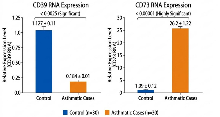
Relative expression of CD39 and CD73 in controls vs. bronchial asthma patients. CD39: cluster of differentiation 39 (ecto-adenosine triphosphate diphosphohydrolase); CD73: cluster of differentiation 73 (ecto-5’-nucleotidase).
There was significant difference value regarding the level of sCD39 in the bronchial asthma serum, it was lower compared to healthy controls (p < 0.0001). Opposite to sCD39 serum level reduction, the value of soluble sCD73 serum level was significantly higher than that of controls (p < 0.0001) (Figure 2).
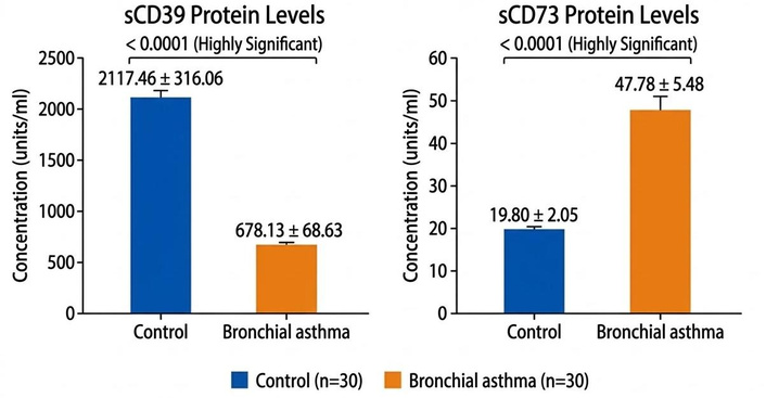
ELISA detection of sCD39 and sCD73 in asthmatic patients and control subjects. sCD39: soluble cluster of differentiation 39 (ecto-adenosine triphosphate diphosphohydrolase); sCD73: soluble cluster of differentiation 73 (ecto-5’-nucleotidase).
The diagnostic performance of sCD39, sCD73, CD39, and CD73 was evaluated using ROC curve analysis, and the corresponding results are presented in Table 3.
ROC curve results of sCD39, sCD73, CD39, and CD73 expressions as markers of asthma.
| Diagnostic marker | AUC | Cutoff | Sensitivity | Specificity | p-value | 95% CI | ||
|---|---|---|---|---|---|---|---|---|
| Lower | Upper | |||||||
| sCD39 | 0.998 | 1,410.2 | 100% | 96.7% | < 0.0001* | 0.992 | 1.000 | |
| sCD73 | 0.954 | 25.67 | 91.7% | 83.3% | < 0.0001* | 0.899 | 1.000 | |
| CD39 | 0.862 | 0.35 | 80% | 83.3% | < 0.0001* | 0.760 | 0.963 | |
| CD73 | 0.981 | 2.52 | 94.7% | 94.1% | < 0.0001* | 0.948 | 1.000 | |
*: statistically significant p < 0.05. AUC: area under the curve; CD39: cluster of differentiation 39 (ecto-adenosine triphosphate diphosphohydrolase); CD73: cluster of differentiation 73 (ecto-5’-nucleotidase); ROC: receiver operating characteristic; sCD39: soluble CD39; sCD73: soluble CD73.
For predicting asthma, all four indicators showed statistically significant results. sCD39 demonstrated 100% sensitivity and 96.7% specificity [area under the curve (AUC) = 0.998] at cutoff 1,410.2; sCD73 demonstrated 91.7% sensitivity and 83.3% specificity (AUC = 0.954) at cutoff 25.67; CD39 expression demonstrated 80% sensitivity and 83.3% specificity (AUC = 0.862) at cutoff 0.35; and CD73 expression demonstrated nearly equal sensitivity of 94.7% and specificity of 94.1% (AUC = 0.981) at cutoff 2.52.
Bronchial asthma is a long-term inflammatory disease of the airways characterized by recurrent episodes of coughing, tightness in the chest, wheezing, and dyspnea, often at night or in the early morning. The complex underlying pathophysiology of asthma includes airway hyperresponsiveness and immune-mediated inflammation. More recent studies have focused on the role of purinergic signaling, specifically the enzymes CD39 and CD73, in regulating inflammation and immune responses in the airways [1, 14]. Thus, investigating subjective indicators for diagnosing asthma and reflecting the severity of the condition continues to be a prominent area of research.
In this work, to assess the levels of cell-bound enzymes, we used qRT-PCR to measure the expression of CD39 and CD73 and to determine the quantities of their soluble forms in the serum of bronchial asthma patients compared with healthy controls.
CD39 is a typical cell surface member of the E-NTPDase family. Initially, CD39 was found to be a B cell activation marker [15, 16].
When comparing asthmatic patients to controls, we discovered that their levels of CD39 expression were considerably lower (p = 0.0025) (Figure 1 and Table 2). This finding is in line with that of Chen et al. [17], who found that extracellular ATP accumulation caused by allergens reduced CD39 expression and eliminated the CD39-mediated suppression of TSLP, interleukin-25 (IL-25), and IL-33 production as well as group 2 innate lymphoid cell proliferation. Additionally, a study by Wang et al. [16] found that CD39 mRNA expression was downregulated in allergic asthma because asthma patients had lower levels of CD39 mRNA in CD4+ T cells and peripheral blood mononuclear cells than did healthy people. They hypothesized that a decrease in CD39 may result in inflammatory damage in asthma by raising serum IL-4 and IL-17A, which mediate airway inflammation in asthma, and that the absence of CD39’s anti-inflammatory qualities may lead to the development of asthma [16]. Wang et al. [18] investigated how the purinergic enzyme machinery influences the development of asthma in a different study. They found that asthma patients have lower percentages of CD39+ Tregs among all Tregs compared to healthy individuals [18].
According to the results of these investigations, insufficient CD39-mediated immune suppression might be the reason, which would hasten the onset of asthma. Additionally, previous studies by Yegutkin et al. [19] demonstrated that the deletion of the CD39 and CD73 genes enhanced the production of cytokines and the recruitment of eosinophils, which exacerbated allergic airway inflammation in mice.
On the other hand. According to Huang et al. [20], CD39, which was thought to be a crucial regulator in airway inflammation, might reduce airway hyperresponsiveness, eosinophilia, mucus deposition, and T-helper type 2 cells (Th2) cytokine production.
Interestingly, it has been proposed that the higher ATP levels observed during asthma flare-ups may be caused by decreased leukocyte expression of ectonucleotide pyrophosphatase/phosphodiesterase 1 (ENPP1), another ATP-degrading enzyme [21].
Extracellular ATP is broken down into adenosine monophosphate (AMP) and adenosine by an enzyme known as sCD39, which can lower immunological activity and control inflammation and immune responses [5].
Yegutkin et al. [19] have shown that a soluble catalytically active form of CD39 circulates in both human and mouse blood.
The soluble form of CD39 was substantially lower in asthmatic patients than in healthy controls in our study (Figure 2). In a similar vein, Jiang et al. [22] discovered that serum CD39 levels were inversely correlated with symptom scores and were lower in people with AR. Additionally, they discovered that serum CD39 showed good disease-reflective and diagnostic capacities for AR. Consequently, nucleotide and nucleoside-mediated CD39 signaling was linked to the homeostatic regulation of eosinophils, inhibited eosinophil accumulation and extravasation, and decreased allergy symptoms in AR [22].
Our study’s ROC results (Table 3, Figure 3) showed that the best outcome for diagnosing asthma patients was values for sCD39. sCD39 demonstrated statistically significant performance in predicting asthma with sensitivity of 100% and specificity of 96.7% at cutoff 1,410.2 (AUC = 0.998). When measuring O2 dissolved in plasma, Díaz-García et al. [23] discovered a negative correlation between CD39 and PaO2/FiO2 (r = –0.351, p = 0.002), suggesting that sCD39 was linked to the degree of hypoxemia in COVID-19 patients. Furthermore, there was a high correlation between the duration of hospital stay and the plasma level of sCD39 on the day of admission (r = 0.264, p = 0.0337). Additionally, the COVID-19 patients who required ICU admission throughout their stay were successfully identified by their CD39 plasma levels at admission [23].
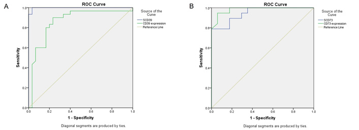
ROC curve of CD39 (A) and CD73 (B) expression, and their soluble forms as markers of asthma. CD39: cluster of differentiation 39 (ecto-adenosine triphosphate diphosphohydrolase); CD73: cluster of differentiation 73 (ecto-5’-nucleotidase); ROC: receiver operating characteristic.
The enzyme CD73 has recently become a “star molecule” in the study of immunology and tumor biology. Hu et al. [24] reported that CD73 modifies downstream inflammatory signaling transduction and controls the balance of ATP, adenosine diphosphate (ADP), AMP, and adenosine release in the extracellular space.
In this study, it was discovered that, in contrast to the downregulation of CD39 expression in bronchial asthma patients. The patients had considerably higher levels of CD73 expression than healthy controls (Figure 1). According to Caiazzo et al. [25], this overexpression may be an adaptive response because CD73 is necessary for initiating anti-inflammatory and tissue repair pathways in bronchial asthma. The increased expression of CD73 may be related to the increased production of adenosine, which is known to control inflammation and airway hyperreactivity in asthma [25].
Experimental results indicate that in animal models of allergic airway inflammation, CD73-derived adenosine is important in controlling the inflammatory/immune response.
The increased expression and activity of CD73 in the lungs of ovalbumin (OVA)-sensitized wild type (WT) mice may be a self-defense system that regulates tissue damage and lung injury brought on by inflammation. Following these findings, the lungs of CD73 knock-out mice exhibit significantly higher levels of Th2 cytokines, IL-4, and IL-5 than those of OVA-sensitized WT mice. CD73 may be a prognostic biomarker for allergic asthma [25]. Garcia-Garcia et al. [26] added that it’s intriguing to observe that enhanced CD73 expression and activity in inflammatory tissue coexist with chronic extracellular adenosine buildup. Over time, this buildup may promote pathological tissue remodeling, resulting in fibrosis and chronic inflammation [26].
Tian et al. [27] discovered that adenosine production was stimulated and CD73 was markedly up-regulated in a separate participant utilizing the long-term cigarette smoke paradigm. This led to the activation of the A2B receptor, which has been demonstrated in numerous studies to have pro-inflammatory properties [27].
Understanding CD73’s function is crucial for developing treatment strategies for pulmonary inflammatory injury, as Cronstein and Sitkovsky [28] have shown that CD73’s contribution to inflammation is sensitive to change as the sickness worsens.
As a result, it has been proposed that CD73 is crucial for immune-inflammatory cells and epithelial barriers, possibly functioning at the interface of innate and adaptive responses during allergic airway inflammation and sensitization. In this work, CD73 expression on immune cells or in its soluble form was examined as a biomarker predictive of allergy and atopy. The results may have implications for allergic airway inflammation prevention and treatment strategies [29]. Evidence suggests that increased CD73 expression and activity in inflammatory tissue may result in a long-term buildup of extracellular adenosine, which may be detrimental and cause the tissue to become chronically inflamed [28].
In addition to CD73 that is linked to cells. sCD73 is an enzyme that cells release into body fluids like blood. Like membrane-bound CD73, it hydrolyzes AMP to adenosine. Its extracellular presence allows it to impact a wider area and may alter systemic effects [30].
Asthma patients in this study had a considerably higher amount of sCD73 form than the healthy control group, p-value of less than 0.0001 (Figure 2). According to Heuts et al. [31], proteolytic cleavage can release a soluble version of CD73 from the membrane, and Airas et al. [32] reported that the soluble form of CD73 had comparable activity to its membrane-bound form.
Remarkable values of sCD73 in the serum of asthmatic patients were shown by the ROC results of this investigation (Table 3, Figure 3). With a statistically significant performance in predicting asthma at cutoff 25.67, sCD73 demonstrated sensitivity of 91.7% and specificity of 83.3%. Furthermore, according to Maksimow et al. [33], the soluble form of CD73 was more predictive of severe pancreatitis than either C-reactive protein or creatinine. The activity of the soluble form of CD73 in a group of patients with moderately acute pancreatitis had an area under the ROC curve value of 0.65 (95% CI, 0.51–0.80) [33]. This was corroborated by Doherty et al. [34], who discovered that whereas a small percentage of circulating CD4+ T cells in healthy people express CD73, this percentage dramatically rises in patients with chronic inflammation, such as those with inflammatory bowel disease (IBD).
In summary, the present study demonstrated that asthmatic patients exhibited significantly lower CD39 expression and its serum sCD39 levels. Whereas CD73 expression and its serum sCD73 levels were significantly higher compared to healthy controls. These findings highlight the differential roles of CD39 and CD73 in asthma pathophysiology. All four biomarkers showed good diagnostic performance by ROC curve (AUC > 0.8), suggesting their potential utility as diagnostic markers.
Notably, the soluble forms of CD39 and CD73 besides cellular CD73 expression demonstrated prominent diagnostic performance on depicted ROC curve (AUC > 0.9). Inspite that sCD39 shows the best specificity and sensitivity as AUC nearly one means outstanding discrimination. And, sCD73 also showed outstanding result. Hence, either sCD39 or sCD73 can be recommended to be promising diagnostic biomarker for asthma because besides their high accuracy there is relevant simplicity and rapidity of ELISA-based measurement.
The limitations of the study are: this study was conducted on a relatively small sample size and at a single center, which may limit the generalizability of the results, lack of previous of research studies on the similar topic and no funding resources.
ADP: adenosine diphosphate
AMP: adenosine monophosphate
AR: allergic rhinitis
ATP: adenosine triphosphate
AUC: area under the curve
CD39: cluster of differentiation 39 (ecto-adenosine triphosphate diphosphohydrolase)
CD73: cluster of differentiation 73 (ecto-5’-nucleotidase)
ELISA: enzyme-linked immunosorbent assay
ENPP1: ectonucleotide pyrophosphatase/phosphodiesterase 1
E-NTPDase: ectonucleoside triphosphate diphosphohydrolase
FEV1: forced expiratory volume in 1 second
GINA: Global Initiative for Asthma
IBD: inflammatory bowel disease
IL-25: interleukin-25
OVA: ovalbumin
qRT-PCR: quantitative real-time PCR
ROC: receiver operating characteristic
sCD39: soluble cluster of differentiation 39 (ecto-adenosine triphosphate diphosphohydrolase)
sCD73: soluble cluster of differentiation 73 (ecto-5’-nucleotidase)
Th2: T-helper type 2 cells
Tregs: regulatory T cells
WT: wild type
AGT, WE, and NNT: Conceptualization, Methodology, Writing—original draft, Writing—review & editing. YE: Conceptualization, Methodology, Writing—original draft, Writing—review & editing, Data curation, Formal analysis, Resources, Investigation,Visualization. All authors read and approved the submitted version.
The authors declare no conflict of interest.
Ethics approval was obtained from the Ethical Committee of Chest Hospital, Assiut, Egypt (08-2022-24). All procedures performed in studies involving human participants were in accordance with the ethical standards of national research committee and with the 2013 Helsinki Declaration.
Informed consent was obtained from all individual participants included in the study.
Not applicable.
The data supporting the findings of this study are available from the corresponding author upon reasonable request.
No funding was received for this study.
© The Author(s) 2026.
Open Exploration maintains a neutral stance on jurisdictional claims in published institutional affiliations and maps. All opinions expressed in this article are the personal views of the author(s) and do not represent the stance of the editorial team or the publisher.
Copyright: © The Author(s) 2026. This is an Open Access article licensed under a Creative Commons Attribution 4.0 International License (https://creativecommons.org/licenses/by/4.0/), which permits unrestricted use, sharing, adaptation, distribution and reproduction in any medium or format, for any purpose, even commercially, as long as you give appropriate credit to the original author(s) and the source, provide a link to the Creative Commons license, and indicate if changes were made.

View: 302
Download: 17
Times Cited: 0
