 Original Article


Affiliation:
1Food Security Research Laboratory, Faculty of Food Science and Nutrition, Universiti Malaysia Sabah, Jalan UMS, Kota Kinabalu 88400, Sabah, Malaysia
ORCID: https://orcid.org/0000-0002-7304-2311

 ,
,
Affiliation:
1Food Security Research Laboratory, Faculty of Food Science and Nutrition, Universiti Malaysia Sabah, Jalan UMS, Kota Kinabalu 88400, Sabah, Malaysia
ORCID: https://orcid.org/0000-0001-5469-566X
,
Affiliation:
2Biotechnology Research Institute, Universiti Malaysia Sabah, Jalan UMS, Kota Kinabalu 88400, Sabah, Malaysia
ORCID: https://orcid.org/0000-0002-2052-9572
,
Affiliation:
3Higher Institution Centre of Excellence, Borneo Marine Institute, Universiti Malaysia Sabah, Jalan UMS, Kota Kinabalu 88400, Sabah, Malaysia
ORCID: https://orcid.org/0000-0003-1548-3327
,
Affiliation:
4Program of Fishery Product Technology, Faculty of Fisheries and Marine Science, Universitas Brawijaya, Malang 65145, Indonesia
ORCID: https://orcid.org/0000-0001-7121-4055
,
Affiliation:
1Food Security Research Laboratory, Faculty of Food Science and Nutrition, Universiti Malaysia Sabah, Jalan UMS, Kota Kinabalu 88400, Sabah, Malaysia
Email: jslee@ums.edu.my
ORCID: https://orcid.org/0000-0002-0249-7749

Explor Foods Foodomics. 2026;4:1010181 DOI: https://doi.org/10.37349/eff.2026.1010181
Received: April 23, 2026 Accepted: July 23, 2026 Published: August 05, 2026
Academic Editor: Filomena Nazzaro, Institute of Food Sciences, Italy
Aim: The fish filleting industry often generates substantial quantities of by-products such as fish bones, heads, scales, and viscera, which are frequently discarded or repurposed for low-value applications. Hydrolysate derived from fish processing wastes have demonstrated various beneficial bioactivities such as ACE-inhibitory, antioxidant, and antidiabetic activities, underscoring their potential as high value functional ingredients or foods. The potency of hydrolysate is determined by peptide composition, which is modulated by the degree of hydrolysis. This study aims to investigate the impact of hydrolysis duration on the ACE-inhibitory and antioxidant capacities of gelatine hydrolysate from hybrid grouper bones.
Methods: Gelatine extracted from hybrid grouper fish heads and bones underwent hydrolysis with 1% Alcalase for varying duration (1 h, 2 h, 4 h, 6 h, 24 h, 48 h). The molecular distribution of the hydrolysates was monitored using sodium dodecyl sulfate-polyacrylamide gel electrophoresis (SDS-PAGE) electrophoresis. ACE-inhibitory and antioxidant capacities (hydroxyl radical scavenging, reducing power, metal chelating activities) of the resultant hydrolysates from different hydrolysis durations were evaluated.
Results: SDS-PAGE analysis revealed that Alcalase hydrolysis of bones gelatine yielded peptides of lower molecular weight. A moderate negative correlation (r = –0.703) (p < 0.01) was reported between the degree of hydrolysis and ACE-inhibitory activity, suggesting that excessive hydrolysis diminishes ACE-inhibitory capacity. Conversely, antioxidant activity of the gelatine hydrolysates was unaffected by hydrolysis extent, as indicated by weak correlations.
Conclusions: These findings indicate that optimising hydrolysis duration maximises the bioactivities of gelatine hydrolysate from hybrid grouper bones.
There is growing epidemiological evidence linking the prevalence of lifestyle-related diseases such as hypertension, obesity, and cancer to dietary factors [1]. This has led to emergence of functional foods that aim to provide health benefits. Food-derived bioactive peptides (composed of 2–20 amino acids residues) in particular demonstrate substantial promise for incorporation into functional foods formulations or as nutraceuticals [1]. These compounds are widely recognised for their various health promoting benefits including antihypertensive [2, 3], antioxidative [4, 5] and antidiabetic properties [3, 6]. Furthermore, bioactive peptides were also believed to have fewer side effects compared to synthetic compounds upon long term consumption [7]. The benefits of protein hydrolysate/bioactive peptides in food products have been documented. For example, the inclusion of 3% of salmon hydrolysate in pasta formulation has been shown to enhance the protein and omega-3 fatty acid of the final products [8]. Similarly, study by Zakaria and Sarbon [9] found that the addition of snakehead fish hydrolysate to sausages resulted in prolonged shelf life, owing to the antioxidative properties of the hydrolysate. The successful commercialization of fish-derived peptides supplements such as PeptACE, Vasotensin, Nutripeptin and Protizen in some countries serves to validate their therapeutic potential and market viability. Notably, PeptACE, which comprises seven peptides isolated from bonito, has demonstrated clinical efficacy in reducing SBP and DBP of mildly hypertensive patients by 11.7 mmg Hg and 6.9 mmg Hg respectively, after a 5-weeks treatment [10].
Bioactive peptides may be generated through diverse approaches including acid/alkali hydrolysis, fermentation, and enzymatic hydrolysis [11]. Enzymatic hydrolysis is preferred to chemical methods because the enzyme employed demonstrate specificity in their catalytic activity and substrate selectivity, thereby allowing precise control over the process [12, 13]. Factors during enzymatic hydrolysis such as protease type, reaction duration, enzyme-to-substrate ratio, and pH greatly influence the degree of hydrolysis (DH) in protein hydrolysate. Careful modulation of these variables facilitates the production of peptides with targeted molecular sizes and enhanced biofunctional attributes [4, 14]. The DH exert a significant impact on the bioactivities of hydrolysate. Extended hydrolysis durations typically yield smaller peptide fragments, which may exhibit enhanced bioactivities. Conversely, excessive hydrolysis can degrade key functional groups, thereby reducing the bioactivities [14]. For instance, a study found that trypsin-generated hydrolysate from giant catfish skin, exhibiting a lower DH demonstrated higher ACE-inhibition and reducing capacities, but lower ferrous chelating ability [15]. In contrast, another study indicated that antioxidant activities (DPPH, ABTS, ORAC, reducing power, metal chelating) of gelatine hydrolysate derived from blacktip shark skin escalated with rising DH [5].
Fish processing yields substantial by-products, including head, skin, trimmings, fins, frames, viscera, bones, and roe accounting for approximately 60% of the total processing output [13, 16]. These discards are rich in proteins, lipids, minerals, and polysaccharides, offering potential for transforming into high-value products, rather than being discarded or used for low-value applications such as animal feed and fertiliser. Valorising fish by-products into bioactive peptides is therefore regarded as a promising strategy to improve economic return, reduce environmental impact and contribute to global food sustainability [17]. Hybrid grouper, the world’s first successfully cultured hybrid grouper produced by crossing Epinephelus fuscoguttatus (tiger grouper) and Epinephelus lanceolatus (giant grouper) has become an economically important aquaculture species since its establishments by the Borneo Marine Research Institute, Universiti Malaysia Sabah in 2006 [18]. Our previous research demonstrated that hybrid grouper bones contain high protein content (55.49%), making them suitable substrates for protein hydrolysate production [19]. Additionally, numerous novel ACE-inhibitory and antioxidant peptides have been isolated from hydrolysate generated through direct hydrolysis of these bones [20, 21]. This has prompted our interest to further explore the potential of hybrid grouper bones by extracting gelatine and generating hydrolysates therefrom. To date, no published studies have investigated the ACE-inhibitory and antioxidant properties of gelatine-derived peptides from this species. Given that our prior work identified Alcalase as the most efficient enzyme for producing bioactive peptides from hybrid grouper bones [20]. Therefore, this study utilised Alcalase to hydrolyse the hybrid grouper bones gelatine for varying durations (1 h, 2 h, 4 h, 6 h, 24 h and 48 h) to identify the optimal hydrolysis time capable of yielding highly potent bioactive peptides. The objective of this study is to investigate the impact of hydrolysis durations on the ACE-inhibitory and antioxidant activities of hybrid grouper bone gelatine hydrolysate, thereby contributing new insights into the valorisation of this economically important aquaculture by-product.
Alcalase (from Bacillus licheniformis, ≥ 2.4 units/g), angiotensin I-converting enzyme (from rabbit lung, ≥ 2 units/mg protein), hippuryl-his-leu hydrate (HHL), o-phthaldialdehyde (OPA), ethylenediaminetetraacetic acid tetrasodium salt dihydrate (EDTA), β-mercaptoethanol, ferrous sulfate, 2-deoxyribose, thiobarbituric acid, ferrous chloride, ferrozine, potassium ferricyanide, ferric chloride and glycerol were purchased from Sigma Aldrich (St. Louis, MO, USA). Coomassie Blue G-250 and Tris-Tricine gel used for electrophoresis were purchased from Bio-Rad Laboratories (Hercules, CA, USA). PageRuler unstained low range protein ladder (3.4–100 kDa), ethyl acetate and trichloroacetic acid were purchased from Thermo Fisher Scientific (San Jose, CA, USA). All chemicals were of analytical grade.
Fish bones (including fish heads) from hybrid grouper (TGGG), were collected from the Borneo Marine Research Institute, Universiti Malaysia Sabah. After cleaning with distilled water, the bones were immediately frozen using liquid nitrogen to inhibit proteolytic reaction and to avoid formation of big ice crystals that could damage tissues structures [22]. Subsequently, the frozen bones were freeze-dried, blended, packed in a sealed bag and stored at –20°C until further use [21].
Gelatine was extracted from the freeze dried bones of TGGG using previously described methods [23, 24]. Briefly, the bones were pretreated with 0.1 M NaOH (1:20, w/v) for 4 h at 4°C to remove non-collagenous protein. The pretreated bones were then demineralised by soaking in EDTA (0.2 M) for 24 h at 4°C, followed by treatment with 0.05 M acetic acid for 3 h at 4°C. Gelatine extraction was subsequently performed by incubating the swollen bones in distilled water for 45°C for 24 h in a shaking incubator. The mixture was then filtered through cheesecloth and the filtrate was collected. The pooled filtrate, designated as bones gelatine was lyophilised and stored at –20°C for further use.
Freeze-dried bone gelatine was dissolved in sodium phosphate buffer (pH 8.0) and subjected to enzymatic hydrolysis using Alcalase (EC 3.4.21.14) with an enzyme-substrate ratio of 1:100 (w/w) at 50°C [20]. The hydrolysis was carried out for varying durations of 1 h, 2 h, 4 h, 6 h, 24 h, and 48 h as shown in Figure 1. The enzymatic reaction was terminated by heating the mixture at 95°C for 15 min in a water bath (Isotemp GDP 20, Fisherbrand, Leicestershine, UK). The mixture was then centrifuged at 10,000× g for 20 min at 4°C. The collected supernatant, referred to as bone gelatine hydrolysate was lyophilised and used for ACE-inhibitory and antioxidant analyses.
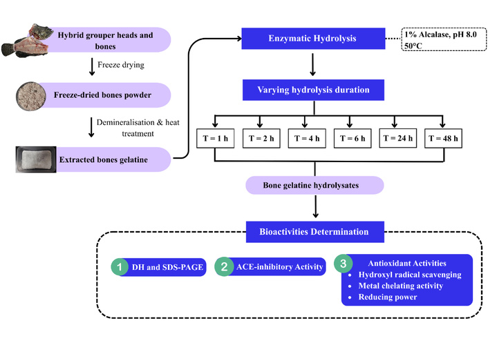
Sequential process for the preparation of bones gelatine hydrolysates at varying hydrolysis durations and their subsequent bioactivities evaluation.
The DH of bone gelatine hydrolysate was determined using the OPA method previously described [25]. Briefly, 0.4 mL of hydrolysate was mixed with 3 mL of OPA reagent, and the mixture was then incubated at room temperature for 2 min. The absorbance of the mixture was measured at 340 nm using a UV-Vis spectrophotometer (Perkin Elmer, Lambda 25, USA). The DH was calculated according to the following formula: DH (%) = ht – h0 / htot × 100%, where ht is the amount of amino group released at the time t, h0 is the amount of amino group in the supernatant at the time 0 h, and htot is the total amount if amino group obtained after acid hydrolysis.
The SDS-PAGE analysis was performed on hydrolysates collected from different time points [26]. The hydrolysates were mixed at 1:1 ratio with sample buffer (200 mM Tris-HCl (pH 6.8)), 40% glycerol, 2% SDS, 2% β-mercaptoethanol, 0.04% Coomassie Blue G-250 and then heated at 100°C for 5 min. Subsequently, 5 µL of each sample was loaded onto a Tris-Tricine gel (4% stacking, 16% separating) along a PageRuler unstained low range protein ladder (3.4–100 kDa). Electrophoresis was performed at a constant voltage of 120 V until the dye front reached the bottom of the gel. The gel was then stained with Coomassie brilliant blue solution.
The ACE-inhibitory activity of hydrolysate was assessed according to previously described methods [23, 27]. Hydrolysate (50 µL, 0.05 mg/mL) was pre-incubated with HHL (150 µL, 8.3 mM) at 37°C for 5 min. Subsequently, ACE (50 µL, 25 mU/mL) was added and the reaction continued for 60 min at 37°C. The reaction was then terminated by the addition of 250 µL of HCl (1 N). Hippuric acid was extracted using 1.5 mL of ethyl acetate, the mixture was vortexed for 5 min and centrifuged at 2,000× g at 4°C for 10 min. The upper organic layer (1 mL) was evaporated to dryness in a vacuum oven set at 40°C for 2 h. The resulting residue was then reconstituted in distilled water, and the absorbance was measured at 228 nm using a UV-Vis spectrophotometer. PeptACE (0.05 mg/mL), a commercially available antihypertensive supplement derived from bonito peptides was served as standard.
The hydroxyl radical scavenging activity of the hydrolysate was evaluated following the method described by Ktari et al. [28]. In brief, the reaction mixture containing ferrous sulfate (0.1 mL, 10 mM), EDTA (0.1 mL, 10 mM), 2-deoxyribose (0.5 mL, 10 mM), and sodium phosphate buffer (0.9 mL, 0.1 M, pH 7.4) was combined with the hydrolysate (0.2 mL, 20 mg/mL). The reaction was initiated by the addition of hydrogen peroxide (0.2 mL, 10 mM), followed by incubation at 37°C for 90 min. Subsequently, the reaction was stopped by the addition of cold trichloroacetic acid (1 mL, 2%) and thiobarbituric acid (1 mL, 1%), and the mixture was heated in a boiling water bath for 15 min. The absorbance of the resulting pink colour was measured at 532 nm using a UV-Vis spectrophotometer. Ascorbic acid (1 mg/mL) was served as standard, as its well-established antioxidant properties provide a reliable reference for comparison purposes.
Metal chelating activity was measured following the method described by Suwal et al. [29]. The hydrolysate (0.25 mL, 1 mg/mL) was first mixed with distilled water (0.925 mL) and ferrous chloride solution (25 µL, 2 mM). The reaction was initiated by the addition of ferrozine (50 µL, 5 mM), followed by 20 min incubation at room temperature. The absorbance of the mixture was subsequently measured at 562 nm using a UV-Vis spectrophotometer. EDTA (0.02 mg/mL), a powerful metal chelator was employed as a standard.
Reducing power of hydrolysate was determined following method described by Lassoued et al. [2]. The hydrolysate (0.2 mL, 20 mg/mL) was mixed with sodium phosphate buffer (0.5 mL, 0.2 M, pH 6.6) and potassium ferricyanide solution (0.5 mL, 1%), then incubated at 50°C for 20 min. Subsequently, trichloroacetic acid (0.5 mL, 10%) was added and the mixture was centrifuged at 3,500× g for 10 min at 4°C. The resulting supernatant was mixed with distilled water and ferric chloride (0.1 mL, 0.1%). After 2 min incubation, the absorbance was measured at 700 nm using a UV-Vis spectrophotometer. The results were expressed as absorbance unit at 700 nm. Ascorbic acid (0.1 mg/mL) was served as standard.
All experiments were performed in triplicates and results are presented as mean ± standard deviation (SD). The Shapiro-Wilk and Levene’s tests were employed to assess normality and the homogeneity of variance respectively, with no violations of these assumptions observed. The data were then analysed using one-way analysis of variance (ANOVA) followed by Tukey multiple range test to determine whether the differences between groups were statistically significant. Correlations between bioactivities and degree of hydrolysis were obtained by using Pearson’s correlation coefficient. Differences between the means were considered significant at p < 0.05. All statistical analyses were performed using the Statistical Package for Social Sciences, SPSS version 25 for Windows.
The DH was used to estimate the extent of bone gelatine degradation by Alcalase. Hydrolysis increased rapidly during the first four hours of reaction, followed by a slower rate of increase up to 24 h (Figure 2A). The reaction then reached a plateau at 48 h, with a DH of 17.17%. The initial rapid hydrolysis was attributed to the high availability of substrate for cleavage, whereas the subsequent slowing down toward the end indicated a limitation in the number of remaining cleavage sites. Figure 2B illustrates the electrophoretic profile of bone gelatine hydrolysate treated with Alcalase. The electrophoretic pattern revealed a progressive decrease in molecular weight distribution of the hydrolysates corresponding to extended hydrolysis durations. The majority of the peptide bands were concentrated within the low molecular weight range, specifically below 15 kDa (lane 4–8) after 2 h of hydrolysis. This observation indicates the efficacy of Alcalase in hydrolysing high molecular weight bone gelatine into smaller peptide fragments. These results align with typical hydrolysis curve reported for thornback ray gelatine [2], razor clam [30], Pacific cod skin gelatine [31], giant catfish skin gelatine [15] and sea cucumber [32]. Although the electrophoretic profile indicates a reduction of molecular weight of the gelatine hydrolysate, this technique provides qualitative data only. Therefore, future study utilising gel filtration chromatography are warranted to be more precisely characterise the molecular weight distribution of the hydrolysate at various time points.
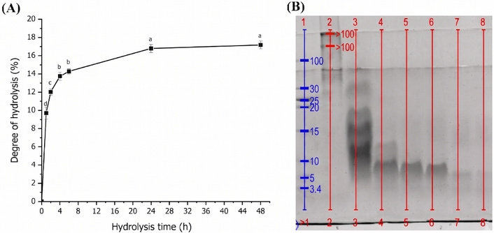
The DH and SDS-PAGE profile of bone gelatine hydrolysate. (A) The DH of bone gelatine treated with Alcalase. (B) SDS-PAGE of bone gelatine hydrolysate of vary hydrolysis durations: protein markers (lane 1), unhydrolysed bone gelatine (lane 2), hydrolysate after 1 h (lane 3), hydrolysate after 2 h (lane 4), hydrolysate after 4 h (lane 5), hydrolysate after 6 h (lane 6), hydrolysate after 24 h (lane 7), hydrolysate after 48 h (lane 8). DH: degree of hydrolysis.
Bone gelatine was hydrolysed by Alcalase for varying durations to assess the impact of hydrolysis duration on the ACE-inhibitory capacity of the resulting hydrolysate. As depicted in Figure 3, the ACE-inhibitory activity of hydrolysate did not exhibit significant differences (p > 0.05) during the initial 24 h of hydrolysis. However, prolonged hydrolysis beyond 24 h significantly reduced the ACE-inhibitory capacity of the hydrolysate. Bone gelatine hydrolysate produced at 48 h, with a DH of 17.70% exhibited the lowest ACE-inhibitory capacity (30.68 ± 2.34%). This finding is corroborated by the moderately significant negative correlation (r = –0.703) (p < 0.01) observed between the DH and ACE-inhibitory activity. Notably, all the hydrolysates demonstrated ACE-inhibition capacity comparable to that of PeptACE (29.82 ± 1.86%), suggesting the potential of gelatine derived from hybrid grouper bones as a functional ingredient particularly in the management of blood pressure.
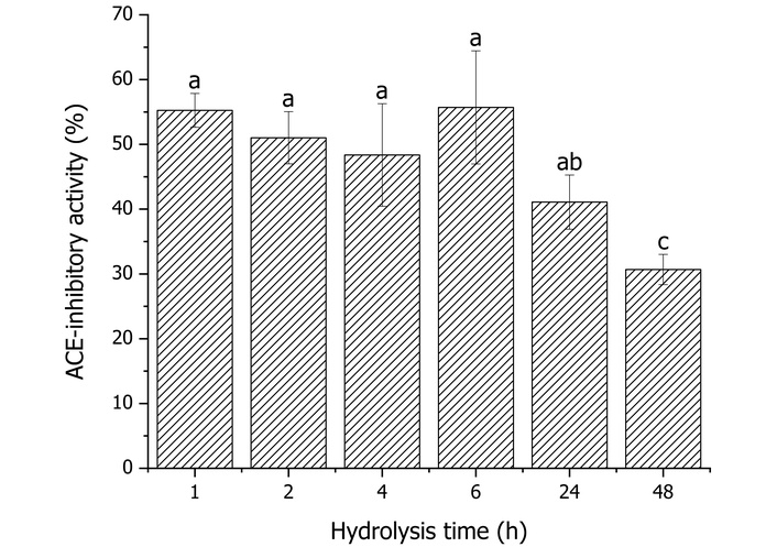
The ACE-inhibitory activity of bone gelatine hydrolysate over different hydrolysis times. Means with different letters (a–c) indicate significant difference at p < 0.05.
Figure 4 demonstrated that the hydroxyl radical scavenging activity of the gelatine hydrolysate remained relatively constant throughout the hydrolysis process. Hydrolysis continuously generates novel peptides with varying potencies that can either enhance or attenuate overall bioactivity. The collective effects of these emergent peptides sustained the hydroxyl radical scavenging capacity of bone gelatine hydrolysate over the 48-h hydrolysis period. This observation suggests that the hydroxyl radical scavenging capacity is minimally influenced by peptide chain length, as evidenced by the weak negative correlation (r = –0.0408) (p > 0.05) observed between the DH and hydroxyl radical scavenging ability. In contrast, Ko et al. [33] reported the hydroxyl radical scavenging ability of flounder fish hydrolysate on the extent of hydrolysis, with longer hydrolysis duration leading to hydrolysate with weaker capacity to scavenge hydroxyl radicals. Furthermore, a study on blacktip shark skin showed that hydrolysate with a DH of 40% exhibited stronger hydroxyl radical scavenging activity than that with a DH of 10% [5]. Conversely, Zhou et al. [34] found that the hydroxyl radical scavenging of papain-hydrolysed scallop muscle initially declined within the first 30 min, but peaked after 60 min and was maintained until the end of hydrolysis (180 min). Although these gelatine hydrolysates displayed scavenging activity, they were less effective than ascorbic acid, which achieved a hydroxyl radical scavenging capacity of 33.84 ± 1.15% at a concentration of only 1 mg/mL.
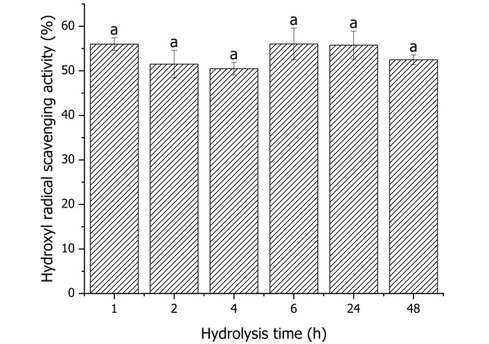
The hydroxyl radical scavenging activity of bone gelatine hydrolysate over different hydrolysis times. Means with different letters indicate significant differences at p < 0.05.
The metal chelating capacity of hydrolysate fluctuated with the duration of hydrolysis (Figure 5). Unlike the hydroxyl radical scavenging activity, the metal chelating capacity of the bone gelatine hydrolysate initially decreased slightly from 1 h to 4 h of hydrolysis, then rose significantly to 65.50% after 6 h of reaction. However, further hydrolysis up to 24 h reduced the metal chelating ability (p < 0.05). The chelating capacity then increased again, reaching a level similar to the 1 h hydrolysate after 48 h of incubation. Notably, the metal chelating activity of hydrolysates was consistently lower than that of EDTA, which displayed metal chelating capacity of 46.62 ± 0.08% at concentration of 0.02 mg/mL. The weak negative correlation between DH and metal chelating ability (r = –0.068) (p > 0.05) indicates that the extent of hydrolysis did not substantially influence the metal chelating ability of gelatine peptides. A similar finding was reported for skipjack roe hydrolysate, where the metal chelating activity rose as the DH increased from 5% to 30%, but excessive hydrolysis up to a DH value of 40% to 50% significantly reduced the chelating ability of the hydrolysate [35]. In another study on skin gelatine derived from Cyprinus carpio, metal chelating capacity decreased significantly when the hydrolysis duration increased from 1 h to 2 h but improved significantly with extended hydrolysis up to 3 h [4].
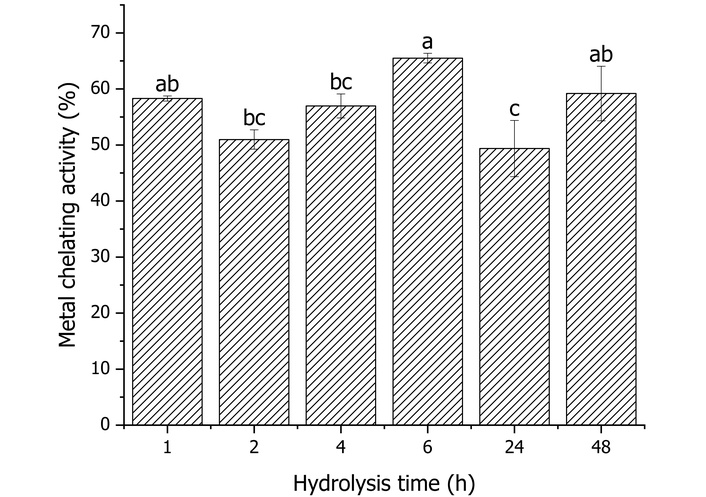
The metal chelating activity of bone gelatine hydrolysate over different hydrolysis times. Means with different letters (a–c) indicate significant difference at p < 0.05.
The reducing power of bone gelatine hydrolysate exhibited a different trend compared to its hydroxyl radical scavenging and metal chelating activities in relation to hydrolysis duration. The reducing potential of the bone gelatine hydrolysate remained similar during the initial 24 h of hydrolysis (Figure 6), but prolonged hydrolysis beyond this point led to a significant increase in the reducing power, reaching a value of 0.1201 (DH = 17.17%). A moderate positive correlation (r = 0.451) (p > 0.05) was observed between the DH and reducing power of bone gelatine hydrolysate, suggesting that the reducing power depends on the accumulation of smaller peptides resulting from extended hydrolysis. Similar observations were reported for gelatine hydrolysate prepared from brownstripe red snapper [36] and red tilapia [37], where the reducing power increased significantly as the DH increased from 20% to 40%. In contrast, a study on gelatine hydrolysate from the skin of giant catfish reported that hydrolysate with a lower DH value exhibited better reducing power than those with higher DH value [15]. Similarly, in another study on hydrolysate prepared from skipjack roe indicated that a DH of 5% demonstrated stronger reducing capacity compared to a DH 50% [35]. Similar to hydroxyl radical scavenging activity, the reducing powers of these gelatine hydrolysates was also lower than of ascorbic acid (0.9284 ± 0.0274).
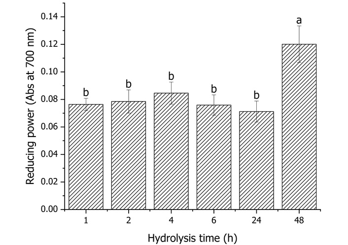
The reducing power of bone gelatine hydrolysate over different hydrolysis times. Means with different letters (a–b) indicate significant difference at p < 0.05.
Hydrolysis duration is known to influence the extent of hydrolysis and subsequently affecting the bioactivities of the resultant hydrolysate. In the present study, gelatine extracted from hybrid grouper bones was hydrolysed using Alcalase to investigate the impact of hydrolysis durations on the antioxidant capacities of the resulting hydrolysate. Analysis of the DH and SDS-PAGE profiles confirmed that Alcalase effectively hydrolysed the bone gelatine, yielding smaller peptides.
The influence of hydrolysis duration on ACE-inhibitory and antioxidant activities varies. Prolonged hydrolysis resulted in a reduction of the ACE-inhibitory activity of bone gelatine hydrolysate. This decline may be attributed to excessive hydrolysis, which cleaves proteins into smaller peptides or free amino acids that are less effective in inhibiting ACE activity. This finding aligns with previous study reported on ribbon jellyfish collagen hydrolysate, where ACE-inhibitory activity remained stable between 1 h and 5 h, but declined upon extended hydrolysis beyond 5 h [38]. Similarly, Ketnawa et al. [15] observed that the ACE-inhibitory activity of giant catfish skin gelatine decreased with increasing hydrolysis duration. Conversely, these results contrast with studies on goby [39] and tilapia [40], where ACE-inhibitory capacities were found to increase alongside extended hydrolysis duration. These variations in hydrolysis duration yield peptides with distinct sequences, which subsequently determine their ACE-inhibitory performance. Peptides possessing hydrophobic amino acids, such as proline, tyrosine, and tryptophan at the C-terminus, have been postulated to exhibit ACE-inhibitory effect [41]. For instance, a comparative study of two peptides isolated from snakehead fish hydrolysate demonstrated that peptide containing three proline residues was more potent than the one containing only two [42].
The antioxidant capacity of a compound can vary depending on the selected assay system, as these methods rely on distinct mechanisms of action and varying reaction conditions. For example, an effective metal ion chelator might fail to exhibit activity in radical scavenging assay. Given these variations, employing a multi-assay approach is essential to obtain a comprehensive evaluation of the antioxidant capacity of food proteins [43]. The present study evaluated the antioxidant potential of bone gelatine hydrolysate using three different antioxidant assays, namely the hydroxyl radical scavenging, metal chelating and reducing power. The hydroxyl radical scavenging assay determined the hydrolysate capacity to neutralise highly reactive hydroxyl radicals implicated in oxidative damage to DNA, proteins and lipids [44]. While assays such as DPPH and ABTS are commonly used to assess radical scavenging activity, they rely on synthetic radicals that may not accurately reflect biologically relevant oxidative environments. The metal chelating assay was included to assess the hydrolysate capacity to bind transition metals such as Fe2+, thereby inhibiting Fenton-type reactions and reducing hydroxyl radical generation through indirect antioxidant mechanism. Additionally, the reducing power assay measured the electron-donating ability of the hydrolysate, offering insight into its overall redox potential and its ability to stabilise oxidised intermediates [45, 46]. These three assays collectively provide a broader antioxidant mechanism by capturing both preventive and chain-breaking activity.
Hydrolysates derived from varying hydrolysis durations displayed comparable hydroxyl radical scavenging capacities, suggesting that the peptide profile generated across these time points possesses a uniform ability to inhibit hydroxyl radical formation from hydrogen peroxide. In contrast, hydrolysis durations up to 24 h did not significantly affect the reducing power of gelatine hydrolysates, with peak reducing power observed only after 48 h. Furthermore, no definitive trend was observed regarding the influence of hydrolysis duration on the metal chelating capacity of the gelatine hydrolysate. Collectively, these findings indicate that DH is not the sole determinant of bioactive properties in protein hydrolysate, rather, factors such as peptide sequences, molecular size and amino acid composition are equally critical for the expression of these biological activities [36]. Amino acids including histidine, proline, and leucine contribute to antioxidant efficacy by facilitating hydrogen and electron transfer, thereby neutralising free radicals [11, 47]. The antioxidative properties of gelatine hydrolysate might be attributed to the presence of these hydrogen donating compounds within the peptides. However, this needs to be further confirmed by more studies in the future.
In conclusion, the current findings demonstrate that Alcalase hydrolysis facilitates the release of ACE-inhibitory and antioxidant peptides from bone gelatine, resulting in a hydrolysate with varied bioactivities. The influence of hydrolysis duration on each bioactivity varies. Prolonged hydrolysis negatively impacted the ACE inhibition but enhanced the reducing power of hydrolysate. In contrast, hydroxyl radical scavenging and metal chelating activities remained relatively unaffected by the extent of hydrolysis. Consequently, the optimal conditions should be tailored according to the specific desired bioactivity. Future research including the characterisation of free amino acid profiles, peptide sequences and molecular weight distribution is essential to elucidate the structural determinants underlying these observed bioactivities. In addition, further studies involving in vivo evaluation and application-based research are recommended to validate the functional potential of the hydrolysate in food or nutraceutical systems.
DH: degree of hydrolysis
HHL: hippuryl-his-leu hydrate
OPA: o-phthaldialdehyde
SDS-PAGE: sodium dodecyl sulfate-polyacrylamide gel electrophoresis
PTC: Conceptualization, Investigation, Methodology, Data curation, Formal analysis, Writing—original draft, Writing—review & editing. PM: Methodology, Resources, Validation. CB: Methodology, Resources, Validation. RS: Resources, Validation. AAJ: Validation. JSL: Conceptualization, Funding acquisition, Methodology, Resources, Supervision, Validation, Writing—review & editing. All authors have read and agreed to the published version of the manuscript.
The authors declare that there is no conflict of interest regarding the publication of this paper.
Not applicable.
Not applicable.
Not applicable.
The data that support the findings of this study are available from the corresponding author upon reasonable request.
This work was supported by the Higher Institution Centre of Excellence (HiCoE) Research Grant Scheme [approval letter no. JPT (BKPI) 1000/016/018/35 (2), grant code HIC2403] and Niche Research Grant Scheme (NRGS) [grant code NRGS0006], provided by the Ministry of Higher Education, Malaysia. The funders had no role in study design, data collection and analysis, decision to publish, or preparation of the manuscript.
© The Author(s) 2026.
Open Exploration maintains a neutral stance on jurisdictional claims in published institutional affiliations and maps. All opinions expressed in this article are the personal views of the author(s) and do not represent the stance of the editorial team or the publisher.
Copyright: © The Author(s) 2026. This is an Open Access article licensed under a Creative Commons Attribution 4.0 International License (https://creativecommons.org/licenses/by/4.0/), which permits unrestricted use, sharing, adaptation, distribution and reproduction in any medium or format, for any purpose, even commercially, as long as you give appropriate credit to the original author(s) and the source, provide a link to the Creative Commons license, and indicate if changes were made.



View: 604
Download: 31
Times Cited: 0


