 Review


Affiliation:
1Shifa College of Pharmaceutical Sciences, Shifa Tameer-e-Millat University, Islamabad 46000, Pakistan
Email: mhhammad497@gmail.com

 ,
Affiliation:
2Department of Pharmaceutical Sciences, Riphah International University, Islamabad 46000, Pakistan
Affiliation:
3HBS Medical and Dental College, Islamabad 45550, Pakistan
Affiliation:
4Department of Cardiology, Mohammed Bin Khalifa Bin Salman Al Khalifa Specialist Cardiac Centre, Awali 946, Bahrain
Explor Endocr Metab Dis. 2026;3:101486 DOI: https://doi.org/10.37349/eemd.2026.101486
Received: August 06, 2026 Accepted: September 20, 2026 Published: November 20, 2026
Academic Editor: Amedeo Lonardo, Universitaria di Modena (-2023), Italy
Metabolic dysfunction-associated steatotic liver disease (MASLD) is the most prevalent chronic liver disorder worldwide, with insulin resistance representing a central pathogenic determinant. Extracellular vesicles (EVs) have emerged as potential mediators of inter-organ communication that may help explain the tissue-specific metabolic reprogramming underlying MASLD. This narrative review synthesized experimental, translational, and clinical evidence examining EV involvement in the bidirectional relationship between insulin resistance and MASLD, prioritizing mechanistic studies with gain- or loss-of-function designs and corroborative human data. Experimental evidence indicates that adipose- and macrophage-derived EVs can promote systemic insulin resistance, whereas lipotoxic hepatocyte-derived EVs may propagate hepatic inflammation and fibrogenesis through bioactive lipids, proteins, and microRNAs. Conversely, selected immune-derived EVs exhibit hepatoprotective effects, highlighting context-dependent signalling. Vesicular routes proposed to link the steatotic liver with the vasculature, the endocrine pancreas, and the spleen are examined alongside the hepatic evidence, since cardiovascular disease is the principal cause of death in this population and type 2 diabetes is reciprocally related to hepatic steatosis. Human studies associate circulating EV profiles with disease severity and clinical outcomes. Collectively, EVs may constitute an important mechanistic link between insulin resistance and MASLD, although causal human evidence remains limited and methodological heterogeneity, lipoprotein contamination, and unresolved cargo stoichiometry continue to impede clinical translation. Reliance on rodent models, supraphysiological vesicle dosing, and interspecies differences in microRNA repertoire constitute further constraints upon translation.
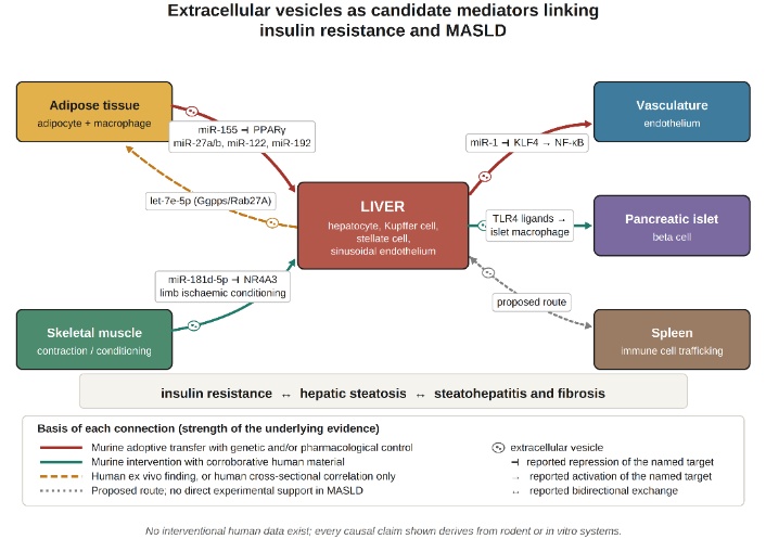
Metabolic dysfunction-associated steatotic liver disease (MASLD) is the most prevalent chronic hepatic disorder worldwide. Meta-analytic synthesis places its prevalence at approximately 30% of the adult population, the highest regional estimates having been reported in Latin America and in the Middle East and North Africa [1]. An earlier meta-analysis derived a pooled global prevalence of approximately 25% [2], and the intervening increase, together with projected growth in disease burden and associated expenditure, has been documented in subsequent analyses [3, 4]. This trajectory has paralleled that of obesity and of type 2 diabetes mellitus with sufficient fidelity that the three conditions are frequently considered interlocking manifestations of a single cardiometabolic phenotype [3].
MASLD is not confined in its consequences to the liver. Excess cardiovascular mortality and an association with malignant complications have been documented [5], and an elevated risk of both hepatic and extrahepatic malignancy has been described specifically [6]. The disorder is accordingly characterised in contemporary guidance as a multisystem condition [7, 8]. This conceptualization carries a mechanistic implication, namely that an adequate account of pathogenesis must address not only the intrahepatic accumulation of lipid but also the apparent coordination of metabolic dysfunction across anatomically remote tissues.
The cardiovascular dimension of this multisystem phenotype warrants particular emphasis, since cardiovascular disease rather than liver-related events accounts for the largest share of deaths in this population. Contemporary syntheses describe associations between MASLD and subclinical atherosclerosis, coronary artery disease, heart failure with preserved ejection fraction, atrial fibrillation and adverse myocardial remodelling [9–11]. Whether the excess risk is attributable to MASLD independently of shared cardiometabolic determinants remains contested, and reported effect estimates vary with the covariates entered into adjustment [10]. The mechanistic question raised by these observations is whether the diseased liver exports signals capable of modifying vascular and myocardial phenotype directly. Vesicular transfer of regulatory microRNA and of bioactive lipid has been proposed as one such route, and the evidence bearing upon it is examined in the Section Vesicular signalling and the cardiovascular consequences of MASLD.
Insulin resistance occupies a central position within this network. In the insulin-resistant liver, suppression of gluconeogenesis by insulin is attenuated whilst stimulation of de novo lipogenesis is preserved or augmented, a dissociation designated pathway-selective hepatic insulin resistance [12]; the consequence is concurrent inappropriate hepatic glucose output and accelerated triglyceride synthesis. Impaired insulin-mediated restraint of adipocyte lipolysis increases delivery of non-esterified fatty acids to the liver, whilst reduced insulin-stimulated glucose disposal in skeletal muscle redirects substrate towards hepatic lipogenic pathways [7, 8, 13, 14]. Insulin resistance is therefore positioned as both antecedent and consequence of hepatic steatosis.
The relationship between MASLD and type 2 diabetes mellitus is likewise reciprocal rather than unidirectional. Hepatic steatosis is present in the majority of patients with type 2 diabetes, the presence of type 2 diabetes is associated with more advanced hepatic fibrosis and with greater liver-related morbidity, and MASLD is in turn associated with subsequent incident diabetes [9, 15]. Hepatic lipotoxicity, pathway-selective hepatic insulin resistance and progressive beta-cell dysfunction have been proposed as the pathogenic substrate shared by the two conditions, and glucose-lowering classes including glucagon-like peptide-1 receptor agonists, dual glucose-dependent insulinotropic polypeptide and glucagon-like peptide-1 receptor agonists, and sodium-glucose cotransporter-2 inhibitors have accordingly been evaluated for concurrent hepatic benefit [15]. Whether vesicular signalling contributes to this reciprocity is considered in Sections Reciprocal hepatic vesicular output and Vesicles as therapeutic agents.
The mediators customarily invoked in explanation of this circularity are predominantly soluble, comprising non-esterified fatty acid flux, altered adiponectin and leptin secretion, and elevated tumour necrosis factor-alpha and interleukin-6 concentrations. The pathogenic relevance of these mediators is well established [16]. The explanatory model they sustain is, however, essentially one of diffuse humoral signalling, and such a model accounts less readily for three observations: the installation of a specific transcriptional programme within one recipient tissue and not another; the traversal of the extracellular space by regulatory information encoded within nucleic acids without attendant degradation; and alteration in the informational content of a signal without corresponding alteration in its bulk concentration.
Extracellular vesicles (EVs) are relevant to each of these considerations. EVs comprise lipid bilayer-delimited particles secreted by most cell types, and encompass endosome-derived exosomes, plasma membrane-derived microvesicles or ectosomes, and apoptotic bodies [17–19]. Their cargo, which includes proteins, lipids, metabolites and several RNA classes, is not a random sampling of the cytosol but is loaded through partially characterised sorting machinery, and its composition varies with the state of the parent cell [20, 21]. Since the vesicular membrane both protects the enclosed cargo and bears surface determinants that influence tissue tropism, EVs afford a candidate mechanism for addressed, information-dense signalling distinct from the diffusion of a soluble cytokine.
The present review examines the evidence bearing upon the proposition that EVs contribute to the relationship between insulin resistance and MASLD. Consideration is given successively to vesicular biogenesis and cargo selection; to the insulin signalling substrate upon which vesicular cargo is proposed to act; to vesicle-mediated communication among adipose tissue, skeletal muscle, liver and pancreatic islets; to intrahepatic vesicular networks; and to diagnostic and therapeutic prospects. Throughout, the experimental design underlying each claim is stated, since the strength of the inference that may be drawn varies substantially between studies.
Three considerations account for the timing of this review and for its format. First, the vesicular literature on metabolic liver disease has expanded rapidly whilst the reporting standards governing it have been revised, most recently in 2023, with the consequence that a substantial proportion of earlier work does not satisfy current characterisation requirements and warrants appraisal on that basis rather than uncritical aggregation [17, 22]. Second, the adoption of MASLD nomenclature in 2023 altered the composition of study cohorts, so that the correspondence between older and newer evidence requires explicit examination rather than assumption [23]. Third, existing reviews of EVs in metabolic liver disease have been organised principally by vesicle source or by cargo class [24]; the present review is instead organised around the insulin resistance axis, and for each claim states the experimental design from which it derives together with the strength of inference that design supports. A narrative rather than systematic format was adopted because the constituent studies differed irreducibly in species, separation method, administered dose and endpoint, such that quantitative pooling would impose a spurious commensurability upon them. The corresponding limitation is that study selection, although guided by the criteria stated in Section Methodological approach to the literature, was not governed by a protocol-driven screening process, and the possibility of selection bias cannot be excluded.
This work constitutes a narrative rather than a systematic review, and no formal quantitative synthesis was undertaken. The literature base was assembled through searches of PubMed, Scopus and Web of Science, in which terms denoting the vesicular compartment (EV, exosome, microvesicle, ectosome, microparticle) were combined with terms denoting the metabolic phenotype [insulin resistance, MASLD, metabolic dysfunction-associated fatty liver disease (MAFLD), non-alcoholic fatty liver disease (NAFLD), metabolic dysfunction-associated steatohepatitis (MASH), non-alcoholic steatohepatitis (NASH), hepatic steatosis, steatohepatitis, obesity, type 2 diabetes] and with terms denoting candidate cargo classes (microRNA, ceramide, sphingosine-1-phosphate, proteomics, lipidomics). Reference lists of retrieved reviews and primary reports were screened manually, and consensus documents issued by the International Society for Extracellular Vesicles and by the principal hepatology societies were retrieved directly from their official websites.
Precedence was accorded to primary investigations employing interventional designs, comprising genetic deletion, pharmacological inhibition of vesicular release, cargo depletion using antagomirs, or adoptive transfer of purified vesicles, since these designs support inference concerning causation more strongly than observational designs. Preference was likewise accorded to reports in which findings in animal models were accompanied by corroborative measurements in human tissue or plasma. Individual reports were appraised against the reporting standards articulated in the successive Minimal Information for Studies of Extracellular Vesicles statements [17, 22], and investigations lacking basic particle characterization, or lacking control for co-isolating non-vesicular material, were treated with greater circumspection. Where a finding is cited from a secondary source rather than from the primary report, this is stated explicitly in the text.
In preparing the present version, findings previously cited from secondary sources were re-examined against the primary reports wherever those reports could be retrieved and verified, and the corresponding statements have been revised to reflect the primary evidence together with its methodological limitations. Where a primary report could not be verified, the secondary attribution has been retained, identified as such at the point of use, and assigned correspondingly lower evidentiary weight.
Three overlapping disease designations appear within the literature under consideration. The historical term NAFLD rested upon an exclusionary definition requiring absence of significant alcohol intake and of competing hepatic disorders [25]. In 2020, an international expert panel proposed MAFLD, in which exclusion was replaced by positive criteria comprising hepatic steatosis together with overweight or obesity, type 2 diabetes mellitus, or evidence of metabolic dysregulation, and in which coexistence with other hepatic disorders was permitted [26, 27]. In 2023, a multisociety Delphi process adopted MASLD, defined as steatosis with at least one of five cardiometabolic criteria; steatohepatitis was retained as MASH, and the category of metabolic and alcohol-related liver disease (MetALD) was introduced for patients exhibiting cardiometabolic risk together with greater alcohol consumption [23].
For vesicular research, these distinctions are of practical consequence. The three frameworks enroll overlapping but non-identical populations, and since circulating EV concentration and cargo composition vary with adiposity, glycemic status, and alcohol exposure, discrepant biomarker findings across studies may in part reflect differences in cohort definition. Investigations published before 2023 almost invariably employed NAFLD criteria; the majority of such patients satisfy MASLD criteria [23], which supports broad continuity of the evidence base, although the correspondence is not exact. MASLD is employed herein as the preferred designation, with MAFLD or NAFLD retained where fidelity to a cited investigation requires it. The frameworks are summarised in Table 1.
Evolution of diagnostic frameworks for metabolic fatty liver disease and their relevance to extracellular vesicle research.
| Designation (year) | Originating framework | Diagnostic logic | Consequence for EV research |
|---|---|---|---|
| NAFLD (from 1980) | Descriptive; subsequently codified in society guidance | Hepatic steatosis with exclusion of significant alcohol intake and of competing aetiologies | Exclusionary criteria yield cohorts heterogeneous with respect to metabolic burden, impairing comparability of vesicular endpoints |
| MAFLD (2020) | International expert consensus, Asia-Pacific-led | Steatosis with overweight or obesity, type 2 diabetes, or two or more markers of metabolic dysregulation; coexisting liver disease permitted | Positive metabolic criteria enrich for the insulin-resistant phenotype of principal relevance to vesicle-mediated communication |
| MASLD, MASH, MetALD (2023) | Multisociety Delphi consensus (AASLD, EASL, ALEH) | Steatosis with at least one of five cardiometabolic criteria; MetALD delineates the group with higher alcohol intake | Delineation of MetALD separates a group in which alcohol exposure independently influences hepatocyte vesicular release |
AASLD: American Association for the Study of Liver Diseases; ALEH: Latin American Association for the Study of the Liver; EASL: European Association for the Study of the Liver; MetALD: metabolic and alcohol-related liver disease.
Accordingly, each clinical cohort discussed below is described using the diagnostic framework applied in the original report, and that framework is stated explicitly wherever a cohort finding is cited. Terminology has not been substituted retrospectively: an investigation conducted under NAFLD criteria is described as such, and its findings are not represented as MASLD findings. Where results obtained under different frameworks are discussed together, no equivalence of phenotype is assumed, since the exclusion of significant alcohol intake under NAFLD criteria, the permissive treatment of concomitant liver disease under MAFLD criteria and the separation of MetALD under MASLD criteria each define a different population [23, 25, 26]. This constraint bears directly upon the comparability of vesicular endpoints, since circulating vesicle concentration and cargo composition vary with adiposity, glycaemic status and alcohol exposure.
EVs are conventionally divided into exosomes, generated within the endosomal system, and microvesicles or ectosomes, which bud from the plasma membrane; apoptotic bodies constitute a larger class released during programmed cell death [18, 19, 28]. Exosome diameters are customarily cited as approximately 30 to 150 nm and microvesicle diameters as approximately 100 to 1,000 nm, although reported ranges vary between investigations and the populations overlap substantially [21, 29].
This overlap carries a methodological consequence. Since no currently available preparative method separates vesicles strictly according to biogenetic route, the successive Minimal Information for Studies of Extracellular Vesicles statements recommend that authors employ operational descriptors reflecting the property actually measured, such as small or large EVs defined by a stated size threshold, or vesicles defined by density or by a specified surface marker, rather than assigning biogenetic labels such as exosome on the basis of size alone [17, 22]. Much of the literature discussed below employs the term exosome for preparations obtained by differential ultracentrifugation or polymer precipitation, procedures recovering heterogeneous small-EV populations. Statements concerning exosome-specific mechanisms should therefore be read as statements concerning small-EV fractions, unless independent evidence of endosomal origin was presented. The classification adopted herein is summarised in Table 2.
Operational classification of EV subclasses and their salient features in insulin resistance and MASLD.
| Operational class | Approximate size | Principal biogenesis | Representative markers | Reported relevance to insulin resistance and MASLD |
|---|---|---|---|---|
| Small EVs (exosome-enriched) | 30–150 nm | Inward budding of the endosomal membrane forming intraluminal vesicles within multivesicular bodies; release on fusion with the plasma membrane | CD9, CD63, CD81, TSG101, ALIX, syntenin-1 | Principal fraction studied in microRNA-mediated inter-organ signalling; adipose tissue identified as a quantitatively major systemic source |
| Medium and large EVs (microvesicles, ectosomes) | 100–1,000 nm | Direct outward budding and scission of the plasma membrane; cytoskeletal and calcium-dependent | Annexin A1, integrins, tissue-specific surface antigens, exposed phosphatidylserine | Amenable to flow cytometric detection, permitting phenotyping of cellular origin; hepatocyte-derived populations reported to track injury |
| Apoptotic bodies | 800–5,000 nm | Membrane blebbing during apoptosis | Phosphatidylserine, histones, fragmented organelles and DNA | Reported to increase in advanced steatohepatitis; carriers of mitochondrial DNA and other damage-associated molecular patterns |
| Non-vesicular co-isolates | Variable | Lipoproteins, protein aggregates, exomeres | Apolipoprotein B, apolipoprotein A-I, albumin | Principal confounder in MASLD, dyslipidaemia increasing lipoprotein burden and potentially inflating apparent vesicle concentration and RNA yield |
ALIX: ALG-2-interacting protein X; CD9, CD63, CD81: cluster of differentiation 9, 63 and 81; TSG101: tumour susceptibility gene 101.
This convention is applied consistently throughout. Where a cited investigation employed differential ultracentrifugation, polymer precipitation or a proprietary capture reagent without demonstrating endosomal derivation, the preparation is described as a small-EV fraction; the term exosome is reserved for preparations in which endosomal origin was established by independent means. The distinction is not merely terminological. Mechanisms attributed to exosome biology on the basis of small-EV data may instead reflect the behaviour of co-isolated ectosomes, exosomes or lipoprotein particles, and this ambiguity constrains the strength of the inferences drawn in Sections Vesicle-mediated inter-organ communication and insulin resistance and Intrahepatic vesicular networks in the progression of MASLD. Statements concerning exosome-specific mechanisms should therefore be read as provisional wherever the underlying separation protocol was non-specific. The relationship between biogenetic route, separation method and the particle population actually recovered is summarised in Figure 1.
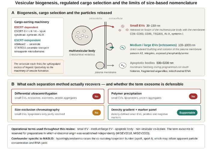
Vesicular biogenesis, regulated cargo selection and the limits of size-based nomenclature. (A) Intraluminal vesicles destined to become small EVs are generated within multivesicular bodies through ESCRT-dependent routes and through ESCRT-independent routes, the latter including the neutral sphingomyelinase 2-ceramide pathway and STARD11-mediated ceramide transport, which are of particular relevance to hepatic lipotoxicity. Small EVs are released on fusion of the multivesicular body with the plasma membrane; medium and large EVs arise by direct outward budding and scission of the plasma membrane; apoptotic bodies arise by membrane blebbing during programmed cell death. Approximate size ranges and representative markers are stated for each class. (B) Because no preparative method separates vesicles strictly according to biogenetic route, the particle population actually recovered is determined by the separation protocol employed, and only density-gradient separation combined with a positive and negative marker panel supports an inference of endosomal origin. The term exosome is accordingly reserved in this review for preparations in which endosomal origin was established independently. A confounder specific to this disorder is noted: hypertriglyceridaemia raises the lipoprotein burden co-isolating with EVs and may inflate apparent particle concentration and RNA yield. ESCRT: endosomal sorting complex required for transport; STARD11: StAR-related lipid transfer domain protein 11.
Intraluminal vesicles that subsequently constitute small EVs are generated principally through the endosomal sorting complex required for transport (ESCRT). ESCRT-0 clusters ubiquitinated cargo upon the endosomal membrane; ESCRT-I and ESCRT-II deform the membrane and concentrate cargo within the forming bud; and ESCRT-III, acting with the AAA-ATPase vacuolar protein sorting-associated protein 4 (Vps4), executes scission [30–33]. Recruitment kinetics of the late-acting machinery are signal-responsive, providing one mechanism whereby extracellular stimuli may modulate vesicular output [34]. An accessory pathway involving the heparan sulfate proteoglycan syndecan, the cytoplasmic adaptor syntenin and the ESCRT-associated protein ALG-2-interacting protein X (ALIX) supports intraluminal budding independently of the canonical sequence [35].
ESCRT-independent routes are of particular relevance to metabolic disease, several being lipid-driven. Ceramide generated by neutral sphingomyelinase promotes coalescence of membrane microdomains and induces negative membrane curvature, favouring intraluminal vesicle formation; tetraspanin-organised microdomains and lipid rafts afford additional sorting platforms [20, 21]. In hepatocytes, ceramide transport mediated by StAR-related lipid transfer domain protein 11 was shown to be required for vesicular biogenesis [36], establishing a biochemical relationship between the sphingolipid excess characteristic of hepatic lipotoxicity and the machinery of vesicle formation [37].
Following release, EVs engage recipient cells through receptor–ligand interaction, direct membrane fusion, or endocytosis, and this tropism is determined principally by surface composition rather than by enclosed cargo [18, 29]. Identical cargo conveyed by vesicles bearing different surface determinants may therefore yield dissimilar tissue distributions, a consideration bearing upon interpretation of biomarker data and upon the design of engineered therapeutic vesicles.
Insulin binding activates the receptor tyrosine kinase, which phosphorylates insulin receptor substrate (IRS) proteins, recruiting phosphoinositide 3-kinase (PI3K) and generating phosphatidylinositol-3,4,5-trisphosphate. Protein kinase B (AKT) is thereby activated and mediates translocation of glucose transporter type 4 (GLUT4) to the plasma membrane in muscle and adipose tissue, inactivates forkhead box protein O1 with consequent suppression of hepatic gluconeogenic gene transcription, and inhibits glycogen synthase kinase 3 [14].
Three features of this architecture are relevant to the manner in which vesicular cargo might perturb it. First, the pathway is a phosphorylation cascade with a small number of obligatory nodes, such that modest reductions in IRS-1 or AKT abundance yield disproportionate loss of downstream signal. Second, several nodes are subject to post-transcriptional regulation by microRNA, IRS-1, AKT, phosphatase and tensin homolog and peroxisome proliferator-activated receptor-gamma (PPAR-gamma) each carry validated microRNA recognition elements, and each is therefore accessible in principle to vesicular delivery of a single regulatory species [38]. Third, the pathway bifurcates downstream of AKT, suppression of gluconeogenesis and stimulation of lipogenesis being subserved by separable downstream branches, a configuration permitting the pathway-selective hepatic insulin resistance described above [12, 14].
Superimposed upon transcriptional regulation is direct lipid interference. Ceramide species antagonise insulin signalling through protein phosphatase 2A-mediated dephosphorylation of AKT and through protein kinase C-zeta-dependent sequestration, whilst diacylglycerol accumulation activates protein kinase C isoforms that inhibit IRS-1 by serine phosphorylation [14]. Since ceramide is both a component of the vesicular membrane and a driver of vesicular biogenesis, EVs may represent simultaneously a vehicle for, and a product of, sphingolipid excess [36, 37].
EVs are best regarded not as an alternative to established mechanisms but as an additional signalling modality operating alongside hormones, cytokines and circulating metabolites, distinguished by the capacity to deliver combinatorial cargo to defined recipient populations. This distinction bears upon the interpretation of negative findings, since a vesicle preparation may fail to alter systemic glucose tolerance whilst nonetheless altering the phenotype of a specific recipient cell type.
Mice bearing adipose-specific deletion of the microRNA-processing enzyme Dicer, together with human subjects affected by lipodystrophy, exhibited substantially reduced circulating small-EV microRNA. Transplantation of adipose tissue, the brown depot proving most effective, restored circulating microRNA concentrations in association with improved glucose tolerance and reduced hepatic fibroblast growth factor 21 expression; the transcriptional effect was reproduced by administration of serum small-EV fractions from control but not from Dicer-deleted animals. Expression of a human-specific microRNA in donor brown adipose tissue regulated a corresponding reporter in recipient liver, which the authors interpreted as evidence of functional inter-organ transfer [39]. These experiments support the designation of adipose tissue as a quantitatively major source of circulating small-EV microRNA in the mouse; the corresponding quantitative contribution in humans has not been established.
Human ex vivo work has addressed functional consequences directly. EVs released from subcutaneous and omental adipose tissue explants inhibited insulin-induced AKT phosphorylation in hepatocytes for the majority of donors, and the degree of inhibition correlated inversely with expression of gluconeogenic genes; interleukin-6 and macrophage migration inhibitory factor concentrations were higher in omental than in subcutaneous vesicles and related to the magnitude of the hepatocyte effect. The response in myotubes was inconsistent, and vesicles from a minority of donors enhanced rather than suppressed hepatic insulin signalling [40]. This heterogeneity indicates that adipose vesicular output is not uniformly deleterious and that donor-level variation is appreciable.
Circulating small-EV microRNA profiles in diet-induced obese mice showed enrichment of miR-122, miR-192, miR-27a-3p and miR-27b-3p. Repeated administration to lean animals of vesicles loaded with mimics of these species produced glucose intolerance, dyslipidaemia, adipose inflammation and hepatic steatosis, peroxisome proliferator-activated receptor-alpha (PPAR-alpha) being identified as the candidate target, its expression falling in white adipose tissue but not in liver, and vesicles transfected with a PPAR-alpha-directed small interfering RNA reproducing the phenotype; the phenotype was attenuated by a lipolysis inhibitor or a PPAR-alpha agonist [41]. Since loaded vesicles were administered at defined doses, this design supports a causal contribution of the transferred species within the model employed, though not necessarily at endogenous vesicular concentrations. Obesity has additionally been reported to modify adipose EV protein cargo [42].
Small EVs secreted by adipose tissue macrophages of obese mice induced glucose intolerance and insulin resistance upon administration to lean recipients, whereas transfer of vesicles from lean donors into obese recipients improved glucose tolerance and insulin sensitivity. miR-155, over-represented in obese macrophage vesicles, was implicated through repression of PPAR-gamma; miR-155-deficient animals were insulin-sensitive and glucose-tolerant, and reconstitution with wild-type bone marrow reinstated the phenotype. Pre-treatment of donor macrophages with the neutral sphingomyelinase 2 inhibitor GW4869 abolished cargo transfer [43].
The design of this investigation combines purification and transfer of the putative mediator, reproduction of the phenotype in a naive host, bidirectionality, identification of a candidate cargo species, genetic deletion of that species, and pharmacological confirmation of vesicle dependence. Within the murine system employed, these elements support a causal interpretation more strongly than is typical of the field. Two qualifications nonetheless apply: GW4869 inhibits sphingomyelinase activity broadly rather than vesicle release specifically, and no equivalent human experiment has been performed.
A single hour of cycling exercise in healthy volunteers increased the circulating abundance of more than three hundred proteins, with enrichment for exosomal and small-vesicle constituents; pulse-chase and intravital imaging indicated that vesicles liberated by exercise localise preferentially to the liver and transfer protein cargo, and arteriovenous balance measurements across the contracting limb identified candidate myokines released independently of the classical secretory pathway [44]. This study establishes that exercise alters the circulating vesicular proteome in humans; it does not establish a metabolic consequence of that alteration.
Two further reports have been appraised from the primary literature rather than from secondary sources. In an investigation combining a twelve-week aerobic exercise intervention in patients with MAFLD with high-fat diet-fed mice, exercise reduced hepatic fat content and improved insulin sensitivity; small-EV miR-324 was lower in trained than in sedentary participants, and precursor and mature miR-324 increased in adipose tissue whilst decreasing in muscle after exercise, which the authors interpreted as indicating an adipose rather than a myocellular origin. Luciferase reporter and immunoblot assays identified Rho-associated coiled-coil-containing protein kinase 1 as a direct target through which miR-324 modified insulin resistance [45]. Separately, remote limb ischemic conditioning attenuated steatohepatitis in several murine models; the effect was mediated by muscle-to-liver transfer of small EVs with consequent hepatic accumulation of miR-181d-5p, hepatic overexpression of miR-181d-5p reproduced the histological and transcriptional benefit through suppression of nuclear receptor 4A3, and circulating EVs obtained from human volunteers undergoing limb ischemic conditioning improved steatohepatitis in animal models and transcriptomic perturbation in primary human hepatocytes [46]. The latter finding is sometimes attributed to exercise; the intervention studied was transient limb ischaemia, and the two stimuli are not equivalent. Neither report demonstrates that the endogenous vesicular flux generated by habitual physical activity in humans is sufficient to modify hepatic phenotype.
Hepatic involvement in vesicular communication is not confined to reception. Lipid overload upregulated hepatic geranylgeranyl diphosphate synthase, which, through geranylgeranylation of Rab27A (Ras-related protein Rab-27A), modified secretion of microRNA-loaded vesicles; among the species released, let-7e-5p promoted lipid accumulation in adipocytes. Plasma EV let-7e-5p concentrations correlated with hepatic geranylgeranyl diphosphate synthase expression, body mass index, and hepatic triglyceride content in patients with NAFLD, the diagnostic framework in force when that cohort was reported [47]. The murine component of this work is interventional; the human component is cross-sectional and correlative.
A second reciprocal axis engages the endocrine pancreas. Lipotoxic hepatocyte-derived EVs induced inflammation in islet macrophages through Toll-like receptor 4 (TLR4)-dependent signalling, with consequent impairment of beta-cell function and insulin secretion; dependence upon macrophage TLR4 was established using genetic and pharmacological approaches [48]. A related report implicating vesicular miR-126a-3p in beta-cell apoptosis is cited from secondary literature [24]. These observations describe a candidate route by which hepatic steatosis might contribute to progressive beta-cell failure, though the quantitative contribution relative to established mechanisms such as glucolipotoxicity is unknown.
These vesicular observations sit within a broader bidirectional relationship between the two conditions. Type 2 diabetes and MASLD share hepatic lipotoxicity, pathway-selective hepatic insulin resistance and progressive beta-cell failure as pathogenic substrate, and each is associated with a worse trajectory of the other [15]. Non-invasive assessment in this population currently proceeds by sequential application of the fibrosis-4 index followed by elastography or a proprietary serum panel, an approach that excludes advanced fibrosis with acceptable negative predictive value but performs less well within the intermediate range [9, 15]. A vesicular contribution to the reciprocity is plausible on the evidence reviewed above, in that lipotoxic hepatocyte vesicles impaired islet function in mice [48] whilst adipose and macrophage vesicles impaired hepatic insulin signalling [40, 43]. It has not, however, been shown in humans that vesicular flux is either necessary for, or quantitatively sufficient to account for, the progression of either condition, and no vesicular measurement has been demonstrated to add predictive information beyond the established non-invasive tests.
The evidentiary basis is not uniform across the axes described. The proposition that hepatocyte-derived EVs directly impair insulin signalling in skeletal muscle and adipose tissue, although mechanistically plausible, is supported by considerably less direct evidence than the adipose-to-hepatic and hepatic-to-islet directions. Representative cargoes, together with the designs from which the associated claims derive, are collated in Table 3.
Representative EV cargoes implicated in the relationship between insulin resistance and MASLD, with the experimental design underlying each claim.
| EV cargo | Principal source | Proposed target or pathway | Reported effect | Design and strength of inference |
|---|---|---|---|---|
| miR-155 | Adipose tissue macrophages | Repression of PPAR-gamma | Glucose intolerance and insulin resistance in lean recipients; reversed with lean-donor vesicles | Adoptive transfer, genetic deletion, marrow reconstitution, GW4869; strongest causal design in the field, murine only [43] |
| miR-122, miR-192, miR-27a/b | Adipose tissue and liver | PPAR-alpha (Ppara) in white adipose tissue | Glucose intolerance, dyslipidaemia, adipose inflammation, hepatic steatosis after mimic-loaded transfer | Loaded-vesicle transfer with pharmacological rescue; causal within model, supraphysiological dosing [41] |
| Adipokine-bearing EVs (IL-6, MIF, MCP-1) | Human adipose explants | Insulin-induced AKT phosphorylation | Reduced hepatic insulin signalling; response heterogeneous, a minority of donors showing the opposite effect | Ex vivo human explant vesicles on cell lines; associative, no in vivo endpoint [40] |
| let-7e-5p | Hepatocytes (Ggpps/Rab27A-dependent) | Adipocyte lipogenic programme | Increased adipocyte lipid accumulation; plasma levels track BMI and hepatic triglyceride | Murine interventional plus human cross-sectional correlation [47] |
| TLR4 ligands | Lipotoxic hepatocytes | Islet macrophage TLR4 | Islet inflammation, impaired insulin secretion | Murine, with genetic and pharmacological TLR4 dependence demonstrated [48] |
| Ceramide and sphingosine-1-phosphate | Palmitate-stressed hepatocytes | S1P receptor 1 on macrophages | Macrophage chemotaxis; cargo depletion abrogates the effect | Knockout hepatocytes, chemotaxis assay, human plasma correlation [37, 49] |
| TRAIL | Lipotoxic hepatocytes (DR5/ROCK1) | Macrophage death receptor 5 | IL-1-beta and IL-6 induction; ROCK1 inhibition reduced vesicle release, injury and fibrosis in vivo | In vitro pathway inactivation plus in vivo pharmacological intervention [50] |
| CXCL10 | Lipotoxic hepatocytes (MLK3) | Macrophage CXCR3 | Innate immune chemotaxis; macrophage-associated inflammation in murine MASH | Genetic and pharmacological MLK3 inhibition; CXCL10-deficient models [51, 52] |
| miR-192-5p; miR-9-5p | Lipotoxic hepatocytes | Rictor/AKT/FoxO1 | M1 polarisation; serum miR-192-5p correlates with inflammatory activity | Murine mechanism plus human serum correlation [53, 54] |
| miR-128-3p | Fat-laden hepatocytes | Stellate cell PPAR-gamma | Stellate activation, proliferation, chemotaxis; upregulated collagen I, alpha-SMA, TIMP-2 | Loss- and gain-of-function in cultured stellate cells; in vitro only [55, 56] |
BMI: body mass index; CXCL10: C-X-C motif chemokine ligand 10; CXCR3: C-X-C motif chemokine receptor 3; DR5: death receptor 5; FoxO1: forkhead box protein O1; Ggpps: geranylgeranyl diphosphate synthase; IL-6: interleukin-6; MCP-1: monocyte chemoattractant protein-1; MIF: macrophage migration inhibitory factor; MLK3: mixed lineage kinase 3; Rab27A: Ras-related protein Rab-27A; ROCK1: Rho-associated coiled-coil-containing protein kinase 1; S1P: sphingosine-1-phosphate; TIMP-2: tissue inhibitor of metalloproteinases 2; TLR4: Toll-like receptor 4; TRAIL: tumour necrosis factor-related apoptosis-inducing ligand.
Cardiovascular disease is the leading cause of death among patients with MASLD, and the mechanistic question is whether hepatic and extrahepatic vesicular flux contributes to vascular injury beyond the contribution of shared cardiometabolic risk factors [9, 10].
Direct evidence derives principally from murine work. EVs released by steatotic hepatocytes induced an inflammatory endothelial phenotype and accelerated atherogenesis; miR-1 was identified as the operative cargo, acting through suppression of Kruppel-like factor 4 with consequent nuclear factor-kappa B activation, and administration of a miR-1 antagomir attenuated atherosclerotic lesion formation in apolipoprotein E (apoE)-deficient mice [57]. Further hepatocyte-derived species reported to act upon the vasculature, including miR-30a-3p, described as inhibiting adenosine triphosphate (ATP)-binding cassette transporter A1-mediated cholesterol efflux and thereby favouring foam cell formation, together with vesicular contributions to osteogenic differentiation of vascular smooth muscle cells, are cited from a recent comprehensive review rather than appraised from the primary reports [24].
Three qualifications attach to this evidence. First, the experiments were conducted in mice, predominantly on apoE- or low-density lipoprotein receptor-deficient backgrounds that model an atherogenic lipid profile unrepresentative of most patients with MASLD. Second, the administered vesicle doses exceeded plausible endogenous flux, so that the demonstrations concern sufficiency under imposed conditions rather than necessity at physiological concentrations. Third, no human study has shown that a circulating vesicular measure predicts cardiovascular events in MASLD independently of established risk factors. The vesicular account is therefore best regarded at present as mechanistically coherent but awaiting human corroboration, rather than as an established contributor to the excess cardiovascular risk observed in this population [10, 11].
Attention has recently been directed towards the spleen as a participant in metabolic liver disease. The spleen contributes to the clearance of circulating apoptotic material and to the differentiation and trafficking of monocyte and lymphocyte populations, and splenic volume and stiffness have been reported to track hepatic disease severity; a bidirectional spleen-liver axis has accordingly been proposed, encompassing splenic modulation of hepatic immune tone, hepatic modulation of splenic immunometabolism, and the therapeutic implications that would follow [58].
A vesicular formulation of this axis is mechanistically plausible but remains largely inferential. Its demonstration would require that hepatocyte- and adipocyte-derived EVs reach splenic macrophage and natural killer T cell populations in functionally relevant quantities, that the resulting splenic output alters hepatic macrophage and stellate cell behaviour, and that interruption of vesicular traffic between the two organs modifies hepatic phenotype. None of these requirements has been demonstrated directly in MASLD. A specific interpretative caution applies: the spleen receives a substantial proportion of intravenously administered vesicle preparations through uptake by its resident macrophage populations, so that splenic accumulation in biodistribution studies is expected on that basis and does not by itself indicate a signalling relationship. The axis is included here because it identifies a testable gap rather than because a vesicular mechanism has been established, and the description of a continuous inter-organ loop should not be read as implying demonstrated causation.
The inter-organ vesicular relationships discussed in the Section Vesicle-mediated inter-organ communication and insulin resistance, together with the basis on which each has been proposed, are summarised in Figure 2.
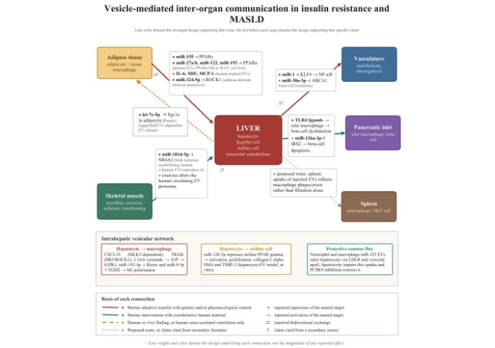
Vesicle-mediated inter-organ communication in insulin resistance and MASLD. Line colour and style denote the strongest experimental design supporting each route, and the coloured dot preceding each named cargo denotes the design supporting that particular claim; neither denotes the magnitude of any reported effect. Four tiers are distinguished: murine adoptive transfer with genetic and/or pharmacological control; murine intervention accompanied by corroborative human material; human ex vivo findings or human cross-sectional correlation alone; and proposed routes, together with claims cited from secondary literature, which are additionally marked with a dagger. Cargo species are named where a specific molecular mediator has been nominated, and the barred symbol denotes reported repression of the indicated target. The section in which each route is examined is stated within the corresponding label. Broken grey lines denote routes that have been proposed but for which direct experimental support in MASLD is absent, including the spleen-liver axis discussed in the Section The spleen-liver axis as a candidate site of vesicular exchange. The lower panel summarizes the intrahepatic vesicular network considered in the Section Intrahepatic vesicular networks in the progression of MASLD.
Within the liver, lipotoxicity has been reported to alter vesicular biology in two respects. Hepatocytes exposed to saturated fatty acids, lysophosphatidylcholine, oxidative stress or hypoxia increased vesicular release; separately, the molecular composition of those vesicles was altered, specific species being enriched rather than the basal cargo being proportionately scaled [50, 59]. If confirmed, this bears upon biomarker design, since particle enumeration alone would capture only part of the available information.
Several stress-responsive pathways have been characterised. Palmitate-induced endoplasmic reticulum stress drove vesicular release through inositol-requiring enzyme 1-alpha and X-box binding protein 1, the resulting vesicles being enriched in ceramide and its metabolite sphingosine-1-phosphate; the requirement for inositol-requiring enzyme 1-alpha was established using knockout hepatocytes [37]. The same axis was subsequently examined in vivo, inositol-requiring enzyme 1A stimulating release of pro-inflammatory hepatocyte vesicles in murine steatohepatitis, with correlative human data reported [59]. Ceramide loading required StAR-related lipid transfer domain protein 11 [36]. Vesicular sphingosine-1-phosphate acted through sphingosine-1-phosphate receptor 1 to drive macrophage chemotaxis, and depletion of vesicular sphingosine-1-phosphate abrogated the effect [49].
Lipid stimulation of death receptor 5 with downstream Rho-associated coiled-coil-containing protein kinase 1 activity promoted release of vesicles bearing tumour necrosis factor-related apoptosis-inducing ligand, which induced interleukin-1-beta and interleukin-6 expression in bone marrow-derived macrophages through death receptor 5 and receptor-interacting protein kinase 1. Administration of the Rho-associated kinase inhibitor fasudil to mice with steatohepatitis reduced serum vesicle concentrations together with hepatic injury, inflammation, and fibrosis [50]. Since fasudil has actions independent of vesicle release, this experiment is consistent with, but does not isolate, a vesicular mechanism. A third axis operates through mixed lineage kinase 3, which mediates release of C-X-C motif ligand 10-bearing vesicles chemotactic for innate immune cells [51]; the contribution of this chemokine was subsequently attributed specifically to macrophage-associated inflammation in murine steatohepatitis [52].
Beyond chemotaxis, hepatocyte-derived vesicles have been reported to influence macrophage phenotype. Vesicular miR-192-5p activated macrophages through Rictor/AKT/forkhead box protein O1 signalling, and serum concentrations of this species correlated with hepatic inflammatory activity in patients [53]. Vesicular miR-9-5p was likewise associated with macrophage activation [54]. Further hepatocyte-derived cargoes reported to activate macrophages, including mitochondrial DNA, retinol-binding protein 4 and integrin beta-1 mediating monocyte adhesion to sinusoidal endothelium, are cited from secondary literature [24].
Hepatocyte-derived miR-122 warrants separate comment. Enriched in and largely specific to hepatocytes, it was released in vesicles during hepatocellular stress and sensitized recipient monocytes to lipopolysaccharide in an alcohol-exposure model [60]; vesicle-associated matrix metalloproteinase 2 was required for transfer to hepatic macrophages, and inhibition of transfer reduced inflammation [61]. miR-122 may therefore function both as an injury marker and as a signalling molecule, which indicates that circulating concentrations should not be interpreted as purely passive indices [38].
Fibrogenesis requires transdifferentiation of quiescent hepatic stellate cells into an activated myofibroblast-like phenotype. Vesicles released by fat-laden hepatocytes were internalised by cultured stellate cells and induced activation, proliferation, chemotaxis and wound-healing responses, with upregulation of collagen I, alpha-smooth muscle actin and tissue inhibitor of metalloproteinases 2. These changes were associated with suppression of stellate cell PPAR-gamma by vesicle-borne microRNA; miR-128-3p was transferred most efficiently, and loss- and gain-of-function studies identified it as a principal modulator [55]. Small EVs from palmitate-treated hepatocytes similarly induced fibrotic activation of stellate cells [56]. Both experiments were conducted in vitro, and neither establishes that this pathway operates at physiological vesicle concentrations in vivo.
Additional vesicular mediators of stellate activation, including mannan-binding lectin serine protease 1, LIM domain and actin binding 1, miR-27a and miR-1297, are cited from secondary literature [24]. The convergence of multiple candidate cargoes upon stellate activation suggests redundancy, from which follows the therapeutic implication that strategies directed against a single cargo species may prove less effective than strategies directed against vesicular release or uptake.
An exclusively pathogenic account of vesicular signalling is not supported by the available data. Neutrophil-derived EVs enriched in miR-223 were selectively internalised by hepatocytes through a mechanism dependent upon hepatocyte low-density lipoprotein receptor and upon apoE carried on the vesicle surface, and this transfer attenuated experimental steatohepatitis; deletion of apoE in immune cells accelerated disease, whereas pharmacological upregulation of hepatocyte low-density lipoprotein receptor using a proprotein convertase subtilisin/kexin type 9 (PCSK9) inhibitor enhanced transfer and ameliorated disease [62]. Myeloid interleukin-6 signalling promoted release of miR-223-enriched small EVs from macrophages and neutrophils, which limited fibrosis through downregulation of the transcriptional co-activator with PDZ-binding motif [63]. Lipotoxic conditions have been reported to impair this route by inducing endoplasmic reticulum stress and PCSK9-mediated downregulation of hepatocyte low-density lipoprotein receptor; that experiment concerned macrophage-derived rather than neutrophil-derived vesicles, and the two sources should not be conflated [24]. Each of these experiments was conducted in mice or in cultured cells, and no human interventional evidence exists for a protective vesicular route.
Macrophage-derived vesicles appear bidirectional in effect, miR-155 aggravating hepatocyte pyroptosis whilst vesicles from M2-polarised macrophages enriched in oxysterol-binding protein-related protein 8 mitigated lipotoxicity. Liver sinusoidal endothelial cell vesicles restrained stellate and Kupffer cell activation under homeostatic conditions, whereas with capillarization the cargo shifted towards insulin-like growth factor binding protein 7 and a disintegrin and metalloproteinase with thrombospondin motifs 1. These findings are cited from a recent comprehensive review rather than appraised from the primary reports, and are assigned correspondingly lower evidentiary weight [24]. Macrophage neurogenic locus notch homologue protein 1 (Notch1) signalling has been reported to modulate regulatory T cells through a small-EV miR-142a-3p and transforming growth factor-beta receptor 1 axis in early disease [64].
It follows that a therapeutic objective of global suppression of vesicular traffic is unlikely to be appropriate, and that selective correction of the balance between injurious and protective flux constitutes the more defensible target.
The evidence reviewed is compatible with a model in which EVs participate in a feed-forward circuit rather than a linear conduit. This model is presented as a synthesis of predominantly rodent and in vitro data, and should be regarded as a hypothesis for testing rather than an established sequence in human disease. The model is depicted schematically in Figure 3.
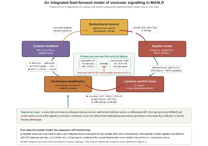
An integrated feed-forward model of vesicular signalling in MASLD. The model is presented as a hypothesis for testing rather than as an established sequence in human disease, and synthesises predominantly rodent and in vitro data; dot colours follow the evidence tiers defined in Figure 2. The central panel depicts the protective counter-flux and its failure: neutrophil- and macrophage-derived miR-223-bearing vesicles are internalised by hepatocytes through a mechanism dependent upon hepatocyte low-density lipoprotein receptor and vesicular apolipoprotein E, and lipotoxicity together with PCSK9 downregulates that receptor. Muscle-derived vesicles released during exercise and remote limb ischaemic conditioning are shown as an opposing input. Two features would render the sequence self-reinforcing: hepatic vesicular output feeds back upon adipose tissue and upon pancreatic islets, and lipotoxicity concurrently impairs uptake of protective miR-223-bearing vesicles, so that the ratio of injurious to protective flux would deteriorate more rapidly than either flux considered alone. Neither feature has been demonstrated in human disease.
Dysfunctional visceral adipose tissue, together with its macrophage infiltrate, exports vesicles reported to suppress insulin signalling in hepatocytes and myocytes, principally through repression of PPARs and of proximal nodes within the IRS/PI3K/AKT cascade [39–41, 43]. Hepatic insulin resistance accelerates de novo lipogenesis whilst impairing suppression of gluconeogenesis, and concurrent failure of insulin-mediated restraint of lipolysis increases delivery of non-esterified fatty acids [7]. Hepatocellular lipid excess generates lipotoxicity and endoplasmic reticulum stress, associated with increased hepatocyte vesicular output and altered cargo composition [37, 50, 51, 59]. Such vesicles have been reported to recruit and polarise macrophages, to activate hepatic stellate cells and to alter endothelial phenotype [49, 53, 55].
Two features would convert this sequence into a self-reinforcing circuit. First, hepatic vesicular output feeds back upon adipose tissue through let-7e-5p [47] and upon pancreatic islets through TLR4-dependent signalling [48]. Second, lipotoxicity has been reported concurrently to impair uptake of protective miR-223-bearing vesicles, such that the ratio of injurious to protective signalling would deteriorate more rapidly than either flux considered alone [24]. Muscle-derived vesicular species released in response to exercise may act upon the same circuit in the opposing direction [44].
The principal implication of this model, if substantiated, is that disease trajectory would be determined not solely by the quantity of circulating metabolic substrate but additionally by the informational content of signals exchanged between organs.
Liver biopsy remains imperfect as a reference standard, is invasive, and is unsuited to repeated assessment, whilst existing non-invasive scores perform only moderately for identification of steatohepatitis with clinically significant fibrosis. EVs have been proposed as candidate biomarkers by virtue of their stability in plasma, their reflection of parent cell state and their possession of surface determinants permitting inference of cellular origin [65, 66].
Circulating microRNA signatures encompassing miR-122, miR-34a and miR-192 have been associated with histological severity [67], and vesicle-associated liver microRNA together with proteomic signatures discriminated experimental fatty liver disease from control [68]. Serum EV proteomics identified fibulin-3 as a predictor of liver-related events in a prospective cohort [69]. Additional candidates, comprising EV size and concentration models, EpCAM-positive and CD133-positive vesicles, GLUT1-positive hepatogenic small EVs, leucocyte-derived vesicle counts, asialoglycoprotein receptor 1-positive vesicular microRNA and urinary EV lipidomic panels, are cited from a recent review [24]. Representative candidates are summarised in Table 4.
Representative EV-based candidate biomarkers in MASLD, with the design and stage of validation stated for each.
| Candidate marker | Compartment | Reported role | Design and stage of validation |
|---|---|---|---|
| Circulating miR-122, miR-34a, miR-192 | Serum or plasma, not vesicle-restricted | Associated with histological severity and hepatocyte ballooning | Cross-sectional, multiple independent cohorts; not EV-specific |
| Vesicular liver microRNA and EV proteome | Plasma, experimental model | Discriminated fatty liver disease from control | Murine discovery study; not validated in humans |
| Fibulin-3 | Serum EV proteome | Correlated with fibrosis progression; predicted liver-related events | Prospective outcome cohort; single cohort, external validation outstanding |
| EV concentration and size distribution | Plasma, total | Staging and identification of severe steatosis within machine-learning models | Single-cohort model development; cited from secondary literature |
| EpCAM-positive, CD133-positive EVs | Serum | Distinguished steatohepatitis from simple steatosis | Cross-sectional, biopsy-anchored; cited from secondary literature |
| GLUT1-positive hepatogenic exosomes | Plasma | Elevated in MASH; proposed for staging | Cross-sectional, biopsy-anchored; cited from secondary literature |
| ASGR1-positive vesicular miR-122, miR-192, miR-128-3p | Plasma, liver-specific vesicles | Tracked disease severity | Cross-sectional, biopsy-anchored; cited from secondary literature |
| Urinary EV lipidomic panel | Urine | Discriminated MASH from simple steatosis (AUC approximately 92%) | Single-cohort discovery with machine learning; overfitting not excluded |
ASGR1: asialoglycoprotein receptor 1; AUC: area under the receiver operating characteristic curve.
Two constraints attend interpretation. First, the majority of these candidates derive from single cohorts without external validation, and none has been shown to confer incremental value over established non-invasive tests within a prospective design. Second, and specific to this disorder, hyperlipidemia increases the lipoprotein burden co-isolating with EVs, such that apparent differences in vesicle concentration or RNA yield may in part reflect lipoprotein contamination [17].
Vesicular release is enzymatically regulated and therefore pharmacologically accessible in principle. Inhibition of Rho-associated kinase with fasudil reduced circulating vesicle concentrations together with hepatic injury, inflammation and fibrosis in murine steatohepatitis, although the agent has vesicle-independent actions [50]. Neutral sphingomyelinase 2 inhibition with GW4869 is the standard tool for establishing vesicle dependence in vitro and abolished macrophage-to-adipocyte cargo transfer [43]; its effects upon sphingolipid metabolism are broad, rendering it unsuitable as a therapeutic prototype. Reports that interleukin-22, a thiazolidinedione derivative and Semaglutide modulate disease in part through vesicular mechanisms are cited from secondary literature [24]; the possibility that agents already entering clinical practice act partly through this route [70] remains to be tested directly.
Mesenchymal stromal cell-derived EVs reduced hepatic lipid accumulation through suppression of mitochondrial fission in a murine model [71], and human induced pluripotent stem cell-derived vesicles reduced stellate activation and fibrosis [72]. The immunomodulatory properties of mesenchymal stromal cell vesicles and their clinical application in liver disease have been reviewed [73]. Clinical activity remains predominantly early-phase and largely outside MASLD; ambiguous regulatory classification, absence of scalable manufacturing with defined critical quality attributes, and uncertain batch-to-batch potency remain the principal obstacles [74].
A distinct translational question concerns whether agents already established in clinical practice for glycaemic control act in part through vesicular mechanisms. Glucagon-like peptide-1 receptor agonists, dual glucose-dependent insulinotropic polypeptide and glucagon-like peptide-1 receptor agonists, sodium-glucose cotransporter-2 inhibitors and thiazolidinediones improve hepatic steatosis and, for certain agents, histological features of steatohepatitis, which supports their consideration in patients with concurrent type 2 diabetes for whom a single intervention may address glycaemic and hepatic endpoints together [15, 70]. Whether any component of this benefit is mediated by alteration of vesicular release or cargo composition has not been established. Reports that interleukin-22, a thiazolidinedione derivative and semaglutide modify disease partly through vesicular routes are cited from secondary literature [24], and no completed trial has incorporated a prespecified vesicular endpoint. Demonstration of such a mechanism would require serial vesicular phenotyping within a randomised design, with formal mediation analysis conducted against histological or event-based outcomes; this has not been undertaken.
Differential ultracentrifugation, size-exclusion chromatography and polymer precipitation recover overlapping but distinct particle populations differing in purity and cargo profile. Combined with variation in anticoagulant, centrifugation protocol, platelet depletion and storage, this renders quantitative comparison across investigations unreliable and constitutes a leading candidate explanation for discordant biomarker reports [17, 22].
Patients with MASLD are characteristically hypertriglyceridaemic, and lipoproteins overlap EVs in both size and density. Without orthogonal confirmation, an apparent increase in circulating EV concentration may substantially reflect lipoprotein contamination [17].
Quantitative estimates indicate that individual vesicles convey, on average, considerably less than one copy of any given microRNA species [75]. The mechanism by which functionally meaningful gene repression is achieved therefore remains unresolved, and candidate explanations, including a rare highly loaded subpopulation, catalytic amplification within the recipient cell, or mediation of effect by cargo other than that assayed, carry distinct interpretative implications. This uncertainty is compounded where transfer experiments employ vesicle quantities exceeding plausible endogenous flux, as in the mimic-loaded transfer experiments considered in Section Adipose tissue as a source of circulating vesicular microRNA [41]; consensus guidance accordingly recommends that functional studies incorporate dose-response and time-course characterisation, together with appropriate vesicular and non-vesicular negative controls, before an observed activity is attributed to EVs [17].
Vesicle preparations contain many thousands of molecular species. Demonstration that a preparation produces an effect, together with demonstration that it contains a candidate microRNA, does not establish that the microRNA mediates the effect. Only a minority of investigations incorporate the antagomir, mimic or genetic controls required, the adipose tissue macrophage miR-155 work constituting an exception [43].
Human MASLD develops over decades under polygenic and environmental influence, whereas murine models compress steatosis, inflammation and fibrosis into weeks to months. Such compression may bias vesicular cargo towards acute stress signals [38].
Three further constraints attach specifically to the inference from rodent vesicular experiments to human pathophysiology. First, the microRNA repertoire is not conserved between species. Sequence, abundance, and tissue distribution differ; conservation of the seed region does not guarantee an equivalent target set, and several species implicated in murine work lack a human orthologue of comparable abundance; a species identified as pro-fibrotic in mice may therefore not be equivalently regulated in human disease [38]. Second, the doses employed in adoptive transfer are typically supraphysiological. Vesicles purified from donor tissue or from conditioned medium are administered in quantities exceeding plausible endogenous flux, frequently by routes and at frequencies without physiological counterpart. Such designs establish that a preparation is sufficient to produce an effect under the conditions imposed; they do not establish that endogenous vesicular signalling is necessary, nor that it is quantitatively adequate [17, 75].
Third, several of the principal claims discussed above derive wholly or predominantly from murine systems. The adoptive transfer of adipose tissue macrophage vesicles and the attribution of the resulting phenotype to miR-155 were conducted entirely in mice [43]; the geranylgeranyl diphosphate synthase and let-7e-5p axis is interventional in mice but cross-sectional and correlative in its human component [47]; and the hepatocyte-to-islet TLR4 route, although examined in human as well as murine islets ex vivo, has not been demonstrated in vivo in humans [48]. Where human data accompany these findings, they consist of correlations between circulating vesicular measures and phenotype rather than of interventional evidence. The integrated model presented in Section “An integrated pathophysiological model” should accordingly be read subject to these constraints, and is offered as a hypothesis for testing rather than as a description of established human pathophysiology.
For much of the human literature, the observation that circulating EVs correlate with disease severity is compatible with vesicles constituting drivers of disease, passive markers of cell turnover, or both. Interventional human data are absent, and the strongest causal claims rest upon murine transfer experiments [41, 43].
Several findings discussed above, particularly in relation to protective vesicular signalling, liver sinusoidal endothelial cell biology and emerging biomarker candidates, have been cited from a recent comprehensive review rather than appraised from the primary reports [24]. Independent appraisal of those primary sources would be required before the corresponding claims are relied upon.
Progress is likely to depend less upon identification of additional cargo species than upon methodological and design improvements, of which five may be specified.
First, uniform adherence to consensus reporting standards, with explicit documentation of separation method, particle characterisation and controls for non-vesicular co-isolates, is a precondition for cumulative knowledge [17].
Second, cell type-resolved analytics are required. Single-vesicle approaches combined with surface-marker-based assignment of cellular origin would permit hepatocyte- or adipocyte-derived signal to be distinguished from the larger platelet- and erythrocyte-derived background dominating plasma preparations [65].
Third, biomarker development should proceed from cross-sectional discovery to prospective validation with predefined thresholds, demonstrating incremental value over established non-invasive tests rather than statistical association with histology alone [69].
Fourth, mechanistic investigations should adopt cargo-specific interventions as standard and should, where feasible, employ conditional or tissue-specific manipulation of vesicular release machinery to test the necessity of endogenous vesicular signalling at physiological doses.
Fifth, therapeutic development should be directed towards rebalancing rather than abolishing vesicular traffic, exploiting the distinction between injurious and protective flux [24].
The available evidence is consistent with a contribution of EVs to the relationship between insulin resistance and MASLD. EVs possess properties that soluble mediators lack, namely protected transport of regulatory nucleic acids, combinatorial cargo composition and surface-encoded tissue addressing. Adoptive transfer experiments in mice, in which vesicles from adipose tissue macrophages of obese donors induced insulin resistance in lean recipients, and interventional studies in which lipotoxic hepatocyte vesicles recruited macrophages and activated stellate cells through pharmacologically interruptible pathways, provide the strongest support currently available for a pathogenic contribution.
That support nonetheless remains confined to rodent and in vitro systems. Interventional human data are absent; the majority of biomarker candidates await external validation; the confounding influence of lipoprotein co-isolation is pronounced in this dyslipidaemic population; and the stoichiometry of functional cargo delivery is unresolved. The contribution of the field to date may therefore be principally conceptual, in reframing MASLD as a disorder in which the informational content of inter-organ signals, and not the quantity of circulating substrate alone, may determine disease trajectory. Establishing whether this reframing yields measurable clinical benefit, whether through earlier identification of progressive disease or through therapeutic targets capable of altering its course, now requires adequately powered interventional studies in human populations.
AKT: protein kinase B
ApoE: apolipoprotein E
BMI: body mass index
ESCRT: endosomal sorting complex required for transport
EV: extracellular vesicle
GLUT4: glucose transporter type 4
IRS: insulin receptor substrate
MAFLD: metabolic dysfunction-associated fatty liver disease
MASH: metabolic dysfunction-associated steatohepatitis
MASLD: metabolic dysfunction-associated steatotic liver disease
MetALD: metabolic and alcohol-related liver disease
NAFLD: non-alcoholic fatty liver disease
PCSK9: proprotein convertase subtilisin/kexin type 9
PI3K: phosphoinositide 3-kinase
PPAR-alpha: peroxisome proliferator-activated receptor-alpha
PPAR-gamma: peroxisome proliferator-activated receptor-gamma
Rab27A: Ras-related protein Rab-27A
TLR4: Toll-like receptor 4
During the preparation of this work, the author(s) used Grammarly to correct grammatical and language errors. After utilizing the tool, the author(s) reviewed and edited the content as necessary.
MH: Conceptualization, Investigation, Validation, Supervision, Writing—original draft, Writing—review & editing. SF, KS & AS: Investigation, Validation, Writing—original draft, Writing—review & editing. All authors read and approved the submitted version.
The authors declare that they have no competing interests. Dr Khadija Shakoor and Dr Abdul Shakoor share a common surname; they are not related and the shared surname is coincidental. This relationship had no bearing upon the design, conduct or reporting of this review, and the authors declare that it gives rise to no competing interest.
Not applicable.
Not applicable.
Not applicable.
All data discussed in this review are available in the cited published sources.
Not applicable. This work received no specific grant from any funding agency in the public, commercial or not-for-profit sectors.
© The Author(s) 2026.
Open Exploration maintains a neutral stance on jurisdictional claims in published institutional affiliations and maps. All opinions expressed in this article are the personal views of the author(s) and do not represent the stance of the editorial team or the publisher.
Copyright: © The Author(s) 2026. This is an Open Access article licensed under a Creative Commons Attribution 4.0 International License (https://creativecommons.org/licenses/by/4.0/), which permits unrestricted use, sharing, adaptation, distribution and reproduction in any medium or format, for any purpose, even commercially, as long as you give appropriate credit to the original author(s) and the source, provide a link to the Creative Commons license, and indicate if changes were made.



View: 60
Download: 4
Times Cited: 0


