 Case Report


Affiliation:
Heart and Vascular Critical Care Unit, Milton S. Hershey Medical Center, Hershey, PA 17033, United States
Email: Kdangelo1@pennstatehealth.psu.edu
ORCID: https://orcid.org/0000-0003-1506-4106


 ,
,
Affiliation:
Heart and Vascular Critical Care Unit, Milton S. Hershey Medical Center, Hershey, PA 17033, United States
ORCID: https://orcid.org/0009-0002-7433-273X
,
Affiliation:
Heart and Vascular Critical Care Unit, Milton S. Hershey Medical Center, Hershey, PA 17033, United States
ORCID: https://orcid.org/0000-0002-7876-6570
,
Affiliation:
Heart and Vascular Critical Care Unit, Milton S. Hershey Medical Center, Hershey, PA 17033, United States
ORCID: https://orcid.org/0009-0003-6271-5597
,
Affiliation:
Heart and Vascular Critical Care Unit, Milton S. Hershey Medical Center, Hershey, PA 17033, United States
ORCID: https://orcid.org/0000-0002-4328-7254
,
Affiliation:
Heart and Vascular Critical Care Unit, Milton S. Hershey Medical Center, Hershey, PA 17033, United States
ORCID: https://orcid.org/0000-0003-4320-6618
Explor Cardiol. 2026;4:1012125 DOI: https://doi.org/10.37349/ec.2026.1012125
Received: May 18, 2026 Accepted: August 07, 2026 Published: September 19, 2026
Academic Editor: Eugenio Picano, Italian National Research Council, Italy
Extracorporeal membrane oxygenation (ECMO) is a life-saving intervention for patients with refractory cardiac or respiratory failure. Complications associated with ECMO include bleeding, thrombosis, and coagulopathy. Overt disseminated intravascular coagulation (DIC) in ECMO patients is a rare but severe complication, yet its presentation, timing, and outcomes remain poorly portrayed in the literature. We present a case series of three patients who developed overt DIC while receiving veno-arterial (V-A ECMO) or veno-venous (V-V ECMO) at a tertiary academic medical center between 2023 and 2025. Overt DIC was diagnosed using the International Society on Thrombosis and Haemostasis (ISTH) scoring system, with scores ≥ 5 considered diagnostic. All patients met the ISTH criteria for overt DIC during ECMO support upon case review. Laboratory findings demonstrated severe thrombocytopenia, prolonged prothrombin time (PT)/international normalized ratio (INR), and hypofibrinogenemia. Despite supportive management, including blood product replacement, targeted anticoagulation adjustments, and correction of underlying triggers, overall, 2 out of 3 patients in the series died during their index hospital admissions. Overt DIC during ECMO is a life-threatening complication associated with risk of bleeding and poor prognosis. These cases emphasize the importance of vigilance for early indicators of impending DIC, including thrombus formation within the ECMO oxygenator or circuit. Once identified, a timely oxygenator exchange may improve circuit performance and oxygenation, potentially limiting further activation of the coagulation cascade. Urgent decannulation should be considered when extracorporeal support is no longer essential for survival. In conclusion, these cases highlight the importance of early recognition and multidisciplinary management of ECMO-associated DIC and support the need for further research to develop strong evidence-based strategies for prevention and treatment.
Disseminated intravascular coagulation (DIC) is a life-threatening condition that can be defined as an extensive hypercoagulable state leading to vascular clotting and compromised blood flow [1]. DIC is characterized by the uncontrolled activation of the coagulation cascade, triggering fibrin deposition in the vasculature, consumption of clotting factors and platelets, and can cause severe hemorrhage [2]. DIC can occur in patients with presentations such as sepsis, malignancies, and trauma. There have been limited case reports of DIC in patients receiving veno-arterial extracorporeal membrane oxygenation (V-A ECMO) and veno-venous extracorporeal membrane oxygenation (V-V ECMO), which can make treating these challenging patients more difficult. Therapeutic approaches to the treatment of DIC include elimination of the contributing factors and treatment of the underlying diseases.
V-A and V-V ECMO are known to cause a range of hemostatic changes including the consumption of coagulation factors, platelet dysfunction, thrombocytopenia, and the reduction of anti-thrombin levels. Significant bleeding can occur in more than 30% of patients on ECMO support, and thrombotic complications are possible in 8–17% of this patient population [3]. Not only the underlying conditions requiring ECMO, such as cardiac arrest, cardiogenic shock, and ARDS, but also the ECMO circuit itself may cause overt DIC [4, 5].
The diagnosis of DIC should encompass both clinical assessment and pertinent laboratory values. Microvascular thrombosis and bleeding are physical manifestations of this acute process. The International Society on Thrombosis and Haemostasis (ISTH) has developed a diagnostic scoring system to identify overt DIC, called the ISTH DIC scale (Table 1) [6]. This scoring system includes a basic coagulation panel that includes a platelet count (Plt), prothrombin time (PT), fibrinogen, and a fibrin-related marker (D-dimer) or fibrinogen degradation products (FDPs) [7]. This scale can be used as a tool for predicting mortality in patients suspected of overt DIC.
The International Society on Thrombosis and Haemostasis (ISTH) scoring chart.
| Laboratory parameter | Result | Points |
|---|---|---|
| Platelet count | > 100 × 109/L | 0 |
| ≤ 100 × 109/L | 1 | |
| < 50 × 109/L | 2 | |
| Fibrin-related markers | No increase | 0 |
| Moderate increase | 2 | |
| Strong increase | 3 | |
| Prothrombin time | < 3 seconds prolongation | 0 |
| ≥ 3 but < 6 seconds prolongation | 1 | |
| ≥ 6 seconds prolongation | 2 | |
| Fibrinogen | > 100 mg/dL | 0 |
| ≤ 100 mg/dL | 1 |
Final score ≥ 5 indicates overt DIC, while a score of < 5 is compatible with non-overt DIC. DIC: disseminated intravascular coagulation.
This case series presents three patients who required ECMO placement for either respiratory failure or cardiogenic shock, and subsequently developed DIC. Their DIC is characterized by thrombocytopenia and hypofibrinogenemia, leading to an array of complications such as acute hemorrhage, emboli, and ischemia of extremities. In two of the cases, emergent ECMO decannulation was performed as a therapeutic intervention in response to DIC. In the remaining case, the patient was kept on ECMO as other therapies were withdrawn as per family wishes.
Despite the retrospective nature of the study and use of de-identified data, informed consent was obtained from patients and/or the next of kin. Clinical, laboratory, and outcome data were extracted from medical records.
The timelines for cases 1–3 appear below in Figures 1, 2, and 3.
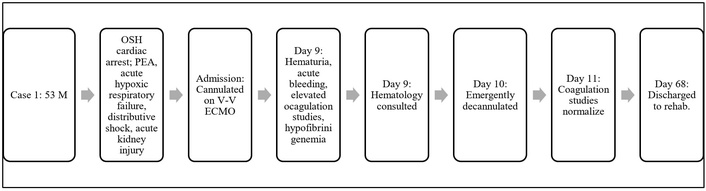
Case 1 timeline. M: male; OSH: outside hospital; PEA: pulseless electrical activity; V-V ECMO: veno-venous extracorporeal membrane oxygenation.
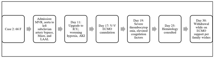
Case 2 timeline. F: female; LAAL: left atrial appendage ligation; MVR: mitral valve replacement; ICU: intensive care unit; V-V ECMO: veno-venous extracorporeal membrane oxygenation.
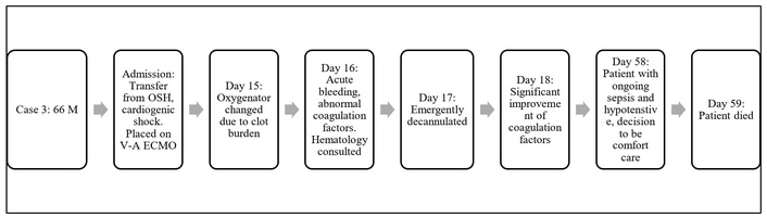
Case 3 timeline. M: male; OSH: outside hospital; V-A ECMO: veno-arterial extracorporeal membrane oxygenation.
A 53-year-old man (case 1) with a history of bicuspid aortic valve status post-surgical replacement with a bioprosthetic valve, hypertension, and opioid use disorder, experienced a witnessed out-of-hospital cardiac arrest. Bystander cardiopulmonary resuscitation (CPR) was initiated, with an estimated time to return of spontaneous circulation of fifty minutes. His initial rhythm was pulseless electrical activity (PEA). He was intubated on scene and transported to an academic hospital emergency department, where he was found to have combined respiratory and lactic acidosis, as well as acute kidney injury. He was profoundly hypotensive and started on norepinephrine before being admitted to the medical intensive care unit. Despite escalating hemodynamic and respiratory support—including high-dose norepinephrine, vasopressin infusion, and maximal ventilatory settings with inhaled nitric oxide therapy, his condition worsened. ECMO was requested given refractory hypoxemia; V-V ECMO was initiated via bicaval cannulation. Post-cannulation echocardiogram showed left ventricular ejection fraction (LVEF) 35–40% secondary to myocardial stunning and severe prosthetic aortic valve stenosis secondary to calcification. A pulmonary artery catheter revealed low cardiac output, prompting epinephrine initiation. Cardiothoracic surgery and structural heart teams were consulted for possible valve intervention. Over the next 3 days, oxygenation improved on ECMO, hemodynamics stabilized, and ventricular function recovered. Anticoagulation was maintained with heparin targeting partial thromboplastin time (PTT) 50–70 seconds. The patient was extubated and approaching readiness for ECMO decannulation. On day 9 of ECMO support, he developed acute bleeding from central lines and hematuria. Coagulation studies were consistent with DIC, demonstrating an international normalized ratio (INR) of 9.5, PTT of 71 seconds, fibrinogen < 60 mg/dL, and anti-Xa activity < 0.1 IU/mL. FDPs and D-dimer were markedly elevated. Antithrombin III activity was elevated at 133%. Factors II, V, VII, IX, and X were also obtained and were unremarkable. Hematology was consulted; leading causes included ECMO-associated hyperfibrinolysis and DIC secondary to cardiac arrest, with a recommendation that removal of ECMO might improve coagulation values. He received a total of one unit of packed red blood cells (PRBC), six doses of platelets, three units of fresh frozen plasma (FFP), 80 units of cryoprecipitate, and 50 mg protamine prior to decannulation. His left ventricular function prior to decannulation improved to 60%, and due to improving pulmonary status and ongoing bleeding, he was decannulated from ECMO on day 10, with aggressive transfusion support peri-decannulation. Post-decannulation, the patient was administered two units of PRBC, 50 units of cryoprecipitate, and tranexamic acid. Repeat labs shortly after decannulation showed improved coagulation: INR 1.4, PTT 36 seconds, and fibrinogen 198 mg/dL, with continued normalization over the next 24 hours, which included INR 1.2, PTT 33 seconds, and fibrinogen > 208 mg/dL. Clinical course after decannulation included the patient undergoing tracheostomy and percutaneous endoscopic gastrostomy (PEG) placement on hospital day 32 and permanent pacemaker placement on hospital day 43 due to a slow/unreliable underlying escape rhythm from complete heart block. On hospital day 68, the patient was deemed medically stable to be discharged to a rehab facility.
A 66-year-old woman (case 2) with a past medical history of hypertension, coronary artery disease with occluded ostial right coronary artery and nonocclusive left coronary artery disease, rheumatic heart disease with mitral stenosis, left subclavian artery stenosis with steal syndrome with prior left subclavian artery stent (thrombosed), heart failure with preserved ejection fraction, atrial fibrillation, peripheral vascular disease, chronic kidney disease (CKD) stage III, former tobacco abuse, pre-diabetes mellitus, gastroesophageal reflux disease (GERD), obesity, basal cell carcinoma, and squamous cell carcinoma of skin presented for a scheduled mitral valve replacement with 27 mm Epic bioprosthetic valve, aorta to left subclavian artery bypass, Maze procedure and left atrial appendage ligation. Postoperatively, the patient required inotropic support for right ventricular insufficiency. She developed acute hypoxic respiratory failure, which was treated with aggressive diuresis and therapeutic bi-level positive airway pressure (BiPap). On hospital day 11, she was upgraded to intensive care for worsening respiratory failure and acute kidney injury. She was placed on V-V ECMO on hospital day 17. On day 19, she developed severe thrombocytopenia with Plt 17 k/uL, INR 2.2, fibrinogen 151 mg/dL, and PTT 41 seconds. HIT Pf4 antibody was negative; therefore, systemic anticoagulation with heparin was initiated at a flat rate of 250 units/h per surgical attending. Hematology was consulted due to concerns for DIC on hospital day 25 following the need for a circuit change. Upon evaluation, the oxygenator had significant fibrin stranding and clots. The patient tolerated the procedure and, per hematology recommendations, repeat HIT Pf4 antibody was negative. Other pertinent laboratory values include Plt 20 k/μL despite multiple transfusions, fibrinogen critically low (< 60 mg/dL), INR elevated (2.4). She also had physical findings concerning for DIC, including tongue necrosis and purpura fulminans. Lower extremity ultrasound was positive for acute non-occlusive deep venous thrombosis within the mid peroneal and mid soleal veins. Peripheral smear demonstrated thrombocytopenia with normal platelet morphology and granularity; RBCs with schistocytes approximately 1 to 2 per high-power field; anisopoikilocytosis with spherocytes, spur cells, burr cells, reticulocytosis, nucleated red blood cells, left-shifted WBCs with reactive neutrophils and bands with toxic granulation. Hematology recommendations for management of DIC were to administer blood products as needed, especially if there were significant bleeding. Hematology also recommended considering restarting anticoagulation once Plt > 20 k/μL if no clinically significant bleeding were present. Her ISTH DIC score was 5 or more, consistent with overt DIC. On hospital day 29, there were significant signs of ECMO oxygenator failure with rapidly declining pump flows despite near-maximal RPMs, and the circuit was exchanged a second time. She continued to have worsening pancytopenia, and due to poor prognosis, her family made the decision to transition to comfort care on hospital day 30.
A 66-year-old man (case 3) presented to an acute care hospital with cough, weakness, and altered mental status, with a past medical history of hypertension and renal transplant. He had severe hypotension and suspected cardiogenic shock, and high-dose vasopressor and inotropic therapy was initiated. He was transferred to an academic medical center and was cannulated for V-A ECMO on hospital day 1. His clinical course was complicated by acute kidney injury requiring continuous renal replacement therapy and supraventricular tachycardia. A transthoracic echocardiogram found his LVEF to be 20–25%, and the right ventricle was normal in size and function. On hospital day 5, the patient was started on systemic anticoagulation with Bivalirudin to maintain a PTT of 50–70 seconds. It was noted on hospital day 15 that the patient’s oxygenator had significant clot burden on both venous and arterial sides. A post-oxygenator arterial blood gas (ABG) showed PaO2 173 mmHg, so the circuit was changed. On hospital day 16, he developed acute bleeding at the ECMO cannula insertion sites. His INR was elevated as high as 4.5, fibrinogen critically low (< 60 mg/dL), PTT elevated (> 200 seconds), and Plt low (66 k/μL). Hematology was consulted and recommended evaluation for vitamin K deficiency (factors II, VII, IX, and X assays) and liver disease (factor V) as causes of coagulopathy. Over the next 24 hours, the patient received nine units of FFP, three doses of platelets, two units of PRBC, 80 units of cryoprecipitate, 5,436 mg of fibrinogen concentrate, and vitamin K 10 mg. He required a massive transfusion protocol on hospital day 17, receiving a total of five doses of FFP, three units of PRBC, and two doses of platelets. He had an emergent computed tomography angiography (CTA) to evaluate for active bleeding, which was unremarkable. Due to worsening hypoxic respiratory failure, the patient was emergently intubated. Per hematology, his factors were within normal limits, and there was no concern that he had congenital or acquired factor deficiency. It was agreed that the patient’s clinical picture of DIC could be caused by a mechanical etiology, and so he was decannulated from ECMO. Additional management recommendations included maintaining fibrinogen > 150 mg/dL, hemoglobin > 7.0 g/dL, and Plt > 20 k/μL. Post-decannulation, he did receive one unit of PRBC, three units of FFP, and 80 units of cryoprecipitate. Twenty-four hours post-decannulation, INR was decreased (1.4), PTT was within normal limits (35 seconds), fibrinogen was normal (327 mg/dL), and platelets were improving (131 k/μL). On hospital day 21, three days after V-A ECMO removal, hematology signed off as the patient’s coagulation lab values were appropriately improving. The clinical course post ECMO decannulation included sepsis and hypotension requiring vasopressor support. On hospital day 58, he decided to proceed to comfort care and died the following day.
The diagnosis of DIC was established through the combination of clinical findings, laboratory values, and ECMO circuit assessment. Laboratory monitoring included complete blood count and coagulation studies, including PT, PTT, INR, and fibrinogen. Additionally, the ISTH overt DIC scoring system was used to support this clinical diagnosis.
Given that thrombocytopenia and coagulation abnormalities are a common occurrence with ECMO support, the ability to determine consumptive coagulopathy from circuit-related hematologic complications could be a potential diagnostic challenge. Other etiologies for the development of DIC, such as sepsis-induced coagulopathies, liver dysfunction, and HIT, were evaluated based on the clinical presentation. This would include HIT antibody testing and obtaining specific levels of factors II, V, VII, IX, and X.
In addition to laboratory evaluation, the ECMO circuit was routinely assessed for evidence of visible clot burden and fibrin stranding within the oxygenator every hour by the multidisciplinary team. Other indicators of imminent pump failure, which included declining oxygenator performance and signs of hemolysis, were monitored daily. Once overt DIC was identified, there was a multidisciplinary discussion regarding either oxygenator exchange or the necessity of continued ECMO support.
In all three patients, progressive oxygenator thrombosis preceded the development of overt DIC in addition to worsening thrombocytopenia, hypofibrinogenemia, and prolonged coagulation factors.
Patient perspectives were not obtained for this case series. Due to the retrospective nature of this case series, follow-up interviews with patients or their families were not conducted. However, future studies incorporating patient- and family-reported experiences may provide insight into the physical, emotional, and psychological impact of ECMO support, including complications.
The pathophysiology of DIC in ECMO patients is not fully understood, and diagnosis in ECMO patients can be particularly challenging as patients can experience coagulopathy for multiple reasons. DIC in critically ill patients can be due to inflammation, infection, and liver failure [8]. Thrombocytopenia can occur as a result of medication side effects or destruction by the ECMO circuit. A recent study in 2024 examined mortality in ECMO patients with DIC, finding that 24% of ECMO patients developed overt DIC, and in-hospital mortality was 55% in the DIC group (p < 0.001) [9]. Improved strategies for early recognition and screening for DIC, and evidence-based interventions for ECMO patients may assist in decreasing mortality in this patient population. Our experience with these patients led us to decannulate them earlier than planned in the hope of resolving the DIC, and we believe this improved the outcomes of several of the patients.
The development of DIC while on ECMO support can lead to difficult management decisions given the fact that ECMO support may perpetuate rather than alleviate the coagulation pathways. The decision of decannulation from ECMO support should not be solely based upon the suspected or confirmed diagnosis of DIC, given the fact that ECMO is a life-sustaining therapy while awaiting cardiac or pulmonary recovery. As soon as recovery has been identified or the prognosis becomes futile, early and prompt decannulation should be considered as described in cases one and three to decrease or prevent the risks of progressive coagulopathy, bleeding, thrombosis, or circuit failure. These decisions involve multidisciplinary collaboration alongside the patient and the patient’s family and should incorporate ethical principles, patient goals of care, and the likelihood of a meaningful recovery.
Major coagulation complications associated with DIC include clot formation and depletion of coagulation factors. Due to the excessive activation of thrombin, fibrinogen cleaves into fibrin, resulting in intravascular clot formation [10]. These microthrombi can lead to hypoperfusion and hypoxia of extremities and organs. Case two demonstrated these complications with the unfortunate development of DVT, tongue necrosis, and purpura fulminans. Patients can also experience an elevated risk of bleeding in DIC due to excessive use and depletion of clotting factors, leading to consumptive coagulopathy. Bleeding at vascular access sites and along the gastrointestinal and pulmonary vasculature can all occur [10]. This was evident in two of the cases presented (1 and 3), where patients developed bleeding at ECMO cannulation and central line insertion sites, cerebral vasculature, and hematuria.
Hypercoagulability and secondary hyperfibrinolysis during ECMO support can increase the rate of thrombotic events and oxygenator membrane failures [11]. Multiple treatments for this major complication have been proposed. For instance, in a single-center, retrospective study, if a patient had hyperfibrinolysis along with signs of oxygenator failure, the ECMO circuit was changed regardless of gas exchange function. Along with the oxygenator exchange, platelet and fibrinogen deficiencies were corrected with blood products. However, patients with active signs of hyperfibrinolysis or DIC without oxygenator failure were treated with tranexamic acid [11]. Although there are no specific guidelines listed for coagulation and management of hyperfibrinolysis through the Extracorporeal Life Support Organization (ELSO), the use of conventional antifibrinolytics is not recommended. Decreased fibrinogen was identified in all three cases; however, cases one and three had critically low levels of < 60 mg/dL (Table 2).
Coagulation factor 24 h range prior to decannulation or withdrawal.
| Case | Age, sex | Cannula type and cannulation site | Days requiring ECMO | ISTH score | Platelet count(k/μL) | PTT(seconds) | PT(seconds) | INR | Fibrinogen(mg/dL) | D-dimer(μg/mL) | FDP(μg/mL) | Anti-Xa(IU/mL) |
|---|---|---|---|---|---|---|---|---|---|---|---|---|
| Case 1 | 53 M | 25 Fr Maquet (L Femoral Vein)17 Fr Maquet (R Internal Jugular) | 10 | Not calculated | 89–110 | 36–100 | 16.7–75 | 1.4–9.5 | < 60–198 | > 20 | > 20 | < 0.1 |
| Case 2 | 66 F | 23 Fr Maquet (R Femoral Vein)17 Fr Maquet (R Internal Jugular) | 11 | 5 or more | 6–15 | 44–57 | 19.9–24.7 | 1.7–2.3 | 87–259 | No result available | No result available | < 0.1 |
| Case 3 | 66 M | 25 Fr Maquet (L Femoral Vein)17 Fr Maquet (L Femoral Artery) | 18 | Not calculated | 66–112 | 47– > 200 | 20.3–40.7 | 1.8–4.5 | < 60–153 | No result available | No result available | No result available |
ECMO: extracorporeal membrane oxygenation; FDP: fibrinogen degradation product; INR: international normalized ratio; ISTH: International Society on Thrombosis and Haemostasis; PT: prothrombin time; PTT: partial thromboplastin time.
Anticoagulation management is an essential piece of ECMO management. At our institution, the standard protocol for ECMO cannulation includes administration of an intravenous heparin bolus ranging from 1,000 to 5,000 units, at the discretion of the cannulating provider, based on the patient’s clinical condition and bleeding risk. In this case series, two patients received a heparin bolus at the time of cannulation, while one patient did not because of thrombocytopenia. All patients were cannulated using BIOLINE® heparin-coated Maquet cannulas, which provide a biocompatible surface that may reduce thrombogenicity and permit delayed initiation of systemic anticoagulation when clinically indicated [12]. Following cannulation, the anticoagulation strategy consists of a continuous unfractionated heparin infusion targeting a PTT of 50–70 seconds. For patients anticipated to undergo advanced therapy work-up or those at an increased risk of HIT, Bivalirudin is used as the primary anticoagulant to minimize the risk of HIT and maintain adequate circuit anticoagulation.
Routine visualization of the circuit, including observing color change, oxygenator assessment, and cannula positioning, is important for early recognition of thrombosis (Figure 4). In our institution, it is standard that these all are checked at least daily by the bedside nurse, the advanced provider, and the Intensivist team. In all three of our cases, there was direct visualization of the oxygenator, and most developed small clots and fibrin stranding on both the arterial and venous side of the oxygenator. If there was a concern for a failing oxygenator, a post-oxygenator gas would be obtained, and if the PaO2 was < 120, the oxygenator was changed. For two of our patients (cases 2 and 3), there was significant clot formation and fibrin stranding on both the arterial and venous sides of the oxygenator, requiring an oxygenator change. Fewer than 24 hours later, those patients started showing signs of overt DIC with associated hyperfibrinolysis and acute hemorrhage. In the literature, there are no suggested coagulation goals or standard treatment for overt DIC aside from treatment of the underlying cause. This may include antibiotics for sepsis, surgical intervention for trauma-related cases, or in this case series, the removal of mechanical circulatory support. For patients with active or high risk of bleeding, platelet, cryoprecipitate, and plasma transfusions are strongly recommended.
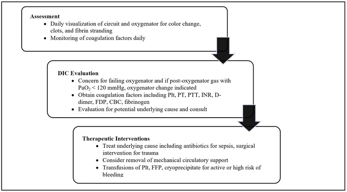
Pathway for DIC evaluation and interventions. CBC: complete blood count; DIC: disseminated intravascular coagulation; FDP: fibrinogen degradation product; FFP: fresh frozen plasma; INR: international normalized ratio; Plt: platelet count; PT: prothrombin time; PTT: partial thromboplastin time.
Case one and case three demonstrated significant improvement in coagulation values 24 hours post-decannulation, and it was suggested that their DIC was induced by mechanically related aspects of the ECMO circuit (Figure 5). This could be due to the artificial surfaces of ECMO triggering both pro-inflammatory and pro-thrombotic states. The inflammatory response caused by ECMO can influence intrinsic and extrinsic coagulation, endothelial cells, platelets, complement pathways, and cytokines [13]. Force from the ECMO pump and circuit shape can also cause shear stress, which can cause the fragmentation of red blood cells, platelets, and von Willebrand multimers [9]. There is also ongoing discussion that systemic coagulation abnormalities may mimic DIC and be an indicator of oxygenator failure [14]. Although it may not be visible on the circuit, thrombus formation on the circuit may consume platelets and fibrinogen, and timely change of the oxygenator may improve coagulation factors [14].
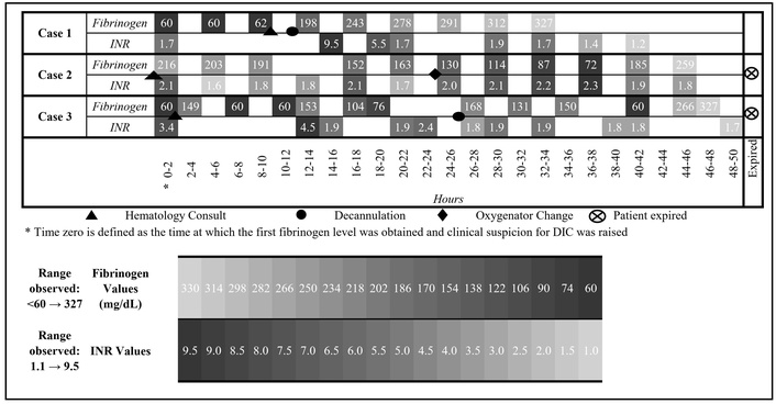
Fibrinogen and INR by case and time. INR: international normalized ratio; DIC: disseminated intravascular coagulation.
We report three patients with ECMO support complicated by DIC. Early recognition using standardized criteria, combined with prompt management of both coagulation abnormalities and underlying triggers, is essential. The ECMO circuit itself can cause overt DIC. If possible, ECMO decannulation may resolve the DIC and improve patient outcomes.
A major limitation to this case series is the small sample size, which can preclude any meaningful statistical analysis. Additionally, we were unable to establish a direct causal relationship between ECMO decannulation and resolution of DIC. However, we hypothesized that ECMO contributed to worsening coagulopathy and may have played a role in the development or persistence of DIC. This hypothesis is supported by the clinical observation that in two patients, coagulopathy improved following ECMO decannulation, although this temporal association does not establish causality.
ABG: arterial blood gas
DIC: disseminated intravascular coagulation
FDPs: fibrinogen degradation products
FFP: fresh frozen plasma
HIT: heparin-induced thrombocytopenia
INR: international normalized ratio
ISTH: International Society on Thrombosis and Haemostasis
LVEF: left ventricular ejection fraction
Plt: platelet count
PRBC: packed red blood cells
PT: prothrombin time
PTT: partial thromboplastin time
V-A ECMO: veno-arterial extracorporeal membrane oxygenation
V-V ECMO: veno-venous extracorporeal membrane oxygenation
KD: Project administration, Writing—original draft, Writing—review & editing, Investigation, Methodology. SH: Project administration, Writing—original draft, Writing—review & editing, Investigation, Methodology, Resources. JO: Writing—original draft. MH: Writing—original draft. JS: Writing—review & editing, Validation, Supervision. AP: Project administration, Supervision, Writing—review & editing. All authors have read and approved the submitted version.
The authors declare that there are no conflicts of interest.
The study was conducted in accordance with institutional guidelines and the principles of the Declaration of Helsinki. A formal ethical approval was not required for this retrospective case report.
Informed consent to participate in the study was obtained from the patients and/or the next of kin.
Written informed consent was obtained from the patients and/or the next of kin for publication of this case report and any accompanying images.
The data supporting the findings of this study are available from the corresponding author upon reasonable request.
Not applicable.
© The Author(s) 2026.
Open Exploration maintains a neutral stance on jurisdictional claims in published institutional affiliations and maps. All opinions expressed in this article are the personal views of the author(s) and do not represent the stance of the editorial team or the publisher.
Copyright: © The Author(s) 2026. This is an Open Access article licensed under a Creative Commons Attribution 4.0 International License (https://creativecommons.org/licenses/by/4.0/), which permits unrestricted use, sharing, adaptation, distribution and reproduction in any medium or format, for any purpose, even commercially, as long as you give appropriate credit to the original author(s) and the source, provide a link to the Creative Commons license, and indicate if changes were made.



View: 324
Download: 11
Times Cited: 0


