 Case Report


Affiliation:
Respiratory Disease Center, Tokyo Shinagawa Hospital, Tokyo 140-8522, Japan

Affiliation:
Respiratory Disease Center, Tokyo Shinagawa Hospital, Tokyo 140-8522, Japan
Email: hidenori.tsh@gmail.com
ORCID: https://orcid.org/0000-0001-9781-258X

 ,
,
Affiliation:
Respiratory Disease Center, Tokyo Shinagawa Hospital, Tokyo 140-8522, Japan
Affiliation:
Respiratory Disease Center, Tokyo Shinagawa Hospital, Tokyo 140-8522, Japan
Affiliation:
Respiratory Disease Center, Tokyo Shinagawa Hospital, Tokyo 140-8522, Japan
Affiliation:
Respiratory Disease Center, Tokyo Shinagawa Hospital, Tokyo 140-8522, Japan
Affiliation:
Respiratory Disease Center, Tokyo Shinagawa Hospital, Tokyo 140-8522, Japan
Affiliation:
Respiratory Disease Center, Tokyo Shinagawa Hospital, Tokyo 140-8522, Japan
Affiliation:
Respiratory Disease Center, Tokyo Shinagawa Hospital, Tokyo 140-8522, Japan
Affiliation:
Respiratory Disease Center, Tokyo Shinagawa Hospital, Tokyo 140-8522, Japan
ORCID: https://orcid.org/0000-0002-0922-8656
,
Affiliation:
Respiratory Disease Center, Tokyo Shinagawa Hospital, Tokyo 140-8522, Japan
Affiliation:
Respiratory Disease Center, Tokyo Shinagawa Hospital, Tokyo 140-8522, Japan
ORCID: https://orcid.org/0000-0002-8101-8653
Explor Med. 2026;7:1001424 DOI: https://doi.org/10.37349/emed.2026.1001424
Received: May 26, 2026 Accepted: July 29, 2026 Published: August 17, 2026
Academic Editor: Anna Maria Spagnolo, University of Genova, Italy
The article belongs to the special issue Legionellosis: From Environmental Exposure to Clinical Disease
Bacterial co-infection in patients with coronavirus disease 2019 (COVID-19) can complicate diagnosis because of overlapping clinicoradiologic findings among pathogens. We report a case of suspected triple co-infection with severe acute respiratory syndrome coronavirus 2 (SARS-CoV-2), Legionella pneumophila (L. pneumophila), and Streptococcus pneumoniae (S. pneumoniae) after hot-spring travel in a post-splenectomy patient. A man in his 70s with a history of subtotal esophagectomy and concomitant splenectomy for esophageal cancer was admitted with fever, worsening productive cough, and altered sensorium. He had recently traveled to a hot spring with a group that included his wife, who also developed a fever and was diagnosed with COVID-19. On admission, results of urinary antigen tests for L. pneumophila and S. pneumoniae were positive, a SARS-CoV-2 nucleic acid amplification test was positive, and sputum culture yielded S. pneumoniae. Chest computed tomography revealed multifocal bilateral ground-glass opacities, small bilateral pleural effusions, and relatively well-defined right middle-lobe consolidation with air bronchograms in a peribronchovascular distribution. These microbiological and radiological findings supported a suspected triple co-infection rather than COVID-19 alone. Treatment with remdesivir, ceftriaxone, and levofloxacin was initiated, and the patient’s condition improved, with better oxygenation and decreased inflammatory markers. Remdesivir and ceftriaxone were discontinued after 5 days, whereas levofloxacin was continued for 14 days. Follow-up imaging revealed marked improvement, and the patient was discharged. Public health investigations did not detect Legionella at the hot-spring facility or identify any additional linked cases. This case highlights the importance of prompt pathogen-directed evaluation when COVID-19 pneumonia is accompanied by a relevant exposure history, host risk factors, and atypical imaging findings.
Bacterial co-infection in patients with coronavirus disease 2019 (COVID-19) can complicate diagnosis and management because of overlapping clinical manifestations, laboratory abnormalities, and chest imaging findings among pathogens [1]. Although pneumonia associated with COVID-19 and multiple bacterial pathogens, including Legionella, has rarely been reported, such cases may follow a severe clinical course [2, 3]. Cases with more than one pathogen may have an atypical presentation, and failure to recognize a treatable bacterial infection may delay appropriate therapy.
We report a case of suspected triple co-infection with severe acute respiratory syndrome coronavirus 2 (SARS-CoV-2), Legionella pneumophila (L. pneumophila), and Streptococcus pneumoniae (S. pneumoniae) that developed after domestic travel to a hot spring in Japan in a patient with a history of splenectomy. This case underscores the importance of considering L. pneumophila involvement and other treatable bacterial co-infections when COVID-19 pneumonia is accompanied by recent hot-spring exposure, host risk factors, and atypical imaging findings. Careful exposure history taking and appropriate pathogen-directed testing are essential for identifying treatable co-infections, guiding timely empirical therapy, and facilitating subsequent antimicrobial adjustment or de-escalation [4, 5].
The patient was in his usual state of health before a 2-day hot-spring trip. After returning home, his wife developed fever first, followed by the patient the next day. Both were diagnosed with COVID-19 at a local clinic, and the patient was transported to our hospital after a worsening productive cough and altered sensorium. Subsequent hospital course, antimicrobial treatments, and laboratory results are summarized in Figure 1.
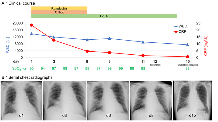
Clinical course and chest radiographs of a patient with pneumonia. (A) Clinical course showing changes in peripheral oxygen saturation, white blood cell count, and C-reactive protein level after initiation of remdesivir, ceftriaxone, and levofloxacin. (B) Serial chest radiographs showing gradual improvement of pulmonary infiltrates. CRP: C-reactive protein; CTRX: ceftriaxone; LVFX: levofloxacin; SpO2: peripheral oxygen saturation; WBC: white blood cell count.
A man in his 70s was admitted to our hospital with fever, worsening productive cough, and altered sensorium. His medical history was notable for esophageal cancer treated with subtotal esophagectomy, followed by reconstruction and concomitant splenectomy 10 years prior to presentation. He had no history of smoking, consumed alcohol daily, and had no known drug allergies. He was not currently receiving corticosteroids, immunosuppressive agents, or anticancer therapy. He was independent in activities of daily living, had previously received pneumococcal vaccination, but had not been vaccinated against COVID-19.
Several days before admission, he traveled to a hot spring for 2 days with a group of nine people, including his wife. After returning home, his wife developed fever, and he developed fever the following day. After being diagnosed with COVID-19 at a local clinic, both were managed conservatively with antipyretics. His productive cough subsequently worsened, and he developed altered sensorium that warranted emergency transport by ambulance to our hospital.
On admission, his body temperature was 38.3°C, blood pressure was 130/60 mmHg, pulse rate was 72 beats/min, respiratory rate was 18 breaths/min, and oxygen saturation was 90% on room air. The patient was mildly confused, with a Glasgow Coma Scale score of 14 (E4V4M6). Physical examination revealed coarse crackles in the right lung. He had cough, sputum production, anorexia, and malaise, but no headache, chills, sore throat, abdominal pain, diarrhea, or myalgia.
Laboratory tests showed leukocytosis (white blood cell count, 13,900/μL) with neutrophil predominance, an elevated C-reactive protein level (23.6 mg/dL), hyponatremia (133 mEq/L), and hypoalbuminemia (2.5 g/dL). Total bilirubin and liver enzyme levels were within normal limits, as was renal function. Urinalysis and urine culture were unremarkable. Urinary antigen tests for L. pneumophila serogroup 1 and S. pneumoniae were positive. These tests were performed using the ImmunoCatch® Legionella (product code E-ET02) and ImmunoCatch® Pneumococcus kits (product code E-ET04), respectively (Eiken Chemical Co., Ltd., Tokyo, Japan), both of which are immunochromatographic assays.
SARS-CoV-2 infection was confirmed by nucleic acid amplification testing on admission. Additional in-house real-time reverse transcription polymerase chain reaction testing performed on hospital day 3 using a QuantStudio 5 system (model A28134, 96-well, 0.2 mL; Thermo Fisher Scientific) with TaqMan™ Fast Virus 1-Step Master Mix (product No. 4444434; Thermo Fisher Scientific), Primer/Probe N2 (2019-nCoV) (product No. XD0008; Takara Bio Inc.), and a custom S-gene primer/probe set (Thermo Fisher Scientific) yielded cycle threshold values of 21.52 for the N2 gene and 22.71 for the S gene, consistent with a high SARS-CoV-2 RNA burden. Mutation screening using TaqPath™ 1-Step RT-qPCR Master Mix, CG (product No. A15299; Thermo Fisher Scientific) and the TaqMan™ SARS-CoV-2 Mutation Panel (product No. A49785; Thermo Fisher Scientific) on the same platform detected the G339D and L452R mutations. According to the institutional testing algorithm in use at that time, the G339D-positive pattern was interpreted as being consistent with an Omicron variant.
Gram staining of purulent sputum showed abundant Gram-positive cocci (4+), and sputum culture yielded S. pneumoniae, including both mucoid and non-mucoid morphotypes (2+ each). Species identification and antimicrobial susceptibility testing of the sputum isolate were performed using the RAISUS ANY automated microbiology system (model RAISUS ANY; Nissui Pharmaceutical Co., Ltd., Tokyo, Japan) with an RSCP2 identification and susceptibility plate (product No. 04135; Nissui Pharmaceutical Co., Ltd., Tokyo, Japan). Blood cultures were negative.
Chest computed tomography (CT) showed multifocal bilateral ground-glass opacities with a nonsegmental distribution, predominantly in the peripheral and subpleural lungs (Figure 2). Superimposed on these opacities, a relatively well-defined area of consolidation with air bronchograms was observed in the right middle lobe in a peribronchovascular distribution with an adjacent faint reticular opacity. Furthermore, trace bilateral pleural effusion was observed. No significant mediastinal or hilar lymphadenopathy was observed.
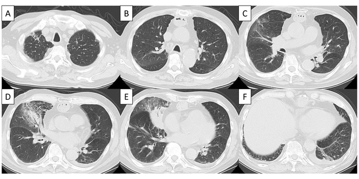
Chest computed tomography on admission. (A–C) Multifocal bilateral ground-glass opacities are distributed predominantly in the peripheral and subpleural lung fields. (D, E) A relatively well-defined consolidation with air bronchograms is present in the right middle lobe in a peribronchovascular distribution, accompanied by adjacent faint ground-glass and reticular opacities. (F) A small subpleural band-like opacity is present in the left lower lobe, with trace bilateral pleural effusions.
Taken together, these microbiological and radiological findings supported the diagnosis of suspected triple co-infection involving SARS-CoV-2, L. pneumophila, and S. pneumoniae.
Treatment was initiated with remdesivir (200 mg IV on day 1 followed by 100 mg IV once daily on days 2–5), ceftriaxone (2 g IV once daily), and levofloxacin (500 mg PO once daily). The patient responded well, with improvement in oxygenation, radiographic abnormalities, and inflammatory markers. After antimicrobial susceptibility testing confirmed that the pneumococcal isolates were susceptible to ceftriaxone, ceftriaxone was discontinued after 5 days, whereas levofloxacin was continued for a total of 14 days. The patient was discharged following clinical improvement.
Follow-up imaging showed regression of the right middle-lobe consolidation to a faint residual opacity, with near-complete resolution of the scattered ground-glass opacities. Subsequent investigations by the local public health authority, including sampling from four bathing areas at the hot-spring facility, did not detect Legionella. No additional cases were identified among the travel group or other facility users; moreover, no obvious alternative environmental source was found. Therefore, the case appeared sporadic, and the source of infection remained unidentified.
At an outpatient follow-up visit after recovery, the patient was informed of the clinical course and favorable outcome. He understood that sharing the details of his case could be educational for clinicians, and provided written informed consent for publication.
This case highlights the difficulty in diagnosing pneumonia in patients with COVID-19 when more than one pathogen is involved. Although bacterial co-infection at the time of COVID-19 presentation is relatively uncommon, treatable bacterial pneumonia may be overlooked if all clinicoradiological findings are attributed solely to SARS-CoV-2 [1]. In this case, neutrophilic leukocytosis, productive cough, focal consolidation, marked inflammation, positive pneumococcal and L. pneumophila urinary antigen tests, growth of S. pneumoniae in sputum culture, and recent hot-spring exposure supported a suspected triple co-infection rather than COVID-19 alone.
Several clinical features further supported suspected L. pneumophila infection. Current guidance recommends testing for Legionnaires’ disease in patients with pneumonia who have severe disease or overnight travel during the incubation period; moreover, the recommendations include combining urinary antigen testing with lower respiratory tract culture or molecular testing when Legionnaires’ disease is suspected [6, 7]. Hot-spring exposure is an important epidemiological clue because hot-spring bath water has been molecularly confirmed as the source of infection in a sporadic case of L. pneumophila pneumonia in Japan [8]. In addition, a relatively well-defined peribronchovascular consolidation with ground-glass opacities constitutes a characteristic CT pattern that is more frequent in L. pneumophila pneumonia than in pneumococcal pneumonia [9]. Urinary antigen testing for Legionella has high specificity; the reported pooled sensitivity and specificity were 79% and 100% overall and 86% and 100% for L. pneumophila serogroup 1, respectively [10]. Although respiratory culture or molecular testing for Legionella was unavailable, the positive urinary antigen result, when interpreted together with the exposure history, clinical features, and CT pattern, supported concomitant legionellosis. Persistent antigen excretion after a previous infection was unlikely because the patient had no known history of prior legionellosis [11]. Incidental false-positive results and pseudoepidemics have been reported [12, 13]. Pontiac fever was also considered but was less consistent with this patient’s radiographic pneumonia, hypoxemia, and focal consolidation [14].
The timing of symptom onset was compatible with the possible acquisition of all three pathogens around the time of hot-spring travel. The reported incubation periods are a mean of 2.6 days for SARS-CoV-2 Omicron infection, 2–14 days for Legionnaires’ disease, and 1–3 days for pneumococcal pneumonia; the low cycle threshold values on hospital day 3 were consistent with a high viral RNA burden and recent active SARS-CoV-2 infection [7, 15, 16].
The clinical presentation may have reflected overlapping but largely independent risk factors: lack of COVID-19 vaccination, recent hot-spring exposure, and prior splenectomy. SARS-CoV-2-related airway epithelial injury and impaired mucociliary clearance may have compromised local airway defenses and facilitated bacterial co-infection [17]. Concomitant L. pneumophila and S. pneumoniae infections occur rarely and have mainly been described in case reports [18, 19]. Thus, this case is best interpreted as involving overlapping risks and possible sequential infections rather than a proven pathogen–pathogen interaction.
The history of splenectomy supports the clinical significance of pneumococcal infection but does not directly explain susceptibility to SARS-CoV-2 or Legionella. This is because asplenia mainly predisposes patients to infections that are caused by encapsulated bacteria, especially S. pneumoniae, through impaired splenic clearance and polysaccharide antigen responses [20]. However, prior pneumococcal vaccination does not eliminate this residual risk [21]. Unlike the fatal case reported by Fukuda et al., in which L. pneumophila and S. pneumoniae co-infection developed during dexamethasone and baricitinib therapy for COVID-19 [3], our patient had a post-splenectomy host risk rather than pharmacological immunosuppression, and improved with early pathogen-directed therapy.
This case underscores the need for early microbiological evaluation when bacterial co-infection is suspected in patients with COVID-19 pneumonia. In such cases, multiplex molecular panels may complement exposure-directed testing by facilitating the simultaneous detection of additional respiratory pathogens [3, 22].
We report a case of suspected triple co-infection involving SARS-CoV-2, L. pneumophila, and S. pneumoniae after hot-spring travel in a post-splenectomy patient. Clinicians should consider Legionella infection when COVID-19 pneumonia is accompanied by recent hot-spring or aerosolized water exposure, atypical imaging findings, or host risk factors, and should promptly evaluate treatable bacterial pathogens.
COVID-19: coronavirus disease 2019
CT: computed tomography
L. pneumophila: Legionella pneumophila
S. pneumoniae: Streptococcus pneumoniae
SARS-CoV-2: severe acute respiratory syndrome coronavirus 2
We would like to thank Editage (www.editage.jp) for English language editing.
RH: Writing—original draft. HT: Conceptualization, Methodology, Investigation, Data curation, Visualization, Project administration, Writing—original draft. TY: Writing—review & editing. KT: Writing—review & editing. KF: Writing—review & editing. YS: Writing—review & editing. HN: Writing—review & editing. NT: Writing—review & editing. MTK: Writing—review & editing. SO: Writing—review & editing. MM: Writing—review & editing. MS: Writing—review & editing. All authors read and approved the submitted version.
The authors declare that they have no conflicts of interest.
This case report was conducted in accordance with the Declaration of Helsinki. Ethical approval was not required for this single case report according to institutional policy because it describes routine clinical care.
Written informed consent to participate was obtained from the patient.
Written informed consent was obtained from the patient for publication of this manuscript and the accompanying images.
The data supporting the findings of this case report are available from the corresponding author upon reasonable request, within the limits of patient privacy and confidentiality.
Not applicable.
© The Author(s) 2026.
Open Exploration maintains a neutral stance on jurisdictional claims in published institutional affiliations and maps. All opinions expressed in this article are the personal views of the author(s) and do not represent the stance of the editorial team or the publisher.
Copyright: © The Author(s) 2026. This is an Open Access article licensed under a Creative Commons Attribution 4.0 International License (https://creativecommons.org/licenses/by/4.0/), which permits unrestricted use, sharing, adaptation, distribution and reproduction in any medium or format, for any purpose, even commercially, as long as you give appropriate credit to the original author(s) and the source, provide a link to the Creative Commons license, and indicate if changes were made.



View: 744
Download: 27
Times Cited: 0


