 Original Article


Affiliation:
1Department of Medical Laboratory Techniques, Al-Mansour Medical Technical Institute, Middle Technical University (MTU), Baghdad 10013, Iraq
Email: Sura.ay@mtu.edu.iq
ORCID: https://orcid.org/0000-0002-4574-8835


 ,
,
Affiliation:
1Department of Medical Laboratory Techniques, Al-Mansour Medical Technical Institute, Middle Technical University (MTU), Baghdad 10013, Iraq
ORCID: https://orcid.org/0000-0001-9313-3192
,
Affiliation:
2College of Science, Al-Karkh University of Science, Baghdad 10011, Iraq
ORCID: https://orcid.org/0009-0002-7855-3302
,
Affiliation:
1Department of Medical Laboratory Techniques, Al-Mansour Medical Technical Institute, Middle Technical University (MTU), Baghdad 10013, Iraq
ORCID: https://orcid.org/0009-0008-0001-1216
,
Affiliation:
3Faculty of Social and Life Sciences, Wrexham University, LL11 2AW Wrexham, United Kingdom
ORCID: https://orcid.org/0000-0002-6066-6328
,
Affiliation:
4Department of Optometry Techniques, Al-Mansour Medical Technical Institute, Middle Technical University (MTU), Baghdad 10013, Iraq
ORCID: https://orcid.org/0000-0001-9747-8120
Explor Immunol. 2026;6:1003266 DOI: https://doi.org/10.37349/ei.2026.1003266
Received: December 17, 2025 Accepted: July 15, 2026 Published: September 15, 2026
Academic Editor: Narinder K. Mehra, All-India Institute of Medical Sciences (AIIMS), India
Aim: The current study was planned to assess the statistical correlation between seroprevalence of human cytomegalovirus (HCMV) and important inflammatory markers: tumor necrosis factor-alpha (TNF-α) and interleukin-18 (IL-18) within Iraqi women diagnosed with polycystic ovary syndrome (PCOS).
Methods: The current case-control study investigated the association between HCMV infection, inflammatory mediators (TNF-α and IL-18), and biochemical profiles in Iraqi women, a cohort of 168. The study population was grouped into four categories: (1) obese with PCOS and (2) non-obese PCOS patients, (3) an obese non-PCOS group, and (4) healthy controls.
Results: In the current study, a marked increase in the concentrations of both TNF-α and IL-18 among women with PCOS compared to controls. This effect was found to be significant regardless of body mass index (BMI). These data support the proposition that inflammation is a key factor in the pathogenesis of the syndrome. Moreover, HCMV seropositivity and PCOS incidence were found to be statistically strongly correlated. Our findings also define the relation of obesity and its role in developing inflammatory responses and underscore the interaction between adiposity and systemic inflammation.
Conclusions: This research paper concludes that a sophisticated interaction occurs between chronic latent viral infection and metabolic dysfunction in PCOS patients. The remarkable increase of both TNF-α and IL-18, mainly in individuals with obesity, highlights the profound inflammatory basis of PCOS. Accordingly, investigating the causal pathways is crucial, and therefore, a strategic approach to optimize patient outcomes could be assimilating viral screening into clinical management protocols.
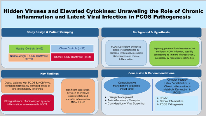
Globally, 6–10% of women of reproductive age are affected by polycystic ovary syndrome (PCOS), which is an endocrine disorder [1]. The PCOS prevalence varies depending on diagnostic criteria, lifestyle, and ethnicity [2]. PCOS extends beyond reproductive difficulties to substantial metabolic and cardiovascular morbidities, including dyslipidemia, insulin resistance, and an augmented risk of type 2 diabetes mellitus [3].
PCOS pathophysiology is multifaceted, with chronic and low-grade inflammation recognized as a central component of disease development [4]. Pro-inflammatory cytokines, particularly interleukin-18 (IL-18), and tumor necrosis factor-alpha (TNF-α) have been implicated in intensifying insulin resistance and hyperandrogenism, which are distinctive characteristics of PCOS [5]. As previously documented elsewhere [6, 7], human cytomegalovirus (HCMV) is a pervasive pathogen with a seroprevalence globally exceeding 60%, and rates often around 80% in developing regions.
As documented by [8], following primary infection, HCMV establishes lifelong latency and can reactivate during systemic inflammation or immunocompromised conditions. Clinically, HCMV infection is often silent in immunocompetent patients; however, it has been related to cardiovascular diseases and specific inflammatory phenomena [9]. Since PCOS is characterized by a state of chronic inflammatory response, a hypothesis arises that the functional dysfunction observed in the patients is attributed to the immune alterations induced by the virus [9]. However, there is a paucity in the scientific literature exploring this direct association. Exploring the interaction between PCOS and HCMV is crucial for developing targeted management strategies, especially in Iraq, where both PCOS and HCMV infections are prevalent [6, 10]. Notwithstanding this fact, local research investigating the association between chronic viral infections and endocrine disorders is limited, highlighting a significant gap in the literature.
After obtaining informed consent from all study groups, blood samples were collected. The study protocol was reviewed and accepted by the Middle Technical University (MTU)/Medical Ethics Committee in Al Zafraniya, Baghdad, Baghdad Governorate, P.C.: 10074. The Ethics Committee Reference (Nr.37) was approved on 27/10/2025. The procedures were performed in strict obedience to the ethical standards set by the WMA Declaration of Helsinki, ensuring the dignity, rights, and privacy of all participants were protected.
The current case-control study was carried out at the Yarmouk Teaching Hospital/Infertility and IVF Center, Baghdad, Iraq. The case-control design was selected due to its efficiency in studying rare outcomes and its ability to assess associations between exposures (inflammation and HCMV infection) and disease presence (PCOS).
G*Power version 3.1 [11] was used to conduct sample size calculation to ensure sufficient statistical power (80%) and to detect a medium effect size (Cohen’s d = 0.5) at a significance level of α = 0.05. Based on the foregoing, a minimum study population of n = 160 was required. Eventually, the sample size was increased to n = 168 participants to account for probable dropouts and incomplete data.
A total of (n = 168) women aged from 18 to 40 years were recruited and grouped into four categories:
Obese women with PCOS (n = 44).
Normal-weight women with PCOS (n = 40).
Obese women without PCOS (n = 38).
Healthy controls (n = 46).
PCOS was detected according to [12], requiring as a minimum two of the following:
Oligo- or anovulation.
Clinical and/or biochemical signs of hyperandrogenism; the morphology of Polycystic ovarian was as follows (≥ 12 follicles measuring (2–9 mm) or ovarian volume > 10 cm3 with ultrasound).
Exclusion criteria were as follows: individuals diagnosed with chronic illnesses and diseases including cardiovascular disease, diabetes mellitus, individuals suffering from current infections, those individuals on hormonal therapy within the last three months, and those individuals with a family history of diabetes or PCOS.
Structured questionnaire was used to reveal probable confounders and were further adjusted in statistical analysis, e.g., dietary habits and physical activity. Bias in measurement was further diminished by blinding the lab workers who carried out biochemical and immunological analyses to participant groups.
Clinical and physical checkups such as weight, height, and body mass index (BMI), were checked by trained healthcare specialists by means of calibrated equipment. BMI was derived from weight (kg) divided by height squared (m2) and categorized based on Asia-Pacific criteria [13]:
Normal weight: 18.5–22.9 kg/m2.
Overweight: 23–24.9 kg/m2.
Obesity: ≥ 25 kg/m2.
Waist circumference and blood pressure were also measured to justify cardiovascular risk factors.
An amount of 10 mL of venous blood samples was drawn between 8:00 and 10:00 AM to examine the daily disparities in hormone and cytokine levels. Samples were then centrifuged at 3,000 rpm for 10 minutes. Serum aliquots were stored at −20°C until further use.
Lipid profiles and glucose: estimated via enzymatic colorimetric methods (Roche Diagnostics, Switzerland) following guidelines of Clinical and Laboratory Standards Institute (CLSI).
Hormone evaluation: follicle-stimulating hormone (FSH), luteinizing hormone (LH), prolactin (PRL), and testosterone levels were detected by enzyme-linked fluorescent assay (ELFA) using the MiniVidas® system [14].
Insulin Resistance: examined using the homeostatic model assessment for insulin resistance (HOMA-IR) formula [15].
HOMA-IR = [Fasting insulin (μU/mL) × Fasting glucose (mg/dL)]/405
Serum TNF-α and IL-18 concentrations were determined using commercial enzyme-linked immunosorbent assay (ELISA) kits following the manufacturer's protocol. Sensitivities of 2.3 pg/mL for TNF-α and 0.31 pg/mL for IL-18, with intra- and inter-assay coefficients of variation < 10%, were documented using this method.
OnSite CMV IgG/IgM Rapid Test (CTK Biotech Inc., 2021) was utilized to measure HCMV IgG and IgM antibodies with a sensitivity of 96% and specificity of 98% [16]. Positive findings were confirmed via ELISA.
Data analysis was performed using IBM SPSS Statistics (Version 26.0; IBM Corp. Armonk, NY, USA). Continuous variables were expressed as mean ± standard deviation (SD), whereas categorical variables were summarized using frequencies and percentages. To evaluate group-wise differences across the four cohorts for continuous parametric variables presented, a one-way Analysis of Variance (ANOVA) was executed. Upon identifying significant main effects (p < 0.05), Tukey’s post-hoc test was applied to perform pairwise multiple comparisons among the groups (obese with PCOS, normal-weight with PCOS, obese controls, and healthy controls). Tukey’s test was specifically selected to strictly control the family-wise type I error rate during multiple testing, thereby confirming specific inter-group statistical differences. Student’s t-test was utilized for two-group comparisons where applicable. Relationships between cytokines, biochemical markers, and HCMV seropositivity were evaluated using Pearson’s correlation coefficient. Furthermore, multivariate regression models were constructed to adjust for potential confounding covariates, such as age and BMI. Statistical significance was defined as p < 0.05 (two-tailed), with corresponding 95% confidence intervals (CIs) provided for key outcomes. Participant flow, eligibility assessment, exclusion criteria (n = 32 excluded out of n = 200 assessed), and final allocations across the four groups (total n = 168; obese with PCOS: n = 44, normal-weight with PCOS: n = 40, obese controls: n = 38, healthy controls: n = 46) are detailed in the CONSORT-style flow chart (Figure 1).
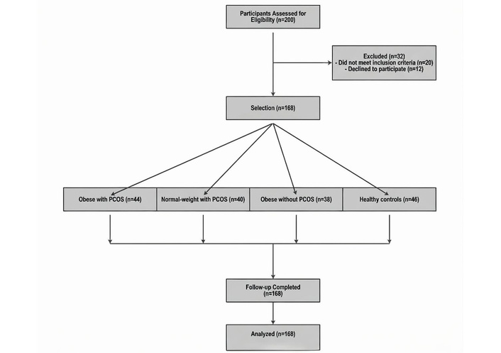
Flow diagram of participant recruitment demonstrating eligibility assessment, follow-up, analysis phases, exclusion criteria, recruitment, randomization, and group allocation. PCOS: polycystic ovary syndrome.
A total of n = 168 participants were eligible and met the inclusion criteria and enrolled in the study out of n = 200 women as an initial number for participants. Baseline characteristics of the study population are illustrated in Table 1.
Baseline demographic and clinical characteristics.
| Variables | Obese with polycystic ovary syndrome (PCOS) (n = 44) | Normal-weight with PCOS (n = 40) | Obese without PCOS(n = 38) | Healthy controls (n = 46) | p-value |
|---|---|---|---|---|---|
| Age (years) | 31.4 ± 4.2 | 30.8 ± 3.9 | 30.1 ± 4.5 | 30.5 ± 4.0 | 0.11 |
| Body mass index (BMI) (kg/m2) | a29.4 ± 3.2 | b21.2 ± 1.9 | a28.9 ± 2.8 | b20.8 ± 2.1 | < 0.001* |
| Systolic BP (mmHg) | 128 ± 10 | 124 ± 9 | 126 ± 11 | 122 ± 8 | 0.06 |
| Diastolic BP (mmHg) | 82 ± 6 | 80 ± 5 | 81 ± 7 | 79 ± 5 | 0.08 |
The four groups were overall compared using the p-values in One-way ANOVA. Asterisk (*) indicates a statistically significant overall difference (p < 0.05). Different superscript letters a and b within the same row indicate significant pairwise differences based on Tukey’s post-hoc test. BP: blood pressure.
Among age groups (p = 0.11; 95% CI: –1.5 to 2.3 years), there were no statistically significant differences.
As anticipated, BMI was significantly higher in the obese PCOS group (mean: 29.4 ± 3.2 kg/m2) compared to the normal-weight PCOS group (21.2 ± 1.9 kg/m2; p < 0.001, Cohen’s d = 2.76), indicating a large size effect. Systolic and diastolic blood pressures were elevated in obese groups; however, it did not reach clinical significance (p = 0.06).
As demonstrated in Figure 2, pro-inflammatory cytokine levels were remarkably higher in the PCOS cohorts than in the healthy control groups.
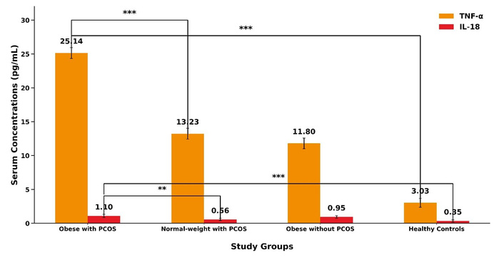
Serum concentrations (pg/mL) of tumor necrosis factor-alpha (TNF-α) and interleukin-18 (IL-18) across the different study groups. Bars represent the mean values ± standard deviation (SD). Statistical significance lines with asterisks indicate pairwise post-hoc comparisons based on Tukey’s test following One-way ANOVA (*** p < 0.001, ** p < 0.01). PCOS: polycystic ovary syndrome.
TNF-α levels: Obese women with PCOS showed the highest TNF-α concentrations (25.14 ± 3.7 pg/mL; 95% CI: 23.8–26.5) compared to normal-weight PCOS individuals (13.23 ± 0.25 pg/mL; p < 0.001, Cohen’s d = 3.10) and controls (3.03 ± 0.38 pg/mL; p < 0.001, Cohen’s d = 5.62), indicating large effect sizes.
IL-18 levels: The highest IL-18 concentrations were found in the obese PCOS group (1.10 ± 0.5 pg/mL; 95% CI: 0.98–1.22), significantly exceeding normal-weight PCOS (0.56 ± 0.06 pg/mL; p = 0.003) and control participants (0.35 ± 0.08 pg/mL; p < 0.001).
The seroprevalence of HCMV-specific IgG and IgM antibodies is presented in Table 2.
Human cytomegalovirus (HCMV) seroprevalence across study groups.
| HCMV status | Obese with PCOS (n = 44) | Normal-weight with PCOS (n = 40) | Obese without PCOS (n = 38) | Healthy controls (n = 46) | p-value |
|---|---|---|---|---|---|
| IgG positive (%) | a68% | a, b65% | a, b54% | b42% | 0.03* |
| IgM positive (%) | a11% | a, b8% | a, b5% | b2% | 0.02* |
p-values were calculated using the Chi-square test to compare frequencies across all groups. Asterisk (*) indicates a statistically significant overall difference (p < 0.05). Different superscript letters (a, b) within the same row indicate statistically significant pairwise differences (p < 0.05) in seroprevalence between specific groups [specifically comparing the polycystic ovary syndrome (PCOS) phenotypes against healthy controls] based on post-hoc pairwise comparisons of column proportions.
HCMV IgG: Higher prevalence was observed in the PCOS groups (68% in obese PCOS; 65% in normal-weight PCOS) compared to controls (42%; p = 0.03; OR: 2.89, 95% CI: 1.23–6.81).
HCMV IgM: A smaller percentage of participants tested positive for HCMV IgM, predominantly in the obese PCOS group (11%), suggesting recent or reactivated infections (p = 0.02).
Women with PCOS exhibited significantly elevated fasting plasma glucose and insulin levels, as explained in Table 3. HOMA-IR was highest in obese PCOS participants (3.8 ± 0.4; 95% CI: 5.5–6.3), significantly exceeding values in normal-weight PCOS individuals (3.0 ± 0.3; p < 0.001; Cohen’s d = 2.45) and healthy controls (2.1 ± 0.2; p < 0.001; Cohen’s d = 3.19). Clinically, these values indicate insulin resistance surpassing the metabolic syndrome threshold (HOMA-IR > 2.5) [15].
Biochemical and hormonal profiles.
| Variables | Obese with PCOS (n = 44) | Normal-weight with PCOS (n = 40) | Obese without PCOS (n = 38) | Healthy controls (n = 46) | p-value |
|---|---|---|---|---|---|
| Fasting glucose (mg/dL) | 102 ± 12 | 98 ± 10 | 100 ± 11 | 94 ± 9 | 0.02* |
| Fasting insulin (μU/mL) | 15.2 ± 3.1 | 12.3 ± 2.9 | 11.1 ± 2.7 | 9.2 ± 2.5 | < 0.001* |
| Homeostatic model assessment for insulin resistance (HOMA-IR) | 3.8 ± 0.4 | 3.0 ± 0.3 | 2.7 ± 0.3 | 2.1 ± 0.2 | < 0.001* |
| Triglycerides (mg/dL) | 139.2 ± 32.6 | 122.6 ± 32.8 | 133.1 ± 30.6 | 96.1 ± 35.2 | 0.03* |
| Free testosterone (ng/dL) | 1.93 ± 0.6 | 1.65 ± 0.5 | 1.10 ± 0.4 | 0.79 ± 0.5 | < 0.001* |
| Luteinizing hormone (LH)/follicle-stimulating hormone (FSH) ratio | 2.4 ± 0.3 | 2.1 ± 0.4 | 1.9 ± 0.2 | 1.4 ± 0.2 | 0.02* |
* Significant differences (p < 0.05) were found between polycystic ovary syndrome (PCOS) groups and healthy controls for all biochemical markers.
Triglycerides: elevated in obese PCOS participants (139.22 ± 32.63 mg/dL; p = 0.03) with moderate effect size (Cohen’s d = 0.75).
LDL/HDL cholesterol: differences were non-significant (p > 0.05), consistent with mixed findings in the literature regarding lipid metabolism in PCOS [3]
Free testosterone levels were elevated in the PCOS groups, with obese participants recording 1.93 ± 0.6 ng/dL compared to 0.79 ± 0.5 ng/dL in controls (p < 0.001; Cohen’s d = 2.12), indicating significant hyperandrogenism. The LH/FSH ratio was also significantly higher in the obese PCOS group (2.4 ± 0.3; p = 0.02), reflecting disrupted ovarian feedback mechanisms.
A robust positive association was demonstrated using Pearson’s correlation analysis between TNF-α and insulin resistance, using HOMA-IR (r = 0.67; p < 0.001), signifying the role of systemic inflammation in deteriorating insulin sensitivity. Furthermore, a significant correlation was found between IL-18 levels and BMI (r = 0.52; p = 0.004), underscoring the role of adiposity in the increase in inflammatory cytokine.
A significant relationship was found between prior HCMV infection and elevated inflammation. HCMV IgG +ve was an independent predictor of TNF-α concentrations, with a regression coefficient (β) of 0.45 (p = 0.01; 95% CI: 0.23–0.67). A significant correlation of adiposity and systemic inflammation was documented in patients with PCOS. BMI was responsible for 32% of the variance in IL-18 levels (R2 = 0.32; p < 0.001).
Multivariate regression analysis findings for BMI and age are presented in Table 4.
Multivariate regression findings exploring levels of inflammatory markers.
| Predictor variables | Tumor necrosis factor-alpha (TNF-α) [β, 95% confidence interval (CI), p-value] | Interleukin-18 (IL-18) (β, 95% CI, p-value) |
|---|---|---|
| Human cytomegalovirus (HCMV) IgG positivity | 0.45 (0.23–0.67, 0.01*) | 0.31 (0.12–0.50, 0.02*) |
| BMI | 0.39 (0.21–0.58, < 0.001*) | 0.44 (0.28–0.60, < 0.001*) |
| Homeostatic model assessment for insulin resistance (HOMA-IR) | 0.52 (0.34–0.70, < 0.001*) | 0.47 (0.30–0.64, < 0.001*) |
| Age | 0.08 (–0.05–0.21, 0.23) | 0.06 (–0.07–0.19, 0.31) |
* Statistical significance at p < 0.05 [the model was set for body mass index (BMI) and age].
This research revealed HCMV IgG positivity, BMI, and HOMA-IR as significant independent prognosticators for both TNF-α and IL-18 levels. However, using this model, age did not reveal any significant statistical correlation.
In this study, we investigated a previously unexplored relationship between PCOS and HCMV infection. Among Iraqi women with PCOS, we looked at metabolic variables, inflammatory markers (TNF-α and IL-18), and HCMV seropositivity. Our findings indicate that TNF-α and IL-18 levels are much greater in women with PCOS, particularly those who are obese. In addition, we also discovered that compared to healthy women, women with PCOS exhibited greater frequencies of HCMV IgG and IgM seropositivity. Interestingly, HCMV seropositivity was associated with increased TNF-α levels even after adjusting for factors including insulin resistance and BMI. These findings imply that viral infections and chronic inflammation may be important factors in PCOS development.
Participants with PCOS and those with obesity exhibited much higher levels of the pro-inflammatory cytokines TNF-α and IL-18. TNF-α levels in obese PCOS patients (25.14 ± 3.7 pg/mL) were about eight times higher than in healthy controls (3.03 ± 0.38 pg/mL, p < 0.001). The rise in TNF-α and IL-18 is consistent with previous research, e.g., [3, 4]. that identified chronic low-grade inflammation as a major characteristic of PCOS. Insulin resistance may have resulted from high TNF-α levels interfering with insulin receptor signalling, e.g., [5]. That might be because of how the body handles fats and glucose; IL-18 also contributes to metabolic issues [17]. In this study, we also discovered a high positive correlation between TNF-α and HOMA-IR, which lends credence to the theory that systemic inflammation exacerbates metabolic problems in PCOS.
The role of HCMV in promoting inflammation remains under discussion, but our results add to the evidence linking HCMV to PCOS. In the current study, we observed that individuals with PCOS had a much higher amount of HCMV IgG antibodies than the control group. In particular, 68% of those obese participants with PCOS tested positive for HCMV, compared to 42% of healthy participants (p = 0.03). Research by [10] findings are in line with our findings who also found that viruses were more prevalent in PCOS cases. This was also asserted by authors, e.g., [6, 8]. HCMV stays dormant in the body and reactivates in response to stress or immunosuppression.
Higher TNF-α levels were independently predicted by HCMV status, as our regression analysis demonstrates (β = 0.45, p = 0.01). This suggests that latent viral infections and the persistent low-level inflammation frequently observed in PCOS may be related. Scientifically speaking, HCMV can cause inflammation by triggering the release of several cytokines through the activation of toll-like receptors [18]. This could explain why our seropositive subjects had higher TNF-α levels. Some researchers, such as [19], suggested that chronic viral infections may affect ovarian function or metabolism via oxidative stress; however, this was not confirmed in our study. It is noteworthy to understand that our research does not allow us to draw strong conclusions about cause and effect. As [20] pointed out, results in this area can vary, which might be because of differences in viral strains or genetics. Further polymerase chain reaction (PCR) research is needed in order to clarify the role of HCMV.
Interestingly, despite the elevated concentrations in inflammation and insulin resistance markers, LDL and HDL cholesterol levels were similar across all groups. This finding is not in alignment with a study that found dyslipidemia is common in PCOS [21]. One explanation might be that the individuals who were younger had received a PCOS diagnosis more recently or had genetic variations unique to this population. Changes in cholesterol levels may have also been avoided by other unmeasured factors such as food or exercise.
Clinically, these results suggest that a team approach is the most effective way to manage PCOS. Reducing inflammation and enhancing insulin sensitivity still depend on controlling weight through diet and increased exercise. In PCOS patients with unexplained inflammation or metabolic issues that do not improve with standard therapies, screening for chronic viruses like HCMV may be helpful because viral infections may contribute to persistent inflammation. Nonetheless, it is important to consider the practicality and affordability of such screening, particularly in nations with limited healthcare resources. Antiviral therapies may be an effective addition to existing treatment regimens. However, further study is required to ascertain their long-term safety, efficacy, and benefits for PCOS.
It can be difficult to use these findings in clinical settings. For example, the expense of HCMV screening and restricted access to antiviral drugs may be barriers in some contexts, and patients’ adherence to complex treatment plans, including antiviral or anti-inflammatory therapies, can also vary. This highlights the need for patient education. Once individuals comprehend the interplay of chronic inflammation and probable viral factors in PCOS, they may act positively in their treatment and make better lifestyle choices. As suggested elsewhere by [1], patient outcomes might be improved by adopting healthier lifestyle changes such as weight loss. Our findings reveal the importance of partnership collaboration of endocrinologists, infectious disease experts, and dietitians to work as a team for better PCOS health outcomes.
This research has some strengths; however, a few limitations can still be noted. Firstly, a direct cause-and-effect relationship between HCMV infection and PCOS cannot be defined as a result of using case-control. Therefore, our findings should be seen as an association, rather than proof of causation. Secondly, in the current study, HCMV status was detected using IgG/IgM serology, which as a result, represents past exposure and potential latency, unlike PCR, and does not present the quantity of infectious agent. As a result, this made it even more difficult to understand the interplay of the virus and persistent inflammation. Thirdly, other possible confounding factors such as physical activity and detailed lifestyle habits in addition to the BMI and age could have been included in our model to obtain a deeper understanding.
The current research demonstrates a robust indication of how chronic inflammation, viral infections, and metabolic problems interact in women with PCOS. Higher levels of TNF-α and IL-18, especially in women with obesity, underscore the role of inflammation in disease development.
The strong association found here between HCMV seropositivity and elevated inflammatory parameters proposes the role that latent viral infections might play in the ongoing inflammation, especially in those individuals suffering from PCOS.
We present here a new research area with probable clinical importance. Health outcomes for patients with PCOS could be improved by monitoring metabolic and inflammatory aspects of these patients and including viral screening and targeted treatments. Further investigations might be needed to reveal the causes and to find an effective, accessible treatment plan for different PCOS patient groups.
BMI: body mass index
CIs: confidence intervals
ELISA: enzyme-linked immunosorbent assay
FSH: follicle-stimulating hormone
HCMV: human cytomegalovirus
HOMA-IR: homeostatic model assessment for insulin resistance
IL-18: interleukin-18
LH: luteinizing hormone
PCOS: polycystic ovary syndrome
PCR: polymerase chain reaction
TNF-α: tumor necrosis factor-alpha
The authors would like to express their gratitude to the staff of the Infertility and IVF Center at Yarmouk Teaching Hospital, Baghdad, for their support during the data collection phase. We also thank all the participants who volunteered for this study.
SOY: Conceptualization, Methodology, Supervision, Visualization. MDA: Conceptualization, Methodology, Supervision. SMA: Investigation, Data curation. IKD: Investigation, Data curation. SM: Formal analysis, Writing—original draft, Writing—review & editing. JS: Formal analysis. All authors read and approved the submitted version.
The authors declare that they have no conflicts of interest.
The study was reviewed and approved by the Middle Technical University Medical Ethics Committee in Baghdad, Iraq (Ethics Reference Number: 37, approved on October 27, 2025). All procedures were conducted in strict adherence to the WMA Declaration of Helsinki.
Informed consent was obtained from all adult participants prior to their inclusion in the study.
Not applicable.
The datasets generated and analyzed during the current study are available from the corresponding author upon reasonable request.
This research received no specific grant from any funding agency in the public, commercial, or not-for-profit sectors.
© The Author(s) 2026.
Open Exploration maintains a neutral stance on jurisdictional claims in published institutional affiliations and maps. All opinions expressed in this article are the personal views of the author(s) and do not represent the stance of the editorial team or the publisher.
Copyright: © The Author(s) 2026. This is an Open Access article licensed under a Creative Commons Attribution 4.0 International License (https://creativecommons.org/licenses/by/4.0/), which permits unrestricted use, sharing, adaptation, distribution and reproduction in any medium or format, for any purpose, even commercially, as long as you give appropriate credit to the original author(s) and the source, provide a link to the Creative Commons license, and indicate if changes were made.


View: 308
Download: 22
Times Cited: 0


