 Case Report


Affiliation:
1Department of Gastroenterology & Hepatology, Dr. Cipto Mangunkusumo National General Hospital, Medical Faculty Universitas Indonesia, Jakarta 10430, Indonesia
2Digestive Disease and Gastrointestinal Oncology Centre, Medistra Hospital, Jakarta 12950, Indonesia
3Gastrointestinal Cancer Centre, MRCCC Siloam Semanggi Hospital, Jakarta 12930, Indonesia
Email: medicaldr2001id@yahoo.com
ORCID: https://orcid.org/0000-0001-9992-9968


 ,
Affiliation:
1Department of Gastroenterology & Hepatology, Dr. Cipto Mangunkusumo National General Hospital, Medical Faculty Universitas Indonesia, Jakarta 10430, Indonesia
4Internal Medicine Division, Siloam Kupang Hospital, Kupang 85111, Indonesia
ORCID: https://orcid.org/0009-0006-5202-0320
,
Affiliation:
1Department of Gastroenterology & Hepatology, Dr. Cipto Mangunkusumo National General Hospital, Medical Faculty Universitas Indonesia, Jakarta 10430, Indonesia
Affiliation:
1Department of Gastroenterology & Hepatology, Dr. Cipto Mangunkusumo National General Hospital, Medical Faculty Universitas Indonesia, Jakarta 10430, Indonesia
ORCID: https://orcid.org/0000-0003-4009-7938
Explor Dig Dis. 2026;5:1005139 DOI: https://doi.org/10.37349/edd.2026.1005139
Received: February 02, 2026 Accepted: August 20, 2026 Published: October 22, 2026
Academic Editor: Jian-Guo Chen, Qidong Liver Cancer Institute, China
The article belongs to the special issue Endoscopic Evaluation in Liver Diseases
Percutaneous thermal ablation is widely used as a locoregional treatment for liver malignancies, but its feasibility and efficacy may be limited by unfavorable anatomy and access constraints. Endoscopic ultrasound-guided radiofrequency ablation (EUS-RFA) has emerged as an alternative approach for selected tumor locations, particularly those located in the left lobe or caudate region. We report a two-center case series describing technical feasibility, safety, and short-term imaging outcomes of EUS-RFA in primary and secondary liver malignancies. We retrospectively reviewed five patients who underwent EUS-RFA for liver malignancy at two tertiary centers. Diagnoses included hepatocellular carcinoma (HCC; n = 2), intrahepatic cholangiocarcinoma (iCCA; n = 1), colorectal liver metastasis (CRLM; n = 1), and pancreatic neuroendocrine tumor (PNET) liver metastasis (n = 1). EUS-RFA was performed using linear echoendoscopes and a radiofrequency generator connected to an EUS-RFA needle with a 1-cm active tip. Outcomes included technical success, procedural safety, and radiologic response at follow-up. Technical success was achieved in all five patients (100%). No immediate major procedure-related complications (bleeding, perforation, infection) were observed. At follow-up imaging, both HCC patients demonstrated stable disease at 1 month. The iCCA and CRLM patients demonstrated stable disease at 4 months. The PNET liver metastasis progressed during the longer follow-up period (8 months). In this small retrospective case series, EUS-RFA was technically feasible in carefully selected patients with liver malignancies, and no immediate major procedure-related complications were observed. EUS-RFA may serve as a complementary technique when percutaneous access is technically challenging or not feasible. Larger prospective studies are needed to better define its role in the multidisciplinary management of liver malignancies.
Liver malignancy, including primary hepatocellular carcinoma (HCC) and secondary metastatic disease, remains one of the most challenging malignancies to manage because treatment depends on tumor biology, disease stage, and underlying liver function. Treatment strategies are influenced not only by tumor burden but also by the degree of liver dysfunction, performance status, and comorbidity profile [1]. Among locoregional treatments for hepato-pancreatobiliary cancers, local tumor ablation, particularly radiofrequency ablation (RFA), has become widely used worldwide, especially for early-stage disease or as a part of multimodal therapy [1, 2]. Tumors located in the caudate lobe, subcapsular region, or adjacent to the diaphragm may be difficult to access safely using a percutaneous approach.
Endoscopic ultrasound (EUS) has therefore emerged as a minimally invasive therapeutic approach. With its high spatial resolution, real-time imaging, and precise needle placement, endoscopic ultrasound-guided radiofrequency ablation (EUS-RFA) has emerged as a potential alternative for selected liver tumors that are not accessible to percutaneous RFA [3, 4]. Although its role in hepatobiliary oncology is still evolving and remains under active investigation, early clinical experience suggests a promising feasibility profile in appropriately selected patients. This study presents a two-center case series and reviews the current literature regarding EUS-RFA for liver malignancies.
This retrospective case series was conducted in two private tertiary referral hospitals in Indonesia over a 3-year period. This study was approved by the local ethics committee. Five of the sixteen EUS-RFA cases were included from the database that fulfilled the inclusion and exclusion criteria. The inclusion criteria were all patients with primary or secondary liver malignancies presenting with hepatic nodules that are considered suboptimal for percutaneous ablation due to anatomical location. The exclusion criteria included technical infeasibility of EUS-guided intervention, uncorrectable coagulopathy, and significant comorbidities precluding the procedure. Treatment selections were determined by the treating multidisciplinary team based on tumor characteristics, anatomical considerations, technical feasibility, and patient comorbidities, expected technical success, and overall clinical judgement. The primary endpoints were technical success (successful needle placement and completion of intended energy delivery), procedural safety (no immediate procedure-related complications such as bleeding, perforation, or bile leak), and radiologic tumor response. Tumor response was assessed using modified Response Evaluation Criteria in Solid Tumors (mRECIST) for HCC and Response Evaluation Criteria in Solid Tumors (RECIST) version 1.1 for non-HCC malignancies [5, 6]. RECIST 1.1 evaluates changes in tumor size, and mRECIST assesses viable tumor based on arterial phase enhancement. Response outcomes were interpreted according to the respective criteria and were not intended for direct comparison across different tumor types.
EUS-RFA was performed using a linear-array echoendoscope under general anesthesia. The target lesion was accessed via a transgastric or transduodenal approach, depending on tumor location. The EUS equipment consisted of an EG-580UT Fujinon (Fujifilm, Japan) or Olympus JF UCT 180 echoendoscope, which was connected to an Aloka ultrasound machine. The generator used was VIVA Combo (Taewoong, South Korea) with an EUSRA 1-cm active tip needle. A dedicated RFA needle was advanced into the distal portion of the lesion under real-time EUS guidance. Radiofrequency energy was applied using a stepwise power escalation strategy (typically 10–15 watts in automatic mode), with sequential ablation performed as the needle was gradually withdrawn to achieve broader coverage. The number of ablation sessions depended on the size and location of the tumor. Ablation continued until echogenic changes suggesting tissue necrosis were observed. All EUS-RFA procedures were performed by an experienced interventional endoscopist and gastroenterologist who had more than 15 years of experience in interventional EUS and percutaneous liver ablation. The EUS-RFA procedure can be seen in Figure 1.
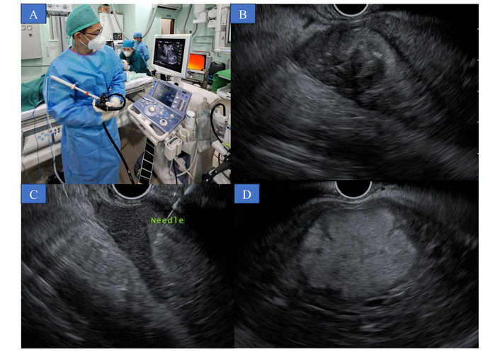
EUS-RFA for hepatocellular carcinoma. (A) Overview of the EUS-RFA procedure; (B) EUS images of the hepatocellular carcinoma before ablation; (C) needle puncture of the target lesion; (D) EUS image of the lesion after RFA. EUS-RFA: endoscopic ultrasound-guided radiofrequency ablation.
This case series consisted of two patients with HCC, one patient with intrahepatic cholangiocarcinoma (iCCA), one patient with colorectal liver metastasis (CRLM), and one patient with pancreatic neuroendocrine tumor (PNET) liver metastasis. All the primary liver cancer patients were diagnosed based on imaging, whereas the liver metastasis was confirmed through computed tomography (CT) scan or magnetic resonance imaging (MRI). All procedures were successfully performed in all patients, with successful needle placement and completion of intended energy delivery. There were no immediate major complications, such as bleeding, infection, and perforation. The intended follow-up duration for all patients was 1 month after EUS-RFA. However, actual follow-up intervals varied because of the retrospective nature of the study and differences in patient adherence to scheduled follow-up visits. Therefore, treatment response was evaluated as the first available post-procedural imaging assessment (first imaging follow-up). The duration of follow-up varied among patients (1–8 months). These assessments describe imaging response at the available follow-up time points rather than oncologic outcome. The treatment plan of the EUS-RFA case series can be seen in Figure 2.
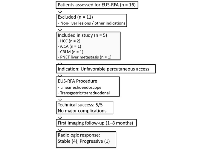
Flow diagram of patient selection, treatment process, and outcomes in this EUS-RFA case series. CRLM: colorectal liver metastasis; EUS-RFA: endoscopic ultrasound-guided radiofrequency ablation; HCC: hepatocellular carcinoma; iCCA: intrahepatic cholangiocarcinoma; PNET: pancreatic neuroendocrine tumor.
The clinical timeline of the five included cases is summarized in Table 1.
Clinical timeline and follow-up outcomes of patients undergoing EUS-RFA.
| Timepoint | Clinical event | ||||
|---|---|---|---|---|---|
| Case 1 | Case 2 | Case 3 | Case 4 | Case 5 | |
| Month 0 | Diagnosis of HCC, unsuitable for percutaneous ablation | Diagnosis of multifocal HCC, unsuitable for percutaneous ablation | Diagnosis of iCCA, unsuitable for percutaneous ablation | Diagnosis of CRLM, unsuitable for percutaneous ablation | Diagnosis of PNET liver metastasis, unsuitable for percutaneous ablation |
| Month 0 | EUS-RFA performed | EUS-RFA performed | EUS-RFA performed | EUS-RFA performed | EUS-RFA performed |
| Month 1 | CT scan → stable disease (mRECIST) | MRI → stable disease (mRECIST) | Not performed | Not performed | Not performed |
| Month 4 | Not performed | Not performed | CT scan → stable disease (RECIST) | CT scan → stable disease (RECIST) | Not performed |
| Month 8 | Not performed | Not performed | Not performed | Not performed | MRI → progressive disease (RECIST) |
CRLM: colorectal liver metastasis; CT: computed tomography; EUS-RFA: endoscopic ultrasound-guided radiofrequency ablation; HCC: hepatocellular carcinoma; iCCA: intrahepatic cholangiocarcinoma; mRECIST: modified Response Evaluation Criteria in Solid Tumors; MRI: magnetic resonance imaging; PNET: pancreatic neuroendocrine tumor.
Case 1: An 80-year-old patient with HCC in segment II, 50 mm, with underlying condition cirrhosis Child Pugh B (score 8). Because of the unfavorable tumor location in the left liver lobe (limited by the respiratory system and ribs), the patient was considered unsuitable for percutaneous ablation. The patient subsequently underwent EUS-RFA, which was completed successfully without complications. At 1-month follow-up, contrast-enhanced CT imaging demonstrated stable disease based on mRECIST criteria. The patient was subsequently started on systemic therapy with atezolizumab and bevacizumab.
Case 2: A 67-year-old patient with Child Pugh A (score 6) cirrhosis was diagnosed with multifocal HCC involving segments III and VIII. Because of anatomical and vascular constraints identified with transabdominal ultrasound, the patient was an unsuitable candidate for percutaneous ablation and underwent EUS-RFA. The procedure was technically successful with no complications. Follow-up MRI at 1 month demonstrated stable disease based on mRECIST criteria. No additional systemic therapy was administered.
Case 3: An 82-year-old patient was diagnosed with iCCA presenting as a 53 mm lesion. Given the prominent mass location at the left liver lobe (segments II and III), closer to the gastric area, and the patient was also planned to undergo biliary duct stenting in the same session, EUS-RFA was the best choice in this condition. At 4-month follow-up, CT imaging demonstrated stable disease based on RECIST 1.1 criteria. The patient subsequently received only immunotherapy with durvalumab.
Case 4: A 65-year-old patient with colorectal cancer was found to have a 23-mm liver metastasis in the caudate lobe. Due to difficulty with needle trajectory for percutaneous access, the patient underwent EUS-RFA. The procedure was technically successful without complications. Follow-up CT imaging at 4 months demonstrated stable disease according to RECIST 1.1 criteria. No additional therapy was administered.
Case 5: An 83-year-old patient with PNET presented with multifocal liver metastases. The planned ablation strategy included treatment of the PNET and liver lesions. The patient therefore underwent EUS-RFA. The procedure was completed successfully without complication. At 8-month follow-up, MRI demonstrated disease progression, with an increase in lesion size consistent with progressive disease based on RECIST 1.1 criteria. No systemic chemotherapy was used.
Baseline imaging findings, procedural characteristics, and follow-up radiology assessments are summarized in Tables 2 and 3.
Baseline and follow-up imaging of patients undergoing EUS-RFA.
| Imaging time point | Case 1 | Case 2 | Case 3 | Case 4 | Case 5 |
|---|---|---|---|---|---|
| Baseline | CT scan: 50 mm lesion in segment II | MRI: 36 mm and 23 mm lesions in segment III; 21 mm lesion in segment VIII | CT scan: 53 mm intrahepatic lesion | CT scan: 23 mm caudate lesion | MRI: 12 mm and 32 mm lesions in the left liver lobe |
| Follow-up | CT scan (1 month): no progression | MRI (1 month): no progression | CT scan (4 months): no progression | CT scan (4 months): no progression | MRI (8 months): increase in size |
CT: computed tomography; EUS-RFA: endoscopic ultrasound-guided radiofrequency ablation; MRI: magnetic resonance imaging.
Clinical characteristics, procedural parameters, and radiologic outcomes of patients undergoing EUS-RFA.
| Case | Age (years) | Diagnosis | Lesion site | Baseline size (mm) | Follow-up duration (month) | Ablation power (watts) | Total ablation time | EUS access route | Imaging modality at follow-up | Response criteria | Response | Additional treatment |
|---|---|---|---|---|---|---|---|---|---|---|---|---|
| 1 | 80 | HCC (Child Pugh score 8) | Segment II | 50 | 1 | 5–20 | 3 min | Transgastric (lesser curvature) | CT scan | mRECIST | Stable disease | Atezolizumab + bevacizumab |
| 2 | 67 | HCC (Child Pugh score 6) | Multifocal (segments III and VIII) | Segment III: 36 and 23; segment VIII: 21 | 1 | 15–25 | 8 min 30 s | Transgastric (lesser curvature) | MRI | mRECIST | Stable disease | None |
| 3 | 82 | iCCA | Intrahepatic | 53 | 4 | 15–30 | 6 min | Transgastric (lesser curvature) | CT scan | RECIST 1.1 | Stable disease | Durvalumab |
| 4 | 65 | CRLM | Caudate lobe | 23 | 4 | 15–30 | 5 min | Transduodenal (second portion of duodenum) | CT scan | RECIST 1.1 | Stable disease | None |
| 5 | 83 | PNET liver metastasis | Multifocal (left liver lobe) | 12 and 32 | 8 | 5–30 | 10 min | Transgastric (lesser curvature) | MRI | RECIST 1.1 | Progressive disease | None |
CRLM: colorectal liver metastasis; CT: computed tomography; EUS-RFA: endoscopic ultrasound-guided radiofrequency ablation; HCC: hepatocellular carcinoma; iCCA: intrahepatic cholangiocarcinoma; mRECIST: modified Response Evaluation Criteria in Solid Tumors; MRI: magnetic resonance imaging; PNET: pancreatic neuroendocrine tumor.
Selected pre- and post-procedural CT images after EUS-RFA procedures from the case series are presented in Figure 3. Complete imaging datasets for all individual cases are not shown in the manuscript.
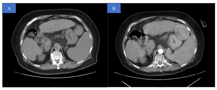
CT imaging before and after EUS-RFA of HCC. (A) Baseline CT image of HCC before EUS-RFA; (B) CT image obtained at the first imaging follow-up (1 month after EUS-RFA) demonstrating post-treatment changes of treated lesions. CT: computed tomography; EUS-RFA: endoscopic ultrasound-guided radiofrequency ablation; HCC: hepatocellular carcinoma.
Patient perspective was not available due to the retrospective nature of this case series.
Liver malignancy management is complex because it depends not only on tumor burden but also on liver function, comorbidities, and feasibility of local therapies. Current guideline therapy for HCC recommends RFA or microwave ablation (MWA) as one of the preferred curative treatments for selected early-stage tumors, especially when surgery is not ideal. For metastatic disease, CRLM management increasingly uses a multidisciplinary strategy, combining systemic therapy with liver-directed approaches in selected patients, including resection or ablation. Liver tumor ablation is a locoregional therapy used for primary liver cancer (especially early-stage HCC) and for selected secondary liver malignancies [1]. This technique destroys tumors by delivering energy or chemical therapies directly into a specific focal tumor to induce coagulative necrosis. The main modalities include RFA, MWA, cryoablation, and chemical injection (e.g., percutaneous ethanol injection) [7]. RFA has historically been the most widely used thermal ablation technique in HCC, supported by extensive clinical experience and evidence. In comparison, MWA offers several technical advantages over RFA, including higher intratumor temperature, larger and more uniform ablation volumes, and reduced susceptibility to the heat-sink effect [8].
RFA produces controlled thermal injury through alternating electrical current, resulting in tissue heating and coagulative necrosis of the targeted tissue. This mechanism is effective for tumor destruction when complete ablation with adequate safety margin is achieved. In carefully selected patients with small, early-stage HCC, RFA can achieve outcomes comparable to hepatic resection, although it is associated with a higher risk of tumor recurrence despite fewer procedure-related complications [9]. Nevertheless, hepatic resection remains the preferred curative treatment for patients with preserved liver function and resectable disease according to current clinical practice guidelines [1]. As tumor size increases beyond 3 cm, achieving complete ablation becomes more challenging because of difficulties in obtaining adequate ablative margins and the potential influence of the heat-sink effect, resulting in a higher risk of incomplete ablation and local recurrence. Therefore, combination treatment strategies may be required in selected patients [10].
In the non-curative setting, the aim is to achieve local tumor control while minimizing the need for systemic therapy and potentially prolonging survival [11]. Nevertheless, RFA may also be considered in selected patients with HCC larger than 3 cm when curative treatment is intended, although treatment efficacy generally decreases as tumor size increases. In a large retrospective cohort of patients with HCC > 3 cm, complete ablation after a single RFA session was achieved in 77 patients from total 100 patients. However, local recurrence (48%) and distant recurrence (82%) remained substantial, highlighting limited durability of RFA in larger tumors. Favorable outcomes were primarily observed in carefully selected patients with preserved liver function (Child Pugh Class A) and lower pre-ablation des-γ-carboxy prothrombin levels [12].
EUS-RFA has recently emerged as an alternative route for percutaneous liver tumor ablation because EUS-RFA may offer a distinct advantage for lesions in the left hepatic lobe or caudate region (that are in close proximity to the stomach or duodenum), tumors with difficult percutaneous access due to interposed structures (ribs, lung, bowel gas), subcapsular or subdiaphragmatic lesions (where percutaneous ablation carries higher procedural risk), patients with obesity that limits percutaneous visualization, or cases where prior percutaneous attempts were unsuccessful [3, 4]. EUS provides high-resolution imaging and precise needle control from a short distance and stable access route, which may improve targeting in these anatomically challenging locations [13]. Although the field still lacks large prospective comparative trials, more recent case series in cirrhotic patients suggest EUS-RFA can achieve good local response in selected HCC lesions with a favorable safety profile when performed in expert centers. The published studies evaluating EUS-RFA in liver malignancy can be seen in Table 4.
Published studies of EUS-RFA for primary and secondary liver malignancies.
| Author | Year | No of patient | Tumor type and location | Reported outcomes |
|---|---|---|---|---|
| Attili et al. [4] | 2018 | 1 | HCC (HCV cirrhosis) segment VIII (25 × 20 mm) | No procedure-related complicationMRI at 1 month: complete disappearance |
| de Nucci et al. [3] | 2020 | 1 | HCC (cirrhosis), large subcapsular lesion (30 × 45 mm) in segment II-III-IVb | Complication: transient and reversible mild liver decompensationTwo EUS-RFA sessions, CT scan: ~70% tumor reduction |
| Katrevula et al. [14] | 2023 | 1 | HCC (HBV cirrhosis), caudate lobe (20 × 16 mm) | Complication: mild self-limiting painMRI at 3 months: complete response |
| De et al. [15] | 2024 | 1 | HCC (MetALD cirrhosis), caudate lobe (16 × 19 mm) | No procedure-related complicationMRI at 3 months: complete response |
| Harwani et al. [16] | 2025 | 5 | HCC (cirrhosis due to HBV and NASH), lesions in segments I, VI, II, and III; multifocal at II and III; multifocal at II, III, VIII, and VII-VIII junction; multifocal at II, III, and VISize ranged from 7 × 11 mm to 34 × 38 mm | No procedural adverse eventCT scan at 1 month: complete response in 4 patients. Partial response in 1 patient due to difficult access (VII/VIII). Repeat EUS-RFA achieved complete response |
| Dancour et al. [17] | 2022 | 14 | Liver metastasis (mainly pancreatic ductal adenocarcinoma, pancreatic NET and colon cancer)24 liver metastases including right lobe (n = 8); median size 20 mm | 2 complications (jaundice and asymptomatic gallbladder hematoma)Reported median overall survival: 16 monthsTechnical success (the definition was > 80% ablation on CT scan) was achieved for 15 of the 20 liver metastases evaluated |
Response assessments were reported according to the criteria used in the original publications and may not be directly comparable across studies. CT: computed tomography; EUS-RFA: endoscopic ultrasound-guided radiofrequency ablation; HBV: hepatitis B virus; HCC: hepatocellular carcinoma; HCV: hepatitis C virus; MetALD: metabolic dysfunction-associated steatotic liver disease with increased alcohol intake; MRI: magnetic resonance imaging; NASH: non-alcoholic steatohepatitis; NET: neuroendocrine tumor.
All patients in this case series were selected based on anatomical limitations to percutaneous ablation, supporting the clinical relevance of EUS-RFA as an alternative technique rather than a first-line modality. EUS-RFA was technically feasible in all treated patients, as reflected by successful needle placement and completion of intended energy delivery. No immediate major procedure-related complications were observed, including bleeding, perforation, or infection. These findings provide preliminary information regarding short-term procedural safety of EUS-RFA when performed by experienced operators in carefully selected patients and are consistent with early reports describing EUS-RFA as a minimally invasive option for selected liver tumors in anatomically favorable locations [4, 16]. However, because of the small sample size, retrospective design, and heterogeneous follow-up, this study cannot reliably assess delayed adverse events or long-term procedural safety.
With respect to treatment response, four of the five patients demonstrated disease stability at the first imaging follow-up, including both HCC patients at 1 month (mRECIST) and the iCCA and CRLM cases at 4 months (RECIST 1.1). Although stable disease does not represent tumor eradication, it may be clinically meaningful as an early indicator of local disease control-particularly in the setting where percutaneous ablation or surgery is limited by unfavorable anatomy, comorbidity, or technical constraints. Notably, one patient with PNET liver metastasis showed progressive disease on later follow-up imaging at 8 months (a 28.13% increase in size). PNET liver metastasis can demonstrate heterogeneous biology and may require multimodality management, including systemic therapy and/or repeated locoregional interventions [18]. A single session of the ablation treatment strategy may be insufficient for biologically aggressive or multifocal metastatic disease, and progression may also reflect systemic tumor behavior rather than failure of local therapy alone.
Current HCC guideline emphasizes that treatment selection depends on tumor burden as well as hepatic functional reserve, making liver malignancy management intrinsically complex in patients with chronic liver disease and cirrhosis [1]. Comparative studies and meta-analyses suggest that ablation can achieve outcomes approaching resection in carefully selected small HCC, often with fewer complications but with an increased risk of recurrence [9]. For secondary malignancy, especially CRLM, local therapies (including ablation) are increasingly integrated into multidisciplinary care. A recent randomized phase 3 trial demonstrated non-inferiority of thermal ablation compared with surgical resection for the primary outcome in selected patients with small (≤ 3 cm), resectable CRLM [19]. Our caudate CRLM case therefore supports the concept that EUS-guided access may expand local treatment options in anatomically difficult lesions. For iCCA, thermal ablation is considered in selected unresectable patients, but data in this setting remain limited and heterogeneous. A recent multicenter study showed that outcomes are better in small lesions, typically ≤ 17 mm [20]. In PNET liver metastasis, ablation is generally used as a local adjunct in selected patients with limited, unresectable hepatic disease, with more favourable outcomes in tumors < 3 cm [21].
In our series, the absence of partial or complete response at the first imaging follow-up may reflect several factors, such as relatively large baseline tumor size in most cases and up to 53 mm. This is clinically relevant, as ablation efficacy is known to decrease with increasing tumor size, particularly beyond 3 cm, where achieving complete tumor necrosis becomes more difficult. The probability of incomplete necrosis is higher because of the heat-sink effect and difficulty obtaining a circumferential safety margin. This makes local tumor control more difficult and usually implies the need for multiple sessions of combined therapy. For EUS-RFA specifically, the published studies are still very limited and have mostly involved small left lobe or caudate lesions, not large tumors. Published case reports and small case series summarized in Table 4 suggest that RFA has primarily been applied to small lesions in anatomically challenging locations, which makes extrapolation for large tumors uncertain. Other contributing factors include heterogeneous tumor biology (primary versus metastatic disease) and the possibility that local ablation alone is insufficient in patients with systemic or biologically aggressive disease.
A key practical limitation of EUS-RFA is segmental accessibility. Because the stomach and duodenum provide the most direct transgastric/transduodenal window to the liver, EUS-guided needle trajectory is generally more favourable for the left hepatic lobe and caudate lobe. In contrast, a lesion located deep in the right lobe may be technically challenging to reach safely due to scope position, limited needle angulation, longer intraparenchymal path, and proximity to major vasculature. This anatomic constraint may result in incomplete ablation coverage or the need for repeat sessions. For example, in a recent study, difficult access to segments VII/VIII was associated with only partial response at first follow-up, but a second EUS-RFA session achieved complete radiologic response [16].
This study has several important limitations. First, the sample size is small, and the study design is a retrospective study, limiting both generalizability and the ability to draw definitive conclusions regarding efficacy of EUS-RFA. Accordingly, this study should not be interpreted as evidence that EUS-RFA is superior or equivalent to hepatic resection, percutaneous thermal ablation, MWA, systemic therapy, or other established treatment modalities. Second, follow-up in our series was relatively short and differed across cases (1–8 months), limiting assessment of delayed adverse events and long-term oncologic outcomes. Third, the primary endpoints focused on technical feasibility and immediate safety. Longer-term outcomes such as local tumor progression, the need for repeat ablation, progression-free survival, and overall survival were not evaluated. Fourth, the clinical applicability of EUS-RFA may be anatomically constrained to the left hepatic lobe and caudate region. Therefore, our findings should be interpreted as preliminary evidence regarding the technical feasibility and short-term procedural safety of EUS-RFA in carefully selected patients with anatomically challenging liver lesions.
Future studies should incorporate standardized protocols, including patient selection criteria, technical ablation parameters (needle type, power, duration, number of applications, sessions), response assessment criteria, imaging modality, and follow-up intervals. Prospective multicenter studies with comparator groups are needed to define the role of EUS-RFA within multidisciplinary liver cancer management and to evaluate long-term oncologic outcomes.
In this small retrospective series, EUS-RFA was technically feasible for selected patients with liver malignancies and no immediate major procedure-related complications were observed. EUS-RFA may serve as a complementary technique in carefully selected cases where percutaneous access is difficult, unsafe, or not feasible. However, the absence of objective tumor response and limited follow-up in this study highlight the need for cautious interpretation of these findings. Larger prospective studies are needed to better define the role of EUS-RFA in the multidisciplinary management of liver malignancies.
CRLM: colorectal liver metastasis
CT: computed tomography
EUS: endoscopic ultrasound
EUS-RFA: endoscopic ultrasound-guided radiofrequency ablation
HCC: hepatocellular carcinoma
iCCA: intrahepatic cholangiocarcinoma
mRECIST: modified Response Evaluation Criteria in Solid Tumors
MRI: magnetic resonance imaging
MWA: microwave ablation
PNET: pancreatic neuroendocrine tumor
RECIST: Response Evaluation Criteria in Solid Tumors
RFA: radiofrequency ablation
CRAL: Conceptualization, Supervision, Writing—original draft, Writing—review & editing. SS: Conceptualization, Formal analysis, Visualization, Writing—original draft, Writing—review & editing. IH: Supervision, Validation. RAG: Supervision, Validation. All authors read and approved the submitted version.
Cosmas Rinaldi Adithya Lesmana, who is the Editorial Board Member and Guest Editor of Exploration of Digestive Diseases, had no involvement in the decision-making or the review process of this manuscript. The other authors declare no conflicts of interest.
This retrospective case series was approved by the Medistra Hospital Health Research Ethics Committee, Number: 221/K-LKJ/ETIK/VI/2025. The approval covered the retrospective review of medical records of patients with liver malignancies who underwent EUS-RFA, including hepatocellular carcinoma, intrahepatic cholangiocarcinoma, colorectal liver metastasis, and pancreatic neuroendocrine tumor liver metastasis. The study was conducted in accordance with the Declaration of Helsinki.
The requirement for individual consent was waived by the Ethics Committee because of the retrospective nature of the study and the use of anonymized data.
The present study is a retrospective case series based on de-identified medical record data, and all CT and EUS images included in the manuscript are fully anonymized and do not contain identifiable patient information.
The datasets analyzed during the current study are available from the corresponding author upon reasonable request.
Not applicable.
© The Author(s) 2026.
Open Exploration maintains a neutral stance on jurisdictional claims in published institutional affiliations and maps. All opinions expressed in this article are the personal views of the author(s) and do not represent the stance of the editorial team or the publisher.
Copyright: © The Author(s) 2026. This is an Open Access article licensed under a Creative Commons Attribution 4.0 International License (https://creativecommons.org/licenses/by/4.0/), which permits unrestricted use, sharing, adaptation, distribution and reproduction in any medium or format, for any purpose, even commercially, as long as you give appropriate credit to the original author(s) and the source, provide a link to the Creative Commons license, and indicate if changes were made.



View: 38
Download: 5
Times Cited: 0


