 Review


Affiliation:
1Faculty of Clinical Sciences, University of Ilorin, Ilorin 240003, Nigeria
Email: patrickashinze@yahoo.com
ORCID: https://orcid.org/0009-0007-9137-5498


 ,
Affiliation:
2Department of Pharmacology and Therapeutics, Faculty of Basic Clinical Sciences, College of Medicine, University of Nigeria, Nsukka 410001, Nigeria
ORCID: https://orcid.org/0009-0004-7009-4396
,
Affiliation:
1Faculty of Clinical Sciences, University of Ilorin, Ilorin 240003, Nigeria
ORCID: https://orcid.org/0009-0004-8522-6160
,
Affiliation:
3St John’s Hospital, Livingston EH54 6PP (NHS Lothian), Scotland
ORCID: https://orcid.org/0009-0007-3052-7590
,
Affiliation:
4Obafemi Awolowo University Teaching Hospital Complex, Ile-Ife 220005, Nigeria
ORCID: https://orcid.org/0000-0002-7170-7623
,
Affiliation:
1Faculty of Clinical Sciences, University of Ilorin, Ilorin 240003, Nigeria
ORCID: https://orcid.org/0009-0006-5123-4423
,
Affiliation:
1Faculty of Clinical Sciences, University of Ilorin, Ilorin 240003, Nigeria
ORCID: https://orcid.org/0009-0004-7309-1721
,
Affiliation:
1Faculty of Clinical Sciences, University of Ilorin, Ilorin 240003, Nigeria
ORCID: https://orcid.org/0009-0001-5424-2377
Explor Cardiol. 2026;4:1012128 DOI: https://doi.org/10.37349/ec.2026.1012128
Received: May 06, 2026 Accepted: June 10, 2026 Published: November 04, 2026
Academic Editor: Eugenio Picano, Italian National Research Council, Italy
Cardio-oncology has emerged as a critical interdisciplinary field addressing the growing burden of cardiovascular complications arising from modern cancer therapies. While advances in chemotherapy, targeted agents, and immunotherapies have substantially improved cancer survival, they have concurrently led to a significant rise in cancer therapy-related cardiac dysfunction (CTRCD), contributing to increased long-term morbidity and mortality among cancer survivors. Conventional surveillance strategies often detect cardiotoxicity only after irreversible myocardial injury has occurred, accentuating the need for more precise, proactive, and personalized approaches. Theranostics, integrating diagnostic imaging, molecular biomarkers, and targeted therapeutics, offers a promising paradigm to enable early detection, risk stratification, and tailored cardioprotective interventions in cardio-oncology. This review synthesizes recent advances in theranostic strategies relevant to cardio-oncology, spanning imaging-based, biomarker-driven, and nanotechnology-enabled approaches. Advanced imaging modalities such as hybrid PET/MRI, AI-enhanced echocardiography, and novel molecular radiotracers [e.g., 68Ga-FAPI, 18F-DHMT, [68Ga]pentixafor] enable early identification of subclinical myocardial injury, inflammation, fibrosis, and metabolic dysfunction before overt declines in left ventricular ejection fraction occur. Complementing imaging, biomarker-driven theranostics including high-sensitivity cardiac troponins, natriuretic peptides, circulating and exosomal microRNAs, and cardiomyocyte-specific cell-free DNA, provide minimally invasive, real-time insights into myocardial stress, injury, and immune-mediated cardiotoxicity. Nanotheranostic platforms, such as liposomal doxorubicin and immunomodulatory nanoparticles, further enhance therapeutic precision by reducing off-target cardiac exposure while preserving antitumor efficacy. Despite these advances, clinical translation remains hindered by interdisciplinary fragmentation, limited access to advanced infrastructure, high development costs, regulatory heterogeneity between agencies, and reliance on surrogate endpoints rather than long-term cardiovascular outcomes. Theranostics represents a transformative approach in cardio-oncology. Integration of advanced imaging, molecular biomarkers, artificial intelligence, and nanotechnology holds significant promise for preserving cardiac health without compromising oncologic success. Embedding theranostic strategies into routine cardio-oncology practice is essential to advancing precision medicine and improving cardiovascular outcomes for cancer patients worldwide.
Cardio-oncology has emerged as a critical subspecialty focused on the intricate relationship between cancer therapies and their associated cardiovascular complications [1]. The field encompasses the prevention, diagnosis, and treatment of cardiac problems arising from chemotherapy and radiation, the protection of cardiac health before, during, and after cancer therapy, and the collaborative management of pre-existing heart conditions alongside oncological care [1, 2]. The rapid evolution of oncological treatments has significantly amplified the interplay between cardiology and oncology, with novel cancer therapies substantially improving survival rates, in some cases conferring additional years of life. This progress has been accompanied by an unintended consequence: an approximately two-to-six-fold increase in cardiovascular mortality among cancer survivors compared to the general population, a burden particularly evident following anthracycline-based chemotherapy, trastuzumab, and thoracic radiotherapy, which collectively represent the highest-risk treatment exposures [2, 3]. Early detection and treatment have demonstrated the capacity to reverse adverse cardiac effects in many oncological contexts; patients at high risk of cardiotoxicity are therefore prioritised for screening and monitoring to enhance early detection and minimise irreversible toxicity [4].
Theranostics, an evolving paradigm rooted in precision oncology, combines diagnostic imaging with targeted therapies to optimise cancer management. By leveraging nuclear medicine diagnostic techniques, clinicians can accurately diagnose, manage, and treat cancer patients through personalised approaches [5]. The field has facilitated the identification of cancer biomarkers using positron emission tomography (PET) and single-photon emission computed tomography (SPECT)-based molecular imaging. These tools enable the design of individualised treatment plans that precisely diagnose and treat cancer patients while minimising adverse effects and improving quality of life and survival rates [6, 7]. The growing complexity of cancer subtypes has driven innovation in precision medicine, tailoring treatments to an individual’s unique genetic, molecular, and environmental profile. Breakthroughs in artificial intelligence (AI)-driven radiomics and nanotheranostics are advancing cancer care by enabling accurate diagnoses and targeted therapies [8].
This review summarises the most current theranostic approaches in cardio-oncology, including advanced imaging-based theranostics, biomarker-guided strategies, targeted drug delivery, and nanotheranostics. Clinical implications and future directions of these approaches are also discussed, with the objective of establishing a foundation for preventive, diagnostic, and therapeutic modalities. In the expanding cardio-oncology literature, numerous narrative reviews have addressed advances in imaging, biomarkers, and nanotechnology in isolation. This review distinguishes itself by synthesising these discrete technological strands into an integrative clinical workflow model centred on theranostics. A unifying framework is articulated that progresses from multi-modal risk assessment to targeted intervention, emphasising the synergistic clinical application of these tools. A consensus-style analysis of immediate translational opportunities and the specific cross-cutting barriers, regulatory, economic, and interdisciplinary, that must be overcome to implement this paradigm is also provided. The objective is to move beyond cataloguing innovations and instead offer a clinically actionable perspective on how integrated theranostic strategies can be embedded into routine cardio-oncology pathways to improve patient outcomes.
Theranostic modalities in cardio-oncology are best understood within a conceptual framework that integrates molecular mechanisms of cardiac injury, diagnostic detection, and targeted therapeutic response. At its core, theranostics operationalises a diagnostic-therapeutic loop: a molecular signal, whether a radiotracer binding to an upregulated receptor, an exosomal microRNA (miRNA) signature, or an elevated biomarker reflecting a specific injury pathway, simultaneously identifies the biological target and informs the selection of the therapeutic agent directed against it. The sections below address the three principal modality domains, imaging-based approaches, biomarker-driven strategies, and nanotheranostics, structured to move from the underlying biology to diagnostic application and then to therapeutic integration.
Advanced imaging technologies are central to theranostics, offering non-invasive means to detect and characterise anatomic, biochemical, and functional pathology, thereby providing critical diagnostic information for precise treatment planning and real-time monitoring. The translation of advanced imaging theranostics faces heterogeneous validation. AI-echocardiography tools for global longitudinal strain (GLS) measurement are progressing through regulatory clearance processes, demonstrating improved reproducibility over manual methods, yet require rigorous external validation across diverse patient populations and ultrasound platforms [9]. Novel radiotracers such as 68Ga-FAPI for fibrosis imaging remain in early-phase trials, with validation pathways longer and more resource-intensive owing to radiopharmaceutical regulatory requirements. A robust cost-effectiveness analysis is also lacking: although hybrid PET/MRI provides unparalleled multi-parametric data, it is surmisably higher in cost per scan compared to sequential standard tests, which must be justified by proven impact on treatment decisions and hard outcomes, evidence that is not yet mature [8–10].
Cardiovascular magnetic resonance (CMR) is valuable for early cardiotoxicity detection by assessing cardiac anatomy, tissue composition, and function [9, 10]. CMR can detect subtle myocardial changes before a decline in left ventricular ejection fraction (LVEF) and accurately quantifies left ventricular mass changes that correlate with anthracycline dose and cardiac event risk [9]. Hybrid PET/MRI integrates PET’s functional and metabolic capacity with MRI’s enhanced anatomical detail and soft-tissue contrast [11]. This combination allows for enhanced lesion characterisation and precise image co-registration in a single session, providing incremental information beyond individual modalities. MRI additionally offers parameters such as dynamic contrast enhancement (DCE) for vascularisation assessment and diffusion-weighted imaging (DWI) for cellularity quantification, aiding in tumour characterisation [11, 12]. In cardio-oncology, 18F-FDG PET/MRI can identify early anthracycline-induced myocardial damage by assessing glucose uptake, and shows promise for cardiac sarcoidosis by combining late gadolinium enhancement (LGE) MRI for myocardial injury with 18F-FDG PET for active inflammation, offering stronger prognostic value than either modality alone [12, 13].
AI, in particular machine learning (ML) and deep learning (DL), is transforming cardio-oncology by improving risk stratification, optimising treatment, and enabling individualised care. These techniques are also instrumental for early detection of cardiac dysfunction, enabling timely and personalised interventions to prevent irreversible damage [14]. AI models analyse electrocardiogram (ECG), echocardiographic, CT, and MRI data to identify patterns indicative of cardiotoxicity [15]. In echocardiography, AI addresses limitations arising from user-dependency and measurement variability by automating complex assessments, enhancing diagnostic precision, and increasing accessibility [16]. Specific applications include improving the accuracy and efficiency of LVEF assessment and automating GLS measurement, a validated early marker for cardiotoxicity, thereby improving reproducibility and predictive accuracy [16, 17].
Novel radiotracers are designed with high specificity for molecular targets and optimal kinetics for signal detection, enabling paired diagnostic and therapeutic agents to target identical mechanisms, which is the core principle of theranostics [18]. Table 1 (see below) summarises key radiotracers with potential applications in cardio-oncology. Clinically, 68Ga-DOTATATE and [68Ga]pentixafor are most suited for imaging myocardial inflammation in the context of immune checkpoint inhibitor (ICI)-associated myocarditis, where CXCR4-positive immune cell infiltration can be quantified. The 18F-DHMT tracer targets superoxide production and is most applicable to early anthracycline cardiotoxicity surveillance, while 68Ga-FAPI provides direct imaging of fibroblast activation protein, facilitating the characterisation of myocardial fibrosis in post-therapy cardiomyopathy. The advantage of each tracer lies in its mechanistic specificity; the disadvantage is the limited availability of radiopharmaceutical infrastructure outside specialist centres.
| Radiotracer | Target | Diagnostic advantage | Potential application in cardio-oncology |
|---|---|---|---|
| 68Ga-DOTATATE | Somatostatin receptors (SSTR) on activated inflammatory cells | High sensitivity for inflammatory infiltration | Imaging myocardial inflammation in ICI myocarditis |
| [68Ga]pentixafor | Chemokine Receptor CXCR4 on pro-inflammatory immune cells | Quantification of CXCR4+ immune cell burden | Imaging coronary inflammation and remodelling |
| 18F-DHMT | Superoxide production (reactive oxygen species) | Direct oxidative stress detection | Early detection of anthracycline-induced cardiotoxicity |
| 68Ga-FAPI | Fibroblast activation protein (FAP) | Direct imaging of active fibrosis | Imaging myocardial fibrosis in post-therapy cardiomyopathy |
Biomarker-driven strategies offer sensitive, cost-effective, and minimally invasive approaches for cardiotoxicity surveillance, often providing insights before overt functional impairment is detectable by imaging. Critically, these biomarkers are not mere indicators of injury; each reflects a distinct mechanistic pathway, enabling their clinical utility to extend beyond detection to the guidance of targeted interventions.
Anthracyclines exert their cardiotoxic effect primarily through topoisomerase IIβ inhibition, triggering mitochondrial reactive oxygen species (ROS) generation, double-strand DNA breaks within cardiomyocytes, and subsequent apoptosis. Trastuzumab, by contrast, impairs cardiomyocyte survival through ErbB2 receptor blockade, disrupting downstream survival signalling pathways including neuregulin-1/ErbB2 and PI3K/Akt. Cardiac troponins (cTnI and cTnT) are specific proteins released upon cardiomyocyte necrosis, providing a sensitive indicator of myocardial damage from these mechanisms [19]. High-sensitivity troponin (hs-Tn) assays enable earlier detection of subtle injury and predict subsequent irreversible CTRCD when elevated [20]. Natriuretic peptides (NPs), specifically B-type natriuretic peptide (BNP) and NT-proBNP, are released in response to myocardial stress and wall stretch from increased intracardiac pressures associated with ventricular dysfunction. Both troponins and NPs are recommended at baseline for patients receiving potentially cardiotoxic agents [20]. Limitations include their non-specificity to the underlying cause of damage and a lack of robust comparative analyses between different hs-Tn assays, hindering standardisation [20].
Circulating miRNAs (cmiRNAs) show promise as early biomarkers for anthracycline-induced cardiotoxicity. Specific cmiRNAs (e.g., miR-34a, miR-1, miR-423, miR-126, miR-130a) may predict cardiotoxicity development [20]. Myeloperoxidase (MPO), implicated in inflammation and oxidative stress, is a promising early biomarker for CTRCD in breast cancer patients receiving anthracyclines and trastuzumab [20, 21]. Combining multiple biomarkers, such as MPO and troponin, may improve predictive accuracy.
Liquid biopsy, a minimally invasive approach that analyses circulating tumour cells (CTCs), circulating tumour DNA (ctDNA), and other blood components, is primarily used in oncology for monitoring cancer progression [21, 22]. It carries additional potential for monitoring cardiotoxicity [22]. Changes in cardiomyocyte-specific cell-free methylated DNA (cfDNA) may indicate cardiomyocyte death from cardiotoxic therapies including radiation [22–24]. Importantly, cfDNA elevation following anthracycline exposure does not correlate closely with high-sensitivity cardiac troponin I (hs-cTnI), suggesting that these markers reflect distinct pathological processes and provide complementary mechanistic information. When cardiomyocyte cfDNA rises in the context of active cancer therapy, this signal may serve as a trigger for escalating cardioprotective measures, including the initiation of nanoparticle-based cardioprotective strategies, thereby operationalising the theranostic loop. Liquid biopsy enables dynamic tracking of both cancer progression and therapeutic cardiac responses, facilitating timely adjustments to therapy [23, 25].
Exosomes are extracellular vesicles that mediate intercellular communication by carrying heterogeneous cargo, including miRNAs [26]. Exosomal miRNAs (ex-miRs) are stable in circulation and carry potential as non-invasive biomarkers for heart failure and cardiotoxicity [24]. Specific ex-miRs, including miR-107, miR-146a, and miR-502, are potential markers of doxorubicin-induced cardiotoxicity (DIC) [24, 26]. Exosomal miR-34a-5p, derived from PD-1 inhibitor-treated macrophages, is mechanistically linked to ICI-related cardiac dysfunction through induction of cardiac senescence. Beyond diagnostic potential, ex-miRs signatures are emerging as functional guides for therapeutic decisions: elevated miR-34a-5p may indicate a patient phenotype more likely to respond to senolytic or mitochondria-targeted interventions, while miR-107 upregulation may support selection of liposomal doxorubicin formulations with optimised cardiac protection profiles. This diagnostic-to-therapeutic translation exemplifies the operational theranostic loop in biomarker-guided cardio-oncology [24, 26].
cfDNA originates from dead cells releasing genomic DNA into the bloodstream. Its tissue specificity allows identification of cardiomyocyte-derived cfDNA. Elevated cardiomyocyte cfDNA levels following anthracycline chemotherapy are significantly associated with an increased risk of subsequent CTRCD [26]. While hs-cTnI also rises in this context, its poor correlation with cardiomyocyte cfDNA indicates distinct underlying pathological processes, and the two biomarkers therefore provide complementary clinical information.
Nanotechnology offers transformative opportunities in cardio-oncology by enabling precise drug delivery, reducing systemic toxicity, and enhancing therapeutic efficacy [27]. Nanoparticles enhance bioavailability and stability, facilitating precise delivery to cancer cells, minimising cardiotoxicity, and improving efficacy. Their modular construction allows them to carry various components, enabling high local drug concentrations at the disease site while protecting healthy tissues [25, 27].
Encapsulation alters doxorubicin biodistribution, reducing accumulation in the myocardium while promoting tumour accumulation via the enhanced permeability and retention (EPR) effect [28]. This significantly lowers myocardial drug concentrations, reducing the topoisomerase IIβ-mediated oxidative injury that underlies anthracycline cardiotoxicity. Liposomal doxorubicin also induces interferon-stimulated genes (ISGs) in the heart, promoting cell survival and reducing apoptosis [28, 29].
pH-sensitive nanoparticles are engineered to respond to the acidic tumour microenvironment (TME), selectively releasing their payload at the disease site while maintaining stability in healthy tissues. Thermally responsive biopolymers respond to mild local hyperthermia (approximately 41°C) at the tumour site. These biopolymers promote preferential accumulation and drug release within the heated tumour, significantly reducing doxorubicin accumulation in the myocardium and preserving cardiac function [28, 29].
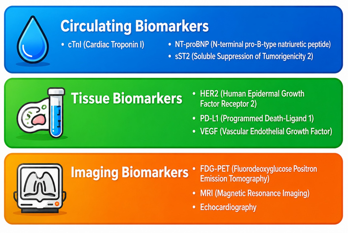
Immunomodulatory nanoparticles are designed to interact specifically with the immune system, enhancing or suppressing its activity for therapeutic outcomes [30]. They address challenges of conventional immunotherapy by delivering tumour antigens and adjuvants to antigen-presenting cells (APCs) and modulating the TME [30]. Nanotheranostics combine diagnostic and therapeutic capabilities, permitting direct delivery of agents to damaged cardiac tissues for targeted, localised treatment with minimal systemic adverse effects. This approach can improve the safety profile of ICIs, which can cause immune-related adverse events including myocarditis, by achieving specific and localised immune response modulation in tumour tissue while reducing systemic side effects [30]. Figure 1 provides an infographic summary of biomarkers of theranostic interest in cardio-oncology [19, 21, 24, 30].
The integration of theranostics into cardio-oncology faces significant translational hurdles despite its transformative potential. This section examines key implementation barriers, illustrative clinical case studies, and critical regulatory and economic considerations shaping real-world adoption.
Interdisciplinary fragmentation remains a fundamental barrier. Cardio-oncology trials such as STOP-CA highlight persistent gaps in collaborative trial design, where cardiologists and oncologists frequently operate independently [31]. Recruitment challenges arise from competing clinical priorities, with a sizable number of eligible patients declining participation owing to overwhelmed care schedules [31]. Exclusion of patients with comorbidities such as hypertension and diabetes creates selection bias, limiting generalisability to real-world populations [31].
Cost and accessibility present substantial hurdles. Advanced theranostic modalities including PET/MRI hybrid imaging and novel radiotracers (e.g., 68Ga-FAPI, 18F-DHMT) require specialised infrastructure unavailable outside tertiary centres [4, 11]. Radiopharmaceutical production faces complex logistical challenges, including radionuclide supply chains and regulatory requirements [4]. Development costs for a single agent may exceed one billion US dollars with timelines spanning 10 to 15 years [4], while patient-specific dosimetry calculations further increase resource demands [4].
Evidence generation barriers include an overreliance on surrogate endpoints such as LVEF changes rather than clinically meaningful outcomes, for example, heart failure hospitalisations [31]. Statistically significant LVEF differences (e.g., 1.3% in STOP-CA) may not translate to tangible patient benefits [31]. Short follow-up durations, typically 12 to 24 months, fail to capture late cardiovascular complications [31].
A central barrier to the routine integration of advanced theranostics is the predominance of proof-of-concept and observational data over high-level clinical evidence. For most novel radiotracers, exosomal biomarker panels, and nanotheranostic platforms, robust validation from multicentre randomised controlled trials (RCTs) demonstrating an impact on hard cardiovascular outcomes, such as heart failure hospitalisation and cardiovascular mortality, is absent. Current clinical guidelines rightly remain anchored to established tools such as serial echocardiographic LVEF. Competing strategies, including the universal prophylactic use of cardioprotectants (e.g., dexrazoxane, ACE inhibitors) in high-risk cohorts, offer a simpler, evidence-supported alternative to complex biomarker-guided approaches. Adoption of precision theranostics must therefore be contingent on generating evidence that it outperforms conventional strategies in both efficacy and cost-effectiveness. Future trial designs must move beyond surrogate endpoints toward pragmatic studies in which theranostic-guided management is compared against standard care for primary endpoints of patient survival and morbidity.
ICIs trigger fulminant myocarditis via CD8+ T-cell-driven inflammation [30]. Emerging approaches combine [68Ga]pentixafor PET, targeting CXCR4-positive inflammatory cells, with cardiac MRI to detect subclinical inflammation [13]. Biomarker-guided strategies using exosomal miR-34a-5p provide mechanistic insight into cardiac senescence, enabling risk stratification [24, 26].
Cytokine release syndrome (CRS) affects 50 to 90% of CAR-T recipients and causes haemodynamic compromise [32]. Theranostic monitoring combines liquid biopsy biomarkers, including endothelial-derived cfDNA and interleukin-6, with echocardiographic strain imaging to guide tiered interventions [25, 26]. Engineering advances incorporating on/off safety switches aim to minimise systemic toxicity [32].
The STOP-CA trial utilised baseline high-sensitivity troponin T (hs-TnT) and NT-proBNP to identify lymphoma patients likely to benefit from atorvastatin prophylaxis [31]. Statin recipients demonstrated reduced CTRCD rates (8% vs. 22% placebo), particularly those with elevated biomarkers [31]. Ongoing research is exploring polygenic risk scores to refine patient selection [20].
Divergent regulatory pathways between the FDA and EMA complicate development. FDA orphan designation requires conditions affecting fewer than 200,000 US patients, versus the EMA threshold of fewer than 5 per 10,000 EU prevalence [4]. Radiopharmaceuticals face additional complexity under FDA investigational new drug (IND) requirements, where diagnostic-therapeutic pairs may require separate approvals despite biological equivalence [4]. Collaborative mechanisms such as the FDA-EMA Parallel Scientific Advice programme show promise in harmonising requirements for rare toxicities [4].
Value-based frameworks must evolve to address theranostic economics. Traditional cost-effectiveness models are not well suited to combined diagnostic-therapeutic agents, where upfront imaging costs (e.g., 68Ga-DOTATATE PET) are offset by optimised patient selection [4, 27]. Patient-specific dosimetry exemplifies this value, enabling personalised dosing to maximise efficacy while minimising toxicity [4].
The economic model and accessibility of theranostics are pivotal to their real-world impact. Infrastructure for advanced modalities, including cyclotron-produced radiotracers and high-field MRI, is concentrated in high-income tertiary centres, creating the risk of a theranostic divide across health systems. A pragmatic, value-based implementation strategy is required, structured across three tiers. At the foundation tier, widespread deployment of validated, low-cost tools such as AI-enhanced point-of-care ultrasound and hs-Tn assays should be achievable across most clinical settings. An advanced tier would involve strategic referral networks for novel biomarker panels or SPECT/CT at regional hubs. The investigational tier would concentrate centralised expertise for PET/MRI and nanotherapeutic trials. Demonstrating that early theranostic intervention reduces downstream costs of heart failure management will be essential for justifying initial investments and achieving equitable access [9, 10, 27].
Advancements in gene editing, AI, and targeted therapeutics are reshaping the landscape of cardio-oncology. Table 2 (see below) outlines the current validation status, key strengths, major limitations, and recommended clinical context for the principal theranostic modalities under evaluation, providing a practical framework for prioritising their integration into cardio-oncology pathways.
Validation status, strengths, limitations, and recommended context for principal theranostic modalities in cardio-oncology.
| Modality | Stage of validation | Key strength | Major limitations/cost | Recommended context |
|---|---|---|---|---|
| AI-GLS (Echocardiography) | Regulatory-cleared devices; multi-society guideline support | High reproducibility; low incremental cost; portable | Algorithm vendor variability; requires quality image acquisition | First-line serial monitoring for patients on anthracyclines or trastuzumab |
| Cardiomyocyte cfDNA | Early-phase clinical studies | High tissue specificity for injury; mechanistic insight | No standardised approved assay; high per-test cost (£300–£700) | Investigational risk stratification in high-dose therapy or clinical trials |
| 68Ga-FAPI PET | Preclinical and early phase I in cardio-oncology | Direct imaging of active fibrosis; quantifiable signal | Limited tracer availability; high scan cost; no outcome data | Research tool for characterising unexplained post-therapy cardiomyopathy |
Gene editing carries significant potential for cardio-oncology. In vivo CRISPR-Cas9 strategies have successfully corrected pathogenic sarcomere gene variants in animal models of hypertrophic cardiomyopathy, reducing cardiac hypertrophy and fibrosis [33]. High-throughput CRISPR screens in human iPSC-derived cardiomyocytes are also identifying genes, including SLCO1A2 and SLCO1B3, that influence doxorubicin sensitivity [32]. Future CRISPR-based strategies may also be applicable to the repair of chemotherapy-induced DNA damage or patient-specific cardiomyopathy mutations, enabling truly personalised cardioprotection [33, 34].
ML constitutes another transformative tool. Models integrating patient data, including demographics, cancer type, and treatment regimens, can identify patients at high risk for cardiotoxicity [35]. DL applied to routine ECGs can detect subclinical declines in ejection fraction following anthracyclines, with reported AUC values of approximately 0.93 [36]. AI algorithms applied to cardiac imaging, including echocardiography, MRI, and CT, improve quantification of cardiac volumes and function, highlighting subtle changes before symptoms emerge [37]. These tools facilitate earlier intervention by identifying at-risk patients and streamlining surveillance.
Mitochondria-targeted antioxidants are a promising pharmacological advance. Compounds such as Mito-TEMPO and MitoQ accumulate in cardiomyocyte mitochondria and scavenge ROS. Preclinical studies demonstrate that Mito-TEMPO administered during chemotherapy significantly reduces doxorubicin- and 5-fluorouracil-induced cardiac injury by preserving mitochondrial function [38]. Mitochondria-targeted coenzyme Q analogues (e.g., MitoQ, SKQ1) have similarly protected animal hearts from drug-induced oxidative damage [37]. Clinical trials are required to confirm the safety and efficacy of these agents as co-administered cardioprotectants.
CAR-T therapy is expanding beyond haematological malignancies into solid tumours, with new designs improving cardiac safety. Recent engineering advances include affinity tuning and logic-gated receptors requiring two tumour signals to activate, thereby sparing healthy tissues. On/off safety switches and methods for local delivery of CAR-T cells aim to confine activity to tumour sites, retaining anti-tumour potency while minimising on-target, off-tumour cardiotoxicity of the type observed in early HER2-directed CAR trials [39].
Hybrid imaging modalities combined with AI analysis represent a further frontier. PET/CT or MRI/PET scans can measure myocardial metabolism and fibrosis in cancer patients, and AI-based analysis can detect early cardiotoxic changes. AI-enhanced hybrid imaging substantially improves the accuracy and efficiency of cardiotoxicity assessment. In practice, such pipelines could allow real-time monitoring of cardiac function during therapy, so that oncologists and cardiologists can adapt treatment, for example by pausing chemotherapy or initiating cardioprotective agents, at the earliest detectable signal [40].
As cardio-oncology continues to evolve, it is essential to address global priorities that ensure equitable access to care, foster collaborative research, and establish comprehensive registries. These initiatives are critical for bridging gaps in knowledge, improving patient outcomes, and promoting health equity across diverse populations. Figure 2 presents a summating theranostic workflow illustration from diagnostics to precision healthcare.
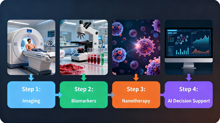
A theranostic workflow illustration from diagnostics to precision healthcare delivery.
Theranostics is redefining cardio-oncology by enabling simultaneous diagnosis and treatment of cancer therapy-related cardiovascular disease, marking a transition toward precision care. Mechanistically grounded advances, including hybrid PET/MRI imaging that targets specific molecular pathways of injury, exosomal biomarker profiling that translates miRNA signatures into therapeutic selection criteria, and nanotheranostic platforms that reduce anthracycline-mediated topoisomerase IIβ injury at the cardiomyocyte level, are collectively establishing a theranostic framework in which diagnostic signal and therapeutic action are functionally linked rather than conceptually adjacent. Real-world integration remains limited by cost, regulatory complexity, and interdisciplinary fragmentation. Two specific recommendations emerge from this evidence synthesis. First, multicentre pragmatic RCTs comparing theranostic-guided cardio-oncology management against standard care should be designed with primary endpoints of heart failure hospitalisation and cardiovascular mortality, rather than surrogate imaging parameters. Second, a tiered implementation model should be formally adopted, committing to universal access to validated low-cost tools such as AI-GLS echocardiography and hs-Tn assays, while restricting novel radiotracers and nanotheranostic platforms to investigational contexts until outcome data mature. Collaborative efforts to establish global registries, develop robust AI-driven decision tools, and promote equitable access to emerging technologies are urgently required. Realising the full potential of theranostics in cardio-oncology demands unified action from the laboratory to the clinic.
AI: artificial intelligence
BNP: B-type natriuretic peptide
cfDNA: cell-free DNA
cmiRNAs: circulating microRNAs
CMR: cardiovascular magnetic resonance
CTRCD: cancer therapy-related cardiac dysfunction
DL: deep learning
ECG: electrocardiogram
ex-miRs: exosomal microRNAs
GLS: global longitudinal strain
hs-cTnI: high-sensitivity cardiac troponin I
hs-Tn: high-sensitivity troponin
ICI: immune checkpoint inhibitor
LVEF: left ventricular ejection fraction
miRNA: microRNA
ML: machine learning
MPO: myeloperoxidase
NPs: natriuretic peptides
PET: positron emission tomography
RCTs: randomised controlled trials
ROS: reactive oxygen species
TME: tumour microenvironment
The authors acknowledge The Lind League, Nigeria, for providing the institutional resources that supported this research project. Some of the subfigures in this manuscript were generated with the assistance of artificial intelligence image generation tools [Perplexity AI]. These images were reviewed and verified by the authors prior to inclusion.
PA: Conceptualization, Methodology, Writing—original draft, Writing—review & editing, Supervision, Validation, Project administration. NOC: Writing—original draft, Writing—review & editing, Data curation. ONO: Writing—original draft, Writing—review & editing, Data curation. WS: Writing—original draft, Writing—review & editing, Data curation. SO: Writing—review & editing, Supervision. YT: Writing—original draft, Writing—review & editing, Data curation. HB: Writing—original draft, Writing—review & editing, Data curation. FM: Writing—original draft, Writing—review & editing, Data curation. All authors read and approved the submitted version.
The authors declare no conflicts of interest.
Not applicable.
Not applicable.
Not applicable.
Not applicable.
No funding was received for this work.
© The Author(s) 2026.
Open Exploration maintains a neutral stance on jurisdictional claims in published institutional affiliations and maps. All opinions expressed in this article are the personal views of the author(s) and do not represent the stance of the editorial team or the publisher.
Copyright: © The Author(s) 2026. This is an Open Access article licensed under a Creative Commons Attribution 4.0 International License (https://creativecommons.org/licenses/by/4.0/), which permits unrestricted use, sharing, adaptation, distribution and reproduction in any medium or format, for any purpose, even commercially, as long as you give appropriate credit to the original author(s) and the source, provide a link to the Creative Commons license, and indicate if changes were made.



View: 88
Download: 3
Times Cited: 0


