 Review


Affiliation:
1Department of General Surgery, Unidade Local de Saúde de Coimbra, 3000-075 Coimbra, Portugal
2Faculty of Medicine, University of Coimbra, 3000-548 Coimbra, Portugal
3Coimbra Institute for Clinical and Biomedical Research (iCBR), Faculty of Medicine, University of Coimbra, 3000-548 Coimbra, Portugal
4Center of Investigation on Environment, Genetics and Oncobiology (CIMAGO), Faculty of Medicine, University of Coimbra, 3000-548 Coimbra, Portugal
5Clinical Academic Center of Coimbra (CACC), 3000-548 Coimbra, Portugal
Email: ritaandrade03@gmail.com
ORCID: https://orcid.org/0000-0003-3650-0640


 ,
Affiliation:
2Faculty of Medicine, University of Coimbra, 3000-548 Coimbra, Portugal
3Coimbra Institute for Clinical and Biomedical Research (iCBR), Faculty of Medicine, University of Coimbra, 3000-548 Coimbra, Portugal
4Center of Investigation on Environment, Genetics and Oncobiology (CIMAGO), Faculty of Medicine, University of Coimbra, 3000-548 Coimbra, Portugal
5Clinical Academic Center of Coimbra (CACC), 3000-548 Coimbra, Portugal
6Germano de Sousa Pathological Anatomy Center, 3000-377 Coimbra, Portugal
ORCID: https://orcid.org/0000-0002-7202-8059
,
Affiliation:
2Faculty of Medicine, University of Coimbra, 3000-548 Coimbra, Portugal
7Clinical Pathology Department, Unidade Local de Saúde de Coimbra, 3000-075 Coimbra, Portugal
ORCID: https://orcid.org/0000-0002-0462-2427
,
Affiliation:
3Coimbra Institute for Clinical and Biomedical Research (iCBR), Faculty of Medicine, University of Coimbra, 3000-548 Coimbra, Portugal
4Center of Investigation on Environment, Genetics and Oncobiology (CIMAGO), Faculty of Medicine, University of Coimbra, 3000-548 Coimbra, Portugal
5Clinical Academic Center of Coimbra (CACC), 3000-548 Coimbra, Portugal
8Cytogenetics and Genomics Laboratory, Institute of Cellular and Molecular Biology, Faculty of Medicine, University of Coimbra, 3000-548 Coimbra, Portugal
9Center for Innovative Biomedicine and Biotechnology (CiBB), University of Coimbra, 3000-548 Coimbra, Portugal
ORCID: https://orcid.org/0000-0002-6096-8705
,
Affiliation:
3Coimbra Institute for Clinical and Biomedical Research (iCBR), Faculty of Medicine, University of Coimbra, 3000-548 Coimbra, Portugal
4Center of Investigation on Environment, Genetics and Oncobiology (CIMAGO), Faculty of Medicine, University of Coimbra, 3000-548 Coimbra, Portugal
5Clinical Academic Center of Coimbra (CACC), 3000-548 Coimbra, Portugal
8Cytogenetics and Genomics Laboratory, Institute of Cellular and Molecular Biology, Faculty of Medicine, University of Coimbra, 3000-548 Coimbra, Portugal
9Center for Innovative Biomedicine and Biotechnology (CiBB), University of Coimbra, 3000-548 Coimbra, Portugal
ORCID: https://orcid.org/0000-0001-6842-1707
,
Affiliation:
1Department of General Surgery, Unidade Local de Saúde de Coimbra, 3000-075 Coimbra, Portugal
2Faculty of Medicine, University of Coimbra, 3000-548 Coimbra, Portugal
3Coimbra Institute for Clinical and Biomedical Research (iCBR), Faculty of Medicine, University of Coimbra, 3000-548 Coimbra, Portugal
4Center of Investigation on Environment, Genetics and Oncobiology (CIMAGO), Faculty of Medicine, University of Coimbra, 3000-548 Coimbra, Portugal
5Clinical Academic Center of Coimbra (CACC), 3000-548 Coimbra, Portugal
ORCID: https://orcid.org/0000-0002-5641-6975
Explor Target Antitumor Ther. 2026;7:1002413 DOI: https://doi.org/10.37349/etat.2027.1002413
Received: May 15, 2026 Accepted: September 17, 2026 Published: November 18, 2026
Academic Editor: Giulia Martini, University of Campania “Luigi Vanvitelli”, Italy
The article belongs to the special issue Liquid Biopsy: Has Already Changed the Clinical Decision-Making in Solid Tumors Treatment?
Cholangiocarcinoma (CCA) is a heterogeneous and often late-diagnosed malignancy in which tissue acquisition, pathological confirmation, and molecular profiling remain clinically challenging. Precision oncology has increased the need for reliable tumour-derived molecular information, particularly when conventional cytology or biopsy is inconclusive or inadequate for next-generation sequencing (NGS). Bile has emerged as a candidate liquid biopsy matrix because it directly drains the biliary tree and may come into contact with malignant or premalignant biliary epithelium, potentially concentrating tumour-derived DNA, RNA, extracellular vesicles (EVs), proteins and metabolites at the site of disease rather than diluting them in the systemic circulation. Bile-derived cell-free DNA (cfDNA) and circulating tumour DNA (ctDNA) are the most mature biomarkers, with matched studies reporting somatic mutation detection in up to 70–71% of bile cfDNA samples compared with 10–54% in plasma, along with higher variant allele fractions and stronger concordance with tumour tissue. The most immediate clinical application is the evaluation of indeterminate biliary strictures, where an NGS-based bile cfDNA assay (Bilemut), evaluated in a mixed cohort of suspicious biliary strictures, achieved an overall sensitivity of 96.4% and a specificity of 69.2%, reaching 100% sensitivity in the 35 patients initially classified as having benign or indeterminate strictures, although this was a single-centre pilot cohort requiring independent validation. DNA methylation, non-coding RNAs, EVs, proteomic and lipidomic signatures are expanding the field beyond mutation detection, with integrated multi-omic approaches showing early promise for diagnosis and risk stratification. Translation to clinical practice remains limited by small cohorts, heterogeneous populations, variable sampling protocols, and the absence of prospective evidence that bile testing changes management or improves outcomes. Its most defensible role is additive rather than substitutive: bile adds molecular information from the local biliary compartment at the moment of an already indicated procedure, in patients in whom tissue is insufficient or non-diagnostic, and plasma is non-informative.
Cholangiocarcinoma (CCA) arises from the biliary epithelium and is anatomically classified into intrahepatic, perihilar and distal subtypes [1]. This distinction is clinically meaningful rather than purely descriptive: these subtypes differ in tissue accessibility, molecular architecture and therapeutic vulnerabilities [1–3], and CCA is increasingly understood as a spectrum of related diseases requiring integrated anatomical, pathological, molecular and clinical assessment [1–3].
Although biliary tract cancers (BTCs) are relatively uncommon in Western countries, they are frequently diagnosed late and remain associated with poor outcomes [1, 4, 5]. Intrahepatic CCA has shown an increasing incidence in several Western populations, while perihilar and distal CCA remain particularly challenging because of their infiltrative growth pattern and close anatomical relationship with the biliary hilum and extrahepatic bile ducts [1, 3]. Surgical resection remains the only potentially curative treatment, but many patients present with unresectable, locally advanced, or metastatic disease [1, 4]. Even after curative-intent surgery, recurrence is common, reflecting both the aggressive biology of the disease and the limitations of current diagnostic and therapeutic strategies [4, 5].
Early and accurate diagnosis remains a major unmet need. Early-stage CCA may be clinically silent, and symptoms, when present, often reflect biliary obstruction rather than tumour-specific biology [1, 5]. Serum biomarkers such as carbohydrate antigen 19-9 (CA19-9) and carcinoembryonic antigen may support diagnostic suspicion, but their specificity is limited, particularly in the presence of cholestasis, cholangitis or benign inflammatory biliary disease [1, 5]. Cross-sectional imaging, magnetic resonance cholangiopancreatography (MRCP), endoscopic ultrasound (EUS), endoscopic retrograde cholangiopancreatography (ERCP) and percutaneous transhepatic cholangiography (PTC) are central to anatomical assessment, staging and biliary drainage, but may still fail to distinguish malignant from benign strictures in early or indeterminate cases [1, 5].
Histological or cytological confirmation is therefore often required, yet tissue acquisition in CCA is technically difficult and diagnostically imperfect [1, 3]. Brush cytology and intraductal biopsy obtained during ERCP are highly specific when positive, but their sensitivity for malignant biliary strictures is limited [1]. EUS-guided sampling, percutaneous biopsy or biopsy of metastatic disease may be useful depending on tumour location and stage, but samples may be small, paucicellular, necrotic or unsuitable for downstream molecular analysis [3]. In perihilar and distal CCA, lesions are often infiltrative and difficult to sample directly; in intrahepatic CCA, desmoplasia, stromal contamination and intratumoural heterogeneity may compromise tissue adequacy for next-generation sequencing (NGS) [2, 3].
These diagnostic limitations have become more consequential in the era of precision oncology. Molecular profiling is recommended in patients with advanced BTC who are candidates for systemic treatment, and NGS is preferred over sequential single-gene testing because multiple clinically relevant alterations may need to be assessed simultaneously [1, 3]. Intrahepatic CCA is enriched for potentially actionable alterations such as isocitrate dehydrogenase 1/2 (IDH1/2) mutations and fibroblast growth factor receptor 2 (FGFR2) fusions or rearrangements, whereas extrahepatic CCA more frequently harbours alterations involving KRAS, TP53, SMAD4 and ERBB2 [2, 3]. Other clinically relevant biomarkers include BRAF alterations, ERBB2/HER2 amplification or overexpression, neurotrophic tropomyosin receptor kinase (NTRK) fusions and microsatellite instability-high (MSI-H)/deficient mismatch repair (dMMR) status [1, 3]. Reliable molecular information is therefore no longer only a research objective; it is becoming part of routine therapeutic planning and clinical trial allocation [1–3].
However, tissue-based molecular profiling is not always feasible. Low tumour cellularity, insufficient biopsy material, necrosis, formalin fixation-related artefacts, and tissue exhaustion during routine diagnostic work-up may compromise NGS [2, 3]. A single tissue sample may also fail to capture spatial tumour heterogeneity, particularly in advanced disease or under therapeutic selective pressure [4, 6]. These limitations have created a need for complementary approaches capable of providing molecular information when tissue is unavailable, insufficient, or no longer representative [3, 4, 6].
Liquid biopsy has emerged as one such strategy. It refers broadly to the analysis of tumour-derived material in biological fluids, including cell-free DNA (cfDNA), circulating tumour DNA (ctDNA), circulating tumour cells, RNA species, extracellular vesicles (EVs), proteins and metabolites [4–6]. In BTC, these approaches have been investigated for diagnosis, molecular profiling, prognostic stratification, treatment monitoring, minimal residual disease detection and identification of resistance mechanisms [4, 5]. Plasma-based liquid biopsy dominates the field because venous blood collection is minimally invasive and repeatable [5, 6]. In advanced disease, plasma ctDNA can capture systemic tumour heterogeneity and provide actionable genomic information when tissue is unavailable or inadequate [4–6].
Still, plasma ctDNA has important limitations in CCA. The tumour-derived fraction of plasma cfDNA may be low, particularly in early-stage, localised or anatomically confined disease, and false-negative results may occur when tumour shedding into the circulation is limited [5, 6]. Plasma ctDNA assays may also have reduced sensitivity for selected alteration types, including fusions and copy number changes, depending on assay design and tumour fraction [6]. Plasma is therefore highly attractive for systemic monitoring and recurrence detection, but it may not always be the most informative compartment for initial local molecular characterisation of biliary tumours [5, 6].
This has led to growing interest in tumour-proximal fluids, particularly bile. Bile is produced by hepatocytes, modified by cholangiocytes and transported through the biliary tree, where it may come into direct contact with malignant or premalignant biliary epithelium [7, 8]. Its composition includes bile acids, phospholipids, cholesterol, bilirubin, proteins, metabolites, EVs, RNA and DNA, and this molecular composition may change in hepatobiliary disease [7, 8]. Because of its anatomical proximity to the tumour, bile may contain tumour-derived material released directly into the biliary lumen, potentially providing a more concentrated and locally informative molecular signal than plasma in selected settings [7–10].
The clinical appeal of bile also lies in procedural opportunity. Bile may be collected during ERCP, endoscopic nasobiliary drainage, percutaneous transhepatic biliary drainage, percutaneous cholangiography, or surgery [7–9]. Although bile sampling is not as simple or repeatable as venous blood collection, it may be efficient when biliary access is already required for drainage, tissue acquisition or evaluation of a stricture [7–9]. Bile liquid biopsy should therefore not be positioned as a replacement for plasma, but as a complementary tumour-proximal approach with a different clinical purpose [6–9].
The most immediate clinical setting is the evaluation of indeterminate biliary strictures. In this context, conventional imaging, CA19-9, cytology and biopsy may fail to establish a definitive diagnosis, delaying treatment in patients with malignancy or exposing patients with benign disease to unnecessary major surgery [1, 9]. NGS of bile cfDNA has shown promising diagnostic performance in suspicious biliary strictures, particularly in cases initially classified as benign or indeterminate by conventional pathology [9]. Combined molecular approaches, including mutation and methylation analysis in bile, have also shown potential for distinguishing malignant from benign pancreatobiliary disease, although this evidence is not specific to CCA alone [10].
Among bile-derived biomarkers, cfDNA and ctDNA currently have the strongest translational rationale. In resectable CCA, matched analysis of tumour tissue, plasma and bile showed significantly higher cfDNA concentrations in bile than in plasma, with somatic mutations detected in all tumour tissue samples, 70% of bile cfDNA samples and only 10% of plasma cfDNA samples [11]. Bile cfDNA also showed higher maximum allele fractions and higher tumour mutational burden than plasma, supporting its potential role as a local source for molecular characterisation [11]. These findings remain preliminary because of the small cohort, but they are consistent with broader BTC studies reporting higher tumour-derived signal and stronger tissue concordance in bile than in plasma [11–13].
The promise of bile liquid biopsy extends beyond mutation detection. Bile may also contain methylated DNA, microRNAs, circular RNAs, EVs, proteins, lipids and metabolites with potential diagnostic, prognostic or predictive relevance [7, 8, 10]. These analytes may support future multimodal biomarker panels integrating genomic, epigenetic, transcriptomic, proteomic and metabolomic information [10]. At present, however, most non-cfDNA approaches remain investigational, and their translation is limited by small cohorts, heterogeneous populations and variable analytical methods [4, 8].
A major barrier to clinical implementation is standardisation. The diagnostic performance of bile-based liquid biopsy depends not only on the presence of tumour-derived material, but also on how bile is collected, processed, preserved and analysed [7, 8, 14]. Sampling route, timing of collection, cholangitis, obstruction, prior drainage, stents, contamination, centrifugation protocol, storage temperature and time to processing may all influence biomarker yield and reproducibility [7, 8, 14]. Recent work suggests that selected bile-derived proteins and mutant KRAS ctDNA can remain stable for up to seven hours at room temperature, supporting the feasibility of real-world bile handling while also reinforcing the need for reproducible standard operating procedures [14].
These observations justify a focused review of the evidence in CCA. The evidence remains early, but it is most compelling in two settings: indeterminate biliary strictures and local molecular profiling when tissue is insufficient, or plasma is non-informative [9, 11–13]. Available studies are still limited by small cohorts, methodological heterogeneity and variation in tumour subtype, disease stage, sampling route and analytical platform [7–9, 11–14]. This review summarises the biological rationale, biomarker landscape, clinical applications, technical limitations and future perspectives of bile-based liquid biopsy in CCA. The overall concept and proposed clinical positioning of bile-based liquid biopsy in CCA are summarised in Figure 1.
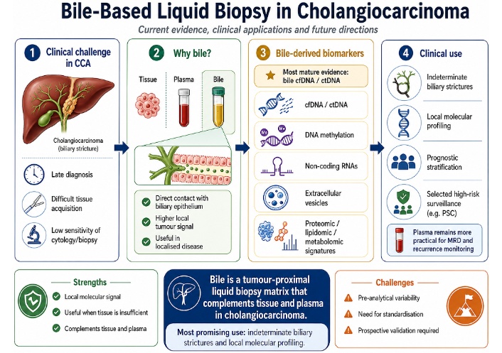
Bile-based liquid biopsy in CCA. Bile is presented as a tumour-proximal matrix that may complement tissue and plasma, particularly in indeterminate biliary strictures and local molecular profiling. CCA: cholangiocarcinoma; cfDNA: cell-free DNA; ctDNA: circulating tumour DNA; MRD: minimal residual disease; PSC: primary sclerosing cholangitis.
The rationale for using bile as a liquid biopsy source in CCA is grounded in anatomy. Unlike plasma, which reflects a systemic compartment where tumour-derived DNA is diluted within a large background of non-tumour cfDNA, bile flows through the biliary tree and may directly contact malignant or premalignant epithelium [6–8]. In CCA, where tumour cells arise from, infiltrate, or obstruct the ductal system, this creates a close interface between tumour tissue and the biliary lumen [1, 7, 8]. Bile is therefore not simply another liquid biopsy fluid; it is a local compartment that may capture tumour-derived material before it becomes diluted in systemic circulation [7, 8].
Bile is also a complex and dynamic biological matrix. It is produced by hepatocytes, modified by cholangiocytes and transported through the intrahepatic and extrahepatic biliary system, carrying bile acids, phospholipids, cholesterol, bilirubin, inorganic salts, proteins, metabolites, EVs and nucleic acids [7, 8]. In malignancy, this composition may be influenced by tumour shedding, epithelial injury, cholestasis, inflammation, infection and microbiome-related changes [7, 8]. This multi-analyte nature helps explain why bile has attracted interest as a liquid biopsy matrix: it may provide information not only on tumour genetics, but also on epigenetic alterations, EV cargo, RNA expression, protein signatures and metabolic remodelling [7, 8, 15].
The tumour-proximal position of bile may also explain why bile-derived cfDNA can show a stronger tumour signal than plasma in selected biliary malignancies. Plasma ctDNA detection depends on tumour burden, vascular invasion, metastatic dissemination, clearance mechanisms and assay sensitivity, and false-negative results may occur when systemic tumour DNA shedding is low [6]. In contrast, DNA released into the biliary lumen may be less diluted and more directly representative of the local tumour compartment [7, 8]. This distinction is especially relevant in localised or duct-confined disease, where plasma ctDNA may be less informative but local biliary shedding may still occur [6, 11–13].
Several studies support this biological model. In BTC cohorts, bile cfDNA consistently showed higher mutation detection rates, higher maximum allele frequencies and stronger concordance with matched tumour tissue than plasma cfDNA, as discussed in detail in cfDNA and ctDNA [12, 13]. These findings support bile as a more tumour-informative matrix for local genomic assessment in biliary malignancies.
Data from CCA support this concept: matched tissue–plasma–bile studies show consistently higher mutation detection and stronger tissue concordance in bile cfDNA than in plasma cfDNA (cfDNA and ctDNA) [11, 12].
Bile may also capture molecular information beyond sequence-level mutation detection. He et al. [10] developed BileScreen, an assay integrating mutations in 23 genes and methylation changes in 44 genes in bile samples from patients with pancreatobiliary disorders. The assay showed high diagnostic performance for pancreatobiliary tract cancer and outperformed serum CA19-9 in specificity, but it was not designed as a CCA-specific test [10]. Its relevance to CCA is therefore mainly conceptual: it supports the use of bile as a matrix for combined genetic and epigenetic tumour detection [10]. Ito et al. [16] further showed that multigene liquid biopsy of bile can identify driver gene alterations in biliary duct cancers, supporting the feasibility of applying panel-based molecular approaches to bile-derived DNA. These studies suggest that bile may provide a broader molecular readout than mutation analysis alone [10, 16].
EVs add another layer to this rationale. EVs are lipid bilayer-delimited particles capable of carrying proteins, DNA, mRNA, microRNAs, long non-coding RNAs, circular RNAs and lipids [15]. In bile, EVs may originate from cholangiocytes, tumour cells, immune cells or stromal components, and their cargo may reflect epithelial injury, inflammation, tumour progression and local tumour–microenvironment communication [15]. Because vesicular membranes protect molecular cargo from degradation, bile-derived EVs may offer a relatively stable compartment for RNA- and protein-based biomarkers in a chemically complex fluid [15]. Although bile EV research is less mature than bile cfDNA analysis, its biological relevance supports its inclusion in future multimodal biomarker strategies [15].
The rationale for bile liquid biopsy is also strengthened by clinical feasibility. Bile can be obtained during ERCP, endoscopic nasobiliary drainage, percutaneous transhepatic biliary drainage, percutaneous cholangiography, biliary stenting or surgery [7–9]. This matters because patients with suspected or confirmed CCA frequently undergo biliary access for drainage, tissue sampling, palliation or evaluation of a biliary stricture [1, 7, 9]. Arechederra et al. [9] showed that bile cfDNA collected during first diagnostic ERCP could be analysed by NGS in patients with suspicious biliary strictures, with the Bilemut assay improving malignancy detection in cases initially classified as benign or indeterminate by conventional pathology. Thus, bile liquid biopsy may add molecular information at the time of an already indicated biliary procedure [9].
However, bile is technically demanding: its complex composition and matrix-specific behaviour, including a broader cfDNA fragment-size distribution than plasma, mean that protocols developed for plasma cannot simply be extrapolated to bile (Technical and pre-analytical challenges) [7, 8, 14, 17, 18]. This technical complexity does not undermine the biological rationale above, but it does mean that translation will depend on reproducible, bile-specific handling and analysis (Technical and pre-analytical challenges).
Pre-analytical handling is therefore central to translation. Sampling route, timing, centrifugation, storage medium, storage duration, preservation tubes and temperature may all affect biomarker yield and reproducibility [14, 17, 18]. Han et al. [14] reported that selected bile-derived proteins, E-cadherin/N-cadherin activity and mutant KRAS ctDNA remained stable for up to seven hours at room temperature in bile samples from patients with CCA. Yang et al. [18] showed that cfDNA preservation tubes-maintained bile cfDNA stability for up to ten days at room temperature and for two months at 4°C, whereas regular tubes were associated with progressive degradation under less favourable conditions. These findings support the operational feasibility of bile biomarker analysis while underlining the need for standardised collection and processing protocols [14, 18].
The biological argument is strongest when bile is sampled during a procedure that is already clinically indicated, such as ERCP, drainage or surgery, and when the question being asked is local rather than systemic. The remaining challenge is operational: the same matrix that is biologically rich is also technically variable, so clinical translation will depend on reproducible handling, preservation and analysis [14, 17, 18].
Bile-derived liquid biopsy in CCA includes several biomarker classes, from cfDNA and ctDNA to methylated DNA, non-coding RNAs, EVs, proteins, lipids and metabolites [7, 8, 15]. This diversity reflects the biological complexity of bile itself. Rather than being only a digestive fluid, bile is a dynamic molecular compartment shaped by hepatocytes, cholangiocytes, tumour cells, inflammation, obstruction, infection and biliary drainage [7, 8]. For CCA, this complexity is both an opportunity and a limitation: bile may capture molecular features that are difficult to access through plasma or small tissue biopsies, but its composition is also influenced by non-malignant processes that can affect biomarker interpretation [7, 8, 14, 18].
Among bile-derived biomarkers, cfDNA and ctDNA currently have the strongest evidence base and are closest to clinical translation [9, 11–13]. The field, however, is expanding beyond mutation detection. DNA methylation [10, 19–21], exosomal microRNAs [22–24], circular RNAs [25], EV cargo [15, 26, 27], proteomic signatures [28, 29] and lipidomic or metabolomic profiles [30–32] may provide complementary information on diagnosis, prognosis, treatment response and tumour biology. These biomarker classes are not competing approaches but potential components of future multimodal bile-based diagnostic panels.
cfDNA is the most extensively studied bile-derived biomarker in CCA and BTC [9, 11–13]. cfDNA refers to extracellular DNA fragments released into biological fluids, whereas ctDNA represents the tumour-derived fraction carrying cancer-associated alterations, including single-nucleotide variants, insertions and deletions, copy number alterations, gene rearrangements, microsatellite instability, tumour mutational burden and methylation patterns [6, 11, 12]. In bile, cfDNA is particularly attractive because tumour-derived DNA may be released directly from malignant biliary epithelium into the ductal lumen, potentially increasing the tumour fraction compared with plasma [7, 8, 11–13].
Several studies support the use of bile cfDNA for local genomic profiling in biliary malignancies. Gou et al. [12] analysed matched tumour tissue, plasma and bile samples from patients with BTC using a 520-gene sequencing panel and detected somatic mutations in 71.4% of bile cfDNA samples compared with 53.6% of plasma cfDNA samples. Bile cfDNA also showed a significantly higher maximum allele frequency and stronger concordance with tumour tissue for single-nucleotide variants and insertions/deletions than plasma cfDNA [12]. Li et al. [13] similarly reported markedly higher cfDNA concentrations in bile supernatant than in plasma, together with higher mutation allele frequencies and greater consistency between bile and tumour tissue than between plasma and tumour tissue. Their observation that bile supernatant and pellet could detect additional genomic alterations not identified in tissue also suggests that bile may capture spatial heterogeneity missed by a single biopsy [13].
In CCA specifically, Liu et al. [11] compared matched tumour tissue, plasma and bile samples from patients with resectable disease and found significantly higher cfDNA concentrations in bile than in plasma. Somatic mutations were detected in all tumour tissue samples, in 70% of bile cfDNA samples and in only 10% of plasma cfDNA samples [11]. Bile cfDNA also showed higher maximum allele fractions, higher tumour mutational burden and stronger concordance with tissue genomic profiles than plasma [11]. Although this was a small cohort, the findings are clinically relevant because they suggest that bile may retain molecular sensitivity when systemic ctDNA shedding is low, a known limitation of plasma-based assays in localised or anatomically confined disease [6, 11].
Bile cfDNA may also support the diagnostic evaluation of indeterminate biliary strictures. Arechederra et al. [9] developed Bilemut, an NGS-based mutational assay using bile cfDNA collected during ERCP in patients with suspicious biliary strictures, including CCA and pancreatic ductal adenocarcinoma. The assay demonstrated that bile cfDNA can add tumour-derived molecular information when conventional pathology is inconclusive, which is precisely the setting where its clinical value is greatest. The diagnostic performance of Bilemut is discussed in detail in Diagnosis of indeterminate biliary strictures [9].
Beyond diagnosis, bile cfDNA may provide information relevant to precision oncology. Clinically relevant alterations in CCA include IDH1 mutations, FGFR2 fusions or rearrangements, BRAF alterations, ERBB2/HER2 alterations, NTRK fusions and MSI-H/dMMR status, although detectability depends on panel design, tumour fraction and analyte quality [1–3, 6]. Fusion detection warrants particular attention: FGFR2 rearrangements are among the most clinically actionable alterations in intrahepatic CCA, but RNA stability and extraction reproducibility in bile remain less established than for cfDNA, which may compromise RNA-based fusion assays; DNA-based approaches can detect fusions through breakpoint-spanning reads but may miss rearrangements with intronic breakpoints and require adequate DNA quality not always achievable from bile [7, 17]. Ito et al. [16] showed that multigene liquid biopsy of bile could identify driver gene alterations in biliary duct cancers, supporting the feasibility of applying panel-based molecular testing to bile-derived DNA. Nevertheless, bile cfDNA is not yet a routine substitute for tissue NGS. Current studies vary in tumour type, stage, sampling route, sequencing platform and availability of matched tissue, and bile cfDNA analysis remains strongly influenced by pre-analytical handling, extraction method and preservation conditions [12, 13, 17, 18]. The key studies of bile-based liquid biopsy in CCA and BTC are summarised in Table 1.
Key studies of bile-based liquid biopsy in CCA and BTC.
| Author (year) | Design | n | Tumour type | Biomarker/Method | Key finding | Bile vs. plasma | Main limitation |
|---|---|---|---|---|---|---|---|
| Arechederra et al. (2022) [9] | Prospective | 68 | CCA, PDAC (biliary strictures) | cfDNA/NGS (Pan-Cancer panel; Bilemut assay) | Overall sensitivity 96.4%, specificity 69.2%; 100% sensitivity in initially benign/indeterminate strictures | Bile cfDNA outperformed initial pathological diagnosis; plasma not directly compared | Pilot study; mixed biliopancreatic stricture cohort; CCA not analysed as an independent validation subgroup; no external validation cohort |
| Liu et al. (2025) [11] | Prospective | 10 | Resectable CCA | cfDNA/NGS | Mutations in 70% bile cfDNA vs. 10% plasma; higher maxAF and TMB in bile | Bile showed higher mutation detection than plasma for local molecular characterisation in this cohort | Very small cohort (n = 10); single centre; limited to resectable disease |
| Gou et al. (2021) [12] | Retrospective | 42 | BTC (mixed) | cfDNA/NGS (520-gene panel) | Mutations in 71.4% bile vs. 53.6% plasma; higher maxAF and tissue concordance in bile | Bile cfDNA showed higher mutation detection and stronger tissue concordance than plasma in this cohort | Mixed BTC population; retrospective; limited matched plasma data |
| Li et al. (2022) [13] | Retrospective | 13 | BTC (mixed) | cfDNA/NGS (425 genes) | Bile cfDNA 1,918 ng/mL vs. 63.1 ng/mL plasma; more alterations per sample in bile | Bile supernatant and pellet detected additional alterations not identified in matched tissue in this cohort | Small cohort; mixed BTC; no prospective validation |
| He et al. (2023) [10] | Retrospective + validation | Not stated for CCA alone | Pancreatobiliary cancer | cfDNA mutations + methylation/BileScreen assay | High diagnostic performance; outperformed CA19-9 in specificity | Bile not directly compared to plasma in same cohort | Not CCA-specific; pancreatobiliary mixed population; methylation markers not independently validated in CCA |
| Shen et al. (2019) [33] | Retrospective | 10 | BTC (mixed) | cfDNA/targeted deep sequencing (150 genes) | SNV/Indel sensitivity 94.7%; CNV sensitivity 75%; Lower CNV sensitivity reported in AJCC stage II and distal bile duct tumours, though cohort size limits interpretation | First study to demonstrate bile cfDNA-based somatic variant detection in BTC using a 150-gene panel | Very small cohort; mixed BTC; n = 1 distal CCA |
| Vedeld et al. (2022) [19] | Retrospective | 273 | CCA, PSC-associated CCA, PSC | DNA methylation/ddPCR (CDO1, CNRIP1, SEPT9, VIM) | 100% sensitivity, 90% specificity for PSC-associated CCA when diagnosed within 12 months of bile collection | Bile not compared to plasma in same cohort | Retrospective; PSC-focused; markers not validated for sporadic CCA; no prospective surveillance pathway |
BTC: biliary tract cancer; CCA: cholangiocarcinoma; cfDNA: cell-free DNA; ctDNA: circulating tumour DNA; maxAF: maximum allele fraction; NGS: next-generation sequencing; PDAC: pancreatic ductal adenocarcinoma; PSC: primary sclerosing cholangitis; TMB: tumour mutational burden; ddPCR: droplet digital polymerase chain reaction; SNV: single nucleotide variant.
DNA methylation is a promising bile-derived biomarker class because epigenetic alterations may occur early in carcinogenesis, potentially preceding the accumulation of driver mutations detectable by sequencing [10, 19]. This is particularly relevant in CCA, where malignant transformation may arise against a background of chronic inflammation, biliary strictures, and dysplasia, especially in patients with primary sclerosing cholangitis (PSC) [1, 19, 20]. Unlike mutation panels, methylation assays may capture cancer-associated epigenetic patterns even when recurrent somatic mutations are absent or heterogeneous; this may be a particular advantage in the inflammatory biliary milieu of PSC [10, 20].
The strongest evidence for bile DNA methylation comes from PSC-associated CCA. Vedeld et al. [19] analysed 344 bile samples from 273 patients with sporadic CCA, PSC-associated CCA, PSC and other non-malignant liver diseases using droplet digital polymerase chain reaction (ddPCR) assays for promoter methylation of CDO1, CNRIP1, SEPT9 and VIM. In patients with PSC, all four markers showed high diagnostic performance for CCA detection, and when analysis was restricted to patients diagnosed with CCA within 12 months after bile collection, the combined marker panel reached 100% sensitivity and 90% specificity [19]. These findings are important because PSC-associated CCA often develops within a background of chronic inflammation and strictures, where imaging, CA19-9, cytology and fluorescence in situ hybridisation (FISH) may be inconclusive [1, 19].
A more recent study by Vedeld et al. [20] evaluated DNA methylation biomarkers in both bile and plasma from patients with PSC. The selected bile methylation panel, including 5-42952178, KCNA6, PRKCB and ZFP82, distinguished CCA from PSC with high accuracy, whereas plasma-based performance was lower [20]. This comparison reinforces a recurring theme in bile liquid biopsy: blood is easier to obtain, but bile may be more informative when the clinical question concerns local malignant transformation within the biliary tree [6, 7, 20].
Methylation may be especially valuable when combined with mutation analysis. He et al. [10] developed BileScreen, a bile-based assay integrating mutations into 23 genes and methylation changes in 44 genes for the diagnosis of pancreatobiliary tract cancer. The model achieved high diagnostic performance in validation cohorts and outperformed serum CA19-9, particularly in specificity [10]. Because the study included pancreatobiliary malignancies rather than CCA alone, its findings should not be interpreted as CCA-specific. Nevertheless, it supports the broader principle that multi-level molecular testing in bile can improve discrimination between malignant and benign pancreatobiliary disease [10]. Cooley et al. [21] also showed that methylated DNA markers can improve the diagnosis of malignant biliary strictures, although their work focused on biliary brushings rather than bile fluid itself. Overall, bile methylation appears most relevant for two clinical scenarios: early detection in high-risk patients and diagnostic clarification of indeterminate strictures [10, 19–21].
MicroRNAs and other non-coding RNAs represent a less mature but biologically compelling group of bile-derived biomarkers. MicroRNAs regulate gene expression post-transcriptionally and are involved in proliferation, apoptosis, angiogenesis, epithelial–mesenchymal transition, immune signalling and treatment resistance [22–24]. In bile, free RNA may be vulnerable to degradation, whereas RNA enclosed within EVs or exosomes is protected by a lipid membrane, increasing its potential stability as a biomarker source [15, 22, 23].
Han et al. [22] provided early proof of concept for bile-derived exosomal microRNAs in CCA. Using next-generation miRNA sequencing, they identified 22 differentially expressed miRNAs between bile-derived exosomes from a patient with perihilar CCA and a control patient with common bile duct stones [22]. Functional pathway analysis suggested enrichment of pathways relevant to CCA biology, including MAPK, VEGF, EGFR, p53 and RAS-related signalling [22]. The study was exploratory and based on a very small sample size, but it demonstrated that bile-derived exosomal miRNAs can carry biologically meaningful tumour-associated signals [22].
Yoshida et al. [23] provided broader clinical evidence in BTC. In 88 bile samples collected during ERCP, exosomal miRNA profiling identified aberrantly expressed miRNAs in bile, whereas serum exosomal miRNAs were less informative [23]. miR-451a and miR-3619-3p were reproducibly upregulated, and their combined diagnostic performance reached an area under the curve (AUC) of 0.819 for BTC detection [23]. High bile miR-3619-3p expression was also associated with worse prognosis, suggesting that bile-derived exosomal miRNAs may have both diagnostic and prognostic relevance [23].
Non-coding RNAs may also have predictive value. Fu et al. [24] studied bile microRNAs associated with chemotherapy efficacy in advanced perihilar CCA treated with hepatic arterial infusion chemotherapy. Bile miR-532-3p, miR-1250-5p and miR-4772-5p were associated with survival after treatment, and mechanistic experiments suggested that miR-532-3p promoted resistance to oxaliplatin and 5-fluorouracil by negatively regulating SPARC expression [24]. This study is notable because it moves bile RNA research beyond diagnosis and towards treatment-response biology [24].
Circular RNAs are another emerging class of non-coding RNA biomarkers. Wen et al. [25] identified an exosomal circRNA signature in bile and serum from patients with CCA-associated biliary obstruction. A three-circRNA panel (hsa-circ-0000367, hsa-circ-0021647 and hsa-circ-0000288) showed strong diagnostic performance, and the bile exosomal circRNA model outperformed CA19-9 [25]. The same study developed models for early recurrence monitoring after curative-intent surgery, suggesting that bile-derived non-coding RNA signatures may eventually contribute to diagnosis, risk stratification and postoperative surveillance [25].
For now, bile-derived RNA signatures are best regarded as biologically informative rather than practice-changing. Their clinical value will depend on whether they add information beyond simpler cfDNA- or methylation-based assays, and whether isolation methods can be reproduced across centres [10, 19–25].
EVs, including exosomes and small EVs, are membrane-bound particles released by living cells and capable of carrying proteins, lipids, DNA, mRNA, microRNAs, long non-coding RNAs and circular RNAs [15, 26]. In CCA, EVs are attractive because they may reflect communication between tumour cells, cholangiocytes, fibroblasts, immune cells and the local biliary microenvironment [15, 26]. Their lipid bilayer protects molecular cargo from enzymatic degradation, which may be especially relevant in bile, a chemically complex fluid containing bile salts, enzymes, inflammatory material and bacterial components [7, 15, 26].
The main biological advantage of bile-derived EVs is that they may combine local anatomical proximity with cargo stability [15, 22, 23, 26]. Studies of bile-derived exosomal miRNAs show that EV cargo can distinguish malignant from benign biliary disease and may carry prognostic information [22, 23]. Muraki et al. [27] linked EV biology with lipidomics by identifying phosphatidylcholine in bile-derived small EVs as a potential biomarker of CCA, though the study included only 13 patients with CCA, limiting the strength of this finding. Wen et al. [25] further showed that exosomal circRNA signatures in bile may support both diagnosis and early recurrence monitoring.
Despite these promising findings, EV-based bile liquid biopsy is not ready for routine clinical use. The main barriers are methodological: small cohorts, heterogeneous populations, variable bile handling and lack of standardised EV isolation and characterisation protocols [15, 26, 27]. Different approaches, including ultracentrifugation, filtration, precipitation, size-exclusion chromatography and affinity-based methods, may isolate different vesicle subpopulations and co-isolated non-vesicular material, affecting reproducibility across studies [15, 26]. EV-based bile biomarker research will therefore require harmonised workflows before its clinical value can be reliably assessed [15, 26].
Proteomic, lipidomic and metabolomic biomarkers provide another dimension of bile liquid biopsy. Because bile is directly involved in bile acid metabolism, lipid handling and biliary epithelial homeostasis, malignant transformation may alter its protein, lipid and metabolic composition [7, 8, 28–32]. These changes may reflect tumour-derived processes, but also stromal remodelling, extracellular matrix turnover, inflammation, cholestasis, microbial metabolism and altered bile acid signalling [7, 28–32].
Bile proteomics has recently produced clinically relevant candidates. Gao et al. [28] identified clusterin as a bile biomarker for CCA and developed an optic fibre biosensor integrated with digestive endoscopy for real-time detection. This study is notable because it moves bile biomarker research from laboratory discovery towards procedure-integrated diagnostics, with the possibility of obtaining molecular information during endoscopic biliary evaluation [28]. Chu et al. [29] used bile proteome profiling to identify upregulated proteins in CCA and integrated these findings with public transcriptomic datasets. POSTN emerged as a candidate biomarker; bile and plasma POSTN levels were higher in CCA than in controls, and plasma POSTN showed diagnostic performance comparable and complementary to CA19-9 [29]. Higher POSTN levels were also associated with epithelial–mesenchymal transition-related biology and worse survival, suggesting potential diagnostic and prognostic relevance [29].
Lipidomics and metabolomics are also gaining momentum. Liu et al. [30] performed a prospective descriptive study including 241 bile samples from patients with CCA or benign bile duct diseases. Using untargeted and targeted lipidomic approaches, they identified distinct bile lipid profiles and developed a six-lipid classifier, BileLipid, with AUCs of 0.943, 0.956 and 0.828 across three independent cohorts [30]. Several lipid classes, particularly lysophosphatidylcholine, were downregulated in CCA, and functional experiments suggested that reduced lysophosphatidylcholine may be biologically relevant rather than merely diagnostic [30]. Choi et al. [31] further examined bile lipidomics in extrahepatic CCA, choledocholithiasis and controls, identifying distinct lipidomic patterns across groups, including lower lysophosphatidylcholine levels in extrahepatic CCA than in choledocholithiasis. These studies suggest that bile lipidomics may help distinguish malignant from benign biliary obstruction, although larger validation cohorts are still needed [30, 31].
The biliary tract harbours a microbial community that may be altered in malignancy, cholestasis and chronic inflammation. Xia et al. [32] characterised the bile microbiome by 16S sequencing in patients with CCA, PSC and benign biliary disease and identified microbial signatures associated with CCA. Bile microbiome data collected during ERCP are, however, susceptible to contamination with duodenal flora introduced during instrumentation, and biliary microbial composition is also shaped by antibiotics, cholangitis, stents and prior drainage, which are the very conditions most common in patients under investigation for CCA. Microbiome-based diagnostic signals therefore require careful decontamination analysis and validation in cohorts with detailed procedural and clinical metadata before they can be reliably interpreted [32].
Integrating multiple analyte classes may capture disease biology that single-platform assays miss. Xia et al. [32] combined microbiome, metabolomic and bile acid data into a diagnostic panel that outperformed cytology-based approaches, and comparable integrative logic underlies BileScreen’s combination of mutation and methylation analysis [10]. This is conceptually attractive because CCA develops within a biliary environment shaped by inflammation, bile acids, microbial communities, epithelial injury and tumour metabolism [10, 32].
These platforms are valuable discovery tools, especially when the aim is to describe the biliary tumour ecosystem rather than a single driver alteration, but their clinical translation is likely to be slower than cfDNA testing because they require complex analytical pipelines, careful control of confounders and external validation [10, 30–32].
Beyond molecular assays, quantitative cytogenetic techniques can enhance the diagnostic yield of conventional brush cytology and biopsy in indeterminate biliary strictures. DNA flow cytometry applied to formalin-fixed, paraffin-embedded tissue can detect aneuploidy as a marker of malignancy: in intrahepatic CCA, Wen et al. [34] found aneuploidy in 47% of malignant samples (22/47) compared with none of 32 benign bile duct adenomas or hamartomas, with aneuploid tumours also showing higher recurrence and mortality on follow-up. Lee et al. [35] extended DNA flow cytometric analysis to bile duct biopsies specifically, supporting its use as an adjunct where cytology alone is equivocal. DNA image cytometry (DNA-ICM), which quantifies nuclear DNA content directly on cytology smears, has also been evaluated at scale: in a retrospective series of 362 patients with indeterminate biliary strictures, Yang et al. [36] reported higher diagnostic accuracy for DNA-ICM than for routine cytology alone (63.3% vs. 42.3%, p < 0.001), with the benefit most pronounced for distal strictures; combining DNA-ICM with routine cytology did not clearly outperform DNA-ICM alone. These techniques do not require sequencing infrastructure and could be adopted as a relatively low-cost adjunct to existing cytology workflows, complementing rather than competing with the bile-based molecular approaches discussed above.
Bile-based liquid biopsy has been investigated across several clinical scenarios in CCA, but its strongest rationale lies in situations where conventional diagnostic or molecular approaches leave uncertainty, particularly for indeterminate biliary strictures and patients in whom tissue is limited, non-diagnostic or insufficient for molecular profiling [4, 5, 7–13]. Other applications, including prognostic stratification, treatment-response assessment and early detection in high-risk populations, are promising but less mature [19–25, 29, 37, 38]. The clinical value of bile therefore depends on the question being asked, the timing of biliary access and what tissue or plasma alternatives are available. Application-specific evidence is presented in this section; Comparison with tissue and plasma-based approaches positions bile relative to tissue and plasma as specimen sources, and Current limitations and future directions synthesises what remains unproven for clinical adoption.
The evaluation of indeterminate biliary strictures is the most immediate clinical application of bile-based liquid biopsy. Despite the use of cross-sectional imaging, MRCP, ERCP, EUS, brush cytology, intraductal biopsy and cholangioscopy, distinguishing benign from malignant biliary strictures remains challenging [1, 9]. Conventional cytology and biopsy are highly specific when positive, but their sensitivity is limited by poor cellularity, sampling error, desmoplastic tumour biology and the infiltrative growth pattern of CCA [1, 9]. This diagnostic uncertainty may lead to repeated invasive procedures, delayed treatment in patients with malignancy or major surgery in patients whose strictures ultimately prove benign [1, 9].
Bile-based molecular testing is attractive in this setting because bile can be obtained during ERCP or other biliary procedures that are already clinically indicated [7, 9]. Instead of relying only on cells sampled directly from the stricture, bile cfDNA may contain tumour-derived DNA shed from malignant or premalignant epithelium along the biliary tract [7, 9]. This may partly overcome the limitations of brush cytology or forceps biopsy, particularly when the lesion is infiltrative, fibrotic or difficult to sample directly [1, 9].
The Bilemut study provides one of the strongest examples of this approach. Arechederra et al. [9] evaluated NGS of bile cfDNA collected during first diagnostic ERCP in a prospective cohort of 68 patients with suspicious biliary strictures. Bilemut achieved an overall sensitivity of 96.4% and specificity of 69.2%, compared with only 60% sensitivity for conventional initial pathological diagnosis. In the 35 patients with an initial diagnosis of benign or indeterminate strictures, where conventional pathology is least informative, Bilemut sensitivity was 100%, with 80% specificity on extended follow-up [9]. The clinical relevance lies precisely in this grey zone: a positive Bilemut result cannot substitute for histological confirmation, but may support closer surveillance, repeat sampling or earlier multidisciplinary reassessment when conventional pathology is inconclusive, pending external validation of this single-centre pilot cohort [1, 9].
Other molecular approaches reinforce the diagnostic potential of bile. He et al. [10] developed BileScreen, a bile-based assay combining mutation analysis and DNA methylation markers for the diagnosis of pancreatobiliary tract cancer. The model showed high diagnostic performance and outperformed serum CA19-9, particularly in specificity, but the cohort was not restricted to CCA [10]. DNA methylation biomarkers also appear promising in PSC-associated CCA, where chronic inflammation, fibrosis and dominant strictures can make conventional diagnosis particularly difficult [19, 20]. Beyond nucleic acids, proteomic, lipidomic and multi-omic approaches (including bile clusterin biosensing, POSTN, BileLipid and ERCP-based bile multi-omics) suggest that future diagnostic tools may integrate genomic, epigenetic, protein, lipid and microbial information rather than relying on a single biomarker class [28–30, 32].
Despite this encouraging data, bile liquid biopsy should be used cautiously in diagnostic reasoning. A positive molecular signal may indicate invasive cancer, high-grade dysplasia, premalignant change or occult disease, but it may also reflect technical artefacts, inflammatory changes or alterations of uncertain significance [9, 19, 20]. Conversely, a negative bile assay cannot currently exclude malignancy, particularly if sampling is inadequate, tumour shedding is low, the sample is degraded or the relevant alteration is not covered by the assay [9, 10, 17, 18]. At this stage, the safest use of bile-based testing is as an adjunct to imaging, cytology, histology and multidisciplinary judgement, particularly when the alternative is prolonged diagnostic uncertainty [1, 9, 10].
Molecular profiling has become a central component of advanced CCA management. Current guidelines and expert recommendations support molecular testing in patients with advanced BTC who are candidates for systemic therapy, preferably using NGS-based approaches capable of assessing multiple clinically relevant alterations simultaneously [1–3]. Actionable or potentially actionable biomarkers include IDH1 mutations, FGFR2 fusions or rearrangements, BRAF alterations, ERBB2/HER2 alterations, NTRK fusions and MSI-H/dMMR status, among others [1–3].
The challenge is that adequate tissue is not always available. Samples in CCA may be small, paucicellular, necrotic, stromal-rich, or exhausted during diagnostic work-up, and low tumour cellularity can compromise NGS success [2, 3]. In this context, liquid biopsy may provide molecular information when tissue is unavailable, insufficient or unsafe to obtain [4, 6]. Plasma ctDNA has an established role in advanced cancer genotyping when validated assays are used and tissue testing is not feasible, but false-negative results remain possible, particularly when ctDNA shedding is low or when the assay has limited sensitivity for fusions or copy number alterations [6].
Bile cfDNA is particularly suited to local molecular profiling because it may contain a higher tumour-derived fraction than plasma in biliary malignancies, with higher mutation detection and stronger tissue concordance across several matched cohorts (cfDNA and ctDNA) [7, 11–13].
Bile may also provide clinically relevant information beyond baseline mutation detection. In the Bilemut study, bile cfDNA analysis detected molecular alterations in patients with suspicious biliary strictures, including alterations with potential therapeutic relevance [9]. Ito et al. [16] further showed that multigene liquid biopsy of bile can identify driver alterations in biliary duct cancers, supporting the feasibility of applying panel-based molecular testing to bile-derived DNA. However, the clinical actionability of bile-derived results still requires validation in treatment-linked studies, particularly for fusion detection, concordance with tissue and plasma, and response to targeted therapy [1–3, 6, 16].
In practice, bile-based molecular profiling is most useful when it solves a practical problem: inadequate tissue, failed or low-yield sampling, or non-informative plasma ctDNA in a patient with suspected local biliary malignancy [6, 7, 9, 11–13]. Tissue remains the preferred material when it is adequate, but bile cfDNA can rescue molecular information in cases where standard sampling falls short [1–3, 9, 11–13, 16].
Bile-derived biomarkers may also contribute to prognostic stratification, although this application remains less mature than diagnosis and molecular profiling. Potential prognostic signals include tumour-derived DNA burden, variant allele fraction, specific genomic alterations, methylation patterns, exosomal miRNA profiles, circRNA signatures and proteomic markers [11, 19, 20, 23–25, 29]. At present, however, most data are exploratory and have not yet been incorporated into validated prognostic models [23–25, 29].
Some studies suggest clinically meaningful associations. Yoshida et al. [23] showed that bile-derived exosomal miRNAs have diagnostic and prognostic potential in BTC, with high bile miR-3619-3p expression associated with worse prognosis. Fu et al. [24] linked bile microRNAs to chemotherapy efficacy in advanced perihilar CCA treated with hepatic arterial infusion chemotherapy, identifying miR-532-3p, miR-1250-5p and miR-4772-5p as survival-associated biomarkers. Mechanistic experiments suggested that miR-532-3p may promote resistance to oxaliplatin and 5-fluorouracil through negative regulation of SPARC, supporting a possible functional role rather than a purely correlative association [24].
Other biomarker classes may also contribute prognostic information. Wen et al. [25] developed bile and serum exosomal circRNA models for diagnosis and early recurrence monitoring, including a recurrence score for patients undergoing curative-intent surgery. Chu et al. [29] identified POSTN through bile proteome profiling and showed that plasma POSTN was associated with epithelial–mesenchymal transition-related biology and worse survival, illustrating how bile may serve as a discovery platform for clinically measurable prognostic biomarkers.
These findings are hypothesis-generating rather than practice-defining. Before bile-derived prognostic biomarkers can influence follow-up intensity or adjuvant treatment decisions, they need to demonstrate independent value beyond stage, nodal status, resection margin, CA19-9, performance status and molecular subtype [1–3, 23–25, 29].
Treatment monitoring and minimal residual disease assessment are among the most attractive clinical applications of liquid biopsy, but the role of bile in this setting is limited by sampling feasibility. Bile may be informative at baseline or during biliary procedures, but repeated sampling usually requires ERCP, a biliary drain, nasobiliary drainage, a biliary catheter or another invasive access route [7, 8]. For this reason, plasma ctDNA is currently better suited to longitudinal monitoring, postoperative surveillance and minimal residual disease assessment [5, 6, 37, 38].
Recent plasma-based studies in BTC support this distinction. Yu et al. [37] evaluated serial tumour-informed ctDNA testing in patients with curatively resected stage I–III BTC. Postoperative ctDNA positivity during the minimal residual disease window and during surveillance was strongly associated with worse relapse-free survival [37]. Among patients with confirmed recurrence, ctDNA identified recurrence in 93.8% of cases, with an average lead time of 3.7 months compared with standard confirmation, whereas CA19-9 did not correlate significantly with relapse-free survival in that cohort [37]. The limited performance of CA19-9 in this setting is not surprising: postoperative biliary complications, cholestasis and residual inflammation can elevate CA19-9 independently of tumour burden, reducing its interpretability as a surveillance marker. These findings support plasma ctDNA as a more specific surveillance biomarker after curative-intent resection [37].
Yoo et al. [38] evaluated longitudinal tumour-informed ctDNA in patients with resected extrahepatic CCA from the STAMP trial. ctDNA positivity before adjuvant chemotherapy, during treatment at 12 and 24 weeks, and at any time after surgery was associated with inferior disease-free survival [38]. ctDNA dynamics were also clinically informative: patients who remained ctDNA-negative had better outcomes, whereas sustained positivity or conversion to positivity predicted recurrence [38]. These data suggest that plasma ctDNA may help identify patients at high risk of recurrence despite surgery and adjuvant therapy, although prospective interventional trials are still needed before ctDNA-guided escalation or de-escalation becomes routine [6, 38].
These studies define the boundary of the field for bile: whether it adds value beyond plasma in patients with drains, recurrent strictures or persistent biliary obstruction remains an open question [6–8].
Early detection is an important goal for bile-based liquid biopsy, particularly in high-risk populations such as patients with PSC [1, 19, 20]. PSC-associated CCA is difficult to diagnose because inflammatory strictures, dominant strictures, dysplasia and early malignancy may overlap clinically, radiologically and endoscopically [1, 19, 20]. CA19-9 can be confounded by cholestasis and cholangitis, imaging may be equivocal, and cytology has limited sensitivity [1, 19]. These limitations provide a strong rationale for bile-derived biomarkers capable of detecting malignant transformation earlier and more accurately [19, 20].
DNA methylation is currently the most promising bile-based approach in this setting. Vedeld et al. [19] analysed bile samples from patients with PSC, PSC-associated CCA, sporadic CCA and non-malignant liver disease and showed that methylation markers in bile could detect CCA in PSC with high accuracy. When analysis was restricted to patients diagnosed with CCA within 12 months of bile collection, the combined marker panel achieved 100% sensitivity and 90% specificity [19]. More recently, Vedeld et al. [20] evaluated DNA methylation biomarkers in both bile and plasma from individuals with PSC and/or CCA and found that selected bile methylation panels discriminated PSC-associated CCA from PSC alone with high accuracy, while plasma performance was lower. These findings support bile as a more informative compartment for local early detection in PSC-associated CCA [19, 20].
NGS-based bile cfDNA may also contribute to early detection. In the Bilemut study, some cases initially considered false positive later developed malignancy or were interpreted as possible premalignant or very early disease signals, suggesting that molecular alterations in bile may precede conventional diagnostic confirmation in selected cases [9]. Multi-omics may further strengthen early detection strategies by integrating microbial, metabolite and bile acid signatures with molecular assays [32]. Xia et al. [32] showed that bile microbiome and metabolite profiles can distinguish CCA from PSC and benign biliary disease, supporting the concept of broader bile-based diagnostic panels for indeterminate strictures and high-risk surveillance.
However, early detection also raises specific risks. In high-risk populations, false-positive results can lead to anxiety, repeated invasive procedures or overtreatment, while false-negative results may delay potentially curative intervention [1, 19, 20]. Biomarkers intended for surveillance must therefore achieve high specificity, be prospectively validated and be embedded in clear clinical algorithms defining when to repeat ERCP, perform cholangioscopy, intensify imaging, consider surgery or refer for transplantation assessment [1, 19, 20]. At present, bile methylation biomarkers are among the most promising candidates for PSC surveillance, but they remain investigational until validated in prospective clinical pathways [19, 20].
Bile is a difficult matrix to standardise, and its clinical value will depend not only on whether tumour-derived biomarkers are present, but on whether they can be collected, preserved, processed and analysed reproducibly [7, 8, 14, 17, 18]. This is a qualitatively different challenge from blood-based liquid biopsy. Bile composition varies with sampling route, obstruction, cholangitis, prior drainage, stents, bacterial contamination, viscosity, bile salt concentration, storage temperature and time to processing [7, 8, 14, 18], and these variables affect different analyte classes differently; a protocol optimised for cfDNA may be unsuitable for EVs, proteins or metabolites [14, 17, 18, 32, 39, 40].
Sampling route is one of the first variables to control. Bile obtained during ERCP, endoscopic nasobiliary drainage, percutaneous transhepatic biliary drainage, percutaneous cholangiography or surgery may represent biologically and technically different specimens [7, 8]. ERCP samples may be influenced by contrast injection, duodenal reflux, instrumentation or dilution; percutaneous samples may reflect a decompressed system after drainage; surgical samples may be less affected by contrast injection, duodenal reflux or endoscopic instrumentation, but are limited to operative cases [7, 8, 14]. Diagnostic performance should not be extrapolated from one collection context to another.
Timing matters too. Bile should ideally be collected before contrast injection, extensive manipulation, stent deployment or irrigation, because these steps dilute the sample or introduce non-tumour contaminants [7, 8]. In patients with cholangitis, inflammatory cells, bacterial products and epithelial injury may substantially alter bile composition [7, 8, 14]. Han et al. [14] collected bile after percutaneous drainage and after resolution of cholangitis to reduce infection-related confounding, a sensible approach that nonetheless introduces another variable: post-drainage bile may differ from bile at first presentation, when obstruction and tumour-derived biomarker concentrations may be highest [14].
The physical and chemical properties of bile create additional challenges. Bile contains bile acids, bilirubin, proteins, lipids, cellular debris, metabolites, bacteria and extracellular particles, all of which may interfere with centrifugation, nucleic acid extraction, PCR amplification, library preparation, protein quantification and mass spectrometry-based analyses [7, 8, 17, 18, 32]. For cfDNA analysis, early removal of cells and debris is important to reduce contamination by genomic DNA released from inflammatory or epithelial cells [17, 40]. However, published protocols vary in centrifugation speed, temperature, duration, number of centrifugation steps and whether bile supernatant, bile pellet or both are analysed [13, 17]. This lack of harmonisation limits reproducibility and makes it difficult to compare results across cohorts [17, 18, 40]. Sample volume is another underreported variable. Studies in the published literature have used bile aliquots ranging from 3–6 mL to 10 mL or more, and because cfDNA yield, tumour fraction and fragment integrity may vary substantially across bile samples, smaller volumes may provide insufficient input for library preparation or approach the limit of detection of downstream assays [8, 13, 14]. Volume should therefore be reported as a standard pre-analytical variable alongside centrifugation protocol and storage conditions.
A key technical issue is that bile cfDNA is not identical to plasma cfDNA. Plasma cfDNA is typically highly fragmented, whereas bile cfDNA may contain a broader fragment-size distribution, including longer DNA fragments [17, 18]. This has practical consequences because extraction kits optimised for plasma may not efficiently recover the full range of bile-derived DNA fragments [17]. Shen et al. [17] addressed this limitation by developing a bile-specific silica membrane-based extraction method, 3D-BCF, designed to recover short, medium and long DNA fragments from bile. Their findings illustrate a central principle for this field: bile should not simply be processed as plasma from another anatomical site but requires matrix-specific extraction and quality-control workflows [17].
Preservation conditions are another determinant of reliability. Han et al. [14] found no significant degradation of protein concentration, E-cadherin/N-cadherin activity or mutant KRAS ctDNA levels over seven hours at room temperature in bile samples from patients with CCA, which is encouraging for real-world workflows where immediate processing after ERCP or percutaneous drainage is not always feasible, though the cohort was small and the biomarker panel limited. Yang et al. [18] extended this work to cfDNA stability, showing that preservation tubes maintained bile cfDNA integrity for up to ten days at room temperature and two months at 4°C, whereas regular tubes showed progressive degradation. When standard tubes are unavoidable, frozen storage at −20°C or −80°C is preferable. These data support practical bile handling but underscore the need for predefined collection, transport and storage protocols, particularly in multicentre studies [14, 18].
Different biomarker classes impose different technical requirements. For cfDNA and ctDNA, the main priorities are rapid removal of cells, prevention of genomic DNA contamination, preservation of fragment integrity, adequate input volume, appropriate extraction methods and validated variant-calling thresholds [17, 18, 40]. For RNA and microRNAs, degradation is a greater concern, although vesicle-protected RNA may be more stable if EV isolation is reproducible [15, 22, 23, 39]. For proteomic, lipidomic and metabolomic analyses, temperature, freeze–thaw cycles, bacterial contamination, bile dilution and time to freezing may substantially alter measured profiles [27–32]. A single processing protocol is therefore unlikely to be optimal for all analyte classes.
EV studies face specific reproducibility challenges. EVs are heterogeneous particles and may co-isolate with lipoproteins, ribonucleoprotein complexes, protein aggregates and other non-vesicular components of biological fluids [39]. The Minimal Information for Studies of Extracellular Vesicles 2023 (MISEV2023) position paper recommends clear EV nomenclature, detailed reporting of source material, sample volume and quality, pre-processing variables, storage conditions, separation and concentration methods, EV characterisation and assessment of co-isolated non-vesicular material [39]. This is particularly relevant for bile, where the terms “exosomes”, “small extracellular vesicles” and “extracellular vesicles” have sometimes been used inconsistently, and where different isolation methods may produce different vesicle populations [15, 22, 23, 25–27, 39].
The broader field of cfDNA testing also provides useful standards for bile-based assays. Consensus recommendations from the Association for Molecular Pathology and the College of American Pathologists emphasise that clinical cfDNA assays should define the intended clinical use, analytical sensitivity, limit of detection, variant classes assessed, potential sources of interference, quality-control criteria, interpretive thresholds and reporting standards [40]. They also recommend documenting key pre-analytical variables, including specimen volume, collection tube, storage conditions, processing details, extraction method, input nucleic acid quantity, sequencing depth or ddPCR parameters and bioinformatic pipeline [40]. Although these recommendations were developed primarily for clinical cfDNA assays in blood, the same principles are highly relevant to bile, where matrix-specific variability is even less standardised [17, 18, 40].
Biological confounding complicates interpretation throughout. Bile composition is shaped not only by tumour biology but also by cholestasis, cholangitis, biliary drainage, antibiotics, stents, stones, liver function, diet and microbiome changes [7, 8, 32]. In PSC, chronic inflammation and dysplasia may overlap with early malignancy at a molecular level [19, 20]. Stents introduce biofilm and bacterial contamination that can obscure tumour-derived signals [41]. These confounders vary between patients and between the time collection points in the same patient, making it difficult to distinguish true signal from biological noise. Analytical heterogeneity compounds this: published bile liquid biopsy studies have used ddPCR [19], NGS-based mutation analysis [9, 11–13, 16], methylation assays [10, 19, 20], miRNA profiling [22–24], proteomics [28, 29], lipidomics [30, 31], and metabolomics and microbiome profiling [32]; these platforms differ in input requirements, detection limits and ability to detect different variant classes [40]. Small, targeted panels miss fusions and copy number changes; hybrid-capture approaches need sufficient DNA input; and assay performance validated in blood cannot simply be assumed to hold in bile [6, 17, 18, 40].
This technical heterogeneity also weakens the published evidence. Many cohorts are small, single-centre and include mixed populations of intrahepatic CCA, perihilar CCA, distal CCA, gallbladder cancer, pancreatic cancer and benign biliary disease [9–13, 30, 32]. Sampling routes, processing times, storage conditions, DNA extraction methods and sequencing platforms are not always reported in sufficient detail [7, 8, 17, 18]. This makes it difficult to determine whether differences in diagnostic performance reflect true biological signal, tumour subtype, disease stage, sampling conditions or pre-analytical variability [7, 17, 18, 40]. Future studies should therefore report minimum technical information, including sampling route, timing relative to contrast injection or drainage, sample volume, bilirubin level, cholangitis status, stent status, antibiotic exposure, tube type, time to centrifugation, centrifugation protocol, storage temperature, freeze–thaw cycles, extraction method, input quantity, quality-control metrics and analytical pipeline [18, 39, 40].
A feasible clinical workflow would ideally include bile collection at the beginning of ERCP or drainage, before contrast injection or major manipulation; immediate documentation of clinical and procedural variables; centrifugation within a defined timeframe; aliquoting according to intended analyte class; storage under validated conditions; and analysis using platforms validated specifically for bile [7, 14, 17, 18]. In multicentre studies, harmonised standard operating procedures or centralised processing will be necessary to ensure comparability [18, 39, 40]. For multi-omics studies, protocols should be designed prospectively according to the intended downstream analyses, because workflows optimised for cfDNA may not preserve EVs, proteins, lipids or metabolites equally well [27–32, 39].
The central technical problem is therefore not whether bile contains tumour-derived material, but whether laboratories can convert it into a reproducible diagnostic specimen. This will require bile-specific standard operating procedures, analytical validation and reporting standards aligned with liquid biopsy and EV guidance [14, 17, 18, 39, 40]. The pre-analytical requirements and published findings by biomarker class are summarised in Table 2.
Pre-analytical requirements and published findings by bile-derived biomarker class.
| Analyte class | Bile volume used (published studies) | Critical pre-analytical variables | Key published findings | Outstanding gaps |
|---|---|---|---|---|
| cfDNA/ctDNA | 1 mL (Arechederra et al. [9]); 3–6 mL (Li et al. [13]); ≥ 10 mL (Han et al. [14]) | Early cell/debris removal; centrifugation protocol; tube type; storage temperature and duration; extraction method (bile-specific vs. plasma kits) | Preservation tubes maintain stability ≤ 10 days at RT and ≤ 2 months at 4°C [18]; 3D-BCF method recovers full fragment-size distribution [17]; stability up to 7 h at RT shown for ctDNA [14] | No standardised minimum volume; optimal centrifugation parameters not established across laboratories; cfDNA yield vs. volume relationship not quantified in bile |
| DNA methylation | Not consistently reported | Similar to cfDNA; input DNA quality critical for ddPCR assays | ddPCR methylation assays (CDO1, CNRIP1, SEPT9, VIM) validated in bile from PSC cohorts [19, 20] | Methylation-specific extraction requirements in bile not validated; freeze-thaw effects unstudied |
| microRNA/non-coding RNA | Not consistently reported | EV isolation method (ultracentrifugation vs. precipitation vs. SEC); RNA extraction kit; freeze-thaw cycles; time to freezing | EV-protected RNA more stable than free RNA in bile [15, 22, 23]; bile miRNA profiles distinguish BTC from benign disease [23] | No bile-specific RNA extraction validation; EV isolation inconsistently reported; miRNA stability in bile not formally assessed |
| EVs | Not consistently reported | Isolation method (ultracentrifugation, SEC, precipitation); bile viscosity and bile salt interference; storage temperature; MISEV2023 reporting standards [39] | EV cargo can distinguish malignant from benign biliary disease [22, 23, 25, 27]; phosphatidylcholine identified in CCA-derived EVs [27] | Heterogeneous isolation methods; inconsistent nomenclature; no bile-specific EV characterisation standards; all studies small (n = 7 malignant, n = 15 total in Muraki et al. [27]) |
| Proteomics/lipidomics/metabolomics | 40–50 mL (bile proteomics [29]); not stated for most lipidomic studies | Immediate freezing; bacterial contamination; bile dilution; freeze-thaw cycles; instrument batch effects | BileLipid classifier (6-lipid panel, AUC 0.828–0.956 across cohorts) [30]; bile microbiome + metabolite signatures distinguish CCA from PSC [32]; POSTN identified via bile proteomics [29] | Complex analytical pipelines; susceptible to ERCP duodenal flora contamination [32]; external validation lacking; no consensus on sample preparation |
cfDNA: cell-free DNA; ddPCR: droplet digital polymerase chain reaction; ERCP: endoscopic retrograde cholangiopancreatography; EV: extracellular vesicle; MISEV2023: Minimal Information for Studies of Extracellular Vesicles 2023; PSC: primary sclerosing cholangitis; RT: room temperature; SEC: size-exclusion chromatography; BTC: biliary tract cancer.
Tissue, plasma, and bile are not interchangeable specimens. Tissue answers the histological question; plasma is best suited to repeated systemic assessment; bile interrogates the local biliary compartment [1–13, 37, 38]. A structured comparison of the three specimen types is provided in Table 3.
Comparison of tissue, plasma ctDNA and bile cfDNA as specimen sources in CCA.
| Comparison criterion | Tissue | Plasma ctDNA | Bile cfDNA |
|---|---|---|---|
| Primary role | Histological diagnosis, grading, immunohistochemistry, baseline NGS | Longitudinal monitoring, MRD detection, recurrence surveillance | Local molecular characterisation, diagnosis of indeterminate biliary strictures |
| Invasiveness | Invasive (biopsy, brushing, surgery) | Minimally invasive (venepuncture) | Procedurally invasive (ERCP, PTBD, surgery) |
| Repeatability | Limited; tissue exhaustion is a real concern | High; suitable for serial sampling | Low; requires biliary access at each time point |
| Tumour fraction | High when cellularity is adequate; low in desmoplastic/necrotic samples | Variable; low in early/localised disease | Higher than plasma in biliary malignancies; enriched in long DNA fragments |
| Spatial heterogeneity | Limited; single biopsy may miss intratumoural heterogeneity | May capture systemic heterogeneity in advanced disease | May capture heterogeneity along biliary tree; affected by sampling site vs tumour location |
| Fusion detection | Good with RNA-based or hybrid-capture NGS | Moderate; dependent on assay design and ctDNA fraction | Potentially limited; RNA stability and extraction reproducibility in bile are less established, and fusion detection depends on assay design and input quality |
| Current evidence | Standard of care; required for histological diagnosis and molecular profiling [1–3] | Strongest evidence for postoperative MRD and recurrence monitoring [37, 38] | Most mature for indeterminate biliary strictures [9] and PSC surveillance [19, 20] |
| When preferred | Whenever adequate material is available; required for histological diagnosis | Serial monitoring after surgery; when tissue is unavailable and rapid results are needed | When biliary access is already indicated and tissue is insufficient or plasma is uninformative |
| Key limitations | Inadequate cellularity (CCA); DNA exhaustion; fails to capture tumour evolution over time | Low ctDNA fraction in early/localised disease; reduced sensitivity for fusions and CNVs | Requires invasive procedure; affected by cholangitis, stents, sampling site; not suitable for serial monitoring |
cfDNA: cell-free DNA; CNV: copy number variation; ctDNA: circulating tumour DNA; ERCP: endoscopic retrograde cholangiopancreatography; MRD: minimal residual disease; NGS: next-generation sequencing; PSC: primary sclerosing cholangitis; PTBD: percutaneous transhepatic biliary drainage.
The practical question is therefore not which specimen is universally superior, but which best answers the specific clinical problem at hand. Tissue remains indispensable in CCA. It provides pathological confirmation, histological classification, tumour grading, immunohistochemistry and exclusion of mimics such as metastatic adenocarcinoma, hepatocellular carcinoma, IgG4-related disease and benign inflammatory strictures [1–3]. Current guidelines recommend tissue acquisition before non-surgical treatment, with ERCP- or PTC-guided biopsies preferred where feasible in perihilar or distal CCA [1]. The problem is not conceptual but practical: CCA samples are often small, paucicellular, stromal-rich, necrotic or exhausted during routine diagnostic work-up, and low tumour cellularity remains a major reason for NGS failure [2, 3]. This is the space where liquid biopsy is most useful: not as a substitute for histology, but as a source of molecular information when tissue falls short [3, 4, 6].
Plasma ctDNA is the most established liquid biopsy approach. It is minimally invasive, repeatable and well suited to monitoring tumour burden, treatment response, molecular relapse and resistance mechanisms [5, 6]. ESMO recommendations support validated plasma ctDNA assays in advanced cancer when tissue biopsy is not feasible or when rapid molecular results are needed [6]. Its limitations are also well recognised: false-negative results occur when tumour shedding is low, and sensitivity may be reduced for fusions and copy number alterations depending on assay design and tumour fraction [6]. These limitations matter in CCA, particularly in localised or anatomically confined diseases, where systemic ctDNA release may be limited [5, 6, 11].
The strongest current evidence for plasma-based liquid biopsy in BTC relates to postoperative surveillance and recurrence risk. Yu et al. [37] showed that postoperative plasma ctDNA positivity during the minimal residual disease window and during surveillance was strongly associated with worse relapse-free survival in patients with curatively resected stage I–III BTC. In that study, ctDNA detected recurrence in 93.8% of patients who recurred, with an average lead time of 3.7 months before standard confirmation, whereas CA19-9 did not correlate significantly with relapse-free survival [37]. Yoo et al. [38] similarly showed that ctDNA status and dynamics predicted recurrence in resected extrahepatic CCA during adjuvant therapy, with sustained positivity or conversion to positivity associated with inferior disease-free survival [38]. This is currently the clearest clinical niche for plasma ctDNA in BTC [37, 38].
Bile-based liquid biopsy has a different strength: anatomical proximity rather than repeatability. Bile directly drains the biliary tree and may contain tumour-derived DNA, RNA, EVs, proteins, lipids and metabolites released locally from malignant or premalignant biliary epithelium [7, 8, 15]. This proximity translates into a stronger tumour-derived signal than plasma across several matched cohorts (cfDNA and ctDNA) [11–13].
This makes bile particularly attractive when the clinical question is local rather than systemic. In indeterminate biliary strictures, the problem is often the inability to prove or exclude malignancy using conventional imaging, cytology and biopsy [1, 9]. Bile can be collected during ERCP, PTC, or biliary drainage, and molecular analysis can be performed on a fluid that directly communicates with the affected biliary segment [7–9]. The Bilemut assay showed high sensitivity for malignancy in suspicious biliary strictures, including cases initially classified as benign or indeterminate by conventional pathology [9]. BileScreen, which integrates mutation and methylation analysis, also showed strong diagnostic performance for pancreatobiliary malignancy using bile samples, although its results should be considered supportive rather than CCA-specific [10]. This is where bile has its most coherent diagnostic niche: not as proof of histology, but as molecular evidence from the affected biliary compartment [9, 10].
Bile may also support molecular profiling for precision oncology, particularly when tissue is insufficient and plasma is negative or low-yield [11–13, 16]. Multigene sequencing of bile cfDNA has identified driver alterations in biliary duct cancers, supporting the feasibility of applying panel-based approaches to bile-derived DNA [16]. Bilemut also identified molecular alterations in bile cfDNA with potential therapeutic relevance, suggesting that bile may provide information beyond diagnosis [9]. However, bile-based molecular profiling still requires clinical validation, especially for fusion detection, actionability, concordance with tissue and plasma, treatment selection and integration into molecular tumour board decision-making [1–3, 6, 9, 16].
The limitations of bile must remain explicit. Unlike plasma, bile is not a truly non-invasive specimen: it usually requires ERCP, percutaneous drainage, nasobiliary drainage, biliary stenting or surgery [7, 8]. This makes it less suitable for repeated monitoring or routine surveillance. Bile is also technically complex, and biomarker yield may be affected by obstruction, cholangitis, prior drainage, stents, bacterial contamination, sampling route, storage conditions and extraction methods [7, 8, 14, 17, 18]. Moreover, bile cannot provide tumour architecture, histological subtype, grading or immunohistochemical context [1–3]. Its most appropriate role is therefore not as a standalone replacement for tissue or plasma, but as an additional tumour-proximal source of molecular information [7, 9–13]. A further limitation that has received little systematic attention is the anatomical relationship between the site of bile collection and the location of the tumour within the biliary tree. Bile sampled at the ampulla during ERCP may not be representative of the biliary segment immediately adjacent to a perihilar tumour; percutaneous drainage of a proximally obstructed system samples a different compartment again; and for intrahepatic CCA, standard biliary access may not reach the affected hepatic segment at all. This spatial mismatch has practical consequences: Shen et al. [33] showed that tumour location in the distal bile duct was associated with significantly decreased sensitivity for copy number variant detection in bile cfDNA, suggesting that the distance between tumour and sampling site can directly affect biomarker yield. Whether the same effect applies to single nucleotide variant (SNV) detection, methylation assays and other analytes remains unstudied, but the principle is biologically plausible. Anatomical proximity between sampling site and tumour should therefore be considered alongside procedural feasibility when planning bile-based molecular analysis.
Specimen choice should therefore start with the clinical question. Histological diagnosis requires tissue [1–3]. Identification of actionable alterations in advanced BTC should use tissue NGS when adequate material is available, with plasma ctDNA as a useful alternative when tissue is unavailable, insufficient or when rapid results are needed [1, 3, 6]. For indeterminate biliary strictures, bile is attractive because it samples the local biliary compartment, especially when ERCP, PTC or drainage is already being performed [7–10, 19, 20]. For minimal residual disease and recurrence monitoring after surgery, plasma remains the better-supported and more practical approach [6, 37, 38].
A realistic workflow would therefore combine specimens rather than rank them. In suspected CCA, tissue should be obtained whenever feasible, while bile collected during the same biliary procedure may support cfDNA, methylation or multi-omic analysis [1, 9, 10, 19, 20]. If tissue is inadequate, bile cfDNA and plasma ctDNA can provide complementary information, with bile weighted toward local disease and plasma toward systemic dynamics [6, 11–13, 16]. The future of liquid biopsy in CCA will depend less on finding a single optimal specimen than on using each compartment for the question it is best suited to answer.
The biological case for bile liquid biopsy is now well established; the clinical case is not. Current data support its potential for diagnosing malignant biliary strictures, improving local molecular characterisation and expanding biomarker discovery in CCA [7, 9–13, 19, 20, 23, 25, 28–32]. What the evidence does not yet provide is proof that bile-based testing improves patient outcomes or reproducibly changes clinical decisions outside research settings [7, 8].
One of the most important limitations is the heterogeneity of existing studies. Many cohorts include mixed populations of intrahepatic CCA, perihilar CCA, distal CCA, gallbladder cancer, pancreatic cancer, and benign biliary diseases [9–13, 30, 32]. This reflects real-world diagnostic uncertainty, especially in patients with biliary obstruction, but it also makes interpretation difficult. CCA subtypes differ in anatomy, biology, molecular profile and clinical presentation [1–3]; they also differ in sampling accessibility, as discussed in Introduction. A biomarker that performs well in distal biliary obstruction may not perform equally well in intrahepatic CCA, PSC-associated strictures or gallbladder cancer [1, 19, 20]. Future studies should therefore report results by anatomical subtype, disease stage, PSC status, presence of obstruction, sampling route and prior biliary intervention [1, 7, 19, 20].
Sample size is another persistent limitation. Several studies provide valuable proof of concept but include small numbers of patients, particularly when subgroup analyses are considered [11, 14, 17, 22, 27]. This is especially relevant for technically important studies on bile cfDNA extraction, bile preservation and biomarker stability, where small cohorts limit generalisability despite the practical importance of the findings [14, 17, 18]. RNA, EV, proteomic, lipidomic and multi-omic studies are also frequently exploratory and require external validation in independent cohorts [22–32]. Larger multicentre studies are needed to distinguish robust disease-associated signals from centre-specific, platform-specific or procedure-specific findings [30, 32, 39, 40].
Pre-analytical and analytical standardisation remain central obstacles, as detailed in Technical and pre-analytical challenges; without harmonised protocols, the same biomarker may perform differently across studies for technical rather than biological reasons [7, 8, 14, 17, 18, 39, 40]. These variables may influence cfDNA integrity [14, 17, 18] and selected bile protein stability [14], while EV analyses require standardised isolation and characterisation procedures [39]; the reproducibility of lipidomic and metabolomic profiles requires further methodological validation [30, 32].
Clinical translation also requires a clear distinction between analytical validity, clinical validity and clinical utility. Many bile-based assays have shown that biomarkers can be detected and may discriminate malignant from benign disease [9, 10, 19, 20, 23, 25, 28–30, 32]. Fewer studies have shown that these assays improve clinical decision-making beyond existing diagnostic pathways. For clinical adoption, it is not enough to demonstrate a high AUC, higher mutation detection than plasma, or better tissue concordance in a selected cohort [11–13, 30]. Future studies must show whether bile testing reduces time to diagnosis, decreases repeat ERCPs or biopsies, improves selection for surgery, reduces benign resections, increases detection of actionable alterations, accelerates treatment allocation or improves patient-relevant outcomes [9, 10, 28, 30, 32].
False-positive and false-negative results require particular attention. In the diagnostic setting, a positive molecular signal in bile may indicate invasive cancer, high-grade dysplasia, premalignant change or occult disease not yet visible by conventional methods [9, 19, 20]. This may be clinically useful if it identifies patients who need closer surveillance or repeat sampling, but it could also lead to anxiety, overtreatment or unnecessary surgery if specificity is insufficient [19, 20]. Conversely, a negative bile assay cannot yet exclude malignancy, particularly if sampling is inadequate, tumour shedding into bile is low, the relevant alteration is not included in the assay, or the sample is affected by degradation or contamination [9, 10, 17, 18]. Future diagnostic algorithms should therefore define how bile liquid biopsy results should be interpreted alongside imaging, CA19-9, cytology, histology, cholangioscopy and clinical evolution [1, 9, 10].
The role of bile liquid biopsy in precision oncology also needs prospective validation. Bile cfDNA can detect tumour-associated and potentially actionable genomic alterations, and several studies suggest that it may outperform plasma for local molecular profiling [9, 11–13, 16]. However, it remains unclear how often bile-derived findings directly change treatment, how reliably they reflect clinically relevant tissue alterations, and whether treatment selected based on bile cfDNA produces expected clinical responses [9, 11–13, 16]. Fusion detection remains a matrix-specific limitation of bile cfDNA rather than an assay-design problem, for the reasons discussed in cfDNA and ctDNA: current protocols make fusion detection from bile cfDNA likely to underperform relative to tissue or plasma RNA-based assays [2, 3, 6, 7, 17]. Future studies should therefore assess not only mutation detection rates, but also actionability, concordance with tissue and plasma, molecular tumour board decisions, treatment allocation and response to targeted therapy [1–3, 6, 16].
DNA fragmentomics, the analysis of cfDNA fragment size, end-motif and nucleosome-footprint patterns, is another dimension that remains largely unexplored in bile. Plasma cfDNA fragmentomics is already in clinical use for colorectal cancer screening (Guardant Shield) and has shown strong diagnostic performance for biliopancreatic cancers using plasma alone, with reported AUCs of 0.94–0.98 rising to 0.995 when combined with CA19-9 [42]. Whether these fragmentomic signals are similarly, or more strongly, present in bile, which already shows a distinct fragment-size profile from plasma (cfDNA and ctDNA), has not yet been studied and represents a concrete opportunity for future work.
Longitudinal monitoring is another area where expectations should remain realistic (Treatment monitoring and minimal residual disease): bile is less suitable than plasma for repeated surveillance because its collection usually requires biliary access [7, 8]. Future research should directly compare bile and plasma at defined clinical time points to clarify their complementary roles, building on the plasma evidence already available [37, 38]: bile for local diagnostic and molecular assessment, plasma for systemic longitudinal monitoring [6, 9, 11–13].
High-risk populations represent an important future direction. Patients with PSC are particularly relevant because chronic inflammation, dominant strictures, dysplasia and early CCA can be difficult to distinguish using conventional tools [1, 19, 20]. Bile methylation markers have shown strong performance for detecting PSC-associated CCA, including cases diagnosed within 12 months of bile collection [19]. More recent data suggest that bile methylation biomarkers may outperform plasma in discriminating PSC-associated CCA from PSC alone [20]. As in other surveillance settings (Early detection in high-risk populations), PSC biomarkers must meet a high specificity threshold before adoption; prospective longitudinal validation within clearly defined PSC surveillance pathways remains essential [1, 19, 20].
Multi-omics approaches are likely to shape the next phase of bile biomarker research. Single-analyte assays may not fully capture the biological complexity of CCA and the biliary microenvironment [30, 32]. Integrated panels combining cfDNA mutations, DNA methylation, miRNA or circRNA signatures, EV cargo, proteins, lipids, metabolites, bile acids and microbiome features may improve diagnostic performance and biological interpretability [10, 23, 25, 28–32]. Xia et al. [32] showed that bile microbiome and metabolite signatures can distinguish CCA from PSC and benign biliary disease, supporting the value of integrated bile-based diagnostic panels. However, multi-omics also increases analytical complexity, cost, dimensionality and risk of overfitting, making external validation and transparent modelling essential [30, 32, 40].
Several emerging directions deserve mention without requiring separate sections. Computational models integrating bile biomarkers with imaging and clinical data have already been developed for BileLipid and bile multi-omics classifiers and may improve diagnostic accuracy but need validation against standard comparators rather than only benign controls [9, 10, 30, 32]. Procedure-integrated assays, such as the optic fibre biosensor for bile clusterin developed by Gao et al. [28], represent a step towards real-time molecular information during ERCP without additional sampling. Whether any of these approaches are ultimately adopted will depend less on biological plausibility than on whether they demonstrably reduce repeat procedures, improve diagnostic yield, prevent unnecessary surgery or increase access to targeted therapy, and whether implementation is practical across the endoscopy units, interventional radiology suites, operating theatres and molecular laboratories that would need to coordinate around them [1, 3, 7, 9, 10, 16, 28, 30, 36].
Future clinical trials should be built around clearly defined clinical questions rather than broad biomarker discovery alone. In indeterminate strictures, trials should compare standard work-up with and without bile liquid biopsy using endpoints such as diagnostic sensitivity, specificity, time to final diagnosis, number of repeat procedures, surgical decision-making and rate of benign resections [9, 10, 28, 30]. In molecular profiling, we propose that future studies measure tissue failure rates, incremental detection of actionable alterations, concordance with tissue and plasma, time to molecular result, treatment allocation and response to targeted therapy. In PSC surveillance, longitudinal studies should evaluate whether bile methylation or multi-omic panels detect cancer earlier than standard surveillance without unacceptable false-positive rates [19, 20, 32]. In monitoring, studies should clarify whether bile adds value beyond the plasma ctDNA evidence already available [37, 38] in selected procedural contexts [25].
Rather than adding another isolated biomarker, the field now needs clinically designed studies that define when bile testing changes management. These studies should focus on the settings where current care remains weakest: indeterminate biliary strictures, inadequate tissue sampling and surveillance of high-risk biliary disease [9, 19, 20].
Bile-based liquid biopsy emerges as a biologically compelling and clinically relevant strategy in CCA because it interrogates a compartment that is anatomically connected to the tumour itself. Unlike plasma, which reflects systemic tumour shedding and may be limited by low tumour fraction in localised disease, bile may contain tumour-derived material released directly from malignant or premalignant biliary epithelium [7, 8]. This tumour-proximal nature makes bile particularly attractive in a disease where tissue acquisition is often difficult, cytology has limited sensitivity, and molecular profiling is increasingly required for therapeutic decision-making [1–3, 7, 8].
The strongest current evidence supports bile cfDNA and ctDNA as the most mature bile-derived biomarkers (cfDNA and ctDNA), especially relevant for indeterminate biliary strictures and for cases in which tissue is insufficient, technically difficult to obtain or non-diagnostic [9, 11–13, 16]. However, bile should not be viewed as a replacement for histology. Its value lies in complementing conventional diagnostic pathways when standard sampling leaves clinically important uncertainty.
Beyond cfDNA, bile offers a broader molecular window into the biliary tumour ecosystem. DNA methylation biomarkers appear particularly promising in PSC-associated CCA [19, 20], while bile-derived RNA signatures [22–25], EVs [26, 27], proteomic markers [28, 29], lipidomic profiles [30, 31] and multi-omic approaches [10, 32] may expand diagnostic and biological insight beyond mutation detection alone. These approaches are still less mature than cfDNA-based assays, but they highlight an important principle: CCA develops within a complex biliary environment shaped by inflammation, cholestasis, bile acids, epithelial injury, microbial communities and stromal remodelling. Future bile-based diagnostics may therefore rely on integrated biomarker panels rather than single-analyte tests [10, 19, 20, 23, 25, 28–32].
Clinical translation will require more than biomarker discovery. Bile is a technically challenging matrix, and its diagnostic performance depends on how it is collected, processed, preserved and analysed [14, 17, 18]. Future studies must establish bile-specific standard operating procedures, robust analytical validation, harmonised reporting standards and prospective multicentre validation [39, 40]. Most importantly, bile-based assays must demonstrate clinical utility: reducing diagnostic delays, limiting repeated procedures, improving molecular profiling, supporting treatment allocation or improving risk stratification in clinically meaningful ways.
The key question is no longer whether bile contains tumour-derived material. It is whether bile testing can be standardised, validated and shown to change clinical decisions in a reproducible way. Its most convincing niche is likely narrow but important: indeterminate biliary strictures, inadequate or non-diagnostic tissue sampling, and high-risk biliary disease where conventional tools remain uncertain [9, 19, 20]. In these settings, bile liquid biopsy could become a meaningful adjunct to existing diagnostic and molecular pathways, not because it is biologically elegant but because it addresses a genuine clinical gap that tissue and plasma cannot reliably fill [14, 17, 18, 39, 40].
AUC: area under the curve
BTC/BTCs: biliary tract cancer(s)
CA19-9: carbohydrate antigen 19-9
CCA: cholangiocarcinoma
cfDNA: cell-free DNA
ctDNA: circulating tumour DNA
ddPCR: droplet digital polymerase chain reaction
dMMR: deficient mismatch repair
DNA-ICM: DNA image cytometry
ERCP: endoscopic retrograde cholangiopancreatography
EUS: endoscopic ultrasound
EV: extracellular vesicle
FGFR2: fibroblast growth factor receptor 2
FISH: fluorescence in situ hybridisation
IDH1/2: isocitrate dehydrogenase 1/2
MISEV2023: Minimal Information for Studies of Extracellular Vesicles 2023
MRCP: magnetic resonance cholangiopancreatography
MSI-H: microsatellite instability-high
NGS: next-generation sequencing
NTRK: neurotrophic tropomyosin receptor kinase
PSC: primary sclerosing cholangitis
PTC: percutaneous transhepatic cholangiography
SNV: single nucleotide variant
ChatGPT (OpenAI) was used to assist in the preparation of Figure 1. The authors reviewed and approved the final figure.
RA: Conceptualization, Investigation, Writing—original draft, Writing—review & editing. RCO: Conceptualization, Supervision, Validation, Writing—review & editing. MFM: Conceptualization, Supervision, Validation, Writing—review & editing. IPR: Conceptualization, Supervision, Validation, Writing—review & editing. IMC: Conceptualization, Supervision, Validation, Writing—review & editing. JGT: Conceptualization, Supervision, Validation, Writing—review & editing. All authors read and approved the submitted version.
The authors declare that they have no conflicts of interest.
Not applicable.
Not applicable.
Not applicable.
Not applicable.
Not applicable.
© The Author(s) 2026.
Open Exploration maintains a neutral stance on jurisdictional claims in published institutional affiliations and maps. All opinions expressed in this article are the personal views of the author(s) and do not represent the stance of the editorial team or the publisher.
Copyright: © The Author(s) 2026. This is an Open Access article licensed under a Creative Commons Attribution 4.0 International License (https://creativecommons.org/licenses/by/4.0/), which permits unrestricted use, sharing, adaptation, distribution and reproduction in any medium or format, for any purpose, even commercially, as long as you give appropriate credit to the original author(s) and the source, provide a link to the Creative Commons license, and indicate if changes were made.



View: 8
Download: 0
Times Cited: 0
Jeanny B. Aragon-Ching
Zhuldyz Myrkhiyeva ... Aliya Bekmurzayeva
James Gutmans, Hiba Mechahougui
Ekaterina S. Kuligina ... Evgeny N. Imyanitov
Alcides Chaux, Andrea Paola Britos Gómez
Alessandro Ottaiano ... Guglielmo Nasti


