Improvement of the sensitivity of circulating tumor DNA-based liquid biopsy: current approaches and future perspectives
Sections
Open Access Review
Improvement of the sensitivity of circulating tumor DNA-based liquid biopsy: current approaches and future perspectives

Affiliation:

1Department of Tumor Growth Biology, N.N. Petrov Institute of Oncology, 197758 St.-Petersburg, Russia

ORCID: https://orcid.org/0000-0002-2396-6540

Ekaterina S. Kuligina
1

Affiliation:

1Department of Tumor Growth Biology, N.N. Petrov Institute of Oncology, 197758 St.-Petersburg, Russia

2Department of Medical Genetics, St.-Petersburg State Pediatric Medical University, 194100 St.-Petersburg, Russia

ORCID: https://orcid.org/0000-0002-9844-4536

Grigoriy A. Yanus
1,2

Affiliation:

1Department of Tumor Growth Biology, N.N. Petrov Institute of Oncology, 197758 St.-Petersburg, Russia

2Department of Medical Genetics, St.-Petersburg State Pediatric Medical University, 194100 St.-Petersburg, Russia

Email: evgeny@imyanitov.spb.ru

ORCID: https://orcid.org/0000-0003-4529-7891

Evgeny N. Imyanitov
1,2*

Explor Target Antitumor Ther. 2025;6:1002333 DOl: https://doi.org/10.37349/etat.2025.1002333

Received: March 19, 2025 Accepted: July 01, 2025 Published: August 08, 2025

Academic Editor: Giulia Martini, University of Campania “Luigi Vanvitelli”, Italy

The article belongs to the special issue ​Liquid Biopsy: Has Already Changed the Clinical Decision-Making in Solid Tumors Treatment?​​

Abstract

Liquid biopsy (LB) is a complex of procedures aimed at the detection of tumor-derived fragments (nucleic acids, proteins, cells, etc.) persisting in the blood or other body fluids. It can be utilized for early cancer diagnosis, analysis of biomarkers of tumor drug sensitivity and prognosis, monitoring of minimal residual disease (MRD), etc. Circulating tumor DNA (ctDNA) is an accessible and reliable LB analyte as it may contain tumor-specific mutations and is amenable to efficient detection by next-generation sequencing (NGS) or droplet digital PCR (ddPCR). High level of ctDNA is typically associated with increased tumor burden and poor prognosis, whereas treatment-related ctDNA clearance increases the probability of a favorable disease outcome. Major efforts have been invested in enhancing the analytical performance of ctDNA detection. Stimulation of apoptosis of tumor cells by irradiation of cancer lumps has been shown to result in a transient but modest increase in ctDNA concentration. There are several sophisticated modifications of ultra-deep NGS protocols, which discriminate between “true” low-copy mutation-specific signals and sequencing artifacts. Slowing physiological ctDNA decay by interfering with liver macrophages and circulating nucleases has shown promise in animal experiments. Reproducibility of ctDNA-based LB assays remains insufficient for samples with ultra-low content of ctDNA; hence, interlaboratory harmonization of ctDNA testing procedures is of paramount importance.

Keywords

Liquid biopsy, cancer therapy, circulating tumor DNA, next-generation sequencing, analytical performance, reproducibility, circulating tumor DNA assays

Introduction

Solid tumors often shed their fragments into the bloodstream and other body fluids. These include live cancer cells or even small multicellular tumor pieces, some tissue-specific proteins, circulating tumor DNA (ctDNA), microRNAs (miRNAs), etc. Detection of these tumor-specific traces is often called liquid biopsy (LB), indicating its potential interchangeability with the invasive extraction of the tumor material. First LB methods were developed several decades ago and relied on the detection of tumor-derived proteins, such as carcinoembryonic antigen (CEA) [1, 2], prostate-specific antigen (PSA) [3, 4], ovarian cancer marker CA-125 [5, 6], etc. These techniques are currently routinely utilized during the diagnostic procedures, the assessment of the efficacy of surgical and therapeutic interventions, and the monitoring of the disease relapse [7, 8]. Furthermore, despite being rather tissue-specific than cancer-specific, protein markers are included in some screening programs [9].

Molecular genetic studies revealed that tumors shed into body fluids not only proteins but also nuclear DNA. This DNA probably originates from cancer cells undergoing apoptosis [1012]. The pattern of driver and passenger mutations in ctDNA is usually identical or highly similar to the one observed in the corresponding primary tumor [1316]. Mutated DNAs are generally more cancer-specific than other biomarkers. Furthermore, current laboratory techniques allow for the detection of single mutated DNA copies in the presence of a huge excess of normal DNA; therefore, ctDNA analysis has become a widely utilized LB option [1721]. In addition, the investigation of ctDNA may serve not only as a proof of the mere presence of cancer disease but provide knowledge on molecular targets present in the tumor tissue [2225]. Elevated concentration of ctDNA in treatment-naïve cancer patients is known to be associated with poor prognosis [2628]. Changes in plasma ctDNA concentrations closely reflect the extent of tumor burden during the natural history of cancer disease and its response to the treatment [2931]. Notably, ctDNA is more frequently detected in tumors with vascular invasion [32]. Collectively, these findings underscore the prognostic utility of ctDNA across a range of tumor types [33].

The content of ctDNA in the bloodstream of cancer patients is vanishingly low, being less than 1–100 copies per 1 mL of plasma [3436]. In early-stage tumors, only a very tiny portion of cells undergo apoptosis and shed DNA [37, 38]. Furthermore, ctDNA is rapidly eliminated from the bloodstream by liver macrophages and soluble nucleases [3942]. Virtually all ctDNA assays are performed at the limit of their technical possibilities.

There are several avenues for the improvement of the performance of ctDNA testing (Figure 1). First, an “ideal” ctDNA assay must be able to detect, say, 1 mutated DNA molecule per 10–25 mL of blood (4–10 mL of plasma) [4345]. Still, this may not be enough for clinical purposes, as plasma obtained from patients with small tumor burden (below 1–10 grams) often contains an order of magnitude less amount of ctDNA [4648]. There are studies suggesting that ctDNA release may be stimulated by a variety of factors such as irradiation, ultrasound, mechanical stress, etc. [4952]. Alternatively, interference with in vivo physiological and pathological processes may influence the proportion of tumor-derived DNA in total circulating cell-free DNA (ccfDNA) [53, 54]. This article provides an overview of practical approaches aimed at increasing the sensitivity of ctDNA detection.

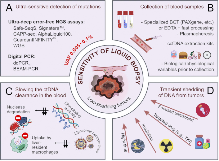
Improvement of the analytical performance of ctDNA-based liquid biopsy. (A) Ultra-sensitive methods of detection of mutated DNA sequences; (B) pre-analytical factors affecting the quality and yield of ctDNA; (C) slowing the ctDNA clearance in the blood (black arrows indicate the inhibition of two routes of ctDNA clearance); (D) transient shedding of DNA from tumors. BCT: blood collection tubes; BEAM: beads, emulsions, amplification, and magnetics; ccfDNA: circulating cell-free DNA; ctDNA: circulating tumor DNA; ddPCR: droplet digital PCR; EDTA: ethylenediaminetetraacetic acid; NGS: next-generation sequencing; TKI: tyrosine kinase inhibitors; VAF: variant allele frequency; WGS: whole-genome sequencing. Some images were adapted using free resources from Flaticon.com

Collection of biospecimens

Normally, blood contains up to several thousand copies of wild-type extracellular DNA per 1 mL of plasma [55, 56]. This background compromises the detection of tumor-derived mutated DNA. The concentration of circulating “normal” DNA is generally higher in the elderly than in young people [57, 58]. Furthermore, some inflammatory or autoimmune diseases, physical exercise, trauma, etc., may increase the overall content of ccfDNA [5966]. Tumor-derived DNA usually constitutes no more than 0.025–2.5% of circulating fluid DNA [34, 36]. This proportion depends on the biological and genetic properties of the tumor, overall amount of malignant cells within the body, and various treatment-related factors [20, 6773]. The release of DNA by blood cells is a major factor confounding ctDNA analysis [35]. At present, all protocols for ctDNA extraction require careful separation of plasma from blood cells and cellular debris, which is achieved by two rounds of centrifugation and manual saving of ctDNA-containing samples. Conventional ethylenediaminetetraacetic acid (EDTA)-containing tubes require almost immediate processing of the blood, with the waiting time not exceeding 2–6 hours at 4°C [74, 75]. There are several blood collection tubes (BCT), e.g., cfDNA (Streck), PAXgene Blood ccfDNA (Qiagen), cfDNA/cfRNA Preservative (Norgene), ImproGene cfDNA (Improve Medical), cfDNA (Roche), etc., which contain some stabilizers of the integrity of nucleated blood cells, thus preventing the release of normal genomic DNA and minimizing the hemolysis. They all allow for the storage and transportation of blood samples for up to 7 days at room temperature [53, 76, 77]. Significant efforts have been invested in head-to-head comparisons of the tubes obtained from different manufacturers, with some minor differences revealed [7880]. Although these tubes are convenient for the ctDNA analysis, they are not always compatible with the multianalyte LB, as they may not permit simultaneous analysis of circulating tumor cells (CTCs), protein markers, extracellular vesicles (EVs), etc. Consequently, many investigators opt for conventional EDTA tubes despite the requirement for immediate blood processing [8185] (Table 1).

 Critical pre-analytical features of ctDNA-based LB workflow

StageRecommendationNotesReferences
Blood collection:
ProcedureUse of butterfly needlesAvoid excessively thin needles and prolonged tourniquet use.[53, 8688]
Plasmapheresis/leukapheresisApply microfluidic enrichment and FACS.[89, 90]
Sample volume2 × 10 mL of blood (for single-analyte LB)Screening, MRD detection, WGS, and testing of multiple analytes may necessitate larger plasma volumes.[81, 91, 92]
BCTEDTA tubesRequire fast processing (within 2–6 hours).[74, 75, 85]
BCT with cell stabilizing preservative agents: cfDNA (Streck), PAXgene Blood ccfDNA (Qiagen), cfDNA/cfRNA Preservative (Norgene), ImproGene cfDNA (Improve Medical), cfDNA (Roche)Preserve the quality of ctDNA samples within 3–7 days at a temperature of 4–25°C.[77, 78, 84]
Biological featuresControl for physical activity, and physiological and pathological status prior to blood collectionChronic or acute diseases (e.g., diabetes, endometriosis, obesity, kidney disease, hypertension, and inflammation) are associated with elevated ccfDNA content.[6163]
Surgical traumaSurgical trauma causes a transient increase in the level of ccfDNA, which persists for up to a few weeks after surgery.[59, 93]
Circadian dynamicsIncrease of CTC and ctDNA content at night.[94, 95]
Induction of transient ctDNA release from tumor before blood takeIrradiationThe spike of ctDNA concentration in 6–24 hours after the procedure.[51, 9699]
UltrasoundSonobiopsy for brain tumors.[50, 52, 100]
Mechanical stressMammography for breast cancer; digital rectal examination for prostate cancer.[49, 101, 102]
Transportation and handlingUse special BCT for long-distance transportation. EDTA tubes are good only for transportation within a hospitalAvoid high temperature, stirring, or violent vibration during transportation.[78, 85, 103]
Plasma processing:
CentrifugationDouble centrifugation:
1st step (slow centrifugal force: 380–3,000 g for 10 min at room temperature), 2nd step (12,000–20,000 g for 10 min at 4°C)
Single low-speed centrifugation is recommended for PEG-mediated enrichment.[53, 85, 104, 105]
Cell-free plasma storageAt –80°C10 years for mutation detection; 9 months for quantitative analysis.[53]
Thawing of stored plasmaSlowly on iceFreeze-thaw cycles must be minimized; it is recommended to store the plasma in small fractions.[106, 107]
ctDNA extraction:
ChemistrySolid phase extraction:
- Silica membrane columns: QIAamp Circulating Nucleic Acids Kit (Qiagen); Cobas ccfDNA Sample Preparation Kit;
- Magnetic beads: QIAamp MinElute ccfDNA Mini Kit (Qiagen); Maxwell RSC LV ccfDNA Kit (Promega); MagNa Pure 24 Total NA Isolation Kit (Roche)
Silica membrane-based kits yield more ctDNA than methods utilizing magnetic beads.[81, 106, 108111]
Liquid phase extractionUtilize the standard phenol-chloroform extraction or specially designed phase-forming aqueous systems.[112, 113]
Microfluidic platformsCost-efficient approach allowing for rapid isolation, detection, and characterization of ctDNA.[114116]
Addition of polymers to the blood sample (e.g., PEG)Improves the quantity and purity of ctDNA; facilitates precipitation of extracellular vesicles, lipoproteins, and ribonucleoprotein complexes, thus providing the access to multianalyte assays.[105, 117, 118]
WorkflowMoving toward standardized automatic methodologies and multianalyte extraction protocolsReduce the hands-on time of the extraction.[106, 119121]

BCT: blood collection tubes; ccfDNA: circulating cell-free DNA; cfDNA: cell-free DNA; CTC: circulating tumor cell; ctDNA: circulating tumor DNA; EDTA: ethylenediaminetetraacetic acid; FACS: fluorescence-activated cell sorting; LB: liquid biopsy; MRD: minimal residual disease; PEG: polyethylene glycol; WGS: whole-genome sequencing

Plasma collection is a serious confounding factor in the real-world clinical setting, as it requires significant manual work and is poorly amenable to standardization. Ideally, an automated device near the blood-draw facility is needed to process the blood and plasma.

Several studies have highlighted a suitability of dried blood spots (DBS) collected via finger-prick microsampling for ctDNA analysis. This approach is compatible with the whole-genome sequencing (WGS) and allows the detection of somatic copy number aberrations, fragment length profiles, tumor-specific single-nucleotide mutations, etc., across various cancer types [122, 123]. The protocol for ctDNA isolation from DBS includes a bead-based size selection step that effectively distinguishes tumor-derived cfDNA from background genomic DNA. Considering that DNA and RNA remain stably preserved on cards for extended periods [124], the use of DBS presents a logistical advantage for practical utilization of ctDNA-based assays.

ctDNA extraction

The overall amount of ctDNA in plasma samples is usually estimated in nanograms [35, 125]. Exhaustive isolation of these tiny quantities of DNA is a challenge, especially given that it is partially degraded (Table 1). Most laboratories utilize kits, which collect DNA on a solid phase. Silica membrane columns [e.g., QIAamp Circulating Nucleic Acids Kit (Qiagen), Cobas cfDNA Sample Preparation Kit (Roche), etc.] require significant manual efforts; however, some comparative studies demonstrated that they produce superior DNA yields [106, 108110]. Protocols involving magnetic beads [e.g., QIAamp MinElute ccfDNA Mini Kit (Qiagen), Maxwell RSC LV ccfDNA Kit (Promega), MagNa Pure 24 Total NA Isolation Kit (Roche)] are more amenable to some automation, although their performance has been questioned because of lower ctDNA recovery [106].

While the majority of laboratories rely on the absorption of ctDNA on membranes or beads [81, 106, 108111], some researchers prefer ethanol-based precipitation of nucleic acids from a liquid phase [112, 113, 126]. Purification of ctDNA may be achieved either by conventional phenol-chloroform extraction or by more sophisticated reagents containing polymers, salts, ionic liquids, and surfactant components. There are data suggesting that these methods offer even higher ctDNA recovery and purity compared to solid-phase systems [112, 113].

The addition of polyethylene glycol (PEG) to the plasma was shown to improve ctDNA isolation [105, 117, 118]. There are also some microfluidic platforms providing rapid and cost-efficient isolation of circulating DNA [114116].

Detection of mutations in ctDNA

There are two major avenues for the analysis of tumor-derived ctDNA [127134]. Post-treatment follow-up often relies on a tumor-informed approach, in which the assay is designed in accordance with the pattern of mutations in the cancer tissue [127, 128]. This procedure may utilize both droplet digital PCR (ddPCR) and next-generation sequencing (NGS), and it is generally less prone to various artifacts [129, 130]. The tumor-agnostic approach is the only viable tool for early cancer detection because it relies on NGS analysis of a wide spectrum of commonly mutated cancer genes or sequencing of the entire genome [131134]. It is essential to emphasize that tumors are not the only source of mutated DNA. Clonal hematopoiesis of indeterminate potential (CHIP) is also characterized by oncogenic mutations and is particularly common in elderly individuals [135, 136].

Earlier ctDNA studies used conventional real-time PCR for the detection of tumor-associated mutations in plasma samples [137, 138]. Real-time PCR has insufficient sensitivity and specificity for ctDNA analysis, and its use is discouraged.

ddPCR is the most appropriate method for tumor-informed ctDNA analysis, as it allows for reliable detection of mutated gene copies in the presence of at least 1,000-fold or even higher excess of normal DNA [20, 139141]. NGS is about an order of magnitude less sensitive than ddPCR; however, this disadvantage is compensated by simultaneous analysis of multiple mutated sites [37, 43, 127, 128, 142144].

Both ddPCR and NGS procedures involve DNA synthesis, which is prone to occasional errors [145]. This is a common drawback; therefore, the presence of a single mutation-specific signal in ddPCR or NGS run cannot be reliably interpreted in favor of mutation. Current NGS technologies utilize elegant modifications, which have been specifically developed for the detection of mosaic mutations, for example, Safe-SeqS [146], Signatera [127], RaDaR [147], Avenio (CAPP-seq) [148], Guardant360 [149], AlphaLiquid100 [23, 150], or some laboratory-developed tests [151].

The performance of NGS-based low-copy ctDNA detection can be improved by the use of so-called unique molecular identifiers (UMIs) [152]. UMIs are random DNA sequences, which are added to individual ctDNA molecules before PCR amplification (Figure 2). Consequently, all PCR products originating from a given DNA molecule (i.e., having the same oligonucleotide identifier) can be recognized and subjected to an individual bioinformatics analysis. Sequence differences between the reads originating from the same template are interpreted as errors. These NGS modifications permit accurate identification of mutations in the presence of 10,000-fold excess of normal DNA [134, 153, 154]. Duplex sequencing employs random tagging of each individual DNA duplexes during library preparation, allowing the identification of all amplified DNA fragments originating from a single strand of the original DNA molecule [155]. The mutation is considered real only if both strands forming a UMI-labelled fragment reveal complementary alterations in a given nucleotide position [156].

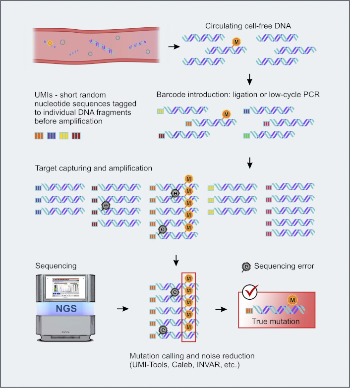
UMI-barcoding strategy for background noise reduction. e: sequencing error; M: true mutation. NGS: next-generation sequencing; UMIs: unique molecular identifiers. Some images were adapted using free resources from Flaticon.com

Combining NGS with artificial intelligence (AI) and machine learning (ML) tools for mutation calling has been shown to reduce the background noise, thus enabling accurate detection of true ctDNA alterations at variant allele frequency (VAF) as low as 10–5 [157160]. Some ML tools, e.g., Random Forest classifiers, K-nearest neighbor algorithms, dual-alignment pipelines, etc., are capable of facilitating ctDNA-based mutation profiling and prognostic stratification [159, 161]. Furthermore, ML/AI-assisted NGS has been successfully applied for the dynamic quantitative monitoring of low-burden cancers and the detection of minimal residual disease (MRD) [159, 162, 163].

In vivo stimulation of ctDNA release

Even if the plasma is optimally processed and the molecular genetic analysis is capable of detecting 1 copy of mutated DNA per preparation, small tumors will remain undetected by the LB [47, 125]. Antitumor therapies, particularly local irradiation and tyrosine kinase inhibitors, induce apoptosis of cancer cells and, therefore, render transient spike of ctDNA concentration [24, 51, 9699, 101, 164166]. The use of cancer drugs for the support of LB is unlikely to have a practical value: in order to warrant the desirable effect, a given drug should a priori have high antitumor activity. In the real-world setting, LB for visible tumor lumps is performed exactly for the selection of the best drugs, while there is no actual need for ctDNA analysis when the best systemic therapy is already known. For example, lung cancer progression under the treatment by first- or second-generation EGFR inhibitors requires the analysis of EGFR T790M mutation, which sometimes can be accomplished by ctDNA testing [139, 144, 167, 168]. Identification of EGFR T790M mutation calls for the administration of osimertinib; however, this drug cannot be administered before the test. Irradiation is potentially more practical for the stimulation of ctDNA shedding. Several studies have shown a transient increase in the ctDNA level right after the beginning of radiotherapy [9699]. However, not all patients demonstrate a ctDNA spike; furthermore, the increase of ctDNA concentration in the bloodstream is generally only within 1.5–2-fold, which may not be sufficient in many clinical situations [51, 169]. All relevant human studies were performed on patients, who received tumor irradiation as a part of the standard treatment plan. It is questionable, whether this intervention can be applied only for the purpose of LB. Furthermore, this approach is potentially feasible only for the analysis of treatment-induced mutations, like EGFR T790M, and is not applicable to early cancer detection and monitoring of MRD.

Brain tumors compose a special category of malignancies, as they are separated from the bloodstream by the blood-brain barrier (BBB). Several studies demonstrated that focused ultrasound may facilitate the detection of ctDNA, probably by the disruption of this barrier [50, 52, 100].

Slowing ctDNA decay

The half-life of ctDNA does not exceed 1–2 hours, with some studies suggesting even shorter estimates [10, 11, 170]. ctDNA undergoes rapid decay. First, cfDNA is efficiently absorbed by liver-resident Kupffer cells and spleen macrophages [40, 42]. In addition, there are circulating nucleases, which accelerate decay of extracellular blood DNA [39, 41]. Martin-Alonso et al. [171] explored these mechanisms of ctDNA clearance in mouse experiments. They injected a succinyl phosphoethanolamine-based liposomal agent to interfere with liver macrophages. In addition, they utilized DNA-binding antibodies in order to protect DNA from nuclease digestion. The combination of these two interventions resulted in a transient increase in the concentration of circulating DNA by more than an order of magnitude [171]. This is theoretically sufficient to allow detection of tumors as small as 1 cm3 in diameter. The described approach is highly promising for cancer screening and monitoring of the MRD. Translation of this methodology from mice to humans requires careful consideration of the safety of injected substances.

Combining ctDNA with other analytes

While ctDNA can be reliably discriminated from non-tumorous DNA by mutation analysis, other intensively studied analytes, like tumor-derived proteins or cells expressing some cell surface markers, are evidently less cancer-specific. Nevertheless, a simultaneous analysis of several groups of biomarkers, for example, ctDNA, messenger RNA (mRNA), miRNA, CTC, tumor-educated platelets, exosomes, and proteins, may provide some added value, especially in tumor-agnostic screening for early cancers [18, 82, 83, 172175]. Another underutilized option is to consider non-blood biological fluids that are proximal to the tumor site, such as urine, cerebrospinal fluid, and pleural or peritoneal effusions [18, 176] (Figure 3).

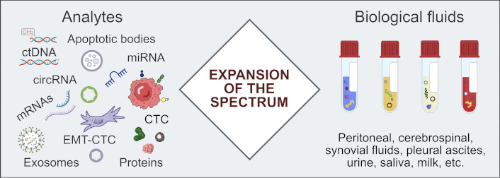
Beyond plasma ctDNA: the reliability of LB can be increased by expanding the range of analytes and involving biological fluids which are proximal to the tumor site. circRNA: circular RNA; CTC: circulating tumor cell; ctDNA: circulating tumor DNA; EMT-CTC: circulating tumor cell undergoing epithelial-to-mesenchymal transition; LB: liquid biopsy; miRNA: microRNA; mRNAs: messenger RNAs

The multianalyte approach has been explored in several blood-based assays, which demonstrated encouraging results in several large-scale investigations [177180]. The best-known is the so-called CancerSEEK panel, which combines quantitative analysis of eight protein cancer biomarkers and detection of over 1,000 tumor-specific mutations in сtDNA. Initially, the assay was tested on 1,005 patients with non-metastatic, clinically confirmed cancers of the ovary, liver, stomach, pancreas, esophagus, colorectum, lung, or breast [181]. The sensitivity of CancerSEEK has been reported within the range of 69–98% for ovarian, liver, stomach, pancreatic, and esophageal cancers, with only 7 of 812 healthy controls being misrecognized as positive. The subsequent DETECT-A prospective study evaluated the performance of this test in more than 10,000 women aged 65–75 years and showed that the CancerSEEK was able to reveal approximately 1 out of 4 “true” early-stage tumors across the entire spectrum of malignant diseases [7].

Multianalyte assays targeted at the detection of a single cancer type understandably render somewhat better performance. For example, a single-tube analysis of ctDNA, exosomal mRNA, and CA19-9 protein in the blood of pancreatic ductal adenocarcinoma patients demonstrated 92% accuracy, 95% specificity, and 88% sensitivity in revealing cancer disease [182]. Single-organ assays have obvious relevance for the post-surgical follow-up of oncological patients aimed at early detection of tumor relapse, while their potential utility in population-based screening is limited to exceptionally common cancer types and high-risk individuals.

The analysis of ctDNA methylation holds a great promise [183185]. Tumors differ from normal cells by the pattern of DNA methylation, and these differences are generally more common than mutations [186, 187]. The technologies for the analysis of methylation of ctDNA rely on bisulfite conversion‐based methods [e.g., whole-genome bisulfite sequencing (WGBS), MCTA-seq, ELISA-seq, targeted bisulfite sequencing, etc.] [188190], enrichment-based approaches combining immunoprecipitation and high-throughput sequencing (SeqCap Epi CpGiant; cfMeDIP‐seq; TET) [191, 192], and various procedures utilizing methyl‐sensitive restriction endonucleases [193, 194]. Several studies demonstrated the utility of methylation analysis of ctDNA for early cancer detection, MRD assessment, estimating treatment response and disease prognosis, etc. [185, 186, 195200]. Methylation-based techniques are potentially proficient in identifying tumor tissue origin, which is particularly important for cancer screening [186, 199, 201203].

Reproducibility of ctDNA assays

Assays that measure ctDNA are influenced by numerous experimental variables and artifacts, many of which remain incompletely understood. Because of the very low concentration in the bloodstream, ctDNA analysis requires PCR amplification and ultra-deep sequencing steps, which may produce various errors. On the other hand, improper blood handling during the collection, transportation, and processing may result in hemolysis and contamination of circulating tumor-derived DNA with genomic DNA. The harmonization of pre-analytical and analytical workflows is essential for the interlaboratory reproducibility of ctDNA-based assays [21, 81, 91, 204].

Several multicenter studies have been conducted to assess the reproducibility of LB results when using different ctDNA extraction methods [205] and various ctDNA detection assays [37, 44, 148]. Circulating mutant fragments, presented with VAF of > 0.5%, were detected with high sensitivity, accuracy, and reproducibility by all participating assays, whereas the detection of VAF at 0.5% or lower proved challenging [44, 206208]. Meanwhile, ctDNA variants with VAF < 0.5% are particularly important, as they characterize early-stage tumors, MRD, or emerging recurrences. Such cases account for 25–30% of all samples submitted for LB [37, 209]. Here, the most reliable approach to avoid false-negative results is the use of ddPCR for the confirmation and tracking of “tumor-informed” mutations. A combination of synthetic DNA spike-in controls (sequins) [210] and cell-line derived reference samples [211] can be utilized for the monitoring of the analytical performance of ctDNA assays.

The use of standardized commercial kits or centralized services may improve the reproducibility of ctDNA-based LB procedures and enable the comparison of various data sets [212215]. Several LB kits and diagnostic pipelines have received FDA or EU approval. Therascreen EGFR RGQ Plasma PCR Kit (Qiagen) is a companion test for detection of EGFR mutations in lung cancer patients [216]. Guardant360 CDx (Guardant Health) [149] and FoundationOne Liquid CDx (Foundation Medicine/Roche) [217] gene profiling platforms have been designed to support the choice of targeted therapies in multiple tumor types. Epi proColon (Epigenomics) is the first FDA-approved blood-based assay, which relies on the use of methylation DNA markers for colorectal cancer screening [218]. Galleri (Grail) [219] and Signatera (Natera) kits [127] are used in Clinical Laboratory Improvement Amendments (CLIA)-certified labs for MRD detection and multi-cancer screening. Syantra DX Breast Cancer (Syantra) assay is suggested to support early diagnosis of breast cancer [220].

Surprisingly, the reproducibility of serial ctDNA tests taken from the same patient within a short time interval has not been rigorously assessed yet. Kuligina et al. [70] investigated serial plasma samples from 82 cancer patients, whose tumors contained common hot-spot oncogenic mutations. Moderate physical exercise, recent food, or time of the day did not significantly influence the content of ctDNA. Strikingly, as many as 24/82 (29%) patients showed the presence of mutated ctDNA in some but not all blood draws. Some degree of instability of various laboratory values is well known to clinicians; therefore, many conventional tests are administered repetitively. It needs to be established whether the same attitude is also of benefit in cancer patients.

Conclusions

ctDNA-based LB is highly important for many areas of clinical oncology, including early cancer detection, prediction of therapeutic response and long-term survival, disease monitoring, and support of various treatment decisions. However, ctDNA analysis is usually performed at the limits of available technologies and, therefore, remains relatively error-prone.

Multiple pre-analytical and analytical factors affect the reproducibility of ctDNA assays. In particular, proper handling of plasma samples as well as the use of ultrasensitive methods for detection of tumor-derived molecules are of paramount significance. Still, the content of ctDNA is usually vanishingly low; therefore, some interventional procedures are currently being assessed. For example, in vivo interference with ctDNA release and decay may lead to a breakthrough in LB performance if proven to be safe. Surprisingly, the reproducibility of ctDNA-based tests has not been rigorously assessed yet; future studies have to put a strong emphasis on this highly important issue.

Abbreviations

AI: artificial intelligence

ccfDNA: circulating cell-free DNA

cfDNA: cell-free DNA

CTCs: circulating tumor cells

ctDNA: circulating tumor DNA

DBS: dried blood spots

ddPCR: droplet digital PCR

EDTA: ethylenediaminetetraacetic acid

LB: liquid biopsy

miRNAs: microRNAs

ML: machine learning

MRD: minimal residual disease

mRNA: messenger RNA

NGS: next-generation sequencing

UMIs: unique molecular identifiers

VAF: variant allele frequency

Declarations

Acknowledgments

We are cordially thankful to Prof. William R. Miller (University of Edinburgh, UK) for his invaluable help in improving this manuscript.

Author contributions

ESK: Conceptualization, Investigation, Visualization, Writing—original draft. GAY: Investigation, Writing—original draft. ENI: Conceptualization, Validation, Writing—review & editing, Funding acquisition, Supervision. All authors read and approved the submitted version.

Conflicts of interest

Evgeny N. Imyanitov who is the Editorial Board Member of Exploration of Targeted Anti-tumor Therapy had no involvement in the decision-making or the review process of this manuscript. The other authors declare that they have no conflicts of interest.

Ethical approval

Not applicable.

Consent to participate

Not applicable.

Consent to publication

Not applicable.

Availability of data and materials

Not applicable.

Funding

This research has been supported by the Russian Science Foundation, grant number [23-45-10038]. The funder had no role in study design, data collection and analysis, decision to publish, or preparation of the manuscript.

Copyright

© The Author(s) 2025.

Publisher’s note

Open Exploration maintains a neutral stance on jurisdictional claims in published institutional affiliations and maps. All opinions expressed in this article are the personal views of the author(s) and do not represent the stance of the editorial team or the publisher.

References

Mohamed E, García Martínez DJ, Hosseini MS, Yoong SQ, Fletcher D, Hart S, et al. Identification of biomarkers for the early detection of non-small cell lung cancer: a systematic review and meta-analysis. Carcinogenesis. 2024;45:122. [DOI] [PubMed]
Nicholson BD, Shinkins B, Pathiraja I, Roberts NW, James TJ, Mallett S, et al. Blood CEA levels for detecting recurrent colorectal cancer. Cochrane Database Syst Rev. 2015;2015:CD011134. [DOI] [PubMed] [PMC]
Liu Y, Hatano K, Nonomura N. Liquid Biomarkers in Prostate Cancer Diagnosis: Current Status and Emerging Prospects. World J Mens Health. 2025;43:827. [DOI] [PubMed] [PMC]
Lilja H, Ulmert D, Vickers AJ. Prostate-specific antigen and prostate cancer: prediction, detection and monitoring. Nat Rev Cancer. 2008;8:26878. [DOI] [PubMed]
Charkhchi P, Cybulski C, Gronwald J, Wong FO, Narod SA, Akbari MR. CA125 and Ovarian Cancer: A Comprehensive Review. Cancers (Basel). 2020;12:3730. [DOI] [PubMed] [PMC]
Bast RC Jr, Badgwell D, Lu Z, Marquez R, Rosen D, Liu J, et al. New tumor markers: CA125 and beyond. Int J Gynecol Cancer. 2005;15:27481. [PubMed]
Lennon AM, Buchanan AH, Kinde I, Warren A, Honushefsky A, Cohain AT, et al. Feasibility of blood testing combined with PET-CT to screen for cancer and guide intervention. Science. 2020;369:eabb9601. [DOI] [PubMed] [PMC]
Veyssière H, Bidet Y, Penault-Llorca F, Radosevic-Robin N, Durando X. Circulating proteins as predictive and prognostic biomarkers in breast cancer. Clin Proteomics. 2022;19:25. [DOI] [PubMed] [PMC]
Barker AD, Alba MM, Mallick P, Agus DB, Lee JSH. An Inflection Point in Cancer Protein Biomarkers: What was and What’s Next. Mol Cell Proteomics. 2023;22:100569. [DOI] [PubMed] [PMC]
Stejskal P, Goodarzi H, Srovnal J, Hajdúch M, van ’t Veer LJ, Magbanua MJM. Circulating tumor nucleic acids: biology, release mechanisms, and clinical relevance. Mol Cancer. 2023;22:15. [DOI] [PubMed] [PMC]
Papadopoulos N. Pathophysiology of ctDNA Release into the Circulation and Its Characteristics: What Is Important for Clinical Applications. Recent Results Cancer Res. 2020;215:16380. [DOI] [PubMed]
Marques JF, Junqueira-Neto S, Pinheiro J, Machado JC, Costa JL. Induction of apoptosis increases sensitivity to detect cancer mutations in plasma. Eur J Cancer. 2020;127:1308. [DOI] [PubMed]
Iams WT, Mackay M, Ben-Shachar R, Drews J, Manghnani K, Hockenberry AJ, et al. Concurrent Tissue and Circulating Tumor DNA Molecular Profiling to Detect Guideline-Based Targeted Mutations in a Multicancer Cohort. JAMA Netw Open. 2024;7:e2351700. [DOI] [PubMed] [PMC]
Nesic M, Rasmussen MH, Henriksen TV, Demuth C, Frydendahl A, Nordentoft I, et al. Beyond basics: Key mutation selection features for successful tumor-informed ctDNA detection. Int J Cancer. 2024;155:92533. [DOI] [PubMed]
Leenanitikul J, Chanchaem P, Mankhong S, Denariyakoon S, Fongchaiya V, Arayataweegool A, et al. Concordance between whole exome sequencing of circulating tumor DNA and tumor tissue. PLoS One. 2023;18:e0292879. [DOI] [PubMed] [PMC]
Vandekerkhove G, Lavoie JM, Annala M, Murtha AJ, Sundahl N, Walz S, et al. Plasma ctDNA is a tumor tissue surrogate and enables clinical-genomic stratification of metastatic bladder cancer. Nat Commun. 2021;12:184. [DOI] [PubMed] [PMC]
Martínez-Vila C, Teixido C, Aya F, Martín R, González-Navarro EA, Alos L, et al. Detection of Circulating Tumor DNA in Liquid Biopsy: Current Techniques and Potential Applications in Melanoma. Int J Mol Sci. 2025;26:861. [DOI] [PubMed] [PMC]
Ma L, Guo H, Zhao Y, Liu Z, Wang C, Bu J, et al. Liquid biopsy in cancer: current status, challenges and future prospects. Signal Transduct Target Ther. 2024;9:336. [DOI] [PubMed] [PMC]
Reina C, Šabanović B, Lazzari C, Gregorc V, Heeschen C. Unlocking the future of cancer diagnosis - promises and challenges of ctDNA-based liquid biopsies in non-small cell lung cancer. Transl Res. 2024;272:4153. [DOI] [PubMed]
Sánchez-Martín V, López-López E, Reguero-Paredes D, Godoy-Ortiz A, Domínguez-Recio ME, Jiménez-Rodríguez B, et al. Comparative study of droplet-digital PCR and absolute Q digital PCR for ctDNA detection in early-stage breast cancer patients. Clin Chim Acta. 2024;552:117673. [DOI] [PubMed]
Pascual J, Attard G, Bidard FC, Curigliano G, De Mattos-Arruda L, et al. ESMO recommendations on the use of circulating tumour DNA assays for patients with cancer: a report from the ESMO Precision Medicine Working Group. Ann Oncol. 2022;33:75068. [DOI] [PubMed]
Øgaard N, Jensen SØ, Ørntoft MW, Demuth C, Rasmussen MH, Henriksen TV, et al. Circulating tumour DNA and risk of recurrence in patients with asymptomatic versus symptomatic colorectal cancer. Br J Cancer. 2024;131:170715. [DOI] [PubMed] [PMC]
Jeong SH, Kyung D, Yuk HD, Jeong CW, Lee W, Yoon JK, et al. Practical Utility of Liquid Biopsies for Evaluating Genomic Alterations in Castration-Resistant Prostate Cancer. Cancers (Basel). 2023;15:2847. [DOI] [PubMed] [PMC]
Sanz-Garcia E, Zhao E, Bratman SV, Siu LL. Monitoring and adapting cancer treatment using circulating tumor DNA kinetics: Current research, opportunities, and challenges. Sci Adv. 2022;8:eabi8618. [DOI] [PubMed] [PMC]
Amato O, Giannopoulou N, Ignatiadis M. Circulating tumor DNA validity and potential uses in metastatic breast cancer. NPJ Breast Cancer. 2024;10:21. [DOI] [PubMed] [PMC]
Moiseenko FV, Volkov NM, Zhabina AS, Stepanova ML, Rysev NA, Klimenko VV, et al. Monitoring of the presence of EGFR-mutated DNA during EGFR-targeted therapy may assist in the prediction of treatment outcome. Cancer Treat Res Commun. 2022;31:100524. [DOI] [PubMed]
Song Y, Hu C, Xie Z, Wu L, Zhu Z, Rao C, et al.; Written on behalf of AME Lung Cancer Collaborative Group. Circulating tumor DNA clearance predicts prognosis across treatment regimen in a large real-world longitudinally monitored advanced non-small cell lung cancer cohort. Transl Lung Cancer Res. 2020;9:26979. [DOI] [PubMed] [PMC]
Pécuchet N, Zonta E, Didelot A, Combe P, Thibault C, Gibault L, et al. Base-Position Error Rate Analysis of Next-Generation Sequencing Applied to Circulating Tumor DNA in Non-Small Cell Lung Cancer: A Prospective Study. PLoS Med. 2016;13:e1002199. [DOI] [PubMed] [PMC]
Yang W, Nguyen R, Safri F, Shiddiky MJA, Warkiani ME, George J, et al. Liquid Biopsy in Hepatocellular Carcinoma: ctDNA as a Potential Biomarker for Diagnosis and Prognosis. Curr Oncol Rep. 2025;27:791802. [DOI] [PubMed] [PMC]
O’Sullivan NJ, Temperley HC, Kyle ET, Sweeney KJ, O’Neill M, Gilham C, et al. Assessing circulating tumour DNA (ctDNA) as a prognostic biomarker in locally advanced rectal cancer: a systematic review and meta-analysis. Int J Colorectal Dis. 2024;39:82. [DOI] [PubMed] [PMC]
Kirchweger P, Wundsam HV, Rumpold H. Circulating tumor DNA for diagnosis, prognosis and treatment of gastrointestinal malignancies. World J Clin Oncol. 2022;13:47384. [DOI] [PubMed] [PMC]
Liao W, Yang H, Xu H, Wang Y, Ge P, Ren J, et al. Noninvasive detection of tumor-associated mutations from circulating cell-free DNA in hepatocellular carcinoma patients by targeted deep sequencing. Oncotarget. 2016;7:4048190. [DOI] [PubMed] [PMC]
Bartolomucci A, Nobrega M, Ferrier T, Dickinson K, Kaorey N, Nadeau A, et al. Circulating tumor DNA to monitor treatment response in solid tumors and advance precision oncology. NPJ Precis Oncol. 2025;9:84. [DOI] [PubMed] [PMC]
Martens GA, Demol J, Dedeurwaerdere F, Breyne J, De Smet K, De Jaeger P, et al. Rational thresholding of circulating tumor DNA concentration for improved surveillance of metastatic breast cancer. ESMO Open. 2024;9:102235. [DOI] [PubMed] [PMC]
Sender R, Noor E, Milo R, Dor Y. What fraction of cellular DNA turnover becomes cfDNA? Elife. 2024;12:RP89321. [DOI] [PubMed] [PMC]
Nishio K, Sakai K, Nishio M, Seto T, Visseren-Grul C, Carlsen M, et al. Impact of ramucirumab plus erlotinib on circulating cell-free DNA from patients with untreated metastatic non-small cell lung cancer with EGFR-activating mutations (RELAY phase 3 randomized study). Transl Lung Cancer Res. 2023;12:170216. [DOI] [PubMed] [PMC]
Li W, Huang X, Patel R, Schleifman E, Fu S, Shames DS, et al. Analytical evaluation of circulating tumor DNA sequencing assays. Sci Rep. 2024;14:4973. [DOI] [PubMed] [PMC]
Abbosh C, Birkbak NJ, Swanton C. Early stage NSCLC - challenges to implementing ctDNA-based screening and MRD detection. Nat Rev Clin Oncol. 2018;15:57786. [DOI] [PubMed]
Han DSC, Lo YMD. The Nexus of cfDNA and Nuclease Biology. Trends Genet. 2021;37:75870. [DOI] [PubMed]
Khier S, Lohan L. Kinetics of circulating cell-free DNA for biomedical applications: critical appraisal of the literature. Future Sci OA. 2018;4:FSO295. [DOI] [PubMed] [PMC]
Kustanovich A, Schwartz R, Peretz T, Grinshpun A. Life and death of circulating cell-free DNA. Cancer Biol Ther. 2019;20:105767. [DOI] [PubMed] [PMC]
Du Clos TW, Volzer MA, Hahn FF, Xiao R, Mold C, Searles RP. Chromatin clearance in C57Bl/10 mice: interaction with heparan sulphate proteoglycans and receptors on Kupffer cells. Clin Exp Immunol. 1999;117:40311. [DOI] [PubMed] [PMC]
Stetson D, Labrousse P, Russell H, Shera D, Abbosh C, Dougherty B, et al. Next-Generation Molecular Residual Disease Assays: Do We Have the Tools to Evaluate Them Properly? J Clin Oncol. 2024;42:273640. [DOI] [PubMed]
Deveson IW, Gong B, Lai K, LoCoco JS, Richmond TA, Schageman J, et al.; SEQC2 Oncopanel Sequencing Working Group. Evaluating the analytical validity of circulating tumor DNA sequencing assays for precision oncology. Nat Biotechnol. 2021;39:111528. [DOI] [PubMed] [PMC]
Wan JCM, Heider K, Gale D, Murphy S, Fisher E, Mouliere F, et al. ctDNA monitoring using patient-specific sequencing and integration of variant reads. Sci Transl Med. 2020;12:eaaz8084. [DOI] [PubMed]
Panet F, Papakonstantinou A, Borrell M, Vivancos J, Vivancos A, Oliveira M. Use of ctDNA in early breast cancer: analytical validity and clinical potential. NPJ Breast Cancer. 2024;10:50. [DOI] [PubMed] [PMC]
Abbosh C, Frankell AM, Harrison T, Kisistok J, Garnett A, Johnson L, et al. Tracking early lung cancer metastatic dissemination in TRACERx using ctDNA. Nature. 2023;616:55362. [DOI] [PubMed] [PMC]
Avanzini S, Kurtz DM, Chabon JJ, Moding EJ, Hori SS, Gambhir SS, et al. A mathematical model of ctDNA shedding predicts tumor detection size. Sci Adv. 2020;6:eabc4308. [DOI] [PubMed] [PMC]
Tosoian JJ, Zhang Y, Meyers JI, Heaton S, Siddiqui J, Xiao L, et al. Clinical Validation of MyProstateScore 2.0 Testing Using First-Catch, Non-Digital Rectal Examination Urine. J Urol. 2025;213:5819. [DOI] [PubMed] [PMC]
Yuan J, Xu L, Chien CY, Yang Y, Yue Y, Fadera S, et al. First-in-human prospective trial of sonobiopsy in high-grade glioma patients using neuronavigation-guided focused ultrasound. NPJ Precis Oncol. 2023;7:92. [DOI] [PubMed] [PMC]
Kuligina E, Moiseyenko F, Belukhin S, Stepanova E, Zakharova M, Chernobrivtseva V, et al. Tumor irradiation may facilitate the detection of tumor-specific mutations in plasma. World J Clin Oncol. 2021;12:121526. [DOI] [PubMed] [PMC]
Meng Y, Pople CB, Suppiah S, Llinas M, Huang Y, Sahgal A, et al. MR-guided focused ultrasound liquid biopsy enriches circulating biomarkers in patients with brain tumors. Neuro Oncol. 2021;23:178997. [DOI] [PubMed] [PMC]
Peng H, Pan M, Zhou Z, Chen C, Xing X, Cheng S, et al. The impact of preanalytical variables on the analysis of cell-free DNA from blood and urine samples. Front Cell Dev Biol. 2024;12:1385041. [DOI] [PubMed] [PMC]
Yuwono NL, Warton K, Ford CE. The influence of biological and lifestyle factors on circulating cell-free DNA in blood plasma. Elife. 2021;10:e69679. [DOI] [PubMed] [PMC]
Bronkhorst AJ, Ungerer V, Holdenrieder S. The emerging role of cell-free DNA as a molecular marker for cancer management. Biomol Detect Quantif. 2019;17:100087. [DOI] [PubMed] [PMC]
Meddeb R, Dache ZAA, Thezenas S, Otandault A, Tanos R, Pastor B, et al. Quantifying circulating cell-free DNA in humans. Sci Rep. 2019;9:5220. [DOI] [PubMed] [PMC]
Ørntoft MW, Jensen SØ, Øgaard N, Henriksen TV, Ferm L, Christensen IJ, et al. Age-stratified reference intervals unlock the clinical potential of circulating cell-free DNA as a biomarker of poor outcome for healthy individuals and patients with colorectal cancer. Int J Cancer. 2021;148:166575. [DOI] [PubMed] [PMC]
Jylhävä J, Kotipelto T, Raitala A, Jylhä M, Hervonen A, Hurme M. Aging is associated with quantitative and qualitative changes in circulating cell-free DNA: the Vitality 90+ study. Mech Ageing Dev. 2011;132:206. [DOI] [PubMed]
Lopes MA, Cordeiro MER, de Alencar Teles Barreto F, de Souza Moreno L, de Medeiros Silva AA, de Loyola MB, et al. Assessment of cfDNA release dynamics during colorectal cancer surgery. Oncotarget. 2025;16:2938. [DOI] [PubMed] [PMC]
Juškevičiūtė E, Neuberger E, Eimantas N, Heinkel K, Simon P, Brazaitis M. Cell-free DNA kinetics in response to muscle-damaging exercise: A drop jump study. Exp Physiol. 2024;109:134152. [DOI] [PubMed] [PMC]
Mareboina M, Deng E, Mouratidis I, Yee NS, Pitteloud N, Georgakopoulos-Soares I, et al. A review on cell-free RNA profiling: Insights into metabolic diseases and predictive value for bariatric surgery outcomes. Mol Metab. 2024;87:101987. [DOI] [PubMed] [PMC]
Zhang M, Cai Y, Zhong X, Liu W, Lin Y, Qiu Z, et al. Effects of cell-free DNA on kidney disease and intervention strategies. Front Pharmacol. 2024;15:1377874. [DOI] [PubMed] [PMC]
Viglianisi G, Santonocito S, Polizzi A, Troiano G, Amato M, Zhurakivska K, et al. Impact of Circulating Cell-Free DNA (cfDNA) as a Biomarker of the Development and Evolution of Periodontitis. Int J Mol Sci. 2023;24:9981. [DOI] [PubMed] [PMC]
Ryu WK, Oh S, Lim JH, Lee SJ, Shin HT, Ryu JS. Monitoring Circulating Tumor DNA in Untreated Non-Small-Cell Lung Cancer Patients. Int J Mol Sci. 2022;23:9527. [DOI] [PubMed] [PMC]
Yuwono NL, Alonso A, Abbott J, Houshdaran S, Henry CE, Rodgers R, et al. Circulating cell-free endometrial DNA level is unaltered during menstruation and in endometriosis. Hum Reprod. 2022;37:25609. [DOI] [PubMed]
Jeong DW, Moon JY, Choi YW, Moon H, Kim K, Lee YH, et al. Effect of blood pressure and glycemic control on the plasma cell-free DNA in hemodialysis patients. Kidney Res Clin Pract. 2015;34:2016. [DOI] [PubMed] [PMC]
Andersen L, Kisistók J, Henriksen TV, Bramsen JB, Reinert T, Øgaard N, et al. Exploring the biology of ctDNA release in colorectal cancer. Eur J Cancer. 2024;207:114186. [DOI] [PubMed]
Rhrissorrakrai K, Utro F, Levovitz C, Parida L. Lesion Shedding Model: unraveling site-specific contributions to ctDNA. Brief Bioinform. 2023;24:bbad059. [DOI] [PubMed] [PMC]
Magbanua MJM, Brown Swigart L, Ahmed Z, Sayaman RW, Renner D, Kalashnikova E, et al. Clinical significance and biology of circulating tumor DNA in high-risk early-stage HER2-negative breast cancer receiving neoadjuvant chemotherapy. Cancer Cell. 2023;41:1091102.e4. [DOI] [PubMed] [PMC]
Kuligina ES, Meerovich R, Zagorodnev KA, Kholmatov MM, Sokolova TN, Laidus TA, et al. Content of circulating tumor DNA depends on the tumor type and the dynamics of tumor size, but is not influenced significantly by physical exercise, time of the day or recent meal. Cancer Genet. 2021;256–7:16578. [DOI] [PubMed]
Chabon JJ, Hamilton EG, Kurtz DM, Esfahani MS, Moding EJ, Stehr H, et al. Integrating genomic features for non-invasive early lung cancer detection. Nature. 2020;580:24551. [DOI] [PubMed] [PMC]
Cho MS, Park CH, Lee S, Park HS. Clinicopathological parameters for circulating tumor DNA shedding in surgically resected non-small cell lung cancer with EGFR or KRAS mutation. PLoS One. 2020;15:e0230622. [DOI] [PubMed] [PMC]
Murtaza M, Dawson SJ, Pogrebniak K, Rueda OM, Provenzano E, Grant J, et al. Multifocal clonal evolution characterized using circulating tumour DNA in a case of metastatic breast cancer. Nat Commun. 2015;6:8760. [DOI] [PubMed] [PMC]
Chandel DS, Tom WA, Jiang C, Krzyzanowski G, Fernando N, Olou A, et al. Preanalytical considerations for clinical assays of circulating human miRNA-451a, miRNA-423-5p and miRNA-199a-3p for diagnostic purposes. PLoS One. 2024;19:e0303598. [DOI] [PubMed] [PMC]
Kang Q, Henry NL, Paoletti C, Jiang H, Vats P, Chinnaiyan AM, et al. Comparative analysis of circulating tumor DNA stability In K3EDTA, Streck, and CellSave blood collection tubes. Clin Biochem. 2016;49:135460. [DOI] [PubMed]
Luo J, Wang S, Zhang S, He Y, Li S, Han J, et al. Performance of ImproGene Cell-Free DNA Tubes for Stabilization and Analysis of cfDNA in Blood Samples. Fetal Pediatr Pathol. 2022;41:77180. [DOI] [PubMed]
Zhao Y, Li Y, Chen P, Li S, Luo J, Xia H. Performance comparison of blood collection tubes as liquid biopsy storage system for minimizing cfDNA contamination from genomic DNA. J Clin Lab Anal. 2019;33:e22670. [DOI] [PubMed] [PMC]
Van Paemel R, De Koker A, Caggiano C, Morlion A, Mestdagh P, De Wilde B, et al. Genome-wide study of the effect of blood collection tubes on the cell-free DNA methylome. Epigenetics. 2021;16:797807. [DOI] [PubMed] [PMC]
Parackal S, Zou D, Day R, Black M, Guilford P. Comparison of Roche Cell-Free DNA collection Tubes® to Streck Cell-Free DNA BCT®s for sample stability using healthy volunteers. Pract Lab Med. 2019;16:e00125. [DOI] [PubMed] [PMC]
Ward Gahlawat A, Lenhardt J, Witte T, Keitel D, Kaufhold A, Maass KK, et al. Evaluation of Storage Tubes for Combined Analysis of Circulating Nucleic Acids in Liquid Biopsies. Int J Mol Sci. 2019;20:704. [DOI] [PubMed] [PMC]
van der Leest P, Schuuring E. Critical Factors in the Analytical Work Flow of Circulating Tumor DNA-Based Molecular Profiling. Clin Chem. 2024;70:22033. [DOI] [PubMed]
Keup C, Kimmig R, Kasimir-Bauer S. Combinatorial Power of cfDNA, CTCs and EVs in Oncology. Diagnostics (Basel). 2022;12:870. [DOI] [PubMed] [PMC]
Keup C, Suryaprakash V, Hauch S, Storbeck M, Hahn P, Sprenger-Haussels M, et al. Integrative statistical analyses of multiple liquid biopsy analytes in metastatic breast cancer. Genome Med. 2021;13:85. [DOI] [PubMed] [PMC]
Schneegans S, Lück L, Besler K, Bluhm L, Stadler JC, Staub J, et al. Pre-analytical factors affecting the establishment of a single tube assay for multiparameter liquid biopsy detection in melanoma patients. Mol Oncol. 2020;14:100115. [DOI] [PubMed] [PMC]
Gerber T, Taschner-Mandl S, Saloberger-Sindhöringer L, Popitsch N, Heitzer E, Witt V, et al. Assessment of Pre-Analytical Sample Handling Conditions for Comprehensive Liquid Biopsy Analysis. J Mol Diagn. 2020;22:107086. [DOI] [PubMed]
Jacob E, Jacob A, Davies H, Jacob D, Jenkins M, Husain M, et al. The impact of blood sampling technique, including the use of peripheral intravenous cannula, on haemolysis rates: A cohort study. J Clin Nurs. 2021;30:191626. [DOI] [PubMed]
Ungerer V, Bronkhorst AJ, Holdenrieder S. Preanalytical variables that affect the outcome of cell-free DNA measurements. Crit Rev Clin Lab Sci. 2020;57:484507. [DOI] [PubMed]
Mouser A, Uettwiller-Geiger D, Plokhoy E, Berube J, Ahuja AJ, Stankovic AK. Evaluation of Pain and Specimen Quality by Use of a Novel 25-Gauge Blood Collection Set With Ultra-Thin Wall Cannula and 5-Bevel Tip Design. J Appl Lab Med. 2017;2:20110. [DOI] [PubMed]
Mishra A, Huang SB, Dubash T, Burr R, Edd JF, Wittner BS, et al. Tumor cell-based liquid biopsy using high-throughput microfluidic enrichment of entire leukapheresis product. Nat Commun. 2025;16:32. [DOI] [PubMed] [PMC]
Bronkhorst AJ, Holdenrieder S. The changing face of circulating tumor DNA (ctDNA) profiling: Factors that shape the landscape of methodologies, technologies, and commercialization. Med Genet. 2023;35:20135. [DOI] [PubMed] [PMC]
Heitzer E, van den Broek D, Denis MG, Hofman P, Hubank M, Mouliere F, et al. Recommendations for a practical implementation of circulating tumor DNA mutation testing in metastatic non-small-cell lung cancer. ESMO Open. 2022;7:100399. [DOI] [PubMed] [PMC]
Heitzer E, Haque IS, Roberts CES, Speicher MR. Current and future perspectives of liquid biopsies in genomics-driven oncology. Nat Rev Genet. 2019;20:7188. [DOI] [PubMed]
Henriksen TV, Reinert T, Christensen E, Sethi H, Birkenkamp-Demtröder K, Gögenur M, et al. The effect of surgical trauma on circulating free DNA levels in cancer patients-implications for studies of circulating tumor DNA. Mol Oncol. 2020;14:16709. [DOI] [PubMed] [PMC]
Herzog C, Jones A, Evans I, Reisel D, Olaitan A, Doufekas K, et al. Plasma cell-free DNA methylation analysis for ovarian cancer detection: Analysis of samples from a case-control study and an ovarian cancer screening trial. Int J Cancer. 2024;154:67991. [DOI] [PubMed] [PMC]
Diamantopoulou Z, Castro-Giner F, Schwab FD, Foerster C, Saini M, Budinjas S, et al. The metastatic spread of breast cancer accelerates during sleep. Nature. 2022;607:15662. [DOI] [PubMed]
MacManus M, Kirby L, Blyth B, Banks O, Martin OA, Yeung MM, et al. Early circulating tumor DNA dynamics at the commencement of curative-intent radiotherapy or chemoradiotherapy for NSCLC. Clin Transl Radiat Oncol. 2023;43:100682. [DOI] [PubMed] [PMC]
Breadner DA, Vincent MD, Correa R, Black M, Warner A, Sanatani M, et al. Exploitation of treatment induced tumor lysis to enhance the sensitivity of ctDNA analysis: A first-in-human pilot study. Lung Cancer. 2022;165:14551. [DOI] [PubMed]
Nygård L, Ahlborn LB, Persson GF, Chandrananda D, Langer JW, Fischer BM, et al. Circulating cell free DNA during definitive chemo-radiotherapy in non-small cell lung cancer patients - initial observations. PLoS One. 2020;15:e0231884. [DOI] [PubMed] [PMC]
Kageyama SI, Nihei K, Karasawa K, Sawada T, Koizumi F, Yamaguchi S, et al. Radiotherapy increases plasma levels of tumoral cell-free DNA in non-small cell lung cancer patients. Oncotarget. 2018;9:1936878. [DOI] [PubMed] [PMC]
Zhu L, Nazeri A, Pacia CP, Yue Y, Chen H. Focused ultrasound for safe and effective release of brain tumor biomarkers into the peripheral circulation. PLoS One. 2020;15:e0234182. [DOI] [PubMed] [PMC]
Förnvik D, Aaltonen KE, Chen Y, George AM, Brueffer C, Rigo R, et al. Detection of circulating tumor cells and circulating tumor DNA before and after mammographic breast compression in a cohort of breast cancer patients scheduled for neoadjuvant treatment. Breast Cancer Res Treat. 2019;177:44755. [DOI] [PubMed] [PMC]
van Netten JP, Cann SA, Glover DW. Mammographic compression: a force to be reckoned with. Br J Cancer. 1999;81:14267. [DOI] [PubMed] [PMC]
Ellervik C, Vaught J. Preanalytical variables affecting the integrity of human biospecimens in biobanking. Clin Chem. 2015;61:91434. [DOI] [PubMed]
van Dorp J, Pipinikas C, Suelmann BBM, Mehra N, van Dijk N, Marsico G, et al. High- or low-dose preoperative ipilimumab plus nivolumab in stage III urothelial cancer: the phase 1B NABUCCO trial. Nat Med. 2023;29:58892. [DOI] [PubMed]
García-Romero N, Madurga R, Rackov G, Palacín-Aliana I, Núñez-Torres R, Asensi-Puig A, et al. Polyethylene glycol improves current methods for circulating extracellular vesicle-derived DNA isolation. J Transl Med. 2019;17:75. [DOI] [PubMed] [PMC]
van der Leest P, Ketelaar EM, van Noesel CJM, van den Broek D, van Boerdonk RAA, Deiman B, et al. Dutch National Round Robin Trial on Plasma-Derived Circulating Cell-Free DNA Extraction Methods Routinely Used in Clinical Pathology for Molecular Tumor Profiling. Clin Chem. 2022;68:96372. [DOI] [PubMed]
van Ginkel JH, van den Broek DA, van Kuik J, Linders D, de Weger R, Willems SM, et al. Preanalytical blood sample workup for cell-free DNA analysis using Droplet Digital PCR for future molecular cancer diagnostics. Cancer Med. 2017;6:2297307. [DOI] [PubMed] [PMC]
Danesi R, Lo YMD, Oellerich M, Beck J, Galbiati S, Re MD, et al. What do we need to obtain high quality circulating tumor DNA (ctDNA) for routine diagnostic test in oncology? - Considerations on pre-analytical aspects by the IFCC workgroup cfDNA. Clin Chim Acta. 2021;520:16871. [DOI] [PubMed]
Lampignano R, Neumann MHD, Weber S, Kloten V, Herdean A, Voss T, et al. Multicenter Evaluation of Circulating Cell-Free DNA Extraction and Downstream Analyses for the Development of Standardized (Pre)analytical Work Flows. Clin Chem. 2020;66:14960. [DOI] [PubMed]
Diefenbach RJ, Lee JH, Kefford RF, Rizos H. Evaluation of commercial kits for purification of circulating free DNA. Cancer Genet. 2018;228–9:217. [DOI] [PubMed]
Sorber L, Zwaenepoel K, Deschoolmeester V, Roeyen G, Lardon F, Rolfo C, et al. A Comparison of Cell-Free DNA Isolation Kits: Isolation and Quantification of Cell-Free DNA in Plasma. J Mol Diagn. 2017;19:1628. [DOI] [PubMed]
Das D, Avssn R, Chittela RK. A phenol-chloroform free method for cfDNA isolation from cell conditioned media: development, optimization and comparative analysis. Anal Biochem. 2024;687:115454. [DOI] [PubMed]
Janku F, Huang HJ, Pereira DY, Kobayashi M, Chiu CH, Call SG, et al. A novel method for liquid-phase extraction of cell-free DNA for detection of circulating tumor DNA. Sci Rep. 2021;11:19653. [DOI] [PubMed] [PMC]
Alexandre L, Araya-Farias M, Nguyen ML, Naoumi N, Gropplero G, Gizeli E, et al. High-throughput extraction on a dynamic solid phase for low-abundance biomarker isolation from biological samples. Microsyst Nanoeng. 2023;9:109. [DOI] [PubMed] [PMC]
Yaghoubi Naei V, Bordhan P, Mirakhorli F, Khorrami M, Shrestha J, Nazari H, et al. Advances in novel strategies for isolation, characterization, and analysis of CTCs and ctDNA. Ther Adv Med Oncol. 2023;15:17588359231192401. [DOI] [PubMed] [PMC]
Jin CE, Koo B, Lee TY, Han K, Lim SB, Park IJ, et al. Simple and Low-Cost Sampling of Cell-Free Nucleic Acids from Blood Plasma for Rapid and Sensitive Detection of Circulating Tumor DNA. Adv Sci (Weinh). 2018;5:1800614. [DOI] [PubMed] [PMC]
Zocco D, Bernardi S, Novelli M, Astrua C, Fava P, Zarovni N, et al. Isolation of extracellular vesicles improves the detection of mutant DNA from plasma of metastatic melanoma patients. Sci Rep. 2020;10:15745. [DOI] [PubMed] [PMC]
Filant J, Nejad P, Paul A, Simonson B, Srinivasan S, Zhang X, et al. Isolation of Extracellular RNA from Serum/Plasma. Methods Mol Biol. 2018;1740:4357. [DOI] [PubMed] [PMC]
Lin T, Lai C, Yeh C. Single-tube two-pronged approach using both cell-free DNA and RNA for multimodal biomarker tests at the time of biopsy. Precis Med Sci. 2023;12:23341. [DOI]
Rothwell DG, Smith N, Morris D, Leong HS, Li Y, Hollebecque A, et al. Genetic profiling of tumours using both circulating free DNA and circulating tumour cells isolated from the same preserved whole blood sample. Mol Oncol. 2016;10:56674. [DOI] [PubMed] [PMC]
Kramer A, Schuuring E, Vessies DCL, van der Leest P, Geerlings MJ, Rozendal P, et al. A Micro-Costing Framework for Circulating Tumor DNA Testing in Dutch Clinical Practice. J Mol Diagn. 2023;25:3645. [DOI] [PubMed]
Roshan A, Talukdar FR, Angela A, Ditter EJ, Zhou Z, Mennea PD, et al. Dried blood sot sampling as a minimally invasive and accessible method for investigating circulating tumor DNA in diverse cancer settings. In: Proceedings of the American Association for Cancer Research Annual Meeting 2025; Part 1 (Regular Abstracts); 2025 Apr 25–30; Chicago, America. Philadelphia (PA): AACR; 2025.
Heider K, Wan JCM, Hall J, Belic J, Boyle S, Hudecova I, et al. Detection of ctDNA from Dried Blood Spots after DNA Size Selection. Clin Chem. 2020;66:697705. [DOI] [PubMed]
Chaisomchit S, Wichajarn R, Janejai N, Chareonsiriwatana W. Stability of genomic DNA in dried blood spots stored on filter paper. Southeast Asian J Trop Med Public Health. 2005;36:2703. [PubMed]
Bettegowda C, Sausen M, Leary RJ, Kinde I, Wang Y, Agrawal N, et al. Detection of circulating tumor DNA in early- and late-stage human malignancies. Sci Transl Med. 2014;6:224ra24. [DOI] [PubMed] [PMC]
Yuan H, Zhu ZZ, Lu Y, Liu F, Zhang W, Huang G, et al. A modified extraction method of circulating free DNA for epidermal growth factor receptor mutation analysis. Yonsei Med J. 2012;53:1327. [DOI] [PubMed] [PMC]
Dickey EM, Martos MP, Yanala U, Corona A, Ezenwajiaku N, Pizzolato J, et al. Utility of tumor-informed circulating tumor DNA for detection of minimal residual disease after curative-intent therapy in localized pancreatic cancer. Surg Oncol Insight. 2025;2:100116. [DOI]
Martínez-Castedo B, Camblor DG, Martín-Arana J, Carbonell-Asins JA, García-Micó B, Gambardella V, et al. Minimal residual disease in colorectal cancer. Tumor-informed versus tumor-agnostic approaches: unraveling the optimal strategy. Ann Oncol. 2025;36:26376. [DOI] [PubMed]
Chan HT, Nagayama S, Otaki M, Chin YM, Fukunaga Y, Ueno M, et al. Tumor-informed or tumor-agnostic circulating tumor DNA as a biomarker for risk of recurrence in resected colorectal cancer patients. Front Oncol. 2023;12:1055968. [DOI] [PubMed] [PMC]
Gong J, Hendifar A, Gangi A, Zaghiyan K, Atkins K, Nasseri Y, et al. Clinical Applications of Minimal Residual Disease Assessments by Tumor-Informed and Tumor-Uninformed Circulating Tumor DNA in Colorectal Cancer. Cancers (Basel). 2021;13:4547. [DOI] [PubMed] [PMC]
Chung DC, Gray DM 2nd, Singh H, Issaka RB, Raymond VM, Eagle C, et al. A Cell-free DNA Blood-Based Test for Colorectal Cancer Screening. N Engl J Med. 2024;390:97383. [DOI] [PubMed]
Nordentoft I, Lindskrog SV, Birkenkamp-Demtröder K, Gonzalez S, Kuzman M, Levatic J, et al. Whole-genome Mutational Analysis for Tumor-informed Detection of Circulating Tumor DNA in Patients with Urothelial Carcinoma. Eur Urol. 2024;86:30111. [DOI] [PubMed]
Honoré N, van der Elst A, Dietz A, van Marcke C, Helaers R, Mendola A, et al. Tumour-agnostic plasma assay for circulating tumour DNA predicts outcome in recurrent and/or metastatic squamous cell carcinoma of the head and neck treated with a PD-1 inhibitor. Eur J Cancer. 2023;195:113372. [DOI] [PubMed]
Parikh AR, Van Seventer EE, Siravegna G, Hartwig AV, Jaimovich A, He Y, et al. Minimal Residual Disease Detection using a Plasma-only Circulating Tumor DNA Assay in Patients with Colorectal Cancer. Clin Cancer Res. 2021;27:558694. [DOI] [PubMed] [PMC]
Reed SC, Croessmann S, Park BH. CHIP Happens: Clonal Hematopoiesis of Indeterminate Potential and Its Relationship to Solid Tumors. Clin Cancer Res. 2023;29:140311. [DOI] [PubMed] [PMC]
Chan HT, Chin YM, Nakamura Y, Low SK. Clonal Hematopoiesis in Liquid Biopsy: From Biological Noise to Valuable Clinical Implications. Cancers (Basel). 2020;12:2277. [DOI] [PubMed] [PMC]
Thierry AR, Mouliere F, El Messaoudi S, Mollevi C, Lopez-Crapez E, Rolet F, et al. Clinical validation of the detection of KRAS and BRAF mutations from circulating tumor DNA. Nat Med. 2014;20:4305. [DOI] [PubMed]
Kim HR, Lee SY, Hyun DS, Lee MK, Lee HK, Choi CM, et al. Detection of EGFR mutations in circulating free DNA by PNA-mediated PCR clamping. J Exp Clin Cancer Res. 2013;32:50. [DOI] [PubMed] [PMC]
Zungsontiporn N, Ouwongprayoon P, Boonsirikamchai P, Leelayuwatanakul N, Vinayanuwattikun C, Moonai K, et al. Detection of EGFR T790M mutation using liquid biopsy for non-small cell lung cancer: Utility of droplet digital polymerase chain reaction vs. cobas real-time polymerase chain reaction. Pathol Res Pract. 2024;255:155213. [DOI] [PubMed]
Crucitta S, Ruglioni M, Novi C, Manganiello M, Arici R, Petrini I, et al. Comparison of digital PCR systems for the analysis of liquid biopsy samples of patients affected by lung and colorectal cancer. Clin Chim Acta. 2023;541:117239. [DOI] [PubMed]
Lee JS, Park SS, Lee YK, Norton JA, Jeffrey SS. Liquid biopsy in pancreatic ductal adenocarcinoma: current status of circulating tumor cells and circulating tumor DNA. Mol Oncol. 2019;13:162350. [DOI] [PubMed] [PMC]
Hashimoto T, Nakamura Y, Oki E, Kobayashi S, Yuda J, Shibuki T, et al. Bridging horizons beyond CIRCULATE-Japan: a new paradigm in molecular residual disease detection via whole genome sequencing-based circulating tumor DNA assay. Int J Clin Oncol. 2024;29:495511. [DOI] [PubMed] [PMC]
Tébar-Martínez R, Martín-Arana J, Gimeno-Valiente F, Tarazona N, Rentero-Garrido P, Cervantes A. Strategies for improving detection of circulating tumor DNA using next generation sequencing. Cancer Treat Rev. 2023;119:102595. [DOI] [PubMed]
Lavdovskaia ED, Iyevleva AG, Sokolenko AP, Mitiushkina NV, Preobrazhenskaya EV, Tiurin VI, et al. EGFR T790M Mutation in TKI-Naïve Clinical Samples: Frequency, Tissue Mosaicism, Predictive Value and Awareness on Artifacts. Oncol Res Treat. 2018;41:63442. [DOI] [PubMed]
Ross MG, Russ C, Costello M, Hollinger A, Lennon NJ, Hegarty R, et al. Characterizing and measuring bias in sequence data. Genome Biol. 2013;14:R51. [DOI] [PubMed] [PMC]
Kinde I, Wu J, Papadopoulos N, Kinzler KW, Vogelstein B. Detection and quantification of rare mutations with massively parallel sequencing. Proc Natl Acad Sci U S A. 2011;108:95305. [DOI] [PubMed] [PMC]
Chen H, Zhou Q. Detecting liquid remnants of solid tumors treated with curative intent: Circulating tumor DNA as a biomarker of minimal residual disease (Review). Oncol Rep. 2023;49:106. [DOI] [PubMed] [PMC]
Verma S, Moore MW, Ringler R, Ghosal A, Horvath K, Naef T, et al. Analytical performance evaluation of a commercial next generation sequencing liquid biopsy platform using plasma ctDNA, reference standards, and synthetic serial dilution samples derived from normal plasma. BMC Cancer. 2020;20:945. [DOI] [PubMed] [PMC]
Lanman RB, Mortimer SA, Zill OA, Sebisanovic D, Lopez R, Blau S, et al. Analytical and Clinical Validation of a Digital Sequencing Panel for Quantitative, Highly Accurate Evaluation of Cell-Free Circulating Tumor DNA. PLoS One. 2015;10:e0140712. [DOI] [PubMed] [PMC]
Yi H, Youk J, Lim Y, Roh H, Kyung D, Kim HP, et al. Analytical and Clinical Validation of a Highly Sensitive NGS-Based ctDNA Assay with Real-World Concordance in Non-Small Cell Lung Cancer. Cancer Res Treat. 2024;56:76573. [DOI] [PubMed] [PMC]
Mitiushkina NV, Yanus GA, Kuligina ES, Laidus TA, Romanko AA, Kholmatov MM, et al. Preparation of Duplex Sequencing Libraries for Archival Paraffin-Embedded Tissue Samples Using Single-Strand-Specific Nuclease P1. Int J Mol Sci. 2022;23:4586. [DOI] [PubMed] [PMC]
Smith T, Heger A, Sudbery I. UMI-tools: modeling sequencing errors in Unique Molecular Identifiers to improve quantification accuracy. Genome Res. 2017;27:4919. [DOI] [PubMed] [PMC]
Salk JJ, Schmitt MW, Loeb LA. Enhancing the accuracy of next-generation sequencing for detecting rare and subclonal mutations. Nat Rev Genet. 2018;19:26985. [DOI] [PubMed] [PMC]
Newman AM, Lovejoy AF, Klass DM, Kurtz DM, Chabon JJ, Scherer F, et al. Integrated digital error suppression for improved detection of circulating tumor DNA. Nat Biotechnol. 2016;34:54755. [DOI] [PubMed] [PMC]
Hallermayr A, Neuhann TM, Steinke-Lange V, Scharf F, Laner A, Ewald R, et al. Highly sensitive liquid biopsy Duplex sequencing complements tissue biopsy to enhance detection of clinically relevant genetic variants. Front Oncol. 2022;12:1014592. [DOI] [PubMed] [PMC]
Schmitt MW, Kennedy SR, Salk JJ, Fox EJ, Hiatt JB, Loeb LA. Detection of ultra-rare mutations by next-generation sequencing. Proc Natl Acad Sci U S A. 2012;109:1450813. [DOI] [PubMed] [PMC]
Maruzani R, Brierley L, Jorgensen A, Fowler A. Predicting high confidence ctDNA somatic variants with ensemble machine learning models. Sci Rep. 2025;15:18384. [DOI] [PubMed] [PMC]
Negoi I. Personalized surveillance in colorectal cancer: Integrating circulating tumor DNA and artificial intelligence into post-treatment follow-up. World J Gastroenterol. 2025;31:106670. [DOI] [PubMed] [PMC]
Zviran A, Schulman RC, Shah M, Hill STK, Deochand S, Khamnei CC, et al. Genome-wide cell-free DNA mutational integration enables ultra-sensitive cancer monitoring. Nat Med. 2020;26:111424. [DOI] [PubMed] [PMC]
Wood DE, White JR, Georgiadis A, Van Emburgh B, Parpart-Li S, Mitchell J, et al. A machine learning approach for somatic mutation discovery. Sci Transl Med. 2018;10:eaar7939. [DOI] [PubMed] [PMC]
Gerratana L, Davis AA, Foffano L, Reduzzi C, Rossi T, Medford A, et al. Integrating machine learning-predicted circulating tumor cells (CTCs) and circulating tumor DNA (ctDNA) in metastatic breast cancer: A proof of principle study on endocrine resistance profiling. Cancer Lett. 2025;609:217325. [DOI] [PubMed]
Widman AJ, Shah M, Frydendahl A, Halmos D, Khamnei CC, Øgaard N, et al. Ultrasensitive plasma-based monitoring of tumor burden using machine-learning-guided signal enrichment. Nat Med. 2024;30:165566. [DOI] [PubMed] [PMC]
Fischer CG, Pallavajjala A, Jiang L, Anagnostou V, Tao J, Adams E, et al. Artificial Intelligence-Assisted Serial Analysis of Clinical Cancer Genomics Data Identifies Changing Treatment Recommendations and Therapeutic Targets. Clin Cancer Res. 2022;28:236172. [DOI] [PubMed] [PMC]
Moiseyenko FV, Kuligina ES, Zhabina AS, Belukhin SA, Laidus TA, Martianov AS, et al. Changes in the concentration of EGFR-mutated plasma DNA in the first hours of targeted therapy allow the prediction of tumor response in patients with EGFR-driven lung cancer. Int J Clin Oncol. 2022;27:85062. [DOI] [PubMed] [PMC]
Katharina P. Tumor cell seeding during surgery-possible contribution to metastasis formations. Cancers (Basel). 2011;3:254053. [DOI] [PubMed] [PMC]
Zagars GK, Sherman NE, Babaian RJ. Prostate-specific antigen and external beam radiation therapy in prostate cancer. Cancer. 1991;67:41220. [DOI] [PubMed]
Bordi P, Del Re M, Minari R, Rofi E, Buti S, Restante G, et al. From the beginning to resistance: Study of plasma monitoring and resistance mechanisms in a cohort of patients treated with osimertinib for advanced T790M-positive NSCLC. Lung Cancer. 2019;131:7885. [DOI] [PubMed]
Goldman JW, Noor ZS, Remon J, Besse B, Rosenfeld N. Are liquid biopsies a surrogate for tissue EGFR testing? Ann Oncol. 2018;29:i3846. [DOI] [PubMed]
Noh JM, Kim YJ, Lee HY, Choi C, Ahn WG, Lee T, et al. Targeted Liquid Biopsy Using Irradiation to Facilitate the Release of Cell-Free DNA from a Spatially Aimed Tumor Tissue. Cancer Res Treat. 2022;54:4053. [DOI] [PubMed] [PMC]
Gauthier VJ, Tyler LN, Mannik M. Blood clearance kinetics and liver uptake of mononucleosomes in mice. J Immunol. 1996;156:11516. [PubMed]
Martin-Alonso C, Tabrizi S, Xiong K, Blewett T, Sridhar S, Crnjac A, et al. Priming agents transiently reduce the clearance of cell-free DNA to improve liquid biopsies. Science. 2024;383:eadf2341. [DOI] [PubMed] [PMC]
Tivey A, Lee RJ, Clipson A, Hill SM, Lorigan P, Rothwell DG, et al. Mining nucleic acid “omics” to boost liquid biopsy in cancer. Cell Rep Med. 2024;5:101736. [DOI] [PubMed] [PMC]
Kuligina ES, Yanus GA, Imyanitov EN. Diversity of the Circulating Tumor Markers: Perspectives of a Multimodal Liquid Biopsy. Biochemistry (Mosc). 2024;89:198597. [DOI] [PubMed]
Hofmann L, Sallinger K, Haudum C, Smolle M, Heitzer E, Moser T, et al. A Multi-Analyte Approach for Improved Sensitivity of Liquid Biopsies in Prostate Cancer. Cancers (Basel). 2020;12:2247. [DOI] [PubMed] [PMC]
Madhavan B, Yue S, Galli U, Rana S, Gross W, Müller M, et al. Combined evaluation of a panel of protein and miRNA serum-exosome biomarkers for pancreatic cancer diagnosis increases sensitivity and specificity. Int J Cancer. 2015;136:261627. [DOI] [PubMed]
Tivey A, Church M, Rothwell D, Dive C, Cook N. Circulating tumour DNA - looking beyond the blood. Nat Rev Clin Oncol. 2022;19:60012. [DOI] [PubMed] [PMC]
Wade R, Nevitt S, Liu Y, Harden M, Khouja C, Raine G, et al. Multi-cancer early detection tests for general population screening: a systematic literature review. Health Technol Assess. 2025;29:1105. [DOI] [PubMed] [PMC]
Di Sario G, Rossella V, Famulari ES, Maurizio A, Lazarevic D, Giannese F, et al. Enhancing clinical potential of liquid biopsy through a multi-omic approach: A systematic review. Front Genet. 2023;14:1152470. [DOI] [PubMed] [PMC]
Patil D, Akolkar D, Nagarkar R, Srivastava N, Datta V, Patil S, et al. Multi-analyte liquid biopsies for molecular pathway guided personalized treatment selection in advanced refractory cancers: A clinical utility pilot study. Front Oncol. 2022;12:972322. [DOI] [PubMed] [PMC]
Xie L, Du X, Wang S, Shi P, Qian Y, Zhang W, et al. Development and evaluation of cancer differentiation analysis technology: a novel biophysics-based cancer screening method. Expert Rev Mol Diagn. 2022;22:1117. [DOI] [PubMed]
Cohen JD, Li L, Wang Y, Thoburn C, Afsari B, Danilova L, et al. Detection and localization of surgically resectable cancers with a multi-analyte blood test. Science. 2018;359:92630. [DOI] [PubMed] [PMC]
Yang Z, LaRiviere MJ, Ko J, Till JE, Christensen T, Yee SS, et al. A Multianalyte Panel Consisting of Extracellular Vesicle miRNAs and mRNAs, cfDNA, and CA19-9 Shows Utility for Diagnosis and Staging of Pancreatic Ductal Adenocarcinoma. Clin Cancer Res. 2020;26:324858. [DOI] [PubMed] [PMC]
Ntzifa A, Lianidou E. Chapter Six - Epigenetics and CTCs: New biomarkers and impact on tumor biology. Int Rev Cell Mol Biol. 2025;392:17798. [DOI] [PubMed]
Wang B, Wang M, Lin Y, Zhao J, Gu H, Li X. Circulating tumor DNA methylation: a promising clinical tool for cancer diagnosis and management. Clin Chem Lab Med. 2024;62:211127. [DOI] [PubMed]
Li L, Sun Y. Circulating tumor DNA methylation detection as biomarker and its application in tumor liquid biopsy: advances and challenges. MedComm (2020). 2024;5:e766. [DOI] [PubMed] [PMC]
Hum M, Lee ASG. DNA methylation in breast cancer: early detection and biomarker discovery through current and emerging approaches. J Transl Med. 2025;23:465. [DOI] [PubMed] [PMC]
Zeng Y, Rong H, Xu J, Cao R, Li S, Gao Y, et al. DNA Methylation: An Important Biomarker and Therapeutic Target for Gastric Cancer. Front Genet. 2022;13:823905. [DOI] [PubMed] [PMC]
Gao Y, Zhao H, An K, Liu Z, Hai L, Li R, et al. Whole-genome bisulfite sequencing analysis of circulating tumour DNA for the detection and molecular classification of cancer. Clin Transl Med. 2022;12:e1014. [DOI] [PubMed] [PMC]
Liang N, Li B, Jia Z, Wang C, Wu P, Zheng T, et al. Ultrasensitive detection of circulating tumour DNA via deep methylation sequencing aided by machine learning. Nat Biomed Eng. 2021;5:58699. [DOI] [PubMed]
Zhang X, Zhao D, Yin Y, Yang T, You Z, Li D, et al. Circulating cell-free DNA-based methylation patterns for breast cancer diagnosis. NPJ Breast Cancer. 2021;7:106. [DOI] [PubMed] [PMC]
Liu Y, Siejka-Zielińska P, Velikova G, Bi Y, Yuan F, Tomkova M, et al. Bisulfite-free direct detection of 5-methylcytosine and 5-hydroxymethylcytosine at base resolution. Nat Biotechnol. 2019;37:4249. [DOI] [PubMed]
Shen SY, Burgener JM, Bratman SV, De Carvalho DD. Preparation of cfMeDIP-seq libraries for methylome profiling of plasma cell-free DNA. Nat Protoc. 2019;14:274980. [DOI] [PubMed]
Gouda MA, Duose DY, Lapin M, Zalles S, Huang HJ, Xi Y, et al. Mutation-Agnostic Detection of Colorectal Cancer Using Liquid Biopsy-Based Methylation-Specific Signatures. Oncologist. 2023;28:36872. [DOI] [PubMed] [PMC]
Li L, Fu K, Zhou W, Snyder M. Applying circulating tumor DNA methylation in the diagnosis of lung cancer. Precis Clin Med. 2019;2:4556. [DOI] [PubMed] [PMC]
Slater S, Bryant A, Aresu M, Begum R, Chen HC, Peckitt C, et al. Tissue-Free Liquid Biopsies Combining Genomic and Methylation Signals for Minimal Residual Disease Detection in Patients with Early Colorectal Cancer from the UK TRACC Part B Study. Clin Cancer Res. 2024;30:345969. [DOI] [PubMed] [PMC]
Symonds EL, Pedersen SK, Yeo B, Al Naji H, Byrne SE, Roy A, et al. Assessment of tumor burden and response to therapy in patients with colorectal cancer using a quantitative ctDNA test for methylated BCAT1/IKZF1. Mol Oncol. 2022;16:203141. [DOI] [PubMed] [PMC]
Bent A, Kopetz S. Going with the Flow: The Promise of Plasma-Only Circulating Tumor DNA Assays. Clin Cancer Res. 2021;27:544951. [DOI] [PubMed] [PMC]
Luo H, Wei W, Ye Z, Zheng J, Xu RH. Liquid Biopsy of Methylation Biomarkers in Cell-Free DNA. Trends Mol Med. 2021:27:482500. [DOI] [PubMed]
Klein EA, Richards D, Cohn A, Tummala M, Lapham R, Cosgrove D, et al. Clinical validation of a targeted methylation-based multi-cancer early detection test using an independent validation set. Ann Oncol. 2021;32:116777. [DOI] [PubMed]
Liu MC, Oxnard GR, Klein EA, Swanton C, Seiden MV; CCGA Consortium. Sensitive and specific multi-cancer detection and localization using methylation signatures in cell-free DNA. Ann Oncol. 2020;31:74559. [DOI] [PubMed] [PMC]
Jamshidi A, Liu MC, Klein EA, Venn O, Hubbell E, Beausang JF, et al. Evaluation of cell-free DNA approaches for multi-cancer early detection. Cancer Cell. 2022;40:153749.e12. [DOI] [PubMed]
Cui P, Zhou X, Xu S, He W, Huang G, Xiong Y, et al. Prediction of methylation status using WGS data of plasma cfDNA for multi-cancer early detection (MCED). Clin Epigenetics. 2024;16:34. [DOI] [PubMed] [PMC]
Chen X, Gole J, Gore A, He Q, Lu M, Min J, et al. Non-invasive early detection of cancer four years before conventional diagnosis using a blood test. Nat Commun. 2020;11:3475. [DOI] [PubMed] [PMC]
Rolfo C, Mack P, Scagliotti GV, Aggarwal C, Arcila ME, Barlesi F, et al. Liquid Biopsy for Advanced NSCLC: A Consensus Statement From the International Association for the Study of Lung Cancer. J Thorac Oncol. 2021;16:164762. [DOI] [PubMed]
Devonshire A, Jones G, Gonzalez AF, Kofanova O, Trouet J, Pinzani P, et al. Interlaboratory evaluation of quality control methods for circulating cell-free DNA extraction. N Biotechnol. 2023;78:1321. [DOI] [PubMed]
Gong B, Deveson IW, Mercer T, Johann DJ Jr, Jones W, Tong W, et al. Ultra-deep sequencing data from a liquid biopsy proficiency study demonstrating analytic validity. Sci Data. 2022;9:170. [DOI] [PubMed] [PMC]
Koessler T, Paradiso V, Piscuoglio S, Nienhold R, Ho L, Christinat Y, et al. Reliability of liquid biopsy analysis: an inter-laboratory comparison of circulating tumor DNA extraction and sequencing with different platforms. Lab Invest. 2020;100:147584. [DOI] [PubMed]
Kuderer NM, Burton KA, Blau S, Rose AL, Parker S, Lyman GH, et al. Comparison of 2 Commercially Available Next-Generation Sequencing Platforms in Oncology. JAMA Oncol. 2017;3:9968. [DOI] [PubMed] [PMC]
Clark TA, Chung JH, Kennedy M, Hughes JD, Chennagiri N, Lieber DS, et al. Analytical Validation of a Hybrid Capture-Based Next-Generation Sequencing Clinical Assay for Genomic Profiling of Cell-Free Circulating Tumor DNA. J Mol Diagn. 2018;20:686702. [DOI] [PubMed] [PMC]
Blackburn J, Wong T, Madala BS, Barker C, Hardwick SA, Reis ALM, et al. Use of synthetic DNA spike-in controls (sequins) for human genome sequencing. Nat Protoc. 2019;14:211951. [DOI] [PubMed]
Jones W, Gong B, Novoradovskaya N, Li D, Kusko R, Richmond TA, et al. A verified genomic reference sample for assessing performance of cancer panels detecting small variants of low allele frequency. Genome Biol. 2021;22:111. [DOI] [PubMed] [PMC]
de Jager VD, Giacomini P, Fairley JA, Toledo RA, Patton SJ, Joosse SA, et al. Reporting of molecular test results from cell-free DNA analyses: expert consensus recommendations from the 2023 European Liquid Biopsy Society ctDNA Workshop. EBioMedicine. 2025;114:105636. [DOI] [PubMed] [PMC]
Fusco N, Jantus-Lewintre E, Serrano MJ, Gandara D, Malapelle U, Rolfo C. Role of the International Society of Liquid Biopsy (ISLB) in establishing quality control frameworks for clinical integration. Crit Rev Oncol Hematol. 2025;209:104619. [DOI] [PubMed]
Batool SM, Yekula A, Khanna P, Hsia T, Gamblin AS, Ekanayake E, et al. The Liquid Biopsy Consortium: Challenges and opportunities for early cancer detection and monitoring. Cell Rep Med. 2023;4:101198. [DOI] [PubMed] [PMC]
Merker JD, Oxnard GR, Compton C, Diehn M, Hurley P, Lazar AJ, et al. Circulating Tumor DNA Analysis in Patients With Cancer: American Society of Clinical Oncology and College of American Pathologists Joint Review. J Clin Oncol. 2018;36:163141. [DOI] [PubMed]
Casula M, Pisano M, Paliogiannis P, Colombino M, Sini MC, Zinellu A, et al. Comparison between Three Different Techniques for the Detection of EGFR Mutations in Liquid Biopsies of Patients with Advanced Stage Lung Adenocarcinoma. Int J Mol Sci. 2023;24:6410. [DOI] [PubMed] [PMC]
Isla D, Álvarez R, Arnal M, Arriola E, Azkarate A, Azkona E, et al. Detection of genomic alterations in liquid biopsies from patients with non-small cell lung cancer using FoundationOne Liquid CDx: a cost-effectiveness analysis. J Med Econ. 2024;27:137987. [DOI] [PubMed]
Caraballo EV, Centeno-Girona H, Torres-Velásquez BC, Martir-Ocasio MM, González-Pons M, López-Acevedo SN, et al. Diagnostic Accuracy of a Blood-Based Biomarker Panel for Colorectal Cancer Detection: A Pilot Study. Cancers (Basel). 2024;16:4176. [DOI] [PubMed] [PMC]
Swanton C, Bachtiar V, Mathews C, Brentnall AR, Lowenhoff I, Waller J, et al. NHS-Galleri trial: Enriched enrolment approaches and sociodemographic characteristics of enrolled participants. Clin Trials. 2025;22:22738. [DOI] [PubMed] [PMC]
Čelešnik H, Potočnik U. Blood-Based mRNA Tests as Emerging Diagnostic Tools for Personalised Medicine in Breast Cancer. Cancers (Basel). 2023;15:1087. [DOI] [PubMed] [PMC]
Cite this Article
Export Citation
Kuligina ES, Yanus GA, Imyanitov EN. Improvement of the sensitivity of circulating tumor DNA-based liquid biopsy: current approaches and future perspectives. Explor Target Antitumor Ther. 2025;6:1002333. https://doi.org/10.37349/etat.2025.1002333
Article Metrics

View: 54

Download: 5

Times Cited: 0