• Open Access
    Perspective

    “Vitaction” deficiency: a possible root cause for multiple lifestyle disorders including Alzheimer’s disease

    Milind Watve 1*
    Ashwini Keskar Sardeshmukh 2

    Explor Neuroprot Ther. 2024;4:108–118 DOI: https://doi.org/10.37349/ent.2024.00074

    Received: January 22, 2024 Accepted: March 07, 2024 Published: April 07, 2024

    Academic Editor: Rafael Franco, Universidad de Barcelona, Spain

    This article belongs to the special issue The Urgent Need for New Hypotheses to Develop Effective Therapeutic Tools Against Alzheimer's Disease

    Abstract

    Behavioural environment and behavioural responses of an individual are known to affect multiple aspects of physiology including neuroendocrine and growth factor signalling, angiogenesis, stem cell dynamics, tissue homeostasis, and maintenance. Despite substantial evidence, the role of behaviour-physiology interface in human health and disease remains underappreciated. The hypothesis proposed here suggests that deficiencies of certain behaviours that have evolved to become essential or “vitactions” can potentially trigger multiple health problems. Altered growth factor expression because of vitaction deficiencies affects angiogenesis and vascular function, neuronal maintenance, transport of glucose and other nutrients to the brain, mitochondrial function, oxidative stress, inflammation, and protein aggregation dynamics all implicated in Alzheimer’s disease (AD). Exercise is already known to be effective in prevention of AD. The hypothesis suggests that it is the behavioural component of exercise over mechanical activity and calorie burning that has crucial effects on brain health through multiple signalling pathways. Similar to vitamin deficiencies, where supplying the deficient vitamin is the only effective solution, for vitaction deficiencies supplying the deficient behavioural stimuli through behaviourally enriched exercise can be the most effective remedy.

    Keywords

    Evolutionary medicine, mismatch hypotheses, growth factor signalling, angiogenesis, Alzheimer’s disease

    The behaviour-physiology interface

    Over half a century, a cluster of non-communicable diseases and conditions are becoming increasingly common in the modern urban industrialized societies including obesity, type 2 diabetes, essential hypertension, cardiovascular disease, stroke, Alzheimer’s disease (AD), certain types of sexual and reproductive disorders including erectile dysfunction and polycystic ovary syndrome (PCOS), osteoporosis, depression, certain types of cancers, chronic liver, and kidney diseases. They are largely believed to be lifestyle disorders. Serious mismatch between the lifestyle for which the human body has evolved versus the sedentary urban lifestyle today is thought to be responsible for the set of pathophysiological mechanisms responsible for these disorders.

    Lifestyle is an inclusive term potentially referring to a wide diversity of factors but only a few factors have been largely focused on, namely diet, lack of physical activity, and substance use such as tobacco and alcohol [14]. There are other elements in lifestyle that can potentially affect physiology and pathophysiology but they are rarely addressed. We suggest here that there is a major mismatch between human ancestral and current lifestyles with respect to the behavioural environment and the corresponding behavioural responses. The behavioural mismatch can potentially be responsible for multiple elements of the pathophysiology of lifestyle disorders [5].

    We state below with available evidence that the behavioural environment and the need for giving appropriate coping responses substantially influences short-term and long-term physiologic states. The behavioural environment includes food related cues, presence or absence of predator, aggressive competitors, and other rewarding as well as risk factors. Appropriate responses to these challenges include physical aggression, defense, agility, quick strategic moves, complex motor coordination, strategic optimization, and the like. Mechanisms behind the behavioural responses commonly involve catecholamines, neurotransmitters, neuropeptides, building and remodeling neuronal pathways, growth factor expression, hormonal responses, angiogenesis, wound healing cascades, immune responses, stem cell dynamics, and tissue microenvironment. The same set of factors are implicated in different ways in different lifestyle associated disorders and therefore it is worth exploring the possibility of behavioural origins of lifestyle disorders.

    In invertebrates, olfactory cues of food, predator, or infection trigger a multitude of responses involving neuropeptides, calcium channels, fat tissue dynamics, Toll-Imd-JNK-JAK/STAT pathways, cyclic nucleotide-gated or transient receptor potential channels, stem cell dynamics, ovulation, and reproductive behaviour [611]. In a social wasp the levels of aggression of a potential new queen appears to trigger her ovarian development along with correlated physiological and behavioural phenotypic traits [12]. Individuals or genotypes vary in their responses to a given environmental or a social behavioural challenge and the responses are also highly flexible and context specific [12, 13]. Many of the signaling pathways linking behaviour to physiology are conserved across animal taxa [7, 11, 14].

    Mammals also show evidence that the behavioural environment and behavioural responses of individuals shape their physiological states and thereby disease risk substantially. One of the best demonstrations of the effect of behavioural environment on pathophysiology is the experiment in which an enriched behavioural environment led to shrinkage of implanted tumors whereas in the control group housed in traditional cages the tumors grew [15]. This experiment and its variations have been reproduced independently by many groups [1620]. Although the mechanisms are not yet completely clear, behavioural environment appears to modulate growth factor expression and tumor immunity in these experiments. Most genes and molecules associated positively with aggression have a negative association with metabolic syndrome, and the ones negatively associated with aggression have a pro-obesity or pro-insulin-resistance action [5, 21]. Social aggression and the dominance subordinate axis in social animals are also linked to physiology through multiple signaling pathways involving serotonin, dopamine, oxytocin, cholesterol, corticosteroids, sex hormones, insulin, insulin-like growth factor 1 (IGF1), and many other growth factors [5]. Subordinate females are known to suppress ovulation in many species involving mechanisms similar to human PCOS [5, 2226]. Aggression, risk taking, and anticipation of injuries stimulate salivary expression of growth factors [27, 28]. Breast feeding and maternal aggression are behaviourally linked through hormones and neuropeptides and together appear to reduce anxiety and blood pressure [29, 30]. Angiogenesis is a complex process affected by a diversity of hormones, growth factors, neuropeptides, and neuroamines that are behaviourally regulated [3139].

    The adaptive role of the behaviour-physiology links

    There have been some attempts to study the evolutionary origins of the behaviour-physiology links and its role in lifestyle disorders. Multiple pathways of food intake regulation in the human body operate through foraging optimization [40]. Predator presence or other foraging risks increase cocaine-amphetamine regulated transcript (CART) expression and the interaction of CART with leptin and glucagon-like peptide 1 (GLP-1) regulate food intake [40, 41]. As a result, body weight regulation is significantly different with and without predators in many animals [4245]. In modern lifestyle, since feeding is detached from foraging, the mechanisms of energy regulation through foraging optimization fail to work. Apart from energy regulation CART, which is responsive to foraging risk, is important in mitochondrial function, and neuronal health and thereby has a protective role in neurodegenerative disorders [4653].

    It is unlikely to be a coincidence that many of the behaviourally regulated growth factors and neuropeptides-neuroamines are involved in wound healing and angiogenesis. These links have adaptively evolved since social conflicts, aggression, adventure, and risk prone behaviours anticipate injuries. Since animals’ lick wounds, salivary glands are the best places to secrete some critical growth factors on receiving a behavioural stimulus [5, 27, 28]. It can be expected therefore that chronic deficiency of these behaviours can lead to impaired regulation of growth factor signaling, wound healing, and angiogenic pathways. Growth factors also influence mitochondrial working and thereby normal function of cells including neurons [5457].

    Although there is ample evidence that the behavioural environment and behavioural responses affect several aspects of physiology, the behavioural mismatches remain largely underappreciated in the pathophysiology of lifestyle disorders. Although the links are demonstrated reproducibly across multiple animal species, the recognition of behaviour as a possible causal factor in non-communicable diseases is largely missing. This hypothesis is based on the exploration of the behaviour-physiology interface.

    Hypothesis

    The mismatch between the ancestral set of behaviours for which the human physiology has evolved and the behavioural repertoire of today’s urban lifestyle is an important causative factor in lifestyle disorders. Many behaviours that are essential for shaping normal physiology are deficient in today’s lifestyle. Since every behaviour is linked to a network of neuronal, endocrine, immunological, and metabolic cascades, chronic deficiency of a behaviour can exert multiple effects on the network (Figure 1). These behavioural deficiencies are comparable to vitamin deficiencies. Similar to vitamins we would like to coin the word “vitactions” for the set of behaviours whose deficiency can potentially cause any kind of physiological problems.

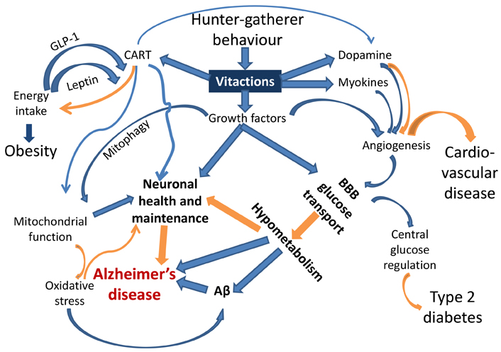
    A schematic representation of the links joining behaviour responsive signaling pathways and the pathophysiology of AD and other lifestyle related disorders. Blue arrows indicate up-regulation, and orange arrows indicate down-regulation or prevention. Each of the links is experimentally demonstrated (references in the text). This is the first attempt to put them together to visualize causal pathways to AD. BBB: blood brain barrier; Aβ: amyloid β

    Of specific relevance to AD are impaired angiogenesis and growth factors required for neuronal health. Neuronal mitochondrial function is evidently impaired in AD [58] and mitochondrial function is dependent on CART and growth factor signaling [45, 5457]. Mitophagy, altered in AD [5961], is also under growth factor regulation [62, 63]. Oxidative stress, also implicated in AD [6466], is generated by defective mitochondria or impaired fission-fusion dynamics [67, 68]. Stimuli from behavioural environments are also suspected to alter the dynamics and distribution of immune cells and thereby systemic inflammatory responses [69] which are also associated with AD [70, 71].

    Subnormal or defective angiogenesis impairs supply of glucose and other nutrients across the blood brain barrier (BBB) [72, 73]. The rate of glucose transport across the BBB is known to be reduced in obesity and type 2 diabetes, which are known risk factors for AD [74, 75]. Subnormal vasculature, reduced transport of glucose through BBB, and lower glucose metabolism in the brain are also known to be associated with AD [7680]. CART has a neuroprotective role in dementia [48, 50]. Dopamine expression is behaviour-responsive and also associated with CART [51], and has complex roles in angiogenesis and neurogenerative conditions [81, 82]. Myokines generated from skeletal muscle activity also promote angiogenesis along with neuroprotection [38, 83].

    With inadequate or defective brain vasculature, the nutrient supply to neurons is expected to be subnormal leading to neuronal death and degenerative changes. However, all parts of the brain need not be equally affected. The brain is expected to have evolved mechanisms by which regions being used more frequently would be selectively spared. Mechanisms by which vasculature in different regions of the brain can be differentially affected have been demonstrated [84]. For a sedentary lifestyle motor coordination functions are underutilized and therefore they would degenerate preferentially sparing the cognitive functions. Therefore, in sedentary lifestyle-related neurogenerative disease, the motor coordination is likely to degenerate first. This may not be noticed in a lifestyle where complex coordination is hardly challenged. Specifically designed tests can detect this degeneration very early which may serve as an early warning or timely diagnosis marker.

    The amyloid plaques, characteristic of AD may be a consequence of reduced glucose metabolism in the brain [85] or triggered by oxidative damage [66]. Amyloid proteins have a tendency to polymerize spontaneously with a small probability. However, if the rate of turnover of proteins is sufficiently large, the polymerization remains limited. If the rate of turnover is reduced below a threshold, proteins can aggregate rapidly [86]. If the reduced rate of metabolism reduces the protein turnover rate, aggregated proteins will be found, whether or not they have any further role in the pathology [87].

    In short, alteration in many pathways radiating from vitaction deficiencies can potentially explain the multiple factors implicated in AD including amyloid plaques, altered vasculature, reduced glucose metabolism, subnormal mitochondrial function, and impaired neuronal maintenance. This could be the most parsimonious explanation for the multiple pathophysiological changes characterizing AD.

    The deficiency of physical activity is well recognized as a risk factor in lifestyle diseases but in the classical view burning calories and achieving energy balance is the main relevance of physical activity. However, the benefits of physical activity are not the same and may even be contradictory under different behavioural contexts [8890]. Therefore, the behavioural context of physical activity needs to be examined carefully. Available literature suggests that different types of exercises may have differential cardiovascular, emotional and metabolic effects [88] as well as growth factor expression [9193]. Most studies appear to report only a limited classification of exercises in terms of low or high intensity and aerobic or resistance exercise types. Exercise can have different behavioural components which generally do not get recorded but they may be largely responsible for the beneficial effects of exercise [88]. Most sports activities mimic various components of a hunter-fighter’s behaviour such as chasing, hitting, aiming, dodging, escaping, risk taking, competing, attacking or defending, and therefore are expected to have different neuroendocrine effects than equal calorie mechanical and monotonous exercise. We hypothesize that the health benefits of exercise and sports are not obtained by burning calories alone but by partially supplementing the deficient vitactions. Apart from physical activity Watve [5] lists physical aggression, agility and rapid action, adventure and injury proneness, skin exposure to the sun, heat, cold, and minor injuries as some of the possible vitactions deficient in the modern lifestyle.

    Existing support to the hypothesis and testable predictions

    Exercises of different types are known to be effective in preventing and managing AD [9298]. The beneficial effects of exercise are likely to be via its links to growth factor expression [99103]. However, the differential effects of the behavioural components of exercise have not been tested in randomized control experiments (RCT). A typical design of RCT should involve comparison of behaviourally enriched exercise with a control group performing energetically comparable mechanical monotonous exercise. Using such a design, the effects of different behavioural components of exercise can be tested individually as well as collectively.

    The prediction of the hypothesis that motor coordination is expected to degenerate first is also supported [104105]. It needs to be explored how this can be systematically used for early warning and timely intervention.

    Clinical implications

    Just as vitamin deficiencies can be prevented or treated by supplementing the deficient vitamin and no other drug works, for vitaction deficiencies supplementing the deficient behaviour in the form of appropriate sports, activities, and exercises is expected to be the only effective solution. Since behaviour-physiology interface has multiple links (Figure 1), all of which may not even be discovered, a single target molecule approach typical of pharma research is unlikely to work.

    Behaviourally enriched exercises are therefore the most promising answer for effective prevention and possibly treatment of AD. The limitation of treatment might be that neurons already dead or irreversibly damaged may not be able to regenerate by behavioural exercises. Nevertheless, prevention of AD by behaviourally enriched exercise is likely to be highly effective.

    Abbreviations

    AD:

    Alzheimer’s disease

    BBB:

    blood brain barrier

    CART:

    cocaine-amphetamine regulated transcript

    Declarations

    Author contributions

    MW: Conceptualization, Writing—original draft. AKS: Data curation, Writing—review & editing.

    Conflicts of interest

    The authors declare that they have no conflicts of interest.

    Ethical approval

    Not applicable.

    Consent to participate

    Not applicable.

    Consent to publication

    Not applicable.

    Availability of data and materials

    Not applicable.

    Funding

    Not applicable.

    Copyright

    © The Author(s) 2024.

    References

    Engelen L, Gale J, Chau JY, Hardy LL, Mackey M, Johnson N, et al. Who is at risk of chronic disease? Associations between risk profiles of physical activity, sitting and cardio-metabolic disease in Australian adults. Aust N Z J Public Health. 2017;41:17883. [DOI] [PubMed]
    Guthrie GE. What is lifestyle medicine? Am J Lifestyle Med. 2018;12:3634. [DOI] [PubMed] [PMC]
    Nyberg ST, Singh-Manoux A, Pentti J, Madsen IEH, Sabia S, Alfredsson L, et al. Association of healthy lifestyle with years lived without major chronic diseases. JAMA Intern Med. 2020;180:7608. [DOI] [PubMed] [PMC]
    Zhang YB, Pan XF, Chen J, Cao A, Xia L, Zhang Y, et al. Combined lifestyle factors, all-cause mortality and cardiovascular disease: a systematic review and meta-analysis of prospective cohort studies. J Epidemiol Community Health. 2021;75:929. [DOI] [PubMed]
    Watve M. Doves, diplomats, and diabetes: a Darwinian interpretation of type 2 diabetes and related disorders. New York: Springer; 2013. [DOI]
    Hart AC, Chao MY. From odors to behaviors in Caenorhabditis elegans. In: Menini A, editor. The neurobiology of olfaction. Boca Raton (FL): CRC Press/Taylor & Francis; 2010.
    Liu Z, Kariya MJ, Chute CD, Pribadi AK, Leinwand SG, Tong A, et al. Predator-secreted sulfolipids induce defensive responses in C. elegans. Nat Commun. 2018;9:1128. [DOI] [PubMed] [PMC]
    Madhwal S, Shin M, Kapoor A, Goyal M, Joshi MK, Ur Rehman PM, et al. Metabolic control of cellular immune-competency by odors in Drosophila. Elife. 2020;9:e60376. [DOI] [PubMed] [PMC]
    Sadanandappa MK, Sathyanarayana SH, Kondo S, Bosco G. Neuropeptide F signaling regulates parasitoid-specific germline development and egg-laying in Drosophila. PLoS Genet. 2021;17:e1009456. [DOI] [PubMed] [PMC]
    Yu S, Luo F, Xu Y, Zhang Y, Jin LH. Drosophila innate immunity involves multiple signaling pathways and coordinated communication between different tissues. Front Immunol. 2022;13:905370. [DOI] [PubMed] [PMC]
    Pribadi A, Rieger MA, Rosales K, Reddy KC, Chalasani SH. Dopamine signaling regulates predator-driven changes in Caenorhabditis elegans’ egg laying behavior. Elife. 2023;12:e83957. [DOI] [PubMed] [PMC]
    Lamba S, Kazi YC, Deshpande S, Natesh M, Bhadra A, Gadagkar R. A possible novel function of dominance behaviour in queen-less colonies of the primitively eusocial wasp Ropalidia marginata. Behav Processes. 2007;74:3516. [DOI] [PubMed]
    Rengarajan S, Hallem EA. Olfactory circuits and behaviors of nematodes. Curr Opin Neurobiol. 2016;41:13648. [DOI] [PubMed] [PMC]
    Pribadi AK, Chalasani SH. Fear conditioning in invertebrates. Front Behav Neurosci. 2022;16:1008818. [DOI] [PubMed] [PMC]
    Cao L, Liu X, Lin EJ, Wang C, Choi EY, Riban V, et al. Environmental and genetic activation of a brain-adipocyte BDNF/leptin axis causes cancer remission and inhibition. Cell. 2010;142:5264. [DOI] [PubMed] [PMC]
    Li G, Gan Y, Fan Y, Wu Y, Lin H, Song Y, et al. Enriched environment inhibits mouse pancreatic cancer growth and down-regulates the expression of mitochondria-related genes in cancer cells. Sci Rep. 2015;5:7856. [DOI] [PubMed] [PMC]
    Takai D, Abe A, Miura H, Tanaka S, Komura JI. Minimum environmental enrichment is effective in activating antitumor immunity to transplanted tumor cells in mice. Exp Anim. 2019;68:56976. [DOI] [PubMed] [PMC]
    Watanabe J, Kagami N, Kawazoe M, Arata S. A simplified enriched environment increases body temperature and suppresses cancer progression in mice. Exp Anim. 2020;69:20718. [DOI] [PubMed] [PMC]
    Xiao R, Ali S, Caligiuri MA, Cao L. Enhancing effects of environmental enrichment on the functions of natural killer cells in mice. Front Immunol. 2021;12:695859. [DOI] [PubMed] [PMC]
    de Sousa Fernandes MS, Santos GCJ, Filgueira TO, Gomes DA, Barbosa EAS, Dos Santos TM, et al. Cytokines and immune cells profile in different tissues of rodents induced by environmental enrichment: systematic review. Int J Mol Sci. 2022;23:11986. [DOI] [PubMed] [PMC]
    Belsare PV, Watve MG, Ghaskadbi SS, Bhat DS, Yajnik CS, Jog M. Metabolic syndrome: aggression control mechanisms gone out of control. Med Hypotheses. 2010;74:57889. [DOI] [PubMed]
    Abbott DH, McNeilly AS, Lunn SF, Hulme MJ, Burden FJ. Inhibition of ovarian function in subordinate female marmoset monkeys (Callithrix jacchus jacchus). J Reprod Fertil. 1981;63:33545. [DOI] [PubMed]
    Wasser SK, Barash DP. Reproductive suppression among female mammals: implications for biomedicine and sexual selection theory. Q Rev Biol. 1983;58:51338. [DOI] [PubMed]
    Abbott DH. Behaviourally mediated suppression of reproduction in female primates. J Zool. 1987;213:45570. [DOI]
    Barrett J, Abbott DH, George LM. Extension of reproductive suppression by pheromonal cues in subordinate female marmoset monkeys, Callithrix jacchus. J Reprod Fertil. 1990;90:4118. [DOI] [PubMed]
    Abbott DH, Saltzman W, Schultz-Darken NJ, Tannenbaum PL. Adaptations to subordinate status in female marmoset monkeys. Comp Biochem Physiol C Pharmacol Toxicol Endocrinol. 1998;119:26174. [DOI] [PubMed]
    Nexø E, Hollenberg MD, Bing J. Aggressive behavior in mice provokes a marked increase in both plasma epidermal growth factor and renin. Acta Physiol Scand. 1981;111:36771. [DOI] [PubMed]
    Aloe L, Bracci-Laudiero L, Alleva E, Lambiase A, Micera A, Tirassa P. Emotional stress induced by parachute jumping enhances blood nerve growth factor levels and the distribution of nerve growth factor receptors in lymphocytes. Proc Natl Acad Sci U S A. 1994;91:104404. [DOI] [PubMed] [PMC]
    Hahn-Holbrook J, Holt-Lunstad J, Holbrook C, Coyne SM, Lawson ET. Maternal defense: breast feeding increases aggression by reducing stress. Psychol Sci. 2011;22:128895. [DOI] [PubMed] [PMC]
    Bosch OJ. Maternal aggression in rodents: brain oxytocin and vasopressin mediate pup defence. Philos Trans R Soc Lond B Biol Sci. 2013;368:20130085. [DOI] [PubMed] [PMC]
    Zukowska-Grojec Z, Karwatowska-Prokopczuk E, Rose W, Rone J, Movafagh S, Ji H, et al. Neuropeptide Y: a novel angiogenic factor from the sympathetic nerves and endothelium. Circ Res. 1998;83:18795. [DOI] [PubMed]
    Möller B, Rasmussen C, Lindblom B, Olovsson M. Expression of the angiogenic growth factors VEGF, FGF-2, EGF and their receptors in normal human endometrium during the menstrual cycle. Mol Hum Reprod. 2001;7:6572. [DOI] [PubMed]
    Kohara H, Tajima S, Yamamoto M, Tabata Y. Angiogenesis induced by controlled release of neuropeptide substance P. Biomaterials. 2010;31:861725. [DOI] [PubMed]
    Shome S, Rana T, Ganguly S, Basu B, Chaki Choudhury S, Sarkar C, et al. Dopamine regulates angiogenesis in normal dermal wound tissues. PLoS One. 2011;6:e25215. [DOI] [PubMed] [PMC]
    Barnabas O, Wang H, Gao XM. Role of estrogen in angiogenesis in cardiovascular diseases. J Geriatr Cardiol. 2013;10:37782. [DOI] [PubMed] [PMC]
    Chodari L, Mohammadi M, Ghorbanzadeh V, Dariushnejad H, Mohaddes G. Testosterone and voluntary exercise promote angiogenesis in hearts of rats with diabetes by enhancing expression of VEGF-A and SDF-1a. Can J Diabetes. 2016;40:43641. [DOI] [PubMed]
    Liu HS, Shen H, Luo Y, Hoffer BJ, Wang Y, Yang Y. Post-treatment with cocaine- and amphetamine-regulated transcript enhances infarct resolution, reinnervation, and angiogenesis in stroke rats – an MRI study. NMR Biomed. 2016;29:36170. [DOI] [PubMed]
    Qi C, Song X, Wang H, Yan Y, Liu B. The role of exercise-induced myokines in promoting angiogenesis. Front Physiol. 2022;13:981577. [DOI] [PubMed] [PMC]
    Yu H, Wang Y, Gao J, Gao Y, Zhong C, Chen Y. Application of the neuropeptide NPVF to enhance angiogenesis and osteogenesis in bone regeneration. Commun Biol. 2023;6:197. [DOI] [PubMed] [PMC]
    Baig U, Lokhande L, Lalwani P, Chawla S, Watve M. Foraging theory and the propensity to be obese: an alternative to thrift. Homo. 2019;70:193216. [DOI] [PubMed]
    Baggio LL, Drucker DJ. Glucagon-like peptide-1 receptors in the brain: controlling food intake and body weight. J Clin Invest. 2014;124:42236. [DOI] [PubMed] [PMC]
    Tidhar WL, Bonier F, Speakman JR. Sex- and concentration-dependent effects of predator feces on seasonal regulation of body mass in the bank vole Clethrionomys glareolus. Horm Behav. 2007;52:43644. [DOI] [PubMed]
    Ramler JP, Hebblewhite M, Kellenberg D, Sime C. Crying wolf? A spatial analysis of wolf location and depredations on calf weight. Am J Agric Econ. 2014;96:63156. [DOI]
    Monarca RI, da Luz Mathias M, Wang D, Speakman JR. Predation risk modulates diet-induced obesity in male C57BL/6 mice. Obesity (Silver Spring). 2015;23:205965. [DOI] [PubMed]
    Mao P, Meshul CK, Thuillier P, Goldberg NR, Reddy PH. CART peptide is a potential endogenous antioxidant and preferentially localized in mitochondria. PLoS One. 2012;7:e29343. [DOI] [PubMed] [PMC]
    Zhang M, Han L, Xu Y. Roles of cocaine- and amphetamine-regulated transcript in the central nervous system. Clin Exp Pharmacol Physiol. 2012;39:58692. [DOI] [PubMed]
    Mao P, Meshul CK, Thuillier P, Reddy PH. Neurotransmitter CART as a new therapeutic candidate for Parkinson’s disease. Pharmaceuticals (Basel). 2013;6:10823. [DOI] [PubMed] [PMC]
    Jin JL, Liou AK, Shi Y, Yin KL, Chen L, Li LL, et al. CART treatment improves memory and synaptic structure in APP/PS1 mice. Sci Rep. 2015;5:10224. [DOI] [PubMed] [PMC]
    Higginson AD, McNamara JM, Houston AI. Fatness and fitness: exposing the logic of evolutionary explanations for obesity. Proc Biol Sci. 2016;283:20152443. [DOI] [PubMed] [PMC]
    Borkar CD, Bharne AP, Nagalakshmi B, Sakharkar AJ, Subhedar NK, Kokare DM. Cocaine- and amphetamine-regulated transcript peptide (CART) alleviates MK-801-induced schizophrenic dementia-like symptoms. Neuroscience. 2018;375:94107. [DOI] [PubMed]
    Lin L, Sun D, Chang J, Ma M, Zhou X, Zhao M, et al. Cocaine‑ and amphetamine‑regulated transcript (CART) is associated with dopamine and is protective against ischemic stroke. Mol Med Rep. 2018;18:3298304. [DOI] [PubMed] [PMC]
    Meng Q, Kim HC, Oh S, Lee YM, Hu Z, Oh KW. Cocaine- and amphetamine-regulated transcript (CART) peptide plays critical role in psychostimulant-induced depression. Biomol Ther (Seoul). 2018;26:42531. [DOI] [PubMed] [PMC]
    Jiang H, Niu F, Zheng Y, Xu Y. CART mitigates oxidative stress and DNA damage in memory deficits of APP/PS1 mice via upregulating β‑amyloid metabolism‑associated enzymes. Mol Med Rep. 2021;23:280. [DOI] [PubMed] [PMC]
    Che TF, Lin CW, Wu YY, Chen YJ, Han CL, Chang YL, et al. Mitochondrial translocation of EGFR regulates mitochondria dynamics and promotes metastasis in NSCLC. Oncotarget. 2015;6:3734966. [DOI] [PubMed] [PMC]
    Poudel SB, Dixit M, Neginskaya M, Nagaraj K, Pavlov E, Werner H, et al. Effects of GH/IGF on the aging mitochondria. Cells. 2020;9:1384. [DOI] [PubMed] [PMC]
    Hu B, Li H, Zhang X. A balanced act: the effects of GH–GHR–IGF1 axis on mitochondrial function. Front Cell Dev Biol. 2021;9:630248. [DOI] [PubMed] [PMC]
    Kan S, Pi C, Zhang L, Guo D, Niu Z, Liu Y, et al. FGF19 increases mitochondrial biogenesis and fusion in chondrocytes via the AMPKα-p38/MAPK pathway. Cell Commun Signal. 2023;21:55. [DOI] [PubMed] [PMC]
    Bhatia S, Rawal R, Sharma P, Singh T, Singh M, Singh V. Mitochondrial dysfunction in Alzheimer’s disease: opportunities for drug development. Curr Neuropharmacol. 2022;20:67592. [DOI] [PubMed] [PMC]
    Fang EF, Hou Y, Palikaras K, Adriaanse BA, Kerr JS, Yang B, et al. Mitophagy inhibits amyloid-β and tau pathology and reverses cognitive deficits in models of Alzheimer’s disease. Nat Neurosci. 2019;22:40112. [DOI] [PubMed] [PMC]
    Kshirsagar S, Sawant N, Morton H, Reddy AP, Reddy PH. Protective effects of mitophagy enhancers against amyloid beta-induced mitochondrial and synaptic toxicities in Alzheimer disease. Hum Mol Genet. 2022;31:42339. [DOI] [PubMed] [PMC]
    Mary A, Eysert F, Checler F, Chami M. Mitophagy in Alzheimer’s disease: molecular defects and therapeutic approaches. Mol Psychiatry. 2023;28:20216. [DOI] [PubMed] [PMC]
    Hou X, Li Z, Higashi Y, Delafontaine P, Sukhanov S. Insulin-like growth factor I prevents cellular aging via activation of mitophagy. J Aging Res. 2020;2020:4939310. [DOI] [PubMed] [PMC]
    Lv D, Yang K, Rich JN. Growth factor receptor signaling induces mitophagy through epitranscriptomic regulation. Autophagy. 2023;19:10345. [DOI] [PubMed] [PMC]
    Huang WJ, Zhang X, Chen WW. Role of oxidative stress in Alzheimer’s disease. Biomed Rep. 2016;4:51922. [DOI] [PubMed] [PMC]
    Cassidy L, Fernandez F, Johnson JB, Naiker M, Owoola AG, Broszczak DA. Oxidative stress in alzheimer’s disease: a review on emergent natural polyphenolic therapeutics. Complement Ther Med. 2020;49:102294. [DOI] [PubMed]
    Tamagno E, Guglielmotto M, Vasciaveo V, Tabaton M. Oxidative stress and beta amyloid in Alzheimer’s disease. Which comes first: the chicken or the egg? Antioxidants (Basel). 2021;10:1479. [DOI] [PubMed] [PMC]
    Yu T, Robotham JL, Yoon Y. Increased production of reactive oxygen species in hyperglycemic conditions requires dynamic change of mitochondrial morphology. Proc Natl Acad Sci U S A. 2006;103:26538. [DOI] [PubMed] [PMC]
    Misrani A, Tabassum S, Yang L. Mitochondrial dysfunction and oxidative stress in Alzheimer’s disease. Front Aging Neurosci. 2021;13:617588. [DOI] [PubMed] [PMC]
    Watve M, Mandani S. Why serum chemokine levels are raised in insulin resistance syndrome: an immune reversal hypothesis. Curr Sci. 2008;95;171–4.
    Akiyama H, Barger S, Barnum S, Bradt B, Bauer J, Cole GM, et al. Inflammation and Alzheimer’s disease. Neurobiol Aging. 2000;21:383421. [DOI] [PubMed] [PMC]
    Kinney JW, Bemiller SM, Murtishaw AS, Leisgang AM, Salazar AM, Lamb BT. Inflammation as a central mechanism in Alzheimer’s disease. Alzheimers Dement (N Y). 2018;4:57590. [DOI] [PubMed] [PMC]
    Hwang JJ, Jiang L, Hamza M, Sanchez Rangel E, Dai F, Belfort-DeAguiar R, et al. Blunted rise in brain glucose levels during hyperglycemia in adults with obesity and T2DM. JCI Insight. 2017;2:e95913. [DOI] [PubMed] [PMC]
    Ojha A, Watve M. Reduced blood to brain glucose transport as the cause for hyperglycemia: a model that resolves multiple anomalies in type 2 diabetes. Qeios. 2023. [DOI]
    Barbagallo M, Dominguez LJ. Type 2 diabetes mellitus and Alzheimer’s disease. World J Diabetes. 2014;5:88993. [DOI] [PubMed] [PMC]
    Nguyen TT, Ta QTH, Nguyen TKO, Nguyen TTD, Giau VV. Type 3 diabetes and its role implications in Alzheimer’s disease. Int J Mol Sci. 2020;21:3165. [DOI] [PubMed] [PMC]
    An Y, Varma VR, Varma S, Casanova R, Dammer E, Pletnikova O, et al. Evidence for brain glucose dysregulation in Alzheimer’s disease. Alzheimers Dement. 2018;14:31829. [DOI] [PubMed] [PMC]
    Religa P, Cao R, Religa D, Xue Y, Bogdanovic N, Westaway D, et al. VEGF significantly restores impaired memory behavior in Alzheimer’s mice by improvement of vascular survival. Sci Rep. 2013;3:2053. [DOI] [PubMed] [PMC]
    Reiman EM, Chen K, Alexander GE, Caselli RJ, Bandy D, Osborne D, et al. Functional brain abnormalities in young adults at genetic risk for late-onset Alzheimer’s dementia. Proc Natl Acad Sci U S A. 2004;101:2849. [DOI] [PubMed] [PMC]
    Jefferies WA, Price KA, Biron KE, Fenninger F, Pfeifer CG, Dickstein DL. Adjusting the compass: new insights into the role of angiogenesis in Alzheimer’s disease. Alzheimers Res Ther. 2013;5:64. [DOI] [PubMed] [PMC]
    Gejl M, Brock B, Egefjord L, Vang K, Rungby J, Gjedde A. Blood-brain glucose transfer in Alzheimer’s disease: effect of GLP-1 analog treatment. Sci Rep. 2017;7:17490. [DOI] [PubMed] [PMC]
    Basu S, Nagy JA, Pal S, Vasile E, Eckelhoefer IA, Bliss VS, et al. The neurotransmitter dopamine inhibits angiogenesis induced by vascular permeability factor/vascular endothelial growth factor. Nat Med. 2001;7:56974. [DOI] [PubMed]
    Lindgren HS, Ohlin KE, Cenci MA. Differential involvement of D1 and D2 dopamine receptors in L-DOPA-induced angiogenic activity in a rat model of Parkinson’s disease. Neuropsychopharmacology. 2009;34:247788. [DOI] [PubMed]
    Lee B, Shin M, Park Y, Won SY, Cho KS. Physical exercise-induced myokines in neurodegenerative diseases. Int J Mol Sci. 2021;22:5795. [DOI] [PubMed] [PMC]
    Parab S, Quick RE, Matsuoka RL. Endothelial cell-type-specific molecular requirements for angiogenesis drive fenestrated vessel development in the brain. Elife. 2021;10:e64295. [DOI] [PubMed] [PMC]
    Lauretti E, Praticò D. Glucose deprivation increases tau phosphorylation via P38 mitogen-activated protein kinase. Aging Cell. 2015;14:106774. [DOI] [PubMed] [PMC]
    Watve M, Bodas A, Diwekar M. Altered autonomic inputs as a cause of pancreatic β-cell amyloid. Med Hypotheses. 2014;82:4953. [DOI] [PubMed]
    Nortley R, Korte N, Izquierdo P, Hirunpattarasilp C, Mishra A, Jaunmuktane Z, et al. Amyloid β oligomers constrict human capillaries in Alzheimer’s disease via signaling to pericytes. Science. 2019;365:eaav9518. [DOI] [PubMed] [PMC]
    Wei Z, Huang Y, Li X, Shao M, Qian H, He B, et al. The influence of aggressive exercise on responses to self-perceived and others’ pain. Cereb Cortex. 2023;33:1080212. [DOI] [PubMed] [PMC]
    Nabe-Nielsen K, Holtermann A, Gyntelberg F, Garde AH, Islamoska S, Prescott E, et al. The effect of occupational physical activity on dementia: results from the Copenhagen Male Study. Scand J Med Sci Sports. 2021;31:44655. [DOI] [PubMed]
    Zotcheva E, Bratsberg B, Strand BH, Jugessur A, Engdahl BL, Bowen C, et al. Trajectories of occupational physical activity and risk of later-life mild cognitive impairment and dementia: the HUNT4 70+ study. Lancet Reg Health Eur. 2023;34:100721. [DOI] [PubMed] [PMC]
    Rojas Vega S, Knicker A, Hollmann W, Bloch W, Strüder HK. Effect of resistance exercise on serum levels of growth factors in humans. Horm Metab Res. 2010;42:9826. [DOI] [PubMed]
    Nishida Y, Matsubara T, Tobina T, Shindo M, Tokuyama K, Tanaka K, et al. Effect of low-intensity aerobic exercise on insulin-like growth factor-I and insulin-like growth factor-binding proteins in healthy men. Int J Endocrinol. 2010;2010:452820. [DOI] [PubMed] [PMC]
    Gulick CN, Peddie MC, Jowett T, Hackney AC, Rehrer NJ. Exercise, dietary protein, and combined effect on IGF-1. Int J Sci Res Methodol. 2020;16:6177. [PubMed] [PMC]
    Thomas VS, Hageman PA. Can neuromuscular strength and function in people with dementia be rehabilitated using resistance-exercise training? Results from a preliminary intervention study. J Gerontol A Biol Sci Med Sci. 2003;58:74651. [DOI] [PubMed]
    Cass SP. Alzheimer’s disease and exercise: a literature review. Curr Sports Med Rep. 2017;16:1922. [DOI] [PubMed]
    Meng Q, Lin MS, Tzeng IS. Relationship between exercise and Alzheimer’s disease: a narrative literature review. Front Neurosci. 2020;14:131. [DOI] [PubMed] [PMC]
    Liu W, Zhang J, Wang Y, Li J, Chang J, Jia Q. Effect of physical exercise on cognitive function of Alzheimer’s disease patients: a systematic review and meta-analysis of randomized controlled trial. Front Psychiatry. 2022;13:927128. [DOI] [PubMed] [PMC]
    Pahlavani HA. Exercise therapy to prevent and treat Alzheimer’s disease. Front Aging Neurosci. 2023;15:1243869. [DOI] [PubMed] [PMC]
    Cotman CW, Berchtold NC, Christie LA. Exercise builds brain health: key roles of growth factor cascades and inflammation. Trends Neurosci. 2007;30:46472. [DOI] [PubMed]
    Frystyk J. Exercise and the growth hormone-insulin-like growth factor axis. Med Sci Sports Exerc. 2010;42:5866. [DOI] [PubMed]
    Gorski T, De Bock K. Metabolic regulation of exercise-induced angiogenesis. Vasc Biol. 2019;1:H18. [DOI] [PubMed] [PMC]
    Jaberi S, Fahnestock M. Mechanisms of the beneficial effects of exercise on brain-derived neurotrophic factor expression in Alzheimer’s disease. Biomolecules. 2023;13:1577. [DOI] [PubMed] [PMC]
    Kim H, Jung J, Park S, Joo Y, Lee S, Sim J, et al. Exercise-induced fibroblast growth factor-21: a systematic review and meta-analysis. Int J Mol Sci. 2023;24:7284. [DOI] [PubMed] [PMC]
    Buchman AS, Bennett DA. Loss of motor function in preclinical Alzheimer’s disease. Expert Rev Neurother. 2011;11:66576. [DOI] [PubMed] [PMC]
    Castillo-Mariqueo L, Giménez-Llort L. Clasping, ledge-score coordination and early gait impairments as primary behavioural markers of functional impairment in Alzheimer’s disease. Behav Brain Res. 2022;435:114054. [DOI] [PubMed]