 Case Report


Affiliation:
1Department of Internal Medicine, Hospital Universitario de Torrejón, 28850 Torrejón de Ardoz, Madrid, Spain

Affiliation:
2Department of Medical Oncology, Hospital Universitario de Torrejón, 28850 Torrejón de Ardoz, Madrid, Spain
Affiliation:
2Department of Medical Oncology, Hospital Universitario de Torrejón, 28850 Torrejón de Ardoz, Madrid, Spain
3Facultad de Ciencias de la Salud, Universidad Francisco de Vitoria, 28223 Pozuelo de Alarcón, Madrid, Spain
Affiliation:
1Department of Internal Medicine, Hospital Universitario de Torrejón, 28850 Torrejón de Ardoz, Madrid, Spain
Affiliation:
1Department of Internal Medicine, Hospital Universitario de Torrejón, 28850 Torrejón de Ardoz, Madrid, Spain
Affiliation:
1Department of Internal Medicine, Hospital Universitario de Torrejón, 28850 Torrejón de Ardoz, Madrid, Spain
Affiliation:
4Department of Pharmacy and Nutrition, Faculty of Biomedical and Health Sciences, Universidad Europea de Madrid, 28670 Villaviciosa de Odón, Madrid, Spain
ORCID: https://orcid.org/0009-0007-5917-9761
 ,
,
Affiliation:
2Department of Medical Oncology, Hospital Universitario de Torrejón, 28850 Torrejón de Ardoz, Madrid, Spain
3Facultad de Ciencias de la Salud, Universidad Francisco de Vitoria, 28223 Pozuelo de Alarcón, Madrid, Spain
Email: lcabezon@torrejonsalud.com
ORCID: https://orcid.org/0000-0002-3468-3626

Explor Med. 2026;7:1001431 DOI: https://doi.org/10.37349/emed.2026.1001431
Received: April 20, 2026 Accepted: July 16, 2026 Published: October 14, 2026
Academic Editor: Ning Li, Chinese Academy of Medical Sciences and Peking Union Medical College, China
Nasopharyngeal carcinoma (NPC) with cutaneous metastases (lethalides) is an exceedingly rare event associated with an aggressive clinical course and poor prognosis, making the management of plurirefractory disease a significant challenge in the evolving landscape of immunotherapy. Here, we report the case of a 59-year-old male diagnosed with stage IVA Epstein–Barr virus (EBV)-positive non-keratinizing NPC who achieved a complete response after induction chemotherapy and concurrent chemoradiotherapy. Twenty-six months later, the patient developed multifocal cutaneous metastases and locoregional recurrence. Over the course of approximately two and a half years, he received six lines of therapy, including platinum-based regimens, pembrolizumab, and cetuximab-paclitaxel, before enrolling in the phase I/II ABILITY-1 trial, where he received the novel interleukin-2 (IL-2) superkine MDNA11 plus pembrolizumab. Despite transient stabilization, the disease progressed, and the patient died 62 months after initial diagnosis. Throughout his clinical course, plasma EBV DNA acted as a precise real-time surrogate of tumor burden, consistently anticipating radiologic progression. This case underscores the aggressive nature of cutaneous NPC and the pivotal role of serial EBV DNA monitoring in guiding therapeutic transitions. Furthermore, the lack of response to multiple agents, including next-generation IL-2 variants, highlights the profound immune exhaustion in late-stage NPC and emphasizes the urgent need for novel targeted strategies, such as antibody-drug conjugates and adoptive T-cell therapies, in the post-immunotherapy setting.
Nasopharyngeal carcinoma (NPC) is a malignant epithelial tumour arising from the mucosal lining of the nasopharynx, with a striking epidemiological clustering in Southern China, Southeast Asia, and North Africa. Globally, approximately 130,000 new cases are diagnosed annually, with an age-standardized incidence of around 2.1 per 100,000 in 2019 [1]. The disease is strongly associated with Epstein–Barr virus (EBV) infection, particularly for the non-keratinising subtypes (World Health Organization, WHO types II and III), which account for the vast majority of cases in endemic regions [2]. Despite a pronounced sensitivity to platinum-based chemoradiotherapy, and 5-year overall survival (OS) rates of approximately 70% for locoregionally confined disease, more than half of patients present with stage III–IV disease, and a substantial proportion eventually relapse [2, 3].
For patients with recurrent or metastatic NPC, the treatment landscape has evolved considerably over the past decade. The phase III GEM20110714 trial established gemcitabine plus cisplatin as the standard first-line regimen in the recurrent/metastatic setting, demonstrating superior progression-free and OS compared with fluorouracil–cisplatin [4]. Recently, the addition of programmed death-1 (PD-1) checkpoint inhibitors, most notably toripalimab (JUPITER-02 trial) and penpulimab-kcqx (AK105-304 trial), to platinum-based chemotherapy has further improved outcomes and established chemoimmunotherapy as the current standard of care for first-line treatment of recurrent/metastatic NPC [5, 6]. Nevertheless, for patients who progress after immunochemotherapy, therapeutic options remain extremely limited, and prognosis is poor.
Cutaneous metastases from NPC (also referred to as lethalides or carcinoma cutis) are a rare and clinically ominous complication, with very few documented cases in the published literature [7, 8]. Their occurrence represents an advanced stage of haematogenous or lymphatic dissemination and typically signals a poor outcome. Concurrently, plasma EBV DNA load has emerged as an indispensable biomarker in NPC management, correlating dynamically with tumour burden, treatment response, and early relapse detection [9, 10].
We present a case of EBV-positive non-keratinizing NPC characterized by an uncommon cutaneous metastatic pattern at first recurrence. Notably, the patient exhibited an extraordinary survival trajectory through six lines of systemic treatment. We emphasize the temporal dynamics of plasma EBV DNA, illustrating its utility in guiding clinical transitions during long-term multimodal management, including immunotherapy.
Table 1 below summarises the patient’s clinical timeline. A comprehensive timeline has been generated using the CARE-writer tool in accordance with journal requirements.
Clinical timeline of the case.
| Date | Event | Details/Visit summary |
|---|---|---|
| Sep 2020 | Initial presentation & diagnosis | 59-year-old Asian male; bilateral epistaxis; right neck mass. CT/PET-CT: NPC left posterolateral wall, bilateral cervical adenopathy. EBER-ISH positive. Stage IVA (cT2N3M0, AJCC 8th ed.) |
| Oct–Dec 2020 | Induction chemotherapy | Cisplatin 80 mg/m2 + gemcitabine 1,000 mg/m2 Q21d × 3 cycles. Major partial response. |
| Jan–Mar 2021 | Definitive chemoradiotherapy (CCRT) | IMRT 69.96 Gy/33 fr to primary + PET+ nodes; 59.40 Gy to high-risk nodes. Concurrent cisplatin 100 mg/m2 → carboplatin AUC4 (grade 2 AKI). |
| Mar 2021 | Complete response confirmed | MRI + PET-CT: complete clinico-radiologic and metabolic response. |
| Apr 2021–Apr 2023 | Surveillance (26 months) | Serial endoscopy, cross-sectional imaging, and plasma EBV DNA monitoring: all unremarkable. |
| May 2023 | First relapse | Multifocal cutaneous tumour deposits (right lateral neck/supraclavicular region); cervical, axillary, and mediastinal lymphadenopathy. Biopsy: recurrent non-keratinising NPC, CPS 70. Plasma EBV DNA 76.4 copies/mL. |
| Jun–Dec 2023 | 1st-line salvage: cisplatin + gemcitabine | Cisplatin 70 mg/m2 + gemcitabine 1,000 mg/m2 (75% dose) Q21d × 6 cycles → partial radiologic response; EBV DNA → undetectable. Gemcitabine maintenance × 6 cycles. |
| Feb 2024 | Biochemical relapse (EBV lead time) | Rising plasma EBV DNA from undetectable to low–positive levels; new nodular skin lesions. Imaging: stable disease by RECIST 1.1. EBV DNA preceded radiologic progression by ~6–8 weeks. |
| Mar–May 2024 | 2nd-line: Pembrolizumab | Compassionate use. Pembrolizumab 200 mg Q3W. Transient EBV DNA clearance; cutaneous inflammatory reaction (possible pseudoprogression). CT cycle 4: confirmed PD; new occipital scalp deposits. |
| Jun–Oct 2024 | 3rd-line: paclitaxel + cetuximab | Biweekly paclitaxel + cetuximab. Complicated by grade 2 acneiform rash and grade 3 inguinal skin toxicity → dose reductions. EBV DNA: 485 → 1,009 → 4,092 → 13,000 copies/mL. CT Oct 2024: unequivocal PD. |
| Dec 2024–Mar 2025 | 4th-line: MDNA11 + pembrolizumab (ABILITY-1 trial) | Phase I/II ABILITY-1 (NCT05086692). MDNA11 IV Q2W + pembrolizumab 400 mg Q6W. iUPD at 8 wks (unconfirmed). EBV DNA rose to 4,062 copies/mL. Clinical PD (worsening cervical ulceration, dysphagia). Trial discontinued after 3 cycles. |
| Apr 2025 | Palliative radiotherapy | 30 Gy/10 fr (HexaSkin technique) to cervical cutaneous deposits. Partial haemostatic and analgesic response. |
| May–Jul 2025 | 5th-line: carboplatin + paclitaxel | Weekly carboplatin AUC2 + paclitaxel 70 mg/m2. EBV DNA: > 4,000 → 252 copies/mL. Partial skin regression. Grade 2 anaemia; 1 episode of febrile neutropenia (G-CSF). Disease re-progression after ~2 months. |
| Aug–Nov 2025 | 6th-line: methotrexate | Methotrexate 40 mg/m2 weekly. No meaningful disease control. PEG tube placement (oropharyngeal tumour encroachment). |
| Nov 2025 | Death | Transition to comprehensive palliative care after shared decision-making. Patient died in November 2025. Total survival: ~62 months from initial diagnosis. |
CCRT: concurrent chemoradiotherapy; EBV: Epstein–Barr virus; G-CSF: granulocyte colony-stimulating factor; IMRT: intensity-modulated radiotherapy; iUPD: unconfirmed progressive disease (iRECIST); PD: progressive disease; PEG: percutaneous endoscopic gastrostomy; Q21d: every 21 days; RECIST: Response Evaluation Criteria in Solid Tumours; AJCC: American Joint Committee on Cancer; AKI: acute kidney injury; AUC: area under the curve; CPS: combined positive score; CT: computed tomography; EBER-ISH: Epstein–Barr encoding region in situ hybridisation; fr: fractions; IV: intravenous; MRI: magnetic resonance imaging; NPC: nasopharyngeal carcinoma; PET-CT: positron emission tomography-computed tomography; Q2W: every 2 weeks; Q3W: every 3 weeks; Q6W: every 6 weeks.
A 59-year-old male of Asian ethnicity with a 24 pack-year smoking history (cessation at age 38) and a background of hypertension presented to the Otorhinolaryngology clinic in September 2020 with a 3-day history of bilateral epistaxis (predominantly right-sided) and a painless, slowly enlarging right neck mass of approximately two months’ duration. Physical examination revealed a firm, approximately 4 cm right cervical lymph node along the posterior border of the sternocleidomastoid, with additional palpable nodes of 2–3 cm in the right posterior triangle and right supraclavicular fossa.
Contrast-enhanced computed tomography (CT) of the neck demonstrated an enhancing nasopharyngeal mass (left posterolateral wall, approximately 30 × 17 mm) extending anteriorly into the left choana, with bilateral cervical lymphadenopathy (levels II–IV bilaterally, right supraclavicular, and right level V; largest node approximately 4 cm). 18F-FDG PET-CT (positron emission tomography-computed tomography) confirmed intense hypermetabolic activity in the left nasopharynx and bilateral cervical adenopathy. Endoscopic biopsy established a diagnosis of poorly differentiated non-keratinising squamous cell carcinoma of the nasopharynx (WHO type II, Regaud pattern). Immunohistochemistry showed p40 positivity (squamous lineage) and negativity for CK7, CK20, TTF-1, and PSA. In situ hybridisation for Epstein–Barr encoding region in situ hybridisation (EBER-ISH) was strongly positive. The disease was staged as cT2 N3 M0, stage IVA per the AJCC/UICC 8th edition classification [11].
Following multidisciplinary team (MDT) discussion, the patient received induction chemotherapy with cisplatin 80 mg/m2 (day 1) and gemcitabine 1,000 mg/m2 (days 1 and 8) every 21 days for 3 cycles, achieving a major partial response. Definitive locoregional treatment comprised intensity-modulated radiotherapy (IMRT) to the nasopharynx and involved nodal regions (69.96 Gy to the primary tumor and PET-positive nodes, and 59.40 Gy to high-risk nodal chains, delivered in 33 fractions) with concurrent platinum-based chemotherapy. Following an initial cycle of high-dose cisplatin (100 mg/m2), the development of grade 2 acute kidney injury (AKI) necessitated a transition to carboplatin (AUC4, area under the curve) for the subsequent two cycles of concurrent chemotherapy, consistent with established protocols for cisplatin-intolerant patients [12]. Post-treatment restaging via MRI and PET-CT in March 2021 confirmed a complete clinico-radiologic and metabolic response.
The patient remained under surveillance for 26 months, during which serial nasopharyngeal endoscopy, cross-sectional imaging, and plasma EBV DNA monitoring remained unremarkable. In May 2023, he presented with rapidly evolving multifocal cutaneous tumour deposits involving the right lateral neck and supraclavicular region, accompanied by right cervical, right supraclavicular, left axillary, and mediastinal lymphadenopathy. Biopsy of the skin lesions confirmed recurrent non-keratinising NPC with the same immunophenotype as the primary tumour. Tumour programmed death-ligand 1 (PD-L1) expression was markedly elevated, with a combined positive score (CPS) of 70. Plasma EBV DNA at relapse was 76.4 copies/mL, a detectable level consistent with active tumour recurrence [9, 13].
Following MDT consensus, first-line salvage therapy was initiated in June 2023 with cisplatin 70 mg/m2 plus gemcitabine 1,000 mg/m2 (days 1 and 8) every 3 weeks, with gemcitabine dose-reduced to 75% on account of prior cumulative exposure. This regimen is consistent with international guidelines for recurrent/metastatic NPC in patients not previously exposed to palliative chemotherapy [4, 14]. After 6 cycles, plasma EBV DNA fell from 76.4 to undetectable, and a partial radiologic response was documented by Response Evaluation Criteria in Solid Tumours (RECIST) 1.1. Given the absence of a suitable clinical trial, single-agent gemcitabine maintenance was administered for an additional 6 cycles, preserving disease control.
In February 2024, routine surveillance identified new nodular skin lesions on the right neck, accompanied by a rise in plasma EBV DNA from undetectable to low-positive levels, despite imaging showing only stable disease by RECIST criteria. This discordance (where liquid biomarker positivity precedes radiologic evidence of progression) exemplifies the superior sensitivity of circulating EBV DNA as an early relapse detector, a phenomenon well-documented in the NPC surveillance literature [9, 15].
In view of the high tumour PD-L1 expression (CPS 70) and EBV-positive tumour biology, pembrolizumab (anti-PD-1) at 200 mg every 3 weeks was initiated via a compassionate use programme in March 2024. After the first infusions, EBV DNA again became undetectable, and the cutaneous lesions exhibited transient erythema and swelling, interpreted as a possible immune-related inflammatory reaction or pseudoprogression, managed conservatively with topical corticosteroids. By cycle 4 (May 2024), however, CT demonstrated clear enlargement of the cutaneous and nodal disease, with new occipital scalp deposits, confirming progressive disease (PD).
Given the unavailability of suitable trials at the time, a third-line regimen of biweekly paclitaxel plus the anti-epidermal growth factor receptor (EGFR) monoclonal antibody cetuximab was initiated in June 2024. EGFR is overexpressed in the majority of NPC tumours, and cetuximab-based combinations have shown modest but documentable activity in both locoregionally advanced and recurrent/metastatic NPC in retrospective and early-phase studies [16, 17]. Treatment was complicated by grade 2 acneiform facial rash and a grade 3 inguinal skin toxicity attributable to cetuximab, a class-effect dermatological toxicity resulting from EGFR inhibition in normal skin keratinocytes, necessitating dose reductions [16]. Despite the dosing modifications, plasma EBV DNA rose progressively over the treatment period (July 2024: 485 copies/mL; August: 1,009 copies/mL; September: 4,092 copies/mL; October: 13,000 copies/mL), and radiologic evaluation in October 2024 documented unequivocal progression with extensive locoregional tumour spread and new axillary, mediastinal, and retrocrural adenopathy. The striking exponential rise in EBV DNA anticipated the radiologic findings by several weeks, once again underscoring its clinical utility as an early surrogate of therapeutic failure [10].
In December 2024, with ECOG PS of 1 and EBV DNA of 1,350 copies/mL, the patient was enrolled in the phase I/II ABILITY-1 trial (NCT05086692) [18] at Hospital Universitario de Torrejón, Madrid, a participating centre. Our patient received MDNA11 intravenously every 2 weeks at an escalating dose level, in combination with pembrolizumab 400 mg Q6W, consistent with the combination arm of the ABILITY-1 protocol. Tolerability was acceptable: observed adverse events included low-grade fever, mild fatigue, grade 2 transient hypotension, electrolyte disturbances, and a grade 4 lymphopenia (transient), none of which required permanent discontinuation. After 8 weeks on treatment (February 2025), imaging suggested unconfirmed progression per immune RECIST (iRECIST) criteria (iUPD), which was not confirmed on subsequent re-evaluation, permitting the patient to continue per protocol. However, EBV DNA had risen to 4,062 copies/mL, indicating increasing tumour activity. Clinical and symptomatic progression, manifested by worsening cervical skin ulceration (Figures 1 and 2) and new-onset dysphagia attributed to epiglottic oedema (treated with a brief corticosteroid course), led to trial discontinuation in March 2025 after three treatment cycles.
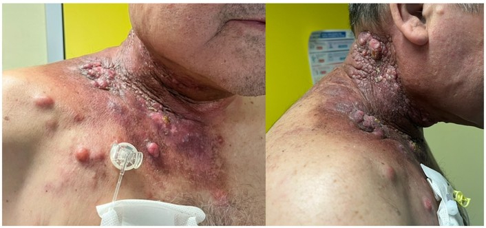
Clinical manifestation of cutaneous metastases (lethalides). Photographs demonstrate multifocal, indurated, and erythematous-to-violaceous skin nodules and plaques. The lesions exhibit a confluent, infiltrative growth pattern involving the right lateral neck, supraclavicular region, and superior chest wall. Focal areas of ulceration and inflammatory crusting are visible, consistent with advanced locoregional lymphatic and hematogenous spread.
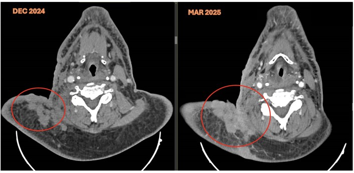
Comparative contrast-enhanced CT images demonstrate significant disease progression between December 2024 and March 2025. At the nodal level, there is a marked increase in the size and confluence of right-sided level II–V cervical and supraclavicular lymphadenopathy, exhibiting central necrosis and extranodal extension. Concurrently, the imaging reveals an unremitting progression of cutaneous and subcutaneous metastatic deposits (lethalides) involving the right cervicosupraclavicular region, characterized by extensive soft-tissue thickening and loss of normal fat planes.
In April 2025, palliative radiotherapy (30 Gy in 10 fractions; HexaSkin technique) was administered to the cervical cutaneous deposits, providing partial haemostatic and analgesic palliation. Fifth-line systemic therapy with weekly carboplatin (AUC2) plus paclitaxel (70 mg/m2) commenced in May 2025 and achieved partial regression of skin lesions and a significant EBV DNA fall (from > 4,000 to 252 copies/mL) (Figure 3). Toxicities included grade 2 anaemia and one episode of febrile neutropenia requiring granulocyte colony-stimulating factor (G-CSF) support. After approximately 8 cycles (~2 months), disease progression recurred. A sixth-line regimen with methotrexate (40 mg/m2 weekly) offered no meaningful disease control. Dysphagia from oropharyngeal tumour encroachment required placement of a percutaneous endoscopic gastrostomy (PEG) tube. In November 2025, following shared decision-making with the patient and his family, a transition to comprehensive palliative care was agreed. The patient died in November 2025, approximately five years after initial diagnosis. Table 2 summarizes the patient’s treatment history by line, and Figure 3 illustrates the longitudinal evolution of plasma EBV viral load across all treatment lines.
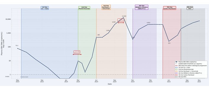
Temporal evolution of plasma EBV viral load relative to successive lines of systemic therapy. The Y-axis is displayed on a log10 scale. Red arrows indicate lead times between EBV DNA rise and subsequent radiological confirmation of progressive disease. Vertical dashed lines denote the initiation of each treatment line.
Patient treatment history by line of therapy.
| Line | Period | Regimen | Best response | EBV DNA (start → end) | Reason for discontinuation |
|---|---|---|---|---|---|
| — | Oct 2020–Mar 2021 | Induction CisGem × 3 cycles + CCRT (IMRT 69.96 Gy + carboplatin) | Complete response | — | Treatment completion |
| 1st | Jun 2023–Feb 2024 | Cisplatin 70 mg/m2 + gemcitabine 1,000 mg/m2 Q21d × 6; then gemcitabine maintenance × 6 | Partial response | 76.4 → undetectable | Disease progression (Feb 2024) |
| 2nd | Mar 2024–May 2024 | Pembrolizumab 200 mg Q3W (compassionate use) | PD | Undetectable → 18 copies/mL | Radiology PD (cycle 4) |
| 3rd | Jun 2024–Oct 2024 | Biweekly paclitaxel + cetuximab (dose-reduced for toxicity) | PD | 485 → 13,000 copies/mL | Unequivocal PD; grade 3 skin toxicity |
| 4th | Dec 2024–Mar 2025 | MDNA11 IV Q2W + pembrolizumab 400 mg Q6W (ABILITY-1, NCT05086692) | PD (iUPD → confirmed PD) | 1,350 → 4,062 copies/mL | Clinical PD: worsening ulceration, dysphagia (3 cycles) |
| 5th | May 2025–Jul 2025 | Weekly carboplatin AUC2 + paclitaxel 70 mg/m2 | Partial response (skin) | > 4,000 → 252 copies/mL | Disease re-progression (~8 cycles) |
| 6th | Aug 2025–Nov 2025 | Methotrexate 40 mg/m2 weekly | PD | Rising (> 3,800 copies/mL) | No disease control; transition to palliative care |
AKI: acute kidney injury; AUC: area under the curve; CCRT: concurrent chemoradiotherapy; CisGem: cisplatin plus gemcitabine; ECOG PS: Eastern Cooperative Oncology Group performance status; EBV: Epstein–Barr virus; G-CSF: granulocyte colony-stimulating factor; IMRT: intensity-modulated radiotherapy; IV: intravenous; iRECIST: immune Response Evaluation Criteria in Solid Tumours; iUPD: unconfirmed progressive disease (iRECIST); PD: progressive disease; PEG: percutaneous endoscopic gastrostomy; Q2W: every 2 weeks; Q3W: every 3 weeks; Q6W: every 6 weeks; Q21d: every 21 days; RECIST: Response Evaluation Criteria in Solid Tumours.
Key diagnostic test results are summarised in Table 3. Plasma EBV DNA measurements were performed throughout the clinical course and served as the primary dynamic biomarker guiding therapeutic transitions.
Summary of key diagnostic test results.
| Test/Finding | Result | Normal range | Interpretation/Relevance |
|---|---|---|---|
| CT neck (Sep 2020) | Nasopharyngeal mass 30 × 17 mm; bilateral cervical adenopathy (max. 4 cm) | — | Staging workup; primary tumour delineation |
| 18F-FDG PET-CT (Sep 2020) | Hypermetabolic activity in the nasopharynx + bilateral cervical nodes | No pathological uptake | Stage IVA confirmed (cT2N3M0) |
| Nasopharyngeal biopsy (Sep 2020) | Non-keratinising SCC, WHO type II, Regaud pattern | — | Histological diagnosis |
| Immunohistochemistry (Sep 2020) | p40+; CK7−, CK20−, TTF-1−, PSA− | — | Squamous lineage confirmed; exclusion of other primary tumours |
| EBER-ISH (Sep 2020) | Strongly positive | Negative | EBV-driven NPC (WHO type II/III) |
| Plasma EBV DNA (May 2023) | 76.4 copies/mL | Undetectable (< 1 copy/mL) | Consistent with tumour recurrence |
| Skin biopsy (May 2023) | Recurrent non-keratinising NPC, same immunophenotype as primary | — | Histological confirmation of cutaneous metastases (lethalides) |
| PD-L1 CPS (May 2023) | 70 | < 1 (negative) | High expression; rationale for pembrolizumab use; predictive value limited at individual level |
| Plasma EBV DNA (Dec 2023) | Undetectable | Undetectable | Response to 1st-line salvage chemotherapy confirmed |
| Plasma EBV DNA (Feb 2024) | Low-positive (rising) | Undetectable | Biochemical relapse ~6–8 wks before radiological evidence of PD |
| CT (Oct 2024) | Extensive locoregional spread; new axillary, mediastinal, retrocrural adenopathy | Stable/Responding | Confirmed PD on 3rd-line therapy; correlated with EBV DNA 13,000 copies/mL |
| Plasma EBV DNA (Jun 2025) | 252 copies/mL (nadir on 5th-line) | Undetectable | Biochemical correlate of partial response to carboplatin–paclitaxel |
EBER-ISH: Epstein–Barr encoding region in situ hybridisation; EBV: Epstein–Barr virus; CPS: combined positive score; CT: computed tomography; NPC: nasopharyngeal carcinoma; PD: progressive disease; PET-CT: positron emission tomography-computed tomography; CK: cytokeratin; PD-L1: programmed death-ligand 1; PSA: prostate-specific antigen; TTF-1: thyroid transcription factor-1; WHO: World Health Organization; SCC: squamous cell carcinoma.
The patient was informed of his diagnosis and prognosis at each clinical transition by the attending oncology team. He consistently expressed a strong desire to pursue all available treatment options, including clinical trial participation. His decision to enroll in the ABILITY-1 trial reflected both his personal determination and his understanding of the investigational nature of the therapy. He and his family were closely supported by the hospital’s palliative care and social work teams throughout the clinical course. The transition to palliative care was made collaboratively, with the patient and his family expressing appreciation for the multidisciplinary support received. In accordance with institutional privacy regulations and given the patient’s subsequent death, this account has been compiled with the informed consent of his next of kin.
This case report illustrates the complex, multiphasic trajectory of a patient with EBV-positive non-keratinising NPC who, after an initial complete response to multimodal treatment, developed the rare complication of cutaneous metastases and ultimately proved refractory to six lines of systemic therapy over an approximately two-and-a-half-year period. Several clinically important themes merit detailed discussion.
Cutaneous metastasis (also referred to as lethalides), as a manifestation of visceral malignancy is itself uncommon, occurring in approximately 5.3% of patients with metastatic carcinoma (range in previous reports, 0.7–9%) [19]. In NPC, skin involvement is exceptionally rare. A retrospective study of 820 NPC patients found cutaneous metastases in fewer than 1% of cases [7]. Published case series from Hong Kong documented four patients with distant skin metastases among a large NPC cohort spanning several years [8]. When skin metastases present at the locoregional level, as in our patient, direct extension from subcutaneous lymphatic tumour deposits appears to be the dominant mechanism, while scalp metastases typically reflect haematogenous seeding [7, 8, 20].
The morphological spectrum of NPC skin metastases is broad. The most common presentation comprises firm, non-tender cutaneous or subcutaneous nodules, often violaceous or erythematous [20]. Less commonly, inflammatory erysipelas-like plaques (carcinoma erysipelatoides) or zosteriform patterns have been reported [21]. In our case, the cutaneous tumour followed an infiltrative, confluent progression, establishing direct locoregional invasion, which correlated with a rapidly escalating EBV DNA trajectory. Cutaneous metastases typically co-occur with multisystem disease and heralded rapid clinical deterioration in our patient, as has been previously described [7, 8]. High PD-L1 expression (CPS 70), as observed in this patient, is the biomarker used to guide immune checkpoint inhibitor selection in head and neck squamous cell carcinoma (SCC) [22]; in EBV-associated NPC, it is typically accompanied by an inflammatory tumour microenvironment that may, paradoxically, coexist with immune evasion [2].
The systemic management of NPC has undergone transformative change over the past five years (Table 4). Gemcitabine–cisplatin, established as the standard first-line doublet chemotherapy for recurrent/metastatic NPC by the landmark GEM20110714 trial [median OS 22 vs. 19 months vs. fluorouracil–cisplatin; hazard ratio (HR) 0.72] [4], has now been superseded as a monotherapy backbone by chemoimmunotherapy combinations.
Summary of key clinical trials of systemic therapies in recurrent/metastatic NPC.
| Trial | Regimen | Setting/Line | n | Key efficacy results | EBV status |
|---|---|---|---|---|---|
| GEM20110714 | Gem + Cis vs. 5-FU + Cis | R/M NPC, 1st-line | 362 | mOS 22.1 vs. 18.6 mo (HR 0.72); mPFS 7.0 vs. 5.6 mo | Not stratified |
| JUPITER-02 | Toripalimab + Gem/Cis vs. Gem/Cis | R/M NPC, 1st-line | 289 | mPFS 21.4 vs. 8.2 mo (HR 0.52); OS benefit (HR 0.63). FDA-approved Oct 2023 | EBV+ majority |
| CAPTAIN-1st | Camrelizumab + Gem/Cis vs. Gem/Cis | R/M NPC, 1st-line | 263 | mPFS 9.7 vs. 6.9 mo (HR 0.54); OS immature at primary analysis | Not stratified |
| RATIONALE-309 | Tislelizumab + Gem/Cis vs. Gem/Cis | R/M NPC, 1st-line | 263 | mPFS 9.6 vs. 7.4 mo (HR 0.52); 3-yr follow-up confirms PFS2 and OS benefit | Not stratified |
| AK105-304 (penpulimab) | Penpulimab + Gem/Cis vs. Gem/Cis | R/M NPC, 1st-line | 291 | mPFS 9.6 vs. 7.0 mo (HR 0.49); OS data immature | EBV+ enriched |
| KEYNOTE-122 | Pembrolizumab vs. chemotherapy | R/M NPC, ≥ 2nd-line | 233 | No OS improvement (HR 0.90); numerically higher ORR in PD-L1-high subgroup | EBV+ majority |
| ABILITY-1 (Phase I/II) | MDNA11 ± pembrolizumab | Advanced solid tumors, ICI-pretreated | Ongoing | 7.7% ORR (monotherapy); responses in anal SCC and MSS-CRC with combo; no DLTs to 120 µg/kg | Mixed histologies |
| BL-B01D1-303 | Izalontamab brengitecan (EGFR/HER3 bisp. ADC) vs. chemo | R/M NPC, pretreated | ~400 | Superior PFS vs. chemotherapy (phase III, Nov 2025) | Not stratified |
5-FU: 5-fluorouracil; ADC: antibody-drug conjugate; EBV: Epstein–Barr virus; Gem/Cis: gemcitabine plus cisplatin; DLT: dose-limiting toxicity; EGFR: epidermal growth factor receptor; HER3: human epidermal growth factor receptor 3; HR: hazard ratio; ICI: immune checkpoint inhibitor; mOS: median overall survival; mPFS: median progression-free survival; MSS-CRC: microsatellite stable colorectal carcinoma; ORR: objective response rate; PD-L1: programmed death-ligand 1; R/M: recurrent/metastatic; SCC: squamous cell carcinoma.
The phase III JUPITER-02 trial (n = 289) demonstrated that the addition of toripalimab (anti-PD-1) to gemcitabine–cisplatin significantly improved progression-free survival (PFS, median 21 vs. 8 months; HR 0.52; 95% CI 0.37–0.73) and OS (HR 0.63; 95% CI 0.45–0.89) in treatment-naïve recurrent/metastatic NPC [5]. These results led to toripalimab’s US FDA approval in October 2023 and its endorsement as a preferred first-line option in international guidelines [14]. The CAPTAIN-1st trial with camrelizumab and the RATIONALE-309 trial with tislelizumab (both combined with gemcitabine–cisplatin) have produced additional confirmatory data, with early OS signals and extended PFS2 benefits that further validate the chemoimmunotherapy paradigm [23, 24]. The recently approved penpulimab-kcqx (AK105-304 trial) provides an additional option, though OS data remain immature [6]. Of note, toripalimab was unavailable in Spain when our patient relapsed; had the current standard been accessible, first-line chemoimmunotherapy would have been the preferred approach.
In the subsequent-line setting, single-agent PD-1 inhibition has proven less uniformly effective. The KEYNOTE-122 trial of pembrolizumab vs. chemotherapy in platinum-pretreated NPC failed to demonstrate an OS advantage, despite a numerically higher response rate among patients with elevated PD-L1 expression [25]. The lack of response in our patient, despite a CPS of 70, is consistent with the unpredictable efficacy of single-agent checkpoint blockade in multiply pretreated NPC, and may reflect prior immune escape mechanisms or an intrinsically exhausted T-cell repertoire. This observation underscores an important limitation of PD-L1 CPS as a predictive biomarker in NPC: while associated with a trend towards immunotherapy benefit, it has insufficient discrimination at the individual patient level and is not currently sufficient to determine patient selection [14, 26].
EGFR is overexpressed in 85–90% of NPC tumours, providing a rationale for EGFR-targeted strategies [16]. Adding cetuximab to platinum-based chemoradiotherapy has shown a trend towards improved locoregional control in retrospective analyses of locoregionally advanced NPC [16]. In the recurrent/metastatic setting, the phase I/II trial of carboplatin plus cetuximab demonstrated an objective response rate (ORR) of approximately 11% in previously treated NPC [17]. In our patient, biweekly paclitaxel plus cetuximab achieved only transient stabilisation (approximately 3 months), consistent with the modest efficacy of this combination in heavily pretreated patients.
Targeting interleukin-2 (IL-2) pathways leverages its role in expanding cytotoxic lymphocytes, yet native IL-2 (aldesleukin) causes severe systemic toxicity (e.g., capillary leak syndrome) by indiscriminately stimulating CD25+ Tregs and endothelial cells [27]. MDNA11 (Medicenna) addresses this via engineered mutations that abolish CD25 binding while enhancing affinity for the IL-2Rβ/γ dimer on CD8+ T and NK cells [18, 28]. This “beta-selective” engineering, coupled with albumin fusion for biweekly dosing, broadens the therapeutic window by avoiding Treg activation and extending half-life. In the ABILITY-1 study (AACR 2025/AACR 2024) [18, 28], MDNA11 monotherapy achieved an ORR of 7.7% (2 of 26 evaluable patients) in immune checkpoint inhibitor (ICI)-pretreated patients (including melanoma CR and MSI-H PDAC PR) with no DLTs up to 120 µg/kg. In combination with pembrolizumab, responses in chemo-refractory anal SCC (CR) and MSS colorectal carcinoma (PR) suggest MDNA11 can sensitize “cold” tumors to PD-1 blockade [18]. While EBV+ NPC’s dense lymphocytic infiltrate and PD-L1 expression favor immune amplification, EBV-driven latency (LMP1/2, EBNA1) facilitates evasion via MHC-I downregulation and TIL exhaustion [2, 29]. Our patient’s lack of response, despite high PD-L1, underscores the profound multifactorial exhaustion in relapsed NPC, highlighting the current limitations of cytokine-mediated strategies in late-stage disease [2].
A particularly instructive aspect of this case is the dynamic correlation between plasma EBV DNA levels and clinical events across six lines of therapy. Quantitative plasma EBV DNA reflects tumour burden in real time, as NPC cells actively secrete EBV-derived genomic DNA into the circulation [9]. Multiple large prospective studies have established that pre-treatment EBV DNA levels are an independent prognostic factor for OS and PFS in both locoregionally advanced and recurrent/metastatic NPC, and that post-treatment EBV DNA clearance correlates strongly with durable tumour response [9, 15]. In our patient, EBV DNA fell from 76.4 to undetectable copies/mL in parallel with clinical and radiologic response to first-line salvage chemotherapy, then became detectable again several weeks before CT imaging confirmed recurrence (February 2024). This pattern (where rising EBV DNA precedes radiologic progression) is consistent with several prospective studies showing that EBV DNA can become detectable before imaging-detectable disease [15]. This observation has meaningful clinical implications: in resource-rich settings, rising plasma EBV DNA during or after treatment could trigger an earlier line change or additional imaging, potentially avoiding disease progression in a window where the tumour is still more amenable to treatment. The exponential EBV DNA elevation during third-line paclitaxel–cetuximab (from 485 to 13,000 copies/mL over 3 months) mirrored the aggressive locoregional expansion documented on CT, reinforcing the agent’s lack of efficacy in this patient. Conversely, the transient, albeit short-lived, EBV DNA decline during fifth-line weekly carboplatin–paclitaxel (from > 4,000 to 252 copies/mL) provided an objective biochemical correlate of the partial clinical response observed. These patterns emphasise the value of integrating EBV DNA into routine clinical monitoring for all patients with EBV-positive NPC under systemic therapy [14].
Regarding the optimal frequency of plasma EBV DNA monitoring, the optimal schedule has not been validated in prospective trials; published practice generally describes periodic measurement during active systemic treatment and every 3–6 months during surveillance after curative-intent therapy [9, 14]. In the present case, EBV DNA measurements were performed approximately every 4–6 weeks during treatment cycles, which proved clinically adequate: rising viral loads consistently preceded radiologic evidence of progression by 4–8 weeks, consistent with the lead-time advantage described in the literature [15]. Based on this experience and current evidence, we propose that a monitoring interval of every 4–6 weeks during active systemic therapy represents a clinically practical and sufficiently sensitive schedule, enabling timely detection of incipient relapse. In the post-treatment surveillance phase, measurements every 3 months appear appropriate for the first 2 years, transitioning to 6-monthly intervals thereafter, in alignment with institutional and guideline recommendations [14].
The therapeutic gap for NPC patients progressing after chemoimmunotherapy remains substantial. Promising strategies include adoptive cell transfer of EBV-specific cytotoxic T lymphocytes (CTLs) [30], which leverages tumour-specific antigens, and therapeutic vaccines (targeting LMP2/EBNA1) designed to boost endogenous CD8+ responses [2]. Bispecific antibodies engaging tumour antigens (e.g., EGFR) and T cells represent further avenues. Targeted therapies like the EGFR-ADC (antibody-drug conjugate) izalontamab brengitecan show promise following recent phase III superiority data [31]. Similarly, becotatug vedotin (MRG003), an EGFR-targeting ADC, received approval in China in October 2025 after achieving an ORR of 30.2% (vs. 11.5% with chemotherapy) in a randomized phase 2 trial in NPC pretreated with chemotherapy and PD-1/PD-L1 inhibitors, supporting the utility of ADC technology in EGFR-overexpressing tumors [32].
For locoregional recurrence, re-irradiation with IMRT can offer long-term survival in selected patients (5-year OS of approximately 41%), but at the cost of substantial severe late toxicity, including mucosal necrosis, as shown in a meta-analysis [33]. Although our patient was ineligible for definitive reirradiation due to prior cumulative doses, palliative hypofractionated radiotherapy (HexaSkin protocol) offered transient symptomatic relief, underscoring radiotherapy’s supportive role in advanced disease [33].
This case underscores the aggressive nature of cutaneous NPC metastases or lethalides and the formidable challenge of managing plurirefractory disease. Our clinical experience highlights three pivotal lessons. First, plasma EBV DNA is an essential real-time biomarker; its dynamic fluctuations anticipated radiologic changes at every clinical transition, providing a superior surrogate for both treatment response and early therapeutic failure. Second, the rapid evolution of the NPC treatment landscape, particularly the shift toward first-line chemoimmunotherapy, necessitates timely implementation of new standards to optimize patient survival. Finally, while participation in the ABILITY-1 trial with the novel IL-2 superkine MDNA11 did not yield a clinical response in this specific late-stage setting, it reinforces the critical importance of ongoing clinical trial enrollment. As the field moves toward personalized oncology, integrating dynamic EBV DNA monitoring with emerging targeted strategies, such as EGFR-directed ADCs and adoptive cell therapies, will be fundamental to overcoming multifactorial immune exhaustion and improving outcomes in advanced NPC.
ADC: antibody-drug conjugate
AUC: area under the curve
CPS: combined positive score
CT: computed tomography
EBV: Epstein–Barr virus
EGFR: epidermal growth factor receptor
HR: hazard ratio
IL-2: interleukin-2
IMRT: intensity-modulated radiotherapy
MDT: multidisciplinary team
NPC: nasopharyngeal carcinoma
ORR: objective response rate
OS: overall survival
PD-1: programmed death-1
PD-L1: programmed death-ligand 1
PET-CT: positron emission tomography-computed tomography
PFS: progression-free survival
RECIST: Response Evaluation Criteria in Solid Tumours
SCC: squamous cell carcinoma
WHO: World Health Organization
We sincerely acknowledge all the patients and their families, whose trust and participation have been the driving force behind this research.
CGR: Conceptualization, Data curation, Methodology, Investigation, Writing—original draft, Writing—review & editing. SCC: Data curation, Validation, Methodology, Investigation, Writing—review & editing. MPK: Data curation, Validation, Methodology, Software. ARR: Methodology, Validation, Writing—review & editing. OJGCM: Validation, Writing—review & editing. SJCS: Validation, Writing—review & editing. BCO: Data curation, Visualization, Investigation, Methodology, Supervision, Writing—original draft, Writing—review & editing. LCG: Conceptualization, Data curation, Validation, Methodology, Investigation, Supervision, Writing—original draft, Writing—review & editing. All authors read and approved the final version of the manuscript.
Luis Cabezón-Gutiérrez declares the following conflicts of interest: Advisory roles: AstraZeneca, Roche, Eisai, and Bristol-Myers Squibb. Speaker’s bureau/honoraria: Roche, AstraZeneca, Bristol-Myers Squibb, Merck Serono, Grünenthal, Pfizer and Eisai. Travel, accommodation, and expenses (Conferences): Merck, Servier. Magda Palka-Kotlowska declares: Advisory roles: GlaxoSmithKline and Esteve. Speaker’s bureau/honoraria: Pfizer, Gilead, Daiichi Sankyo, GlaxoSmithKline, AstraZeneca, and Novartis. Sara Custodio-Cabello declares: Speaker’s bureau / Honoraria: Fresenius, Abbott, Astellas. Claudia García-Ruíz, Otto José Guillermo Chacón-Medina, Samuel Jesús Castañeda-Sánchez, Beatriz Chacón-Ovejero and Adriana Rosero-Rodriguez declare no conflicts of interest.
Ethical approval for this case report was not required, as it has been exempted in accordance with institutional regulations governing retrospective single-patient reports. This study was conducted in accordance with the ethical principles of the Declaration of Helsinki.
Written informed consent for participation in this case report was obtained from the patient’s next of kin (family members), given that the patient is deceased.
Written informed consent for publication of this case report, including accompanying clinical images, was obtained from the patient’s next of kin (family members), given that the patient is deceased.
The data that support the findings of this case report are not publicly available due to patient privacy and confidentiality restrictions, but are available from the corresponding author upon reasonable request and subject to institutional approval.
Not applicable.
© The Author(s) 2026.
Open Exploration maintains a neutral stance on jurisdictional claims in published institutional affiliations and maps. All opinions expressed in this article are the personal views of the author(s) and do not represent the stance of the editorial team or the publisher.
Copyright: © The Author(s) 2026. This is an Open Access article licensed under a Creative Commons Attribution 4.0 International License (https://creativecommons.org/licenses/by/4.0/), which permits unrestricted use, sharing, adaptation, distribution and reproduction in any medium or format, for any purpose, even commercially, as long as you give appropriate credit to the original author(s) and the source, provide a link to the Creative Commons license, and indicate if changes were made.



View: 74
Download: 5
Times Cited: 0


