 Original Article


Affiliation:
1Department of Ophthalmology, Stritch School of Medicine, Loyola University Chicago, Maywood, IL 60153, U.S.
Email: premchudgar@gmail.com
ORCID: https://orcid.org/0009-0009-0021-8476


 ,
,
Affiliation:
2Ophthalmology Post Doctorate, Loyola University Stritch School of Medicine, Maywood, IL 60153, U.S.
ORCID: https://orcid.org/0009-0002-0358-4753
,
Affiliation:
1Department of Ophthalmology, Stritch School of Medicine, Loyola University Chicago, Maywood, IL 60153, U.S.
ORCID: https://orcid.org/0009-0003-7445-7142
,
Affiliation:
3Ophthalmology, Loyola University Medical Center, Maywood, IL 60153, U.S.
ORCID: https://orcid.org/0000-0002-0028-5769
,
Affiliation:
3Ophthalmology, Loyola University Medical Center, Maywood, IL 60153, U.S.
ORCID: https://orcid.org/0000-0002-3269-7880
,
Affiliation:
3Ophthalmology, Loyola University Medical Center, Maywood, IL 60153, U.S.
4Pathology and Laboratory Services, Loyola University Medical Center, Maywood, IL 60153, U.S.
ORCID: https://orcid.org/0000-0002-0791-6957
Explor Med. 2026;7:1001430 DOI: https://doi.org/10.37349/emed.2026.1001430
Received: April 12, 2026 Accepted: August 18, 2026 Published: October 13, 2026
Academic Editor: Lindsay A. Farrer, Boston University School of Medicine, USA
Aim: This preliminary study aims to evaluate the expression of high mobility group box 1 (HMGB-1) in keratoconic corneas, normal corneas, and corneas from donors with Fuchs’ corneal dystrophy using immunohistochemistry.
Methods: Under a Loyola University Institutional Review Board (IRB) approved protocol (LU#219768), archived paraffin-embedded corneal tissues were obtained from the Loyola University Medical Center Department of Pathology. The study included three keratoconic corneas, three normal donor corneas, and three Fuchs’ dystrophy corneas. Tissue sections were deparaffinized in 100% xylene, rehydrated through a graded ethanol series, and subjected to antigen retrieval. Sections were stained with an HMGB-1 primary antibody, followed by a fluorescently conjugated anti-rabbit IgG secondary antibody and 4',6-diamidino-2-phenylindole (DAPI) nuclear counterstain. Immunofluorescence imaging and deconvolution analysis were performed using a Nikon Eclipse Ti2 microscope equipped with a digital camera. Two prespecified comparisons, keratoconus versus normal and Fuchs’ dystrophy versus normal, were evaluated using Welch’s independent-samples t tests with Holm adjustment for multiple testing. Statistical analyses were performed using IBM SPSS Statistics.
Results: HMGB-1 expression was increased in corneal tissues from patients with keratoconus and Fuchs’ corneal dystrophy compared with normal controls. The increased levels of HMGB-1 suggest enhanced activation of pro-inflammatory and tissue-remodeling pathways. Increased HMGB-1 expression in keratoconus may be associated with cellular stress, inflammation, and extracellular matrix remodeling.
Conclusions: These preliminary findings suggest that HMGB-1 expression may be increased in keratoconus; however, due to the small sample size of n = 3 per group, larger studies are needed to confirm this association and clarify its potential role in disease pathogenesis. These findings suggest that HMGB-1 may be associated with corneal cellular stress and inflammatory signaling present in keratoconic corneas and warrant further investigation and validation.
Keratoconus is a progressive, corneal ectatic disorder characterized by thinning and anterior protrusion of the cornea, resulting in a conical shape [1]. This structural alteration leads to irregular astigmatism, myopia, and ultimately, decreased visual acuity [2]. The disease typically manifests during adolescence or early adulthood and may progress gradually over several years [3]. Keratoconus is widely regarded as a multifactorial condition involving genetic, biochemical, mechanical, and environmental factors that trigger an innate immune response associated with allergy and atopy [4]. The association of keratoconus with allergy, atopy, and chronic ocular surface irritation suggests that innate immune activation may contribute directly to the inflammatory microenvironment that promotes corneal stromal weakening and ectatic progression [5].
The disease primarily affects the corneal stroma, which constitutes most of the corneal thickness and is composed of an organized extracellular matrix (ECM) essential for corneal strength and transparency [6]. Studies have demonstrated that patients with keratoconus exhibit increased degradation of ECM components critical for maintaining corneal structure, leading to stromal thinning and ectasia [7]. Histopathological and biochemical analyses have identified several proteins involved in the innate immune response that contribute to ECM breakdown, altered collagen organization, and keratocyte apoptosis and necrosis within the stromal layer [8, 9].
Although keratoconus was classically defined as a non-inflammatory disorder, recent evidence indicates a strong inflammatory component [10, 11]. Elevated levels of cytokines such as interleukin-1α/β (IL-1α/β), IL-6, and tumor necrosis factor-alpha (TNF-α) have been detected in the tears, corneal tissue, and serum of keratoconus patients compared to healthy controls [12]. These proteins are key mediators of innate immune signaling and suggest sustained activation of inflammatory and oxidative pathways within the corneal microenvironment [13]. Importantly, many of these cytokines are downstream of pattern recognition receptor (PRR) engagement, positioning the innate response as a key driver of disease progression [14].
High mobility group box 1 (HMGB-1) is a nuclear chromatin-binding protein, in normal unstressed cells, it is predominantly localized within the nucleus, where it binds chromatin and contributes to DNA organization, transcriptional regulation, replication, repair, and maintenance of genomic stability [15]. HMGB-1 under conditions of cellular stress or injury is released into the extracellular environment where it functions as a damage-associated molecular pattern (DAMP) [16]. Extracellular HMGB-1 activates innate immunity signaling cascades by binding to PRRs such as the receptor for advanced glycation end products (RAGE) and toll-like receptors (TLRs: TLR2 and TLR4), leading to NF-κB activation, cytokine release, and recruitment of immune cells [17]. This positions HMGB-1 as a central mediator of inflammation, fibrosis, and tissue remodeling.
Recent studies have implicated HMGB-1 in several degenerative and fibrotic diseases, though its specific role in keratoconus remains incompletely understood [18]. Given that keratoconus is associated with ECM degradation, oxidative stress, and chronic inflammation, HMGB-1 expression may be associated with the inflammatory, oxidative, and ECM alterations observed in keratoconus [19]. Understanding the expression and localization of HMGB-1 in keratoconic corneas could therefore provide valuable insight into the mechanisms underlying disease progression.
Based on this background, we hypothesize that HMGB-1 expression is increased in keratoconic corneas and is associated with inflammatory and tissue-remodeling pathways. To test this hypothesis, this study investigates the expression patterns of HMGB-1 in corneal tissues from patients with keratoconus and Fuchs’ corneal dystrophy, compared with normal donor corneas, using immunofluorescence microscopy.
Corneal tissue specimens were obtained from the Loyola University Medical Center Pathology Archives under an active Institutional Review Board (IRB) approved protocol (LU#219768). The study included corneal tissues from patients with keratoconus (n = 3), normal donor corneas (n = 3), and donors with Fuchs’ corneal dystrophy (n = 3). The samples are fully anonymous and deidentified. The corneal samples obtained are from advanced stage keratoconus patients who had undergone corneal transplant surgery. All specimens were stored under controlled conditions until sectioning and analysis.
Reagents used for tissue processing and immunofluorescence staining included 100% xylene for deparaffinization; 95%, 70%, and 50% ethanol for graded tissue rehydration; deionized water; phosphate-buffered saline (PBS; pH 7.4); and 10 mM sodium citrate buffer for antigen retrieval. Bovine serum albumin (BSA) was used as a blocking agent to reduce nonspecific antibody binding. Immunostaining was performed using a rabbit anti-HMGB-1 primary antibody, followed by a fluorescently conjugated anti-rabbit IgG secondary antibody. Nuclear counterstaining was performed using 4',6-diamidino-2-phenylindole (DAPI).
Additional consumables included poly-L-lysine-coated glass slides, glass coverslips, microtome blades, hydrophobic barrier pens, humidified staining chambers, and standard laboratory pipetting supplies. Image acquisition and deconvolution were performed using Nikon imaging software, quantitative fluorescence measurements were obtained using Imaris® software, and statistical analyses were performed using IBM SPSS Statistics.
A total of 3 keratoconic corneal samples, 3 normal donor corneas, and 3 corneas from donors with Fuchs’ corneal dystrophy were obtained from the Loyola University Medical Center core pathology archives under an active IRB-approved protocol (LU#219768). Keratoconus is a rare ocular condition, making the acquisition of large quantities of samples difficult, leading to the smaller sample size in this study. All tissues were archived, paraffin-embedded specimens were stored under controlled conditions until use. Each corneal tissue block was sectioned using a microtome set to 6 µm, and three unstained slides were prepared from each specimen. These slides were subsequently used for immunohistochemical staining.
Paraffin-embedded corneal tissue was sectioned at a thickness of 6 µm using a microtome and mounted on poly-L-lysine-coated glass slides. The tissue sections were deparaffinized by immersion in 100% xylene for 20 minutes, followed by sequential 5-minute washes in 95%, 70%, and 50% ethanol. The sections were then rinsed in deionized water for 1 minute and equilibrated in PBS for 10 minutes. Excess buffer was drained before antigen retrieval was performed. Heat-induced epitope retrieval (HIER) was performed using 10 mM sodium citrate buffer. The slides were heated to approximately 100°C for 10 minutes and then allowed to cool for 30 minutes. After blocking, the tissue sections were incubated with a rabbit polyclonal anti-HMGB-1 primary antibody (Abcam, Cambridge, UK; catalog no. ab18256). The primary-antibody solution was prepared by adding 4 µL of primary antibody and 2 µL of Triton X-100 to 2 mL of blocking buffer, corresponding to an approximate antibody dilution of 1:500 and a Triton X-100 concentration of 0.1%. The sections were incubated with the primary antibody for 24 hours at 4°C in a humidified dark chamber. Following PBS washes, the slides were incubated with Alexa Fluor® 594 AffiniPure® Donkey Anti-Rabbit IgG (H + L) secondary antibody (Jackson ImmunoResearch Laboratories, West Grove, PA, USA; catalog no. 711-585-152). The secondary staining solution was prepared by adding 2 µL of secondary antibody and 1 µL of DAPI to 1 mL of blocking buffer, corresponding to a secondary-antibody dilution of 1:500. The slides were incubated with this solution for 60 minutes in the dark. Then the sections were washed 2 times for 10 minutes in PBS and mounted using fluorogel. All stained tissue sections were imaged using a Nikon Eclipse Ti2 fluorescence microscope equipped with a digital camera. Within each imaging session, acquisition parameters were standardized and uniformly applied across all samples. Exposure times were kept constant for each staining channel. Deconvolution of immunofluorescence images was performed using the associated Nikon image analysis software.
Deconvolution immunofluorescence imaging was performed on all corneal tissue samples using a Nikon Eclipse Ti2 fluorescence microscope equipped with a digital camera. Three tissue sections were evaluated from each biological sample, and five images were acquired from each section, resulting in 15 images per sample. Images were deconvolved using Nikon imaging software. Exposure times, magnification, and all other image-acquisition parameters were standardized and maintained across the keratoconus, Fuchs’ endothelial corneal dystrophy, and normal control groups.
HMGB-1 fluorescence intensity was quantified using Imaris® software (Oxford Instruments, Abingdon, UK). Regions of interest were selected to include areas demonstrating a representative range of HMGB-1 expression while maintaining adequate tissue integrity and image quality. When present within the tissue section, regions from the corneal epithelium, stroma, and endothelium were evaluated to provide representative sampling across the corneal layers. The same general region-selection criteria were applied consistently across all experimental groups.
Background autofluorescence was determined from areas within the corresponding image that lacked specific HMGB-1 staining and was subtracted from the measured fluorescence signal before quantitative analysis. HMGB-1 immunofluorescence intensity above background was measured in the red fluorescence channel using the Imaris surface-analysis function, while DAPI nuclear counterstaining was visualized in the blue channel. Image acquisition and quantitative analysis were not performed under blinded conditions; therefore, the possibility of observer bias is acknowledged as a limitation of this preliminary study. Statistical analyses were performed using IBM SPSS Statistics software.
Following image acquisition and deconvolution, three-dimensional image analysis and quantification were performed using Imaris® software (Bitplane, Oxford Instruments). Z-stack images were imported into Imaris®, and fluorescence channels were individually visualized and analyzed. Surface rendering was applied to delineate corneal tissue regions, including the epithelium, stroma, and endothelium, based on intensity thresholds. HMGB-1 fluorescence signal was masked to the corresponding tissue surfaces to exclude background signal and non-relevant regions. Quantitative measurements, including mean fluorescence intensity, were extracted for each region of interest (ROI). Identical rendering parameters and analysis thresholds were applied uniformly across all samples to ensure consistency and reproducibility.
Quantitative HMGB-1 immunofluorescence intensity values were exported from Imaris® software for statistical analysis. Data points were summarized as mean fluorescence intensity in arbitrary units (a.u.). Because only three independent tissue specimens were available per group, the normality of the tissue-level data could not be reliably assessed. Furthermore, the Fuchs’ dystrophy group demonstrated marked heterogeneity and an apparent bimodal distribution, suggesting that the assumption of normality may not have been met. Accordingly, the results of the parametric analyses were interpreted as exploratory and preliminary, and future studies with larger sample sizes are needed to confirm these findings using appropriately validated statistical methods. The distributions of areas of expression were non-normal and demonstrated unequal variances, overall differences among the normal, keratoconus, and Fuchs’ dystrophy groups were evaluated using Welch’s one-way analysis of variance. Two prespecified comparisons relevant to the study question, keratoconus versus normal and Fuchs’ dystrophy versus normal, were evaluated using Welch’s independent-samples t tests. The two p values were adjusted using the Holm method. Mean differences, 95% confidence intervals, and Hedges’ g effect sizes were reported. A two-sided adjusted p value of less than 0.05 was considered statistically significant. Statistical analyses were performed using IBM SPSS Statistics.
Quantitative immunofluorescence analysis revealed clear differences in HMGB-1 expression across normal, keratoconic, and Fuchs’ corneal dystrophy corneal tissues, highlighting disease-specific patterns of inflammatory and stress-associated signaling. Fluorescence intensity values were measured in a.u. and reflected HMGB-1 protein abundance within corneal tissue sections (Figure 1).
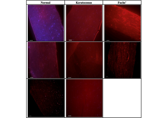
Immunofluorescence analysis of high mobility group box 1 (HMGB-1) expression in human corneal tissue. Representative immunofluorescence (IF) images at 20× magnification of paraffin-embedded human corneal sections from normal, keratoconus, and Fuchs’ corneal dystrophy samples stained with anti-HMGB-1 antibody (red) and counterstained with 4',6-diamidino-2-phenylindole (DAPI) to label nuclei. Normal corneal tissue demonstrates low-level, diffuse HMGB-1 expression with preserved stromal architecture. In contrast, keratoconic corneas exhibit increased and irregular HMGB-1 signal with disrupted stromal organization, while Fuchs’ dystrophy samples show robust HMGB-1 immunoreactivity with widespread distribution throughout the corneal tissue. All images were acquired using identical imaging parameters, including magnification, exposure time, laser intensity, and gain settings, using a Nikon Eclipse Ti2 fluorescence microscope, to allow direct qualitative comparison across conditions.
Normal donor corneas demonstrated low and relatively uniform HMGB-1 fluorescence intensity, with a mean value of 338.6 ± 53.6 a.u. (Figure 2). Signal distribution within this group was narrow, consistent with minimal baseline inflammatory activity and homeostatic cellular turnover in healthy corneal tissue. HMGB-1 localization in normal samples appeared diffuse and low-level, supporting its expected role as a predominantly nuclear protein under non-pathologic conditions.
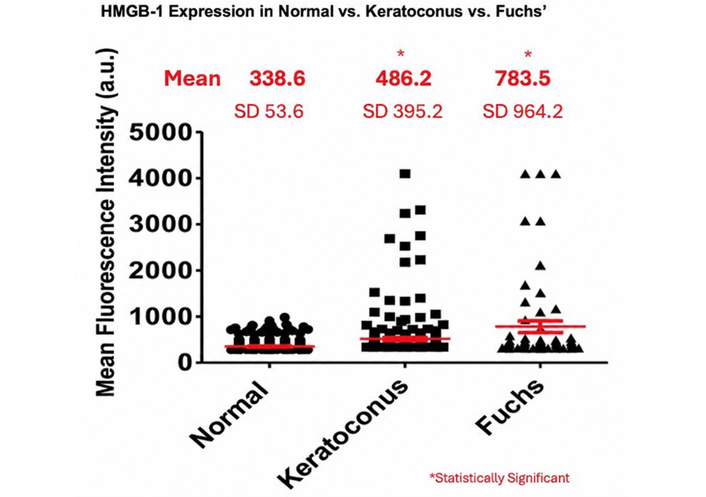
Quantitative analysis of high mobility group box 1 (HMGB-1) immunofluorescence intensity in human corneal tissue. Scatter plot depicting HMGB-1 fluorescence intensity in arbitrary units (a.u.) measured from immunofluorescence images of normal, keratoconus, and Fuchs’ corneal dystrophy corneal samples. Each data point represents an individual region of interest (ROI) from the n = 3 tissues. Red horizontal lines denote group means and the bars represent standard deviation. Mean values were 338.6 a.u. for normal corneas, 486.2 a.u. for keratoconic corneas, and 783.5 a.u. for Fuchs’ endothelial dystrophy corneas. Welch’s one-way ANOVA demonstrated a significant overall group difference, F(2, 80.33) = 10.32, p = 0.00010. The two prespecified comparisons showed higher HMGB-1 intensity in keratoconus versus normal tissue [Welch t(61.17) = 2.87; Holm-adjusted p = 0.0057] and in Fuchs’ dystrophy versus normal tissue [Welch t(59.37) = 3.57; Holm-adjusted p = 0.0014].
In contrast, keratoconic corneas exhibited a significant increase in HMGB-1 expression, with a mean fluorescence intensity of 486.2 ± 395.2 a.u. (Figure 2). In the prespecified comparison with normal corneas, the mean difference was 147.7 a.u. (95% CI, 44.7–250.6), Welch t(61.17) = 2.87, Holm-adjusted p = 0.0057, with a Hedges’ g of 0.52. The broad distribution and high-intensity observations indicate substantial spatial variability in HMGB-1 expression within the keratoconus. This heterogeneity is consistent with the known patchy nature of stromal thinning, focal inflammation, and ECM disruption characteristic of keratoconus. These findings suggest increased cellular stress and inflammatory signaling within keratoconic corneal tissue.
Fuchs’ corneal dystrophy samples demonstrated the highest overall mean HMGB-1 fluorescence intensity 783.5 ± 964.2 a.u. (Figure 2). Examination of the fluorescence intensity distribution revealed pronounced variability within the Fuchs’ group. Three measurements reached the upper recorded intensity value of 4,095 a.u., contributing to the marked variance and right-skewed distribution. In the prespecified comparison with normal corneas, the mean difference was 444.9 a.u. (95% CI, 195.4–694.3), Welch t(59.37) = 3.57, Holm-adjusted p = 0.0014, with a Hedges’ g of 0.65. These findings indicate elevated but highly heterogeneous HMGB-1 immunoreactivity in the Fuchs’ dystrophy corneas. These high-intensity regions corresponded to localized areas of corneal stress, dense guttae formation, and focal inflammatory activation, rather than diffuse, uniform upregulation across the tissue. The presence of these localized HMGB-1 “hot spots” resulted in a substantially increased standard deviation within the Fuchs’ group. This finding highlights the importance of spatial heterogeneity in Fuchs’ corneal dystrophy and suggests that HMGB-1 expression in this condition may be driven by focal cellular injury and stress responses rather than a global increase throughout the cornea.
Welch’s one-way analysis of variance demonstrated a significant overall difference in maximum HMGB-1 fluorescence intensity among the three groups, Welch F(2, 80.33) = 10.32, p = 0.00010. The two prespecified disease-versus-normal comparisons remained statistically significant after Holm adjustment. The observed spatial heterogeneity, broad intensity distributions, and focal regions of high HMGB-1 expression in diseased tissues suggest that HMGB-1 may play an important role in corneal inflammatory signaling, cellular stress responses, and ECM remodeling. These findings support further investigation of HMGB-1 as a potential biomarker of corneal cellular stress and inflammation.
This pilot study demonstrates increased expression of HMGB-1 in corneal tissues from patients with keratoconus and Fuchs’ corneal dystrophy, supporting a role for this protein in corneal inflammation and ECM remodeling. Quantitative immunofluorescence revealed a significant increase in HMGB-1 expression in keratoconic corneas compared with normal corneas, along with a variably elevated and spatially heterogeneous pattern in Fuchs’ corneal dystrophy. Together, these findings suggest that increased HMGB-1 expression may be associated with cellular stress, inflammation, and degenerative changes in corneal tissue.
HMGB-1 could be associated with this inflammatory cascade; however, the present study did not evaluate downstream signaling or establish whether HMGB-1 initiates or amplifies tissue injury. Upon cellular stress or injury, HMGB-1 translocates from the nucleus to the extracellular space, where it engages PRRs such as the RAGE receptor and TLR2 and TLR4 on corneal epithelial cells and keratocytes [19]. Activation of these receptors triggers NF-κB signaling and subsequent upregulation of proinflammatory cytokines including IL-1β, IL-6, and TNF-α [20]. These cytokines have been reported at elevated levels in keratoconic tears and corneal tissue, supporting the biological relevance of HMGB-1-mediated inflammatory signaling [21, 22]. The heterogeneous distribution of HMGB-1 observed in keratoconus aligns with the focal nature of stromal thinning and suggests localized regions of increased cellular stress and ECM degradation [23, 24]. These findings are consistent with prior studies demonstrating increased inflammatory cytokines, oxidative stress markers, and matrix metalloproteinase activity in keratoconic corneas, further supporting a model in which chronic low-grade inflammation contributes to progressive stromal weakening [25]. HMGB-1 may act upstream in this inflammatory cascade, amplifying tissue damage through sustained innate immune activation [26].
Fuchs’ endothelial dystrophy demonstrated the highest mean HMGB-1 fluorescence intensity and a significant ROI-level increase relative to normal corneal tissue. The distribution was markedly heterogeneous, with a broad variance, and three values at the upper recorded intensity of 4,095 a.u. The bimodal distribution of HMGB-1 expression observed in this group likely reflects regional differences in disease severity, with low-intensity regions resembling relatively preserved endothelium and high-intensity regions corresponding to areas of advanced corneal stress and guttae formation. Oxidative stress and mitochondrial dysfunction, both well-established features of Fuchs’ dystrophy, are known triggers of HMGB-1 release and may drive focal inflammatory amplification within affected corneal regions [27, 28]. The significant overall difference in HMGB-1 expression across all groups, as demonstrated by the ANOVA, supports disease-associated expression of HMGB-1 despite biological variability.
Fuchs’ endothelial corneal dystrophy was included as a disease comparison group, rather than as an age- or anatomically matched control. Fuchs’ dystrophy is characterized by endothelial cell loss, oxidative and mitochondrial stress, and degenerative changes involving the corneal endothelium and Descemet’s membrane. These cellular stressors may promote the release or increased expression of HMGB-1. Therefore, inclusion of the Fuchs’ group allowed us to assess whether elevated HMGB-1 expression was specific to keratoconus or was also observed in another corneal disorder associated with cellular injury and tissue degeneration. However, the two diseases affect different corneal compartments: keratoconus primarily involves the epithelium and stroma, whereas Fuchs’ dystrophy predominantly affects the endothelium and Descemet’s membrane. In addition, patients with Fuchs’ dystrophy are generally older than patients with keratoconus, and age-related changes, endothelial cell loss, and cumulative oxidative stress may independently influence HMGB-1 expression. Consequently, the Fuchs’ group should not be interpreted as a directly matched comparator for keratoconus. Rather, its elevated and heterogeneous HMGB-1 expression provides evidence that HMGB-1 may represent a broader marker of corneal cellular stress and injury. The keratoconus-versus-normal comparison therefore remains the primary analysis for evaluating the potential association between HMGB-1 and keratoconus, while the Fuchs’ group provides additional biological context regarding the specificity of HMGB-1 expression.
This study should be interpreted as a pilot study designed to better understand the HMGB-1 expression patterns in the pathogenesis of keratoconus. The small sample size, with three tissue specimens per group, represents a major limitation and restricts the statistical power of the study. Therefore, the findings should be considered exploratory rather than definitive. The small sample size reflects the limited availability of rare archived human corneal tissues, particularly keratoconic corneal specimens suitable for immunofluorescence analysis. Although this restricts statistical power, similarly small sample sizes have been reported in prior keratoconus immunohistochemistry studies, including “Transepithelial riboflavin/ultraviolet. A corneal cross linking in keratoconus: morphologic studies on human corneas.” Therefore, the present findings should be interpreted as preliminary and hypothesis-generating rather than definitive. Additional studies with larger sample sizes are needed to definitively determine whether HMGB-1 is significantly elevated in Keratoconus and Fuchs’ endothelial dystrophy and whether this expression correlates with disease severity or localized endothelial injury.
These findings identify increased HMGB-1 expression in keratoconus and Fuchs’ endothelial dystrophy as a potential marker of cellular stress, inflammation, and ECM remodeling in corneal pathology. The increased concentration in diseased tissues warrants further investigation. The present observational study does not establish that HMGB-1 has an active pathogenic role or that it represents a therapeutic target, further investigation is required.
The biological sample size of three corneas per group substantially limits statistical power and external validity. Therefore, these results should be interpreted as exploratory and hypothesis-generating. Larger studies with greater sample sizes and more complete clinical characterization are needed to confirm the role of HMGB-1 in keratoconus and Fuchs’ endothelial dystrophy.
Since the corneal tissues were obtained as fully anonymized and deidentified archival specimens, patient-level clinical information was not available for analysis. Accordingly, demographic and clinical variables such as age, sex, keratoconus severity, Kmax, thinnest corneal thickness, history of allergy or atopy, eye rubbing, contact lens use, prior corneal cross-linking, corneal scarring, and specific surgical history could not be retrieved. Detailed fixation and storage information beyond the specimens being archived and paraffin-embedded tissues maintained under controlled conditions was also unavailable. These factors may influence HMGB-1 expression and represent an important limitation of the present study. Future studies using prospectively collected specimens with linked clinical data will be needed to evaluate the relationship between HMGB-1 expression, patient characteristics, disease severity, prior treatment, and tissue-processing variables.
Alternative explanations for the observed HMGB-1 staining patterns should also be considered. Increased HMGB-1 expression may not be specific to keratoconus alone and could reflect a broader response to corneal cellular stress, tissue injury, inflammation, or degeneration. Differences in archival tissue preservation, fixation, disease stage, regional sampling, or localized areas of stromal and endothelial damage may also have contributed to variability in fluorescence intensity. Therefore, while the findings support a potential association between HMGB-1 and corneal disease activity, they do not establish a direct causal role for HMGB-1 in keratoconus pathogenesis.
This study demonstrates that HMGB-1 has increased expression in keratoconic tissues measured by immunofluorescence microscopy. HMGB-1 is a DAMP protein whose increased expression may reflect cellular stress and innate inflammatory signaling within the cornea. The observed increase in HMGB-1 expression is consistent with, but does not establish a causal relationship to, the inflammatory and stromal changes associated with keratoconus. Overall, these preliminary results suggest that increased HMGB-1 expression is associated with keratoconus and may serve as a marker of corneal cellular stress or inflammatory activity.
a.u.: arbitrary units
DAMP: damage-associated molecular pattern
DAPI: 4',6-diamidino-2-phenylindole
ECM: extracellular matrix
HMGB-1: high mobility group box 1
IL-1α/β: interleukin-1α/β
IRB: Institutional Review Board
PBS: phosphate-buffered saline
PRR: pattern recognition receptor
RAGE: receptor for advanced glycation end products
ROI: region of interest
TLRs: toll-like receptors
TNF-α: tumor necrosis factor-alpha
The authors would like to thank Dr. Campbell for generously providing access to his laboratory and its resources, and Dr. Yue for providing the corneal tissue samples used in this study.
PC: Conceptualization, Methodology, Formal analysis, Investigation, Data curation, Writing—original draft, Writing—review & editing, Funding acquisition. SKK: Software, Formal analysis, Visualization. CS: Methodology. PB: Resources. CB: Supervision. OI: Conceptualization, Methodology, Investigation, Resources, Writing—review & editing, Supervision, Project administration. All authors read and approved the submitted version.
The authors declare that they have no conflicts of interest.
The study was conducted in accordance with the Declaration of Helsinki and approved by the Institutional Review Board of the Protection of Human Subjects Loyola University Health Sciences Division (protocol code LU#219768 and date of approval 1 July 2025). The study used deidentified, archived human corneal tissue obtained from the Loyola University Medical Center Pathology Archives and was conducted in accordance with applicable institutional and ethical standards.
This study involved retrospective analysis of deidentified archival tissue and no direct interaction with human participants, the requirement for informed consent was waived by the Institutional Review Board.
Consent for publication was not required because the study did not include identifiable patient information, clinical images, or other data that could be used to identify individual participants.
Datasets are available on request: the raw data supporting the conclusions of this manuscript will be made available by the authors, without undue reservation, to any qualified researcher.
The Illinois Society for Prevention of Blindness has funded this project (LU#219768). The funders had no role in study design, data collection and analysis, decision to publish, or preparation of the manuscript.
© The Author(s) 2026.
Open Exploration maintains a neutral stance on jurisdictional claims in published institutional affiliations and maps. All opinions expressed in this article are the personal views of the author(s) and do not represent the stance of the editorial team or the publisher.
Copyright: © The Author(s) 2026. This is an Open Access article licensed under a Creative Commons Attribution 4.0 International License (https://creativecommons.org/licenses/by/4.0/), which permits unrestricted use, sharing, adaptation, distribution and reproduction in any medium or format, for any purpose, even commercially, as long as you give appropriate credit to the original author(s) and the source, provide a link to the Creative Commons license, and indicate if changes were made.



View: 79
Download: 6
Times Cited: 0


