Review


Affiliation:
Wellcome-Wolfson Institute For Experimental Medicine, Queen’s University Belfast, BT9 7BL Belfast, United Kingdom
Email: hnormanbruce01@qub.ac.uk
ORCID: https://orcid.org/0000-0002-0792-5490


 ,
,
Affiliation:
Wellcome-Wolfson Institute For Experimental Medicine, Queen’s University Belfast, BT9 7BL Belfast, United Kingdom
ORCID: https://orcid.org/0009-0004-0458-4311
,
Affiliation:
Wellcome-Wolfson Institute For Experimental Medicine, Queen’s University Belfast, BT9 7BL Belfast, United Kingdom
ORCID: https://orcid.org/0009-0009-6701-5630
,
Affiliation:
Wellcome-Wolfson Institute For Experimental Medicine, Queen’s University Belfast, BT9 7BL Belfast, United Kingdom
ORCID: https://orcid.org/0000-0001-9244-9961
,
Affiliation:
Wellcome-Wolfson Institute For Experimental Medicine, Queen’s University Belfast, BT9 7BL Belfast, United Kingdom
ORCID: https://orcid.org/0000-0001-9452-7716
,
Affiliation:
Wellcome-Wolfson Institute For Experimental Medicine, Queen’s University Belfast, BT9 7BL Belfast, United Kingdom
ORCID: https://orcid.org/0000-0003-2063-7992
Explor Med. 2026;7:1001418 DOI: https://doi.org/10.37349/emed.2026.1001418
Received: January 30, 2026 Accepted: May 28, 2026 Published: July 27, 2026
Academic Editor: Lindsay A. Farrer, Boston University School of Medicine, USA
The article belongs to the special issue Asthma in Children: Origins, Endotypes and Future Perspectives
Acute wheeze is one of the most common paediatric presentations to emergency care. Although blood eosinophils (BEO) are an established biomarker of type 2 inflammation in stable asthma, their role in guiding acute management—particularly systemic corticosteroid use—remains unclear. The aim of this review was to map and synthesise the existing evidence on the measurement and clinical utility of BEO sampled during acute wheeze exacerbations in children. A scoping review was conducted in accordance with Joanna Briggs Institute methodology and Preferred Reporting Items for Systematic Reviews and Meta-Analysis extension for scoping reviews (PRISMA-ScR) guidelines. Medline, Embase, Cochrane Library and Web of Science were searched for studies published in the last 20 years involving children under 16 years presenting acutely with wheeze and undergoing eosinophil testing. Prospective and retrospective studies were included. Data were summarised descriptively according to eosinophil measurement methods, reported values, cut-offs and associations with characteristics and treatment response. Eight heterogeneous studies (n = 855) met inclusion criteria, comprising five observational or retrospective studies and three randomised controlled trials. BEO reporting varied widely, with inconsistent units, cut-offs and poorly described sampling timing. No study utilised point-of-care testing. Across studies, eosinophil dynamics differed by age, wheeze phenotype, disease severity and viral aetiology, preventing meaningful comparison. Evidence supporting eosinophil-guided corticosteroid use was limited. Two studies demonstrated improved outcomes with systemic corticosteroids in children with rhinovirus-associated wheeze and higher BEO, while lower BEO were observed in respiratory syncytial virus (RSV)-associated wheeze. There is a paucity of high-quality data describing BEO during acute wheeze exacerbations in children. Current evidence underpinning eosinophil-guided care is largely derived from stable outpatient cohorts and may not be directly applicable to acute settings. Prospective studies with standardised sampling of BEO during acute presentations are needed to inform precision-based corticosteroid use.
Wheeze is one of the most common paediatric presentations to acute healthcare settings, and asthma remains among the most prevalent chronic conditions in childhood [1, 2]. Wheezing episodes are particularly frequent in preschool-aged children, with up to 50% experiencing at least one episode before the age of six [2–4].
Preschool wheeze (PSW) is a broad umbrella term encompassing a heterogeneous range of clinical phenotypes and underlying pathophysiology. Only a minority of children with PSW will later be diagnosed with asthma, a condition characterised by chronic airway inflammation, mucus hypersecretion, and bronchoconstriction, commonly driven by type 2 (T2) inflammation, which is associated with IgE sensitisation and eosinophilia. In adult airway diseases (asthma and COPD), blood eosinophils (BEO) are a well-established biomarker of T2 inflammation, with higher levels predicting responsiveness to corticosteroids and anti-T2 biologics, while no benefit is seen in non-T2 disease [5, 6]. In PSW, T2 and non-T2 disease coexist and cannot be reliably distinguished clinically. Despite wide heterogeneity, acute management strategies rely on a blanket approach of bronchodilator therapy, supplemental oxygen as required, and a majority of children are treated empirically with high-dose oral corticosteroids (OCSs). The most recent European Respiratory Society (ERS) taskforce for recurrent PSW (2024) demonstrates that personalised management approaches for PSW are a priority [7]. Therefore, using BEO as a biomarker offers a logical, evidence-based approach to identify corticosteroid-responsive T2 inflammation in this population. Two recent trials have demonstrated the feasibility of BEO as a point-of-care (POC) test in paediatric trials in the clinic setting to phenotype pre-school-aged children, where BEO are shown to predict response to inhaled corticosteroid (ICS) [8, 9].
The role of systemic corticosteroids in acute PSW remains particularly controversial, with conflicting RCTs and an individual patient data meta-analysis which identifies a minimal benefit of prednisolone compared to placebo [10–14]. Given the diverse population, it is possible that varying results are due to an unidentified subgroup of children who may benefit from OCS; however, identifying this subgroup remains elusive. The recent meta-analysis evaluating the efficacy of OCSs in PSW included twelve studies [10]. Of these, only two reported BEO, and only one measured BEO during acute exacerbations rather than at baseline [15, 16]. Short course OCS therapy is associated with adverse effects, particularly when associated with cumulative exposure, further highlighting the importance of acute phenotype-driven care in the paediatric population [17–19]. Clinical features alone are unable to identify children most likely to benefit from systemic corticosteroids, and acutely accessible tools to phenotype children should be investigated [7, 10, 11]. Given the established feasibility of BEO as a POC test in the clinic setting, the authors postulate the role of BEO POC as a means to address this clinical need.
There is a substantial body of literature describing normative BEO in children with asthma. To illustrate this, key papers have been selected and summarised in Table 1. There is evidence of the clinical utility of BEO in predicting treatment response, future asthma morbidity, and BEO are proportionate to patient age and gender [20–23]. However, these data are derived from cohorts that were clinically stable prior to trial enrolment. Consequently, the role of BEO as a biomarker to guide corticosteroid use during acute wheeze exacerbations remains poorly defined, particularly in directing corticosteroid therapy.
Summary of large paediatric data sets providing eosinophil values for children.
| Author/year | Population/sampling point | Country/study design | Eosinophil values (× 109/L) | Eosinophil observations |
|---|---|---|---|---|
| Fitzpatrick et al. 2016 [20] | n = 300Age: 12–59 monthsTiming: > 2 weeks after SCS | USAClinical trial | Mean = 0.26 | BEO > 0.3 × 10⁹/L predictor of ICS response |
| Fitzpatrick et al. 2023 [21] | n = 1,074Age: 12–71 monthsTiming: > 4 weeks after SCS | USA3 clinical trials | Median = 0.24 | BEO significantly higher in males and children > 36 monthsBEO a predictor of exacerbation rates |
| Just et al. 2021 [22] | n = 402Age = 1–15 yearsTiming: no current SCS treatment | FranceCohort study | Mean 0.33Q3 0.32 preschool ageQ3 0.60 school-age | BEO vary with age and genderBEO associated with recurrent wheeze |
| Hartl et al. 2020 [23] | n = 1,204Age 6–18 yearsTiming: unknown | AustrianCohort study | Median < 10 yearsFemale 0.18, male 0.24Median 10–18 yearsFemale 0.12, male 0.16 | BEO age and sex dependent with a plateau around puberty |
Q3: upper limit of third quartile of age group. BEO: blood eosinophils, data reported to 2 dp; ICS: inhaled corticosteroid; SCS: systemic corticosteroids.
How are BEO measured and reported in children presenting acutely with wheeze?
What standardised BEO cut-offs are used in the acute setting?
What role may BEO have in predicting acute response to OCS therapy, particularly in the preschool population?
This scoping review followed the framework proposed by both the Joanna Briggs Institute and the Preferred Reporting Items for Systematic Reviews and Meta-Analysis extension for scoping reviews (PRISMA-ScR) [24, 25]. Ethical approval was not required. A search strategy was defined to address the research questions above and run through four databases (Table S1). Eligible papers included any studies published in the last two decades, including patients under 16 years presenting acutely with wheeze who have had BEO testing through any means. Papers examining the role of steroids according to BEO were initially sought, but all acute management strategies were considered, including placebo or standard of care strategies because steroid response was not the sole objective or remit of the review. Both prospective and retrospective studies were included and studies reporting BEO in combined paediatric and adult populations were included assuming paediatric data would be extractable.
Searches were run from 4th to 17th March 2025 across Medline, Embase, Cochrane and Web of Science databases (example: Table S1). There were no language restrictions applied. Reviews, expert consensus documents and guidelines were excluded but interrogated for further unidentified sources. Screening was conducted by three authors (DJM, QSG, HNB) independently using the Rayyan online management programme [26]. Following automatic removal of duplicates and initial screening of titles and abstracts, studies meeting eligibility criteria underwent full-text reviews by at least two authors (with any discrepancies resolved by a third author). All studies were examined for duplicate cohorts, and where a study reported secondary analysis, the original studies were also identified for triangulation or to support data extraction. Data for each study were extracted by a minimum of two authors, using a standardised and agreed data extraction tool relevant to the study objectives. Included studies were described and summarised. Studies were examined by the methods of obtaining BEO, the BEO identified in the population, and then according to the outcome and intervention of each study. The BEO were reported as given in each study, i.e., percentage or absolute values. If used, cut-off values to mark eosinophilia were reported and compared descriptively. Secondly, the studies were examined for the impact of BEO in the acute assessment and management of wheeze. These were summarised and explored into themes identified by the reviewers to address the research questions. As this was a scoping review, the studies were not assessed for the risk of bias.
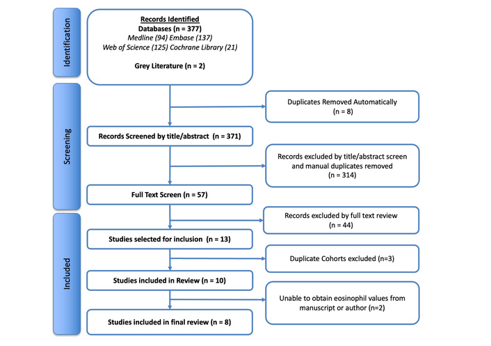
Our search identified 379 studies in the past twenty years pertaining to the role of acute BEO in the management of wheeze exacerbations. Most studies were excluded due to the lack of information on BEO taken in the acute setting and three papers were excluded due to repeat analysis of the same cohort, as per Figure 1. Two studies did not report numerical values for the BEO taken at baseline (data graphically presented or reported after steroid therapy administered) and the required data was not available in supplementary tables or via email contact with the corresponding author.
A summary of the eight selected studies is included in Table 2. Five of the eight studies were small observational or retrospective data analyses, and three were randomised controlled trials. Sample size ranged from 40 to 234 children, with mean ages of 13.2 months to 11.2 years and with a male dominance. In most studies, reporting of BEO was a secondary outcome or simply a means to characterise the population. Populations varied widely from different geographical settings, nature of acute wheeze presentations, wheeze/asthma history, and presence of atopy (which was inconsistently defined).
Summary of the 8 included studies in the review.
| Reference | Holden et al. [27] | Kocak et al. [28] | Elkharwili et al. [29] | Li et al. [30] | Gileles-Hillel et al. [31] | Kim et al. [32] | Jartti et al. [16] | Jartti et al. [15] | ||||||||
|---|---|---|---|---|---|---|---|---|---|---|---|---|---|---|---|---|
| Year | 2017 | 2006 | 2020 | 2024 | 2021 | 2022 | 2006 | 2015 | ||||||||
| Country | UK | Turkey | Egypt | China | Israel | South Korea | Finland | Finland | ||||||||
| Total n | 85 | 40 | 60 | 202 | 234 | 82 | 78 | 74 | ||||||||
| Setting | ED | Ward/OPD | ED | Hospital | Paediatric ward | ED | ED/ward | |||||||||
| Study intervention/design | Observational (patients received OCS as per local protocol) | Observational [patients received PRED 1–2 mg/kg per day for 5 days (maximum, 40 mg/day)] | DBRCT [DEX (0.3 mg/kg) OR multiple doses (0.6 mg/kg) for 2 days or PRED 5-day course (1.5 mg/kg)] | Observational (aerosolizedhigh-dose glucocorticoids given in exacerbation) | Retrospective comparison of: DEX 0.3–0.6 mg/kg/day, 1–5 days or bethamethasone 0.2 mg/kg/day, 3–5 days | Observational (all patients received SCS as part of local protocol) | DBRCT (oral PRED 2 mg/kg per day in three divided doses for 3 days or placebo) | |||||||||
| Study objective | Investigate if there is a difference in BEO during the acute and stable phase of children with preschool wheeze | Evaluate the immune biomarkers and the effects of glucocorticoid treatment on acute asthma exacerbations compared to healthy children | Evaluate the effectiveness of different doses of DEX vs. PRED in managing asthma exacerbations in children | Investigate the role of 14-3-3β in acute asthma exacerbations and evaluate risk factors contributing to asthma exacerbation | Compare the effectiveness of betamethasone versus DEX in managing acute wheezing in preschool children requiring hospital admission | Evaluate the association of EDN with lung function and the prognosis of children hospitalised for severe asthma exacerbation | Evaluate the short- and long-term effects of PRED when given during the first acute and moderate-to-severe rhinovirus-induced wheezing episode in young children | |||||||||
| Acute exacerbation | Exacerbation (> 24 h) of doctor diagnosed wheeze | Acute asthma exacerbation (severity as per NAEPP) | Exacerbation defined according to National Institutes of Health, which requires use of SCS | Asthma exacerbation requiring a change in treatment medication, ED visit or SCS | Acute wheezing requiring hospitalisation | Severe asthma exacerbation | Hospitalised children with acute wheeze caused by respiratory virus. Exacerbation determined by respiratory symptoms score (RSS) | |||||||||
| Severity of exacerbations | Severity not reported | 16% mild64% mod20% severe | Baseline PRAM scores not reported | 61% mild-mod39% severe | 100% hospitalised due to O2 requirement or failure of response to treatment | 100% severe | Mean RSS = 6.2/12 (moderate) | 82% admitted | ||||||||
| Population asthma or wheeze history | At least 1 previous parent-reported wheeze | Asthma as per ATS criteria | Previous history of asthma exacerbation | Asthma diagnosed according to GINA guidelines | Not specified: 41% Previous diagnosis of asthma; 17% using ICS | Severe asthma exacerbation requiring admission and 3 days SCS | 1st or 2nd episode of wheezing | Absence of prior episodes of wheeze | ||||||||
| Subgroups of total population | Acuten = 68 | Controln = 17 | Acuten = 25 | Controln = 15 | DEXGroup I (0.3 mg/kg) n = 20Group 2 (0.6 mg/kg) n = 20 | PREDn = 20 | Acute exacerbationn = 101 | Stablen = 101 | Controln = 65 | Bethamethasonen = 145 | DEXn = 89 | Diagnosis of asthma including evidence of BDR or bronchial hyperresponsiveness in the last 1 y. | Rhinovirusn = 40 | RSVn = 38 | PREDn = 34 | Placebon = 40 |
| Age mean | N/A | N/A | 9.6 years | 8.8 years | G1 = 5.93 ± 2.37 yearsG2 = 6.52 ± 2.64 years | 6.15 ± 2.75 years | N/A | N/A | N/A | N/A | N/A | 11.2 ± 3.6 years | 1.38 ± 0.62 years | 0.86 ± 0.59 years | 13.2 ± 6.9 months | 12.2 ± 5.1 months |
| Age median | 31.2 m | 39.5 m | N/A | N/A | N/A | N/A | 6.8 years | 6.9 years | 7.2 years | 2.5 years | N/A | N/A | N/A | N/A | N/A | |
| Gender Male (%) | 62 | 82 | 52 | 53 | G1 = 40G2 = 50 | 55 | 63 | 60 | 60 | 59 | 67 | 70 | 63 | 47 | 79 | 75 |
| Atopy definition | Parental report of eczema or allergic rhinitis diagnosis | Elevated eosinophil count, IgE and positive RAST | N/A | Serum specific IgE concentration | Through personal history | Presence of a total serum IgE value > 100 IU/mL or sensitization to common allergens | Positive IgE against selected allergens | Positive IgE against selected allergens | ||||||||
| Atopy history (%) | 49 | 29 | 64 | N/A | N/A | N/A | 45 | 37 | N/A | 18 | 28 | 43 | 44 | 8 | 29 | 31 |
m: months in the age category. ATS: American Thoracic Society; BDR: bronchodilator response; BEO: blood eosinophils; DBRCT: double blind randomised controlled trial; DEX: dexamethasone; ED: emergency department; EDN: eosinophil-derived neurotoxin; GINA: Global Initiative for Asthma; ICS: inhaled corticosteroid; N/A: not available; NAEPP: National Asthma Education and Preventing Program; OCS: oral corticosteroid; OPD: Outpatient department; PRAM: pediatric respiratory assessment measure; PRED: prednisolone; RSV: respiratory syncytial virus; SCS: systemic corticosteroids.
The BEO data extracted from the eight studies is summarised in Table 3. Six studies reported BEO counts, but there was inconsistency requiring translation of some values and limiting reliable comparison. Two studies reported BEO as percentages but did not provide comparative white cell counts. Four studies analysed BEO data according to cut-offs to define eosinophilia. Two studies used 0.3 × 10⁹/L whilst the VINKU studies analysed the cut-offs of 0.2 and 0.4 × 10⁹/L which were used to predict risk of future wheeze exacerbations when prednisolone was used to manage rhinovirus (RV) induced wheeze [15, 16]. Holden et al. [27] found the standard 0.3 × 10⁹/L to be a promising cut-off to predict risk of exacerbation in the follow up period but only according to stable BEO post recovery, not according to acute BEO.
Summary of eosinophil values reported during acute wheeze exacerbations across 8 studies.
| Reference | Holden et al. [27] | Kocak et al. [28] | Elkharwili et al. [29] | Li et al. [30] | Gileles-Hillel et al. [31] | Kim et al. [32] | Jartti et al. [16] | Jartti et al. [15] | |||||||||
|---|---|---|---|---|---|---|---|---|---|---|---|---|---|---|---|---|---|
| Population sub-groups | Acute | Control | Acute | Control | G1 | G2 | G3 | Acute | Stable | Control | Dexamethasone | Betamethosone | N/A | RV | RSV | Pred | Placebo |
| Eosinophil count unit/metric/values | × 109/L**Median (range) | × 109/L**Mean ± SD | Eosinophil %*Mean ± SD | Eosinophil %*Median (p25/p75) | × 109/L**Median (IQR) | × 109/L**Mean ± SD | × 109/L**Mean ± SD | ||||||||||
| 0.1 (0.00–2.41)Stable(n = 20)0.43 (0.12–1.5) | 0.17 (0.00–0.83) | Day 1: 0.61 ± 0.43Day 5: 0.16 ± 0.09 | 0.12 ± 0.06 | 8.80 ± 4.99 | 8.25 ± 4.47 | 8.15 ± 4.63 | 3.8 (2.65–4.75) | 2.7 (1.8–4.05) | 2.2 (1.30–3.85) | 0.1 (0.00–0.3) | 0.04 (0.00–0.2) | 0.34 ± 0.34 | 0.44 ± 0.29 | 0.09 ± 0.19 | 0.51 ± 0.45 | 0.51 ± 0.38 | |
| Eosinophil cut-off | 0.3 × 10⁹/L** | Not used/reported | Not used/reported | Not used/reported | Not used/reported | 0.3 × 10⁹/L** | 0.2 × 10⁹/L** | 0.4 × 10⁹/L** | |||||||||
| The percentage of samples above the cut-off value (%) | 71 | 0 | 32 | 52 (n = 67) | 50 | 57 | |||||||||||
| Sample timing | ED Department with < 24 hours of SCS therapyStable: > 4weeks after acute | On admission | On admission | Day 1 (admission) + 5 days post steroids | Admission (prior to treatment) | In clinic | On arrival | Prior to treatment | Prior to randomisation on ward | Prior to randomisation | |||||||
Results reported to 2 dp. *: Studies with eosinophil percentage reported were also identified to have a discrepancy in reporting units between tables within their publications; **: units reported in different units but converted to cell counts (× 109/L) for consistency. N/A: not available; RSV: respiratory syncytial virus; RV: rhinovirus; SCS: systemic corticosteroids.
Five studies offered a comparison of BEO in acute and stable scenarios, albeit the nature of comparison varied. Holden et al. [27] also report multiple cell line absolute values (eosinophils, neutrophils and leukocytes) demonstrating the potential value or limitation of using eosinophil percentages in this setting. In 20 paired samples, BEO were reduced in the exacerbation and then recovered after 4 weeks from enrolment, in contrast to the neutrophils, which were dominant in the exacerbation and reduced after recovery [27]. All BEO were processed via laboratory methods, i.e., none used POC methodology. No study reported their laboratory’s reference values for interpretation. Timing was poorly reported in studies.
Two studies, by the same research group, examined the relationship between BEO and the underlying viral aetiology of the wheeze episode, and the resultant impact of steroids. VINKU1 demonstrated that when given prednisolone, children with RV-induced wheezing (p = 0.03) and BEO ≥ 0.2 × 10⁹/L (p = 0.005) had fewer relapses in the next two months when compared to children with respiratory syncytial virus (RSV)-induced wheezing and BEO < 0.2 × 10⁹/L, respectively [16]. VINKU2 went on to show that when given prednisolone, children with a RV load of greater than 7,000 copies/mL and BEO ≥ 0.4 × 10⁹/L had fewer chances of a new wheezing episode short term (2-month follow-up) (p = 0.01) and long term (12-month follow-up) (p = 0.04) compared to placebo [15]. Together, the VINKU studies were the only included studies to report viral testing and compare BEO in wheeze according to different pathogens, albeit their age group was the lowest of all the studies, including infants as well as preschool aged children.
Reporting of baseline variables was varied across the studies. Atopy was chosen as a comparison in Table 2. Whilst inconsistently measured, the majority of studies used IgE tests for selected common aeroallergens. Between a third and a half of each population were regarded atopic, however, this was more likely to have been evaluated biochemically in older children and through personal history in younger children. No study meaningfully compared clinical or biological definitions of atopy and BEO. Due to different study design, interventions and outcome assessments, there was no direct method of comparing steroid response according to BEO. Therefore, this review was unable to address the research question regarding the role of BEO in predicting steroid response in an acute exacerbation.
This review aimed to evaluate the role of BEO in the acute assessment and management of children with wheeze and asthma, specifically its ability to predict response to OCSs, and to define BEO ranges and thresholds in the acute setting. However, limited data were identified to address these questions. Only eight heterogeneous studies, comprising 855 participants, were included, this is in stark contrast to the data available from stable children, highlighting a significant gap in the current literature.
POC BEO testing is acceptable in paediatric outpatient settings and may support phenotype-driven care, particularly in preschool children. UK studies have shown that POC BEO > 0.3 × 10⁹/L or > 4% during stable disease are associated with increased risk of future wheeze attacks [8, 9]. However, these POC measurements were taken during clinic visits rather than during acute presentations, and one study demonstrated that BEO had poor stability between visits, limiting the reliability of single measurements [33]. All eight studies in this review recruited participants from emergency departments, where blood sampling is frequently performed out of hours. This is clinically relevant, as BEO are subject to circadian variation, and samples taken overnight or outside clinic hours may differ from daytime measurements [34]. The small sample sizes and limited reporting prevented meaningful evaluation of variability related to sampling timing, setting, or method.
BEO may vary between acute and stable disease, particularly when reported as a percentage rather than an absolute count. During acute wheeze exacerbations, inflammation, physiological stress, and shifts in other leukocyte cell counts, may reduce the relative eosinophil proportion, potentially limiting sensitivity in the emergency setting. In the UK preschool aged study, BEO were reduced during acute presentation and normalised in stable disease, with the converse observed for neutrophils; however, interpretation was limited by a small sample size (n = 20) and absence of pathogen testing [27]. Similarly, the South Korean study of severe asthma exacerbations found no significant difference in BEO between admission and discharge despite systemic steroid use (p = 0.267, n = 82) [32]. The UK study excluded patients “receiving more than 24 hours” of systemic steroid before recruitment, introducing potential bias to the BEO data as eosinophils are highly responsive to corticosteroids, as seen in adults with T2 driven asthma [35]. However, Holden et al. [27] reported that there were no significant difference in BEO between children with acute wheeze who had or had not received SCS before blood sampling, albeit this data was not shown.
In contrast, three included studies, in predominantly school-aged populations, reported increased BEO during acute asthma exacerbations. Both Li et al. [30] and Kocak et al. [28] demonstrated significantly higher BEO in children with acute exacerbations compared with those with stable asthma or on discharge and healthy controls. Interestingly, these two studies had exacerbation groups with higher proportions of atopy (64, 45%, respectively), however, the UK study also reported high atopy in the exacerbation group (49%), albeit the latter was a parent reported outcome rather than a biological one. Similarly, Elkharwili et al. [29] observed significant reductions in BEO following systemic corticosteroid therapy, as expected from adult studies [35]. Together, these data suggest that eosinophil dynamics in acute disease may differ by age, disease phenotype, and severity, as well as by treatment received prior to eosinophil measurement.
The studies included in this review spanned a wide age range from infancy to adolescence and approximately two thirds were male. Whilst this reflects the gender distribution of pre-pubescent asthma, it further limits the generalisability of the findings. Population data demonstrate that in healthy children, BEO are age and sex dependent, with higher levels observed in prepubescent children and males (Table 1) [21–23]. The eight studies vary in reporting average BEOs above and below those reported in Table 1. Distinct eosinophil distributions between preschool and school-aged children with wheeze or asthma suggest that these groups represent not only different clinical syndromes but also differing eosinophil profiles. Consequently, data pooled in this review are inadequate to draw meaningful conclusions and highlight the importance of rigorously prospectively designed trials where blood is sampled during exacerbation prior to any OCS.
Jartti et al. [15, 16] demonstrate in the VINKU studies that children with RV-induced wheeze and elevated BEO were more responsive to systemic corticosteroids than those with RSV-induced wheeze who had a reduced eosinophil count. Whilst these two studies comprehensively evaluate the relationship between eosinophils, virus type, viral load and steroid response in a young cohort, a major limitation is the small sample sizes and the notable difference in age group between the RSV and RV groups (p < 0.001) [15, 16]. Kato et al. [36] (a small study originally excluded from review as numerical values were not reported in the manuscript) also demonstrated a significantly higher level of eosinophils in children with RV compared to RSV infection (p = 0.05, n = 33). These studies provide an insight into the complex interplay between host, pathogen and treatment, but larger prospectively driven trials are required. Our review was unable to demonstrate a relationship between eosinophil counts and clinical or biological features of atopy.
BEO may be regarded as a promising biomarker to deliver precision-based, phenotypic-driven care for children with asthma, however, this review highlights a marked absence of high-quality eosinophil data obtained during acute exacerbations. The evidence base that underpins eosinophil-guided care is derived from stable outpatient cohorts, yet acute presentations, where treatment decisions regarding systemic corticosteroids are made, remain poorly characterised. Across the eight small and heterogeneous studies identified in this review, eosinophil dynamics appear to vary by age, wheeze phenotype, disease severity, and viral aetiology. Therefore, the acute paediatrician has to extrapolate data from stable disease, which risks misclassification and inappropriate treatment in this heterogeneous population. There is a need for prospectively collected observational studies that systematically measure BEO during acute wheeze and asthma exacerbations. Such studies should be large enough to represent the full spectrum of the paediatric population, capture all clinically relevant phenotypes, and incorporate pathogen-specific data alongside treatment exposure and response. Given the dearth of available evidence, BEO are not yet feasible for the implementation of acute personalised medicine in a clinical trial. Closing the gap between BEO data in acute exacerbations and stable asthma is an important step to establishing eosinophils as a reliable biomarker to guide corticosteroid use in emergency care.
BEO: blood eosinophils
OCSs: oral corticosteroids
POC: point-of-care
PSW: preschool wheeze
RSV: respiratory syncytial virus
RV: rhinovirus
T2: type 2
The supplementary table for this article is available at: https://www.explorationpub.com/uploads/Article/file/1001418_sup_1.pdf.
HNB: Conceptualization, Investigation, Formal analysis, Writing—original draft, Supervision, Funding acquisition. DJM and QSG: Investigation, Writing—original draft, Formal analysis. HG, TW, and PJM: Writing—review & editing, Supervision. All authors read and approved the submitted version.
PJM reports speaker’s honoraria, participation in steering committees, and support to attend educational events from AstraZeneca and GlaxoSmithKline. The other authors declare no conflicts of interest.
Not applicable.
Not applicable.
Not applicable.
No new data were generated in this review. All eight studies included in the analysis are clearly cited to trace the data presented. All relevant data are contained within the manuscript.
HNB is funded as a doctoral fellow through the Health and Social Care Northern Ireland’s Research and Development division [EAT/5732/22]. The funders had no role in study design, data collection and analysis, decision to publish, or preparation of the manuscript.
© The Author(s) 2026.
Open Exploration maintains a neutral stance on jurisdictional claims in published institutional affiliations and maps. All opinions expressed in this article are the personal views of the author(s) and do not represent the stance of the editorial team or the publisher.
Copyright: © The Author(s) 2026. This is an Open Access article licensed under a Creative Commons Attribution 4.0 International License (https://creativecommons.org/licenses/by/4.0/), which permits unrestricted use, sharing, adaptation, distribution and reproduction in any medium or format, for any purpose, even commercially, as long as you give appropriate credit to the original author(s) and the source, provide a link to the Creative Commons license, and indicate if changes were made.



View: 933
Download: 16
Times Cited: 0
Michele Piazza ... Giorgio Piacentini
Michele Piazza ... Diego Peroni