 Review


Affiliation:
1Department of Life, Health and Environmental Sciences, University of L’Aquila, 67100 L’Aquila, Italy
2Department of Innovative Technologies in Medicine & Dentistry, Dental School, ‘G. D’Annunzio’ University of Chieti-Pescara, 66100 Chieti, Italy
ORCID: https://orcid.org/0000-0002-0080-1743

 ,
,
Affiliation:
1Department of Life, Health and Environmental Sciences, University of L’Aquila, 67100 L’Aquila, Italy
ORCID: https://orcid.org/0000-0003-3731-5463
,
Affiliation:
2Department of Innovative Technologies in Medicine & Dentistry, Dental School, ‘G. D’Annunzio’ University of Chieti-Pescara, 66100 Chieti, Italy
ORCID: https://orcid.org/0009-0003-9601-2822
,
Affiliation:
1Department of Life, Health and Environmental Sciences, University of L’Aquila, 67100 L’Aquila, Italy
ORCID: https://orcid.org/0000-0003-1532-932X
,
Affiliation:
1Department of Life, Health and Environmental Sciences, University of L’Aquila, 67100 L’Aquila, Italy
Affiliation:
2Department of Innovative Technologies in Medicine & Dentistry, Dental School, ‘G. D’Annunzio’ University of Chieti-Pescara, 66100 Chieti, Italy
ORCID: https://orcid.org/0000-0002-7501-0794
,
Affiliation:
1Department of Life, Health and Environmental Sciences, University of L’Aquila, 67100 L’Aquila, Italy
Email: sara.bernardi@univaq.it
ORCID: https://orcid.org/0000-0001-6130-8533

Explor Med. 2026;7:1001405 DOI: https://doi.org/10.37349/emed.2026.1001405
Received: August 02, 2025 Accepted: March 17, 2026 Published: May 25, 2026
Academic Editor: Gaetano Isola, University of Catania, Italy
The recent improvements of the ultrasounds technology and the probes have led the dental community to start to apply this technology at macrovascular and microvascular level. This scoping review aims to investigate the diagnostic and research applications of ultrasonography in periodontics. A comprehensive literature search was conducted in PubMed, Scopus, and Web of Science, to identify relevant studies in periodontology: The search strategy included the following terms: “ultrasonography”, “echography”, “ultrasound”, “dentistry”. The review was conducted in accordance with the PRISMA-ScR guidelines. In addition, a manual search was conducted through the following journals from the last 10 years: Journal of Clinical Periodontology, the Journal of Dental Research and Oral Surgery, and Oral Medicine, Oral Pathology and Oral Radiology. Ten studies were included, covering different applications of ultrasonography in periodontology. Ultrasonography was employed in peri-implant and periodontal diagnosis, assessment of soft tissue thickness and vascularization, palatal wound and bone healing. This review highlights the effectiveness of ultrasonography in diagnosis and surgical evaluation. Further research, standardized protocols, and randomized clinical trials are needed, expanding the investigation to more fields relevant to the maxillofacial district.
Over the last three decades, dentistry has experienced significant progress across all its branches. These developments have highlighted the necessity for highly accurate diagnostic tools, particularly advanced imaging techniques [1]. Alongside clinical examinations, two-dimensional (2D) imaging techniques such as periapical radiography (PR) and panoramic radiography (PANO), as well as three-dimensional (3D) cone beam computed tomography (CBCT), are imaging exams commonly employed. CBCT provides enhanced information compared to standard 2D dental radiographs, particularly concerning the bucco-lingual dimension [2]. The diagnostic process also relies on spiral CT, magnetic resonance imaging (MRI), and ultrasound (US) [1]. Due to the exposure of ionizing radiation, both for patients and healthcare providers, and the consequent potential biological damage, in recent years, awareness regarding the frequency of radiographs has increased [3, 4]. Advancements in technology have recently enabled significant reductions in radiation doses from the latest-generation devices while providing increasingly high-resolution diagnostic imaging and broadening applications across all areas of dentistry [5, 6]. Furthermore, the development of ionizing-radiation-free diagnostic exams in dentistry, aimed at overcoming the limitations of traditional methods, has driven scientific research to achieve promising results for the future. MRI and ultrasound imaging (UI) represent significant advancements in this field, as highlighted by extensive evidence supporting their applications across various branches of dentistry [7].
UI is commonly used in medical practice primarily because of its low cost, low risk for the patient, and capability to deliver real-time images. Frequencies ranging from 2 to 20 MHz are typically operated [8]. Ultrasonography operates by exploiting the acoustic properties of biological tissues to generate cross-sectional images. A transducer emits high-frequency sound waves that are reflected at the interfaces between structures of differing densities, producing echoes [9, 10]. The magnitude of these echoes is directly proportional to the difference in acoustic impedance between adjacent tissues [11]. High-intensity echoes (hyperechoic, appearing white) are typically associated with structures such as bone or gas, whereas low-intensity echoes (hypoechoic, appearing black) are characteristic of fluids. Intermediate intensities are displayed as various shades of gray [11]. Because materials like bone and air impede sound wave transmission, they generate acoustic shadows, preventing the visualization of tissues located beyond these interfaces [12]. This phenomenon represents a key limitation of ultrasonography, particularly when examining tissues with low water content. However, advancements in image-processing software have significantly mitigated these challenges, resulting in markedly improved diagnostic imaging capabilities [13].
The implementation of the Doppler effect has led to the development of techniques for analysing blood flow, starting with continuous and pulsed Doppler methods. This was later followed by Color Doppler, which incorporates directional data and blood velocity into ultrasound images [14]. Furthermore, ultrasound technology has advanced to include 3D and four-dimensional (4D) imaging, delivering real-time volumetric representations of tissues [15]. With the recent technological advancements, ultrasound has become portable. More recently, the introduction of pocket-sized ultrasound devices has facilitated the use of handheld ultrasounds, significantly enhancing the ease of transport to perform examinations [16]. The primary benefit is their portability due to their smaller and lighter design, which facilitates transport to remote or hard-to-reach places [17]. High-intensity focused ultrasound (HIFU) has gained popularity among aesthetic medicine practitioners in recent years. It was initially utilized in oncology for treating cancers of many organs, including the liver (such as hepatocellular carcinoma), prostate, pancreas, kidneys, breasts, thyroid, and bones [18]. Currently, it is also employed in procedures focused on facial and neck rejuvenation as well as the reduction of subcutaneous fat [18]. Moreover, current research is focusing on AI-empowered ultrasonography, although the implementation of AI in medical ultrasonography encounters various challenges [19].
Ultrasonography has been less commonly used in dentistry due to the absence of suitable transducers capable of navigating the confined oral space and the challenges associated with achieving effective coupling between the transducer and target tissues [20]. The intraoral transducer is designed explicitly for intraoral use, featuring an 18-MHz center frequency and a spatial resolution of up to 64 µm [21]. Ultrasound has traditionally been used in dentistry to diagnose major salivary gland conditions, particularly sialolithiasis [22]. In recent years, increasing scientific evidence has highlighted the role of ultrasound in the study of periapical lesions, monitoring their healing, and differentiating between various diagnoses [23]. Additionally, ultrasound examinations have proven valuable for assessing the temporomandibular joint (TMJ), including evaluating the health of the articular disc and detecting possible displacements [24]. Beyond joint evaluation, the ultrasound allows for a detailed examination of soft tissues, including the oral mucosa. Tzoumpas et al. [25] emphasized the importance of measuring the thickness of the maxillary attached gingiva, particularly when planning connective tissue grafts (CTGs) from the palate, making this application highly useful in periodontal surgery planning. Similar evaluations can be applied to implant surgery and peri-implant health, with the benefit of avoiding ionizing radiation and its associated biological risks [26]. Bohner et al. [27] demonstrated no statistical difference in the measurements of buccal bone thickness around dental implants when comparing ultrasound and optical microscopy. Although this study had ex vivo limitations, it provided a strong starting point for studying peri-implant hard tissues. To validate these findings, the same research group simulated peri-implant bone defects in porcine bone and compared measurements from CBCT and ultrasound. The results showed no statistically significant differences in defect width and height, highlighting the potential of ultrasound for diagnosing and monitoring peri-implant bone defects [27]. This technology has also been found useful for evaluating intraosseous lesions, supporting their diagnosis, differential diagnosis, and follow-up after treatment, all without the risks associated with ionizing radiation [28].
Another important application of the ultrasound is in the study of blood vessels. In dentistry, it is particularly valuable for planning oral surgeries near large blood vessels or assessing vascular lesions to determine their nature and blood flow [29].
The aim of this scoping review is to map and synthesize the clinical applications of ultrasonography in periodontology, focusing on evidence derived from human clinical studies evaluating its diagnostic and research use in periodontal and peri-implant contexts. The following research questions were developed to meet the aims of this scoping review:
“What are the main clinical fields of application of ultrasonography in periodontology based on available human studies?” This question aims to explore the various clinical and diagnostic areas within dentistry where ultrasonography has been utilized, identifying its primary indications.
“In which clinical contexts has ultrasonography been applied in periodontics?” This question focuses on the specific uses of ultrasonography in the periodontal field, particularly in relation to diagnosis, soft and hard tissues assessment, and postoperative evaluation.
“What are the reported advantages and limitations of using ultrasonography in periodontal practice?” This question examines the reported benefits, such as the absence of ionizing radiation and real-time imaging, as well as the technical and clinical limitations associated with its use.
“What types of ultrasonographic techniques and technologies have been employed in periodontal research?” This question focuses on identifying the different ultrasound modalities (e.g., Doppler, high-frequency ultrasound, shear wave elastography) and their specific applications in the included studies.
The proposed scoping review will follow the JBI methodology for scoping reviews [30]. This project is registered as DOI: https://doi.org/10.17605/OSF.IO/7MJWA on OSF.
This scoping review screened three different databases, thus Pubmed, Scopus, and Web of Science, to identify articles on the topic, published from 2015 to 2025. Given the focus on periodontology, a dedicated search strategy was developed. The authors adhered to the PRISMA Extension for Scoping Reviews (PRISMA-ScR) [31] to conduct a structured electronic search and select studies concerning the application of ultrasonography in periodontology (Figure 1).
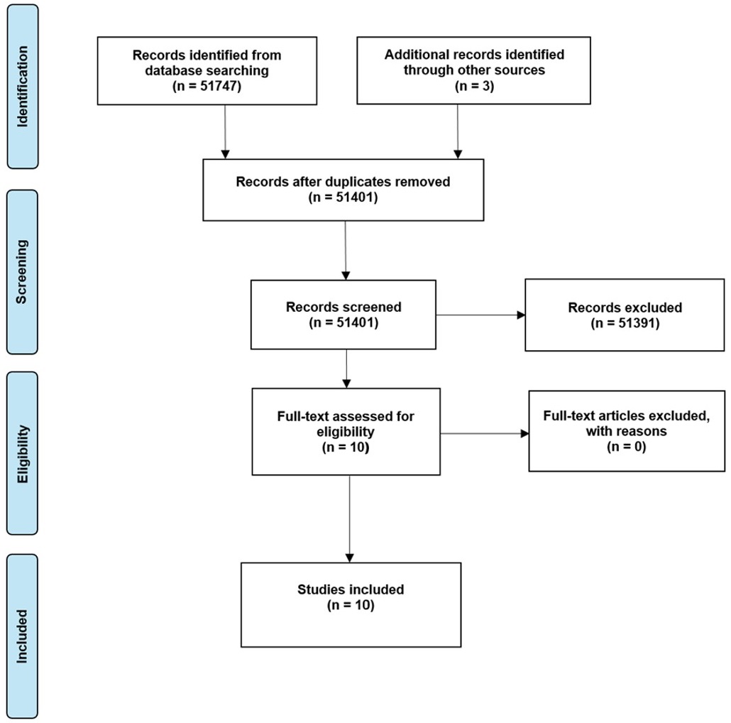
Preferred reporting items for Systematic Reviews and Meta-Analyses (PRISMA) flowchart illustrating the experimental study search and selection process for the application of ultrasonography in periodontology.
Due to the emerging nature of ultrasonography applications in dentistry and the heterogeneity of terminology used across medical and dental literature, manual searching of key journals and reference lists was undertaken to complement the electronic search and ensure comprehensive identification of clinically relevant studies from the last ten years (2015–2025) among the following journals: the Journal of Clinical Periodontology, the Journal of Dental Research and Oral Surgery, and Oral Medicine, Oral Pathology, and Oral Radiology.
An electronic search was conducted using the following search terms, which were adapted for each database. In PubMed, the string used was: ultrasonography AND echography AND ultrasound AND dentistry. In Scopus, the search was performed with: TITLE-ABS-KEY (“ultrasonography” AND “echography” AND “ultrasound” AND “dentistry”). In Web of Science, the strategy applied was: TS = (“ultrasonography” AND “echography” AND “ultrasound” AND “dentistry”).
The PCC (Population, Concept, Context) method was used to develop this scoping review, defining the following parameters:
P: population of living humans
C: applications of ultrasonography
C: periodontology
Studies were considered eligible if they met the following criteria: human clinical studies investigating the application of ultrasonography in periodontology for diagnostic or research purposes, including randomized controlled trials, controlled clinical trials, and clinical diagnostic studies; publications from the last 10 years (2015–2025); in vivo investigations; and articles available in the English language.
Exclusion criteria comprised: in vitro studies, animal studies, abstracts, book chapters, conference proceedings, non-peer-reviewed articles, narrative reviews, systematic reviews, and meta-analyses, as well as studies not available in English.
Two independent authors (D.G. and P.B.) conducted the primary literature research. The data extraction process included the evaluation of the selected titles, excluding studies that did not meet the predefined eligibility and inclusion criteria. Afterwards, the eligible articles were assessed in their full text for final inclusion. In cases of disagreement between the selectors, due to individual evaluations, the final consensus was achieved through re-evaluation and discussion. No studies were excluded following full-text assessment. The selected studies were finally reviewed for qualitative synthesis. To assess inter-rater reliability during the study selection process, Cohen’s Kappa coefficient (κ) was calculated.
Before proceeding with the full data extraction, the process was pilot tested on a small subset of five studies by two independent reviewers. This preliminary step was undertaken to ensure clarity of the eligibility criteria and consistency in the interpretation of the data extraction form. Discrepancies were discussed and resolved, and the refined version of the form was then applied to all included studies.
The first PRISMA diagram schematically shows the selection process for the application of ultrasonography in periodontology (Figure 1). Through digital research, carried out on three different databases (PubMed, Scopus, and Web of Science), 51,747 studies were identified, and 3 records were selected through other sources. 51,401 records were screened after duplicates removal and underwent preliminary analysis based on the evaluation of title and abstract. Inclusion and exclusion criteria were applied, and a total of 51,391 studies were excluded from this first screening survey, reducing the total of studies subjected to the final screening to 10. Finally, 10 studies were included, as no records were excluded based on the full-text evaluation. Inter-rater reliability was assessed using Cohen’s Kappa coefficient, which demonstrated an almost perfect level of agreement between the reviewers, both during the initial screening phase (κ = 0.93) and in the full-text eligibility evaluation (κ = 0.87).
The study of Galarraga-Vinueza et al. [32] included 60 patients with 60 implants evaluated with HFUS, 30 diagnosed as healthy, and 30 affected by peri-implantitis. The aim was to apply HFUS to characterize peri-implant sites as healthy or diseased and to assess the presence of ultrasonographic markers. Characterizing peri-implant tissues using HFUS echo intensity (EI) reveals a notable distinction between healthy and diseased areas. Both HFUS EI and the presence or absence of a hypoechoic supracrestal area (HSA) could serve as effective ultrasonographic diagnostic markers for differentiating the health status of peri-implant tissues [32]. In the study of Majzoub et al. [33], 13 periodontists with different levels of ultrasound experience were asked to measure 3 periodontal diagnostic parameters (PDPs) on 37 human periodontal ultrasound scans taken at the midfacial site of non-molar maxillary teeth. The results suggest that novice learner periodontists can achieve good agreement in measuring ultrasound-derived PDPs, such as soft tissue height, soft tissue thickness, and crestal bone thickness, after just a few weeks of training. Raters recognized the diagnostic value of ultrasound and found it relatively easy to learn; however, there is a necessity to evaluate the cost-benefit ratio [33].
In the Barootchi et al. [34] study, 42 posterior implants in 21 patients with nonadjacent dental implants, diagnosed as healthy, peri-implant mucositis, or peri-implantitis, were scanned and analysed using real-time ultrasonography. For the first time, this study demonstrated the diagnostic potential of color flow/power ultrasound concerning clinical diagnoses of dental implants in both healthy and diseased states. This technology could be an important tool for assessing inflammation levels at implant sites and evaluating the effectiveness of treatments [34]. These findings are resumed in Table 1.
Summary of article manually searched through the following journals from the last 10 years: Journal of Clinical Periodontology, the Journal of Dental Research and Oral Surgery, and Oral Medicine, Oral Pathology and Oral Radiology.
| Authors(year) | Title | Type of study | Sample size | Ethical approval status | Application of US |
|---|---|---|---|---|---|
| Galarraga-Vinueza et al. (2024) [32] | Echo-intensity characterization at implant sites and novel diagnostic ultrasonographic markers for peri-implantitis | Randomized controlled trial (pilot, single-center, comparative, prospective study) | 60 patients with 60 implants | Approved | Diagnosis of implant health and assessing the presence of ultrasonographic markers |
| Majzoub et al. (2022) [33] | Agreement in measurements of ultrasonography-derived periodontal diagnostic parameters among multiple raters: A diagnostic accuracy study | Retrospective diagnostic study | 13 periodontists and 37 human periodontal ultrasound scans | Approved | Periodontal diagnostic parameters (PDPs) measurement |
| Barootchi et al. (2022) [34] | Ultrasonographic tissue perfusion in peri-implant health and disease | Case-control cross-sectional clinical study | 42 posterior implants in 21 patients | Approved | Diagnosis of implant health |
US: ultrasound.
The study of Sameera et al. [35] aimed to compare the clinical effectiveness of the laser-assisted new attachment procedure (LANAP) with the excisional new attachment procedure (ENAP) and to evaluate blood flow in both techniques using ultrasound Doppler flowmetry. A split-mouth, double-blinded, controlled clinical trial was performed involving 15 patients with chronic periodontitis. The LANAP group showed a more significant reduction in all measured parameters than the ENAP group. Despite a slower rate of revascularization in the LANAP group, there was a significant improvement in all clinical and radiographic parameters from baseline to follow-up visits compared to the ENAP group [35]. In a study by Tavelli et al. [36], twenty-eight participants presenting with isolated nonmolar implants exhibiting peri-implant soft tissue dehiscences (PSTDs) were enrolled and randomized to receive either coronally advanced flap (CAF) or tunnel technique (TUN), both with a CTG. Both groups observed a significant increase in ultrasonographic mucosal thickness (UMT) between baseline and any other time point. However, no significant differences were found between CAF and TUN treatments. The mean UMT gains for the CAF group at 1.5, 3, and 5 mm from the soft tissue margin were 1.54, 1.67, and 1.48 mm, while TUN showed gains of 1.30, 1.26, and 1.47 mm. UMT remained stable for both groups from 6 to 12 months. This trial showed that CAF + CTG treatment for isolated PSTDs resulted in better clinical and volumetric outcomes than TUN + CTG [36]. In another study by Tavelli et al. [37] in 2025, twenty-eight patients presenting with isolated healthy PSTDs were treated with either CAF + CTG or TUN + CTG. Ultrasound scans were performed at baseline, 1 week, 1 month, 6 months, and 12 months. Doppler ultrasonography revealed changes in blood flow occurring at implant sites augmented with CTG. The main differences between the two techniques in tissue perfusion were observed at the interproximal sites, with early tissue perfusion outcomes associated with clinical and volumetric outcomes of PSTD therapy at 12 months [37]. In 2024, Torumtay Ci̇n et al. [38] assessed the impact of platelet-rich fibrin (PRF) on tissue thickness and vascularization of the palatal donor site by ultrasound following subepithelial connective tissue harvesting. A total of 20 patients participated in this study. Ultrasound offers a non-invasive and objective way to evaluate the regenerative effects of PRF on palatal wound healing after soft tissue harvesting. The findings showed that using PRF on the donor area after sCTG harvesting improved healing scores with increased vascularization in the early healing period and enhanced palatal tissue thickness for up to 90 days [38]. In a study by Koca-Ünsal et al. [39], the aim was to assess the efficacy of titanium-prepared platelet-rich fibrin (T-PRF) in accelerating wound healing and preventing complications in the donor site. Ten patients were divided into 2 groups: T-PRF and control. In the control group, a gelatin sponge was placed at the donor site. UI showed an increase in vascularization in the T-PRF group, which can be interpreted as better wound healing. However, the groups had no statistical difference in tissue thickness [39]. These findings are summarized in Table 2.
Summary of the studies included in the first search strategy concerning US in periodontology.
| Authors(year) | Title | Type of study | Sample size | Ethical approval status | Application of US |
|---|---|---|---|---|---|
| Sameera et al. (2018) [35] | ENAP vs LANAP: assessment of revascularization using ultrasound Doppler flowmetry-a split-mouth randomized controlled clinical trial | Split-mouth randomized controlled clinical trial | 15 patients | Approved | Assessment of revascularization |
| Tavelli et al. (2023) [36] | Coronally advanced flap versus tunnel technique for the treatment of peri-implant soft tissue dehiscences with the connective tissue graft: A randomized, controlled clinical trial | Randomized controlled clinical trial | 28 patients | Approved | Assessment of mucosal thickness |
| Tavelli et al. (2025) [37] | Doppler ultrasonographic evaluation of tissue revascularization following connective tissue graft at implant sites | Randomized controlled clinical trial | 28 patients | Approved | Assessment of revascularization |
| Torumtay Cin et al. (2024) [38] | Ultrasonographic analysis of palatal donor site healing accelerated with platelet-rich fibrin following subepithelial connective tissue harvesting | Prospective randomized controlled clinical trial | 20 patients | Approved | Assessment of vascularization and palatal tissue thickness |
| Koca-Ünsal et al. (2021) [39] | Ultrasonographic evaluation of the titanium-prepared platelet-rich fibrin effect in free gingival graft procedures | Randomized controlled clinical trial | 10 patients | Approved | Assessment of vascularization and tissue thickness |
| Meschi et al. (2020) [40] | Multi-modular bone healing assessment in a randomized controlled clinical trial of root-end surgery with the use of leukocyte- and platelet-rich fibrin and an occlusive membrane | Randomized controlled clinical trial | 44 patients | Approved | Assessment of bone healing |
| Gholami et al. (2021) [41] | What Are the Effects of Methylprednisolone Injection Into the Masseter and Gluteal Muscle on Pain, Edema and Trismus After Impacted Lower Third Molar Surgery? A Randomized Clinical Trial | Randomized controlled clinical trial | 60 patients | Assessment of edema after impacted lower third molar surgery |
ENAP: excisional new attachment procedure; LANAP: laser-assisted new attachment procedure; US: ultrasound.
The aim of the study of Meschi et al. [40] was to assess in a multi-modular manner the bone healing of 44 patients 1 year after root-end surgery (RES) with the use of leukocyte and platelet-rich fibrin (L-PRF) and an occlusive membrane. The ultrasound was used to assess the amount of periapical bone healing in time and the timeframe in which the cortical bone plate closes. Between the UI taken at baseline and 12 months after RES, all groups showed healing characterized by reduced lesion surface and cortical opening. However, no significant differences were found between the + L-PRF and −L-PRF patients regarding surface or cortical opening and the time required for healing. In contrast, evidence suggested a better outcome with the occlusive membrane. The surface and cortical opening values were significantly lower with the occlusive membrane, with the differences becoming more pronounced after 3 months. These lower values associated with the occlusive membrane correspond to a significantly shorter healing time [40].
Gholami et al. [41] evaluated the effect of methylprednisolone injection into the masseter and gluteal muscles on pain, edema, and trismus after impacted lower third molar surgery. Sixty patients were randomly assigned to 2 test groups, where the injection of methylprednisolone was performed either in a masseter or gluteal muscle, and a control group where no medication was given. UI showed no significant difference in edema among the three groups, and similar results were found for edema and trismus [41]. These findings are summarized in Table 1.
This review investigated the current applications of ultrasonography in dentistry to evaluate its true diagnostic and operational potential. Ultrasonography has proven useful in other dental fields, such as periodontology, where research often focuses on assessing the quality of regenerated soft and hard tissues [42].
Accurate diagnosis of periodontal and peri-implant diseases begins with the evaluation of soft tissues through clinical inspection and periodontal probing, and radiographic imaging techniques such as full-mouth intraoral radiographs, which are useful for assessing the presence of periodontal lesions around natural teeth and the misfit between implant and abutment or abutment and prosthesis [43], and finally CBCT.
Studies by Galarraga-Vinueza et al. [32], Majzoub et al. [33], and Barootchi et al. [34] have highlighted the potential role of ultrasonography in diagnosing periodontal and peri-implant diseases. Indeed, ultrasonographic imaging provides a detailed interpretation of tissue characteristics through ultrasonographic imaging, distinguishing healthy sites from those affected by periodontal disease and enabling precise measurements.
Ultrasound has been employed not only for the assessment of soft tissues but also for hard tissues, including teeth. Kim et al. [44] explored HFUS for measuring caries sizes, comparing the results with those obtained from CT. The promising findings suggest that, with the development of suitable probes, HFUS could become a reliable tool for assessing caries dimensions.
The predictability of a surgical procedure involving soft or hard tissue grafting depends on factors such as graft stability, biocompatibility, and the ability of the graft to revascularize [45]. In the study of Sameera et al. [35], ultrasonography was used to evaluate blood flow in regenerated tissues, comparing different techniques. Blood flow analysis is necessary to investigate the change of the grade of soft tissue, such as muscle, so using ultrasound is helpful, as the study of Yalcin et al. [46] showed, conducting an ultrasonographic analysis to assess the blood flow parameters of the external carotid artery, maxillary, facial, and mental artery before and after splint application on the masseter muscle.
Furthermore, recent studies by Tavelli et al. [36] have evaluated the morphological characteristics of soft tissues, particularly mucogingival grafts, by assessing keratinized mucosa thickness. Similarly, the strain and elasticity of soft tissue could be evaluated through ultrasound strain elastography, comparing these parameters concerning the soft tissue around natural tooth crowns and implant-supported crowns [47].
Since the autologous keratinized mucosa graft requires donor site evaluation and wound management after soft tissue harvesting, some authors, such as Torumtay Cin et al. [38] and Koca-Ünsal et al. [39], have used ultrasonographic imaging to assess tissue vascularization in the palate and the efficacy of platelet concentrates like PRF and T-PRF in improving donor site healing. Meschi et al. [40] also evaluated bone tissue healing following regenerative procedures incorporating L-PRF.
The recent literature demonstrated that ultrasonography could have a potential in other clinical fields involving the maxillofacial district, thanks to its effectiveness in assessing the morphological characteristics of other types of soft tissues, such as glandular and muscular tissue, about different pathological conditions or clinical treatments such as esthetic medicine injection procedures. de Souza Nobre et al. [48] used ultrasonography to assess the reduction of muscle thickness after botulinum toxin injection. Ultrasound probes are commonly used to analyze the clinical anatomy of the target areas in many surgical disciplines, allowing for the identification of soft tissues, particularly during procedures involving injecting drugs such as local anesthetics [49]. As regards the guided anesthesia technique in esthetic medicine, ultrasonography showed promising results, as Hernández et al. [50] used the ultrasound to guide anesthesia of the infraorbital nerve.
Moreover, drug injection procedures are frequently employed in the treatment of gnathological disorders such as myofascial pain, as the study of Saglam et al. [51] aimed to investigate the effects of occlusal splint (OS) and masticatory muscle trigger point (TP) local lidocaine injections of masseter muscle on patients diagnosed with myofascial pain, performing shear wave elastography. Kheder et al. [52] compared ultrasound-guided versus non-guided Dextrose 10% injections in patients affected by internal derangement in the TMJ. In another study conducted by De la Torre Canales et al. [53] on 60 patients affected by myofascial temporomandibular-disorder pain (MFP-TMD) who were treated with BoNT-A, the thickness of both masseters and anterior temporalis muscles during maximum voluntary contraction (MVC) was measured using ultrasonography. Similarly, Alkaya et al. [54] compared the changes in the thickness of the masseter and anterior temporalis muscle of 30 edentulous patients following the reconstruction of implant-supported fixed prostheses with 30 dentate individuals using ultrasonographic probes, revealing an increase in the thickness of the masseter and anterior temporalis muscle, as well as a decrease in asymmetry between left and right muscle.
Besides, Park et al. [55] investigated the effects of synchronized neuromuscular electrical stimulation (NMES) and chewing exercises on bite force and masseter muscle thickness using a portable ultrasound, while Lione et al. [56] evaluated the effects of posterior bite blocks on masseter muscles and facial growth in prepubertal dolichofacial patients through ultrasonographic scans.
This review presents several limitations. First, as a scoping review focused on clinical human studies, the breadth of mapped evidence may be narrower than reviews including preclinical or exploratory research. Second, the heterogeneity of ultrasonographic techniques and outcome measures limited direct comparability across studies.
Furthermore, despite the adoption of a structured electronic search strategy, the emerging and inconsistently indexed nature of dental ultrasonography required substantial manual searching to ensure adequate literature coverage.
Finally, while prioritizing clinical evidence strengthens applicability to practice, relevant preliminary data from pilot, feasibility, or observational investigations may not have been fully represented. Future systematic reviews may further quantify diagnostic performance and clinical effectiveness.
This scoping review mapped the current clinical applications of ultrasonography in periodontology, highlighting its diagnostic and monitoring potential across periodontal and peri-implant contexts. Given the technological evolution that occurred in last years, further studies are needed to deepen the understanding of ultrasonographic techniques and improve their application in the dental and maxillofacial fields, such as the diagnosis and management of gnathological disorders. High awareness should be placed on the development of standardized protocols and an increase in randomized clinical trials is auspicated.
2D: two-dimensional
CAF: coronally advanced flap
CBCT: cone beam computed tomography
CTGs: connective tissue grafts
EI: echo intensity
ENAP: excisional new attachment procedure
LANAP: laser-assisted new attachment procedure
L-PRF: leukocyte and platelet-rich fibrin
MRI: magnetic resonance imaging
PDPs: periodontal diagnostic parameters
PRF: platelet-rich fibrin
PSTDs: peri-implant soft tissue dehiscences
RES: root-end surgery
TMJ: temporomandibular joint
T-PRF: titanium-prepared platelet-rich fibrin
TUN: tunnel technique
UI: ultrasound imaging
UMT: ultrasonographic mucosal thickness
S Bernardi: Conceptualization, Writing—review & editing. DG: Investigation, Writing—original draft. PB: Investigation, Writing—original draft. S Bianchi: Validation, Writing—review & editing, Supervision. LT: Investigation, Writing—original draft. SV: Investigation, Writing—original draft. GV: Validation, Writing—review & editing. All authors read and approved the submitted version.
The authors declare that they have no conflicts of interest.
Not applicable.
Not applicable.
Not applicable.
Data will be available upon reasonable request to the corresponding author.
Not applicable.
© The Author(s) 2026.
Open Exploration maintains a neutral stance on jurisdictional claims in published institutional affiliations and maps. All opinions expressed in this article are the personal views of the author(s) and do not represent the stance of the editorial team or the publisher.
Copyright: © The Author(s) 2026. This is an Open Access article licensed under a Creative Commons Attribution 4.0 International License (https://creativecommons.org/licenses/by/4.0/), which permits unrestricted use, sharing, adaptation, distribution and reproduction in any medium or format, for any purpose, even commercially, as long as you give appropriate credit to the original author(s) and the source, provide a link to the Creative Commons license, and indicate if changes were made.



View: 1041
Download: 19
Times Cited: 0


