 Review


Affiliation:
Department of Translational Research, College of Osteopathic Medicine of the Pacific, Western University of Health Sciences, Pomona, CA 91766, USA
Email: vrai@westernu.edu
ORCID: https://orcid.org/0000-0001-6286-2341


Explor Immunol. 2026;6:1003269 DOI: https://doi.org/10.37349/ei.2026.1003269
Received: April 09, 2026 Accepted: August 18, 2026 Published: September 23, 2026
Academic Editor: Roberto Paganelli, G. d’Annunzio University, Italy
Diabetic foot ulcer (DFU) is a chronic inflammatory disease because of persistent hyperglycemia. The pathophysiology of DFUs is mediated by chronic inflammation, decreased angiogenesis, and altered extracellular matrix (ECM) remodeling. Impaired immune response halts the DFUs in the inflammatory phase of healing without progressing them to the resolution phase, resulting in delayed healing. This suggests that immunomodulation of the DFU microenvironment may be beneficial in promoting wound healing by subsiding chronic inflammation, promoting angiogenesis, and ECM remodeling. This narrative review focuses on summarizing the upcoming strategies and research in immunomodulation to promote healing in DFUs in recent years. The review has discussed the role of small molecules, exosomes, hydrogels, and natural compounds tested in preclinical trials to promote wound healing, followed by the limitations and future directions.
A diabetic foot ulcer (DFU), an open sore typically located on the bottom of the foot in individuals with diabetes, is often caused by neuropathy (diabetic peripheral neuropathy), poor circulation (peripheral arterial disease; PAD), or trauma in the presence of persistent hyperglycemia. Diabetic neuropathy is the most common cause, often combined with PAD. Common signs include skin discoloration, swelling, redness, blisters, or drainage. A key indicator is the presence of a callus surrounding a localized, deep wound, often with a foul odor if infected. Common high-risk factors include high blood sugar, smoking, foot deformities, and previous ulceration [1]. DFUs affect 19–34% of patients over their lifetime, and the annual incidence is approximately 1.9% to 4.0% among people with diabetes. The global prevalence is estimated at 6.3%, with approximately 19–26 million cases each year. The condition carries a high morbidity: 20% require amputation, 10% die within one year of diagnosis, and recurrence rates reach 40% within one year. Patients are typically older, have a longer history of diabetes, and, in some studies, are more commonly male (4.5%) than female (3.5%). Prevalence is higher in developing nations compared to developed nations, often due to high rates of walking barefoot. The prevalence in North America is roughly 13%, and in Africa is roughly 15% [2–4]. Approximately 50–60% of foot ulcers become infected, which significantly raises the risk of hospitalization and amputation [5]. Recurrence, amputation, and death are common clinical concerns even with the standard therapy comprising glycemic control, wound care, dressing, debridement (removing dead skin), off-loading (eliminating pressure on the wound with total contact casts, specialized shoes, or crutches), and infection control with antibiotics [6]. Oxygen therapies, negative pressure wound therapy, acellular bioproducts, skin and bioengineered grafts, human growth factors, energy-based therapies, and systemic therapies (low-molecular-weight heparin, iloprost (a synthetic prostacyclin analog) infusion, vildagliptin, oral pentoxifylline) are emerging with beneficial results of enhanced wound healing, but supportive evidence or data are from small randomized controlled trials with high risks of bias [7]. This indicates the need to design better therapeutics, and for that, it is important to understand the molecular mechanism underlying nonhealing chronic DFUs.
Non-healing DFUs are driven by a molecular, chronic inflammatory state, characterized by persistent hyperglycemia-induced oxidative stress, excessive matrix metalloproteinases (MMPs, especially MMP-9), impaired angiogenesis, and altered immune response contributing to impaired granulation tissue formation, extracellular matrix (ECM) remodeling, and impaired DFU healing [8–10]. A severely dysregulated immune response, characterized by chronic, stalled inflammation, marked by impaired neutrophil function and reduced macrophage switching, rather than a failed immune response, contributes to nonhealing DFUs [8–10]. This suggests that targeting the immune response by immunomodulation may be of therapeutic significance. This narrative review focuses on the immune response during wound healing in normal physiological conditions as well as in DFUs, immunomodulation strategies to promote healing, limitations, and future directions for immunomodulation to promote wound healing in DFUs. This review provides an update on the research in the field of DFU healing from the perspective of immunomodulation in recent years. This review not only discusses the updates but also the limitations and strategies to mitigate these limitations, which are sparsely discussed in previously published reviews.
A literature search was conducted using PubMed and Google Scholar to identify the pathophysiology, mechanistic, and treatment aspects, and immune involvement in DFUs. The keywords DFUs, immune response, infection, inflammation, biofilm, and immunomodulation were used, alone or in combination, to search the literature. The focus for selecting the articles was on full-text articles, but abstracts only for research articles, review articles, case reports, and clinical trials were also looked for and included. The article search was focused on articles published in the last 5 years, but articles from previous years were also included, if needed. Duplicate and non-English articles were removed during the literature search.
The immune response in wound healing is a highly coordinated, multi-stage process where innate immune cells (neutrophils and macrophages) act as key regulators, progressing from pathogen defense to tissue repair. Following injury, neutrophils arrive first to combat infection via phagocytosis and neutrophil extracellular traps (NETs). M1 macrophages then arrive and remove debris, followed by polarization to the M2 phenotype, transitioning the wound from inflammation to the proliferation phase [11–14]. During the inflammatory phase, skin-resident cells activated by damage-associated molecular patterns (DAMPs) release cytokines and chemokines to recruit neutrophils and monocytes to the inflammatory site. Monocytes then differentiate into pro-inflammatory M1 macrophages, releasing more cytokines, which in turn activate downstream signaling regulating acute inflammation, cell survival, and cell metabolism. Mast cells secreting monocyte chemoattractant protein-1 (MCP-1) facilitate monocyte differentiation to M1 macrophages [11–14]. Mast cells are stimulated by keratinocytes to secrete mediators to promote vasodilation, enhancing immune cell recruitment. Activated keratinocytes and neutrophils release cytokines and reactive oxygen species (ROS), respectively, and promote acute inflammation. Neutrophils also release NETs and cytokines to promote bacterial clearance. During the proliferation phase, pro-healing M2 macrophages contribute to tissue repair and the inhibition of inflammation, while growth factors regulate angiogenesis, granulation tissue formation, re-epithelialization, and nerve regeneration. The immune response subsides after the proliferation phase, mediated by M2 macrophages and Th2 cells, and the wound enters the remodeling phase. Table 1 and Figure 1 [11–14] summarize the molecular mechanisms, immune response, and other cells along with secreted factors in various phases of wound healing.
Immune response and molecular mechanisms involved in phases of wound healing.
| Wound healing phase | Involved in molecular mechanisms, immune cells, and other cell types |
|---|---|
| Hemostasis (minutes to hours) |
|
| Inflammatory phase (hours to days) |
|
| Proliferative phase (days to weeks) |
|
| Remodeling phase (weeks to months) |
|
EGF: epidermal growth factor; FGF: fibroblast growth factor; VEGF: vascular endothelial growth factor; TGF-β: transforming growth factor beta; PDGF: platelet-derived growth factor; KGF: keratinocyte growth factor; ECM: extracellular matrix.
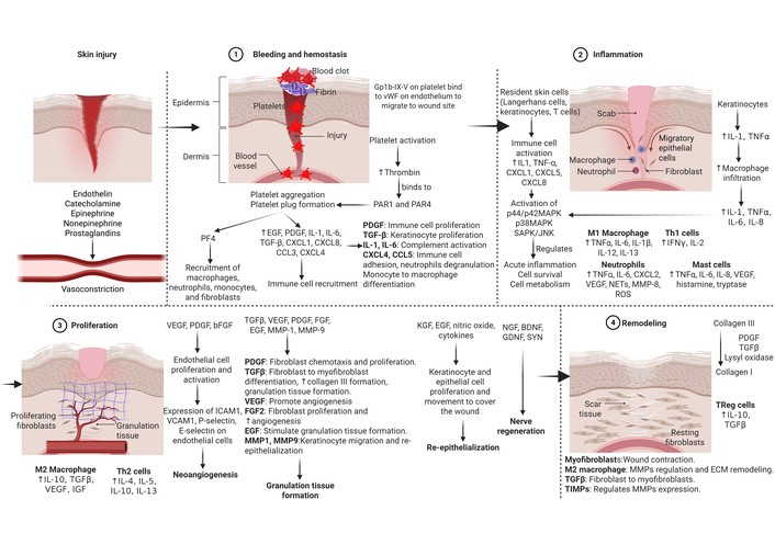
Immune response and molecular mechanisms during wound healing. The immune response is essential for wound healing, initiating a four-phase process including hemostasis, inflammation, proliferation, and remodeling driven by immune cells and signaling molecules. EGF: epidermal growth factor; FGF: fibroblast growth factor; VEGF: vascular endothelial growth factor; TGF-β: transforming growth factor beta; PDGF: platelet-derived growth factor; NGF: nerve growth factor; BDNF: brain-derived neurotrophic factor; GDNF: glial cell line-derived neurotrophic factor; IGF: insulin-like growth factor; KGF: keratinocyte growth factor; SYN: synaptophysin; PF4: platelet factor 4; CXCL: C-X-C motif ligand; CXCR: C-X-C motif chemokine receptor; CCL: C-C motif chemokine ligand; IL: interleukin; TNF-α: tumor necrosis factor alpha; MAPK: mitogen-activated protein kinase; SAPK: stress-activated protein kinase; JNK: c-Jun N-terminal kinases; NETs: neutrophil extracellular traps; ROS: reactive oxygen species; MMP: matrix metalloproteinase; ICAM-1: intercellular adhesion molecule 1; VCAM-1: vascular cell adhesion molecule 1. Created in BioRender. Rai, V. (2026) https://BioRender.com/4bfzni7.
Non-healing DFUs are driven by a molecular environment trapped in chronic inflammation, associated with high MMPs, diminished growth factors, involving impaired angiogenesis, excessive oxidative stress, impaired fibroblast function, and altered ECM remodeling (shown in Figure 1), together delaying wound healing [15]. Key mechanisms include dysfunctional macrophages (M1-skewed) failing to transition to a repair phenotype (M2), resulting in excessive proinflammatory cytokines [interleukin (IL)-1, IL-6, IL-8, tumor necrosis factor (TNF)-α], premature senescence of fibroblasts, and reduced growth factor signaling [e.g., transforming growth factor (TGF)-β]. A decreased concentration of other growth factors, such as epidermal growth factor (EGF), platelet-derived growth factor (PDGF), fibroblast growth factor (FGF), and vascular endothelial growth factor (VEGF), secreted by various cells in the wound microenvironment, also contributes to the nonhealing of DFU [8–10].
Chronic inflammation driven by persistent hyperglycemia-induced immune dysfunction is a major contributing factor to nonhealing DFUs. In the presence of persistent hyperglycemia and chronic inflammation, neutrophils and macrophages are trapped in a proinflammatory state, releasing excess pro-inflammatory cytokines (TNF-α, IL-1β, IL-6, and IL-8) that destroy tissue rather than repairing it. Increased secretion of proinflammatory cytokines is due to skewed polarization of macrophages towards M1 instead of M2 [10, 16].
Neutrophils are key immune cells that, in DFUs, become dysfunctional, leading to delayed healing and chronic inflammation. They exhibit impaired phagocytosis, excessive NETosis (releasing NETs), and prolonged presence at the wound site, causing tissue destruction and increased infection risk due to high glucose. NETs are web-like structures of DNA and proteins released by neutrophils that, when excessive, significantly delay DFU healing by promoting chronic inflammation, tissue damage, and reduced angiogenesis. Elevated NET components in wounds (e.g., neutrophil elastase, citH3) correlate with infection, severe inflammation, and higher amputation rates [17–20].
Dysfunctional macrophage polarization due to hyperglycemia, where macrophages are “stuck” in the pro-inflammatory M1 phenotype and don’t polarize to the anti-inflammatory, tissue-repairing M2-phenotype contributes significantly to persistent inflammation. Poor microcirculation prevents an appropriate macrophage response, exacerbating inflammation. High glucose and advanced glycation end products (AGEs) directly activate the inflammatory M1 phenotype. Overactivation of the NOD-, LRR- and pyrin domain-containing protein 3 (NLRP3) inflammasome pathway promotes persistent M1 polarization. Macrophage polarization is regulated by phosphatidylinositol 3‐kinase (PI3K)/protein kinase B (AKT), peroxisome proliferator‐activated receptors (PPARs), nuclear factor kappa beta (NF‐κB), toll-like receptors (TLRs), Janus kinase (JAK)/signal transducer and activator of transcription (STAT), and Notch signaling [21–23]. Persistently increased numbers of M1 macrophages release high levels of pro-inflammatory cytokines and chemokines, including TNF-α, IL-1β, IL-6, IL-12, type I interferon (IFN), C-X-C motif ligand (CXCL) 1-3, CXCL-5, and CXCL8-10, causing tissue destruction and nonhealing [21, 24].
Not only is fibroblast function impaired, but also phenotypic changes of fibroblasts and their plasticity, with an increased number of CD40+ inflammatory fibroblasts secreting IL-6 and IL-8, and decreased angiogenic fibroblasts, contribute to nonhealing DFUs [25, 26]. Metabolic stress on fibroblasts causes a phenotypic shift towards dysfunction, senescence, and inflammatory signaling. Non-healing DFUs exhibit a depletion of functional fibroblasts [myofibroblasts, alpha smooth muscle actin (α-SMA)+ and TGF-β+] and an accumulation of dysfunctional subsets with decreased proliferation, migration, and secretory function [CD40+, fibroblast specific protein (FSP)–, thrombospondin (TSP)+] that fail to remodel the ECM (due to decreased granulation tissue formation) and promote angiogenesis (TSP+), but promote inflammation (CD40+) [25–28]. Downregulation of Wnt signaling, particularly the Wnt/β-catenin pathway, suppresses fibroblast migration, proliferation, and collagen synthesis, delaying re-epithelialization [29–31]. Wnt/β-catenin signaling is activated during the proliferative phase of wound healing. Elevated Wnt signaling enhances the migration and proliferation of dermal fibroblasts and promotes their differentiation into myofibroblasts. Active Wnt signaling directly increases the expression of type I and type III collagen, which are necessary for constructing the granulation tissue that fills a wound [31]. Deletion of specific Wnt ligands (e.g., Wnt10a) results in significantly larger wound areas and delayed wound healing because of decreased collagen production and reduced fibroblast/myofibroblast activity [30]. The Wnt pathway regulates the migration and differentiation of keratinocytes and is required for the re-epithelialization process. Downregulation of this signaling inhibits this repair, resulting in poor wound closure and reduced structural regeneration [29].
Overexpression of MMPs in chronic DFUs due to persistent inflammation contributes to altered ECM remodeling. MMPs, particularly MMP-9, break down ECM proteins, such as fibronectin, preventing granulation tissue formation and tissue reconstruction. While MMP-8 aids healing by collagen repair, excessive MMP-9/tissue inhibitors of matrix metalloproteinase (TIMP)-1 ratios in wound fluid prevent tissue repair. Elevated MMP-9 levels destroy growth factors and receptors, preventing the wound from progressing beyond the inflammatory phase. Along with MMP-9, nonhealing DFUs also have increased expression of MMP-1 (excessive collagen breakdown), MMP-2 (a marker of impaired healing and persistent ulceration), and MMP-3 (acts as a physiological activator of MMP-9, compounding the proteolytic damage), which degrade the ECM and hinder healing. It should be noted that TIMPs (such as TIMP-2) are significantly lower in chronic wounds [32–34].
Significantly decreased VEGF and reduced endothelial nitric oxide production in chronic DFUs hinder neoangiogenesis, resulting in poor oxygen/nutrient delivery, chronic hypoxia, and impaired wound healing. This impaired process results from hyperglycemia, causing endothelial cell dysfunction, persistent inflammation, and limited angiogenesis [35]. Reduced expression of VEGF and dysfunctional endothelial progenitor cells (EPCs) lead to inadequate capillary growth, causing localized ischemia. Persistently high glucose levels impair EPC number, function, mobilization, migration, and proliferation, resulting in reduced angiogenesis and poor healing. Hyperglycemia leads to dysfunction in the signaling pathways of EPCs, such as Notch pathway dysfunction, which hinders cell differentiation and proliferation, ultimately decreasing the formation of new capillaries [36]. Decreased expression and function of angiogenic factors like FGF-2 are also characteristic of chronic DFUs. FGF-2 is decreased in chronic DFUs because of non-enzymatic glycation of FGF-2 in the presence of hyperglycemia. Decreased FGF-2 levels lead to deficient endothelial cell proliferation, migration, and tube formation, which inhibits the formation of new blood vessels necessary for wound healing. Decreased FGF-2 also correlates with decreased fibroblast mitosis and viability, which further hinders the wound healing process [37].
Hyperglycemia increases ROS, leading to the accumulation of AGEs, which bind to the receptor for AGEs (RAGE) and trigger chronic inflammation, oxidative stress, and impaired cellular function that damage tissues, prevent wound closure, and induce chronic inflammation [38]. AGE-RAGE interaction activates various inflammatory and oxidative stress pathways such as NF-κB, PI3K-AKT, and JAK-STAT signaling, which sustain an inflammatory (M1) phenotype in macrophages. This prevents the necessary transition to the pro-healing (M2) phenotype, leaving the wound trapped in an inflammatory state [39]. By increasing the production of ROS, the AGE-RAGE axis perpetuates chronic inflammation via transcriptional activation of NF-κB, followed by numerous proinflammatory cytokines and adhesion molecules, including endothelin-1, intercellular adhesion molecule 1 (ICAM-1), E-selectin, and tissue factors, causing further tissue damage, more AGE formation, and delayed wound healing [40]. AGEs induce significant apoptosis of fibroblasts and reduce collagen production, disrupting granulation tissue formation. The AGE-RAGE signaling reduces the proliferative capacity of keratinocytes and fibroblasts, reducing re-epithelialization and halting wound closure [41, 42]. The AGE-RAGE axis disrupts autophagy and induces EPC apoptosis, diminishing their ability to migrate and repair damaged blood vessels, reducing angiogenesis crucial for nutrient delivery to the wound, and leading to delayed wound healing. AGE-RAGE axis also downregulates VEGF, a key stimulant for angiogenesis, further reducing the formation of new capillaries [39].
Biofilms are complex, polymicrobial communities embedded in an extracellular polymeric substance (EPS) matrix that protects bacteria from immune responses and antimicrobials, acting as a major driver of nonhealing DFUs. Common pathogens like Staphylococcus aureus and Pseudomonas aeruginosa form biofilms, which are associated with reduced microbiota diversity and increased recurrence rates of infections [43]. The biofilm EPS acts as a physical barrier preventing host immune cells (neutrophils) and antibodies from reaching the bacteria, while also causing dysregulation of the local immune environment. Chronic, polymicrobial biofilms prevent immune clearance and contribute to persistent inflammation by arresting wound healing at the inflammatory phase, preventing transition to the proliferation stage. High levels of proteases and ROS produced by the biofilm bacteria and the resulting persistent inflammation damage tissue, leading to chronic wound stagnation [44, 45]. These molecular failures result in a chronic, nonhealing wound, necessitating targeted therapeutic approaches focusing on rebalancing the immune response in the wound microenvironment [46].
Immunomodulation in DFU healing focuses on targeting altered molecular mechanisms discussed in the previous section. This includes reversing chronic, overactive inflammation by shifting macrophages from a pro-inflammatory (M1) to a pro-regenerative (M2) phenotype, promoting angiogenesis, increased granulation tissue formation, and ECM remodeling, altogether promoting DFU wound healing and remodeling. Advanced strategies include using bio-functional hydrogels, phytochemicals (flavonoids, alkaloids, tannins), stem cells to lower ROS, glucose depletion, sustained drug release devices, reduction of inflammatory cytokines (TNF-α, IL-6), and promoting angiogenesis. Pharmacological agents such as sodium-glucose cotransporter 2 (SGLT2) inhibitors and biologics (e.g., TNF-α inhibitors) are being investigated to reduce systemic and local inflammatory factors [47–50]. Further, reduced migratory and proliferative capacity of mesenchymal stem cells (MSCs) in the wound area limits the regenerative potential. MSCs can modulate immune responses, reduce inflammation, and enhance angiogenesis to accelerate wound healing [51].
As discussed above, skewed polarization towards M1 macrophages is altered in DFU. Targeting macrophage polarization, specifically shifting from a pro-inflammatory M1 phenotype to a pro-healing M2 phenotype, is crucial for DFU healing. Therapies targeting a shift toward the M2 type promote tissue repair, angiogenesis, and collagen deposition, and attenuate inflammation (Table 2, Figure 2).
Therapeutic agents promoting M2 macrophage polarization and wound healing in diabetes.
| Macrophage polarization | Study | Model | Strategy | Mechanisms/Outcome |
|---|---|---|---|---|
| Humans | ADSCs mediated macrophage polarization [52] | Bioinformatics analysis of GSE134431 and GSE80178 datasets of human origin | 30 macrophage polarization-associated differentially expressed genes (MA-DEGs) were identified and analyzed. | ADSCs regulate EREG and CSTA expression to promote M2 macrophage polarization. |
| ON101 cream, a phase 3 randomized clinical trial [53] | Human patients | Twice-daily applications of ON101 or an absorbent dressing were changed once daily or 2 to 3 times a week for 16 weeks, with a 12-week follow-up. | ON101 exhibited better healing efficacy.ON101 regulates macrophage polarization. | |
| Animal model and in vitro | MDSC-dependent macrophage polarization [54] | DFU mouse model | MCC950 was injected every other day into the wound in C57BL/6 diabetic mice. | MCC950 increased M2 macrophages and decreased pro-inflammatory genes. MCC950 recruits MDSCs and significantly accelerates diabetic wound healing. |
| Resveratrol effects on macrophage polarization and wound healing [55] | Diabetic mice modelTHP1 cells | Diabetes was induced with STZ in C57BL/6 mice.THP1 cells were used to evaluate the effects of resveratrol on polarization and the secretion of pro-inflammatory factors. | Resveratrol significantly increased diabetic wound healing.Resveratrol reduces TNF-α, iNOS, and IL-1β secretion and promotes M2 macrophage polarization. | |
| Puerarin [56] | Male C57BL/6 miceRAW264.7 cells | Diabetes was induced using streptozotocin in mice.Puerarin (120 mg/kg i.p.) was administered daily to the mice | Induces M2 macrophage polarization.The effects of puerarin on macrophage polarization are related to NF-κB and MAPK signaling pathways.Puerarin promotes diabetic wound healing. |
ADSCs: adipose-derived stem cells; MDSCs: myeloid-derived suppressor cells; TNF-α: tumor necrosis factor alpha; iNOS: inducible nitric oxide synthase; IL: interleukin; NF-κB: nuclear factor kappa beta; MAPK: mitogen-activated protein kinase.
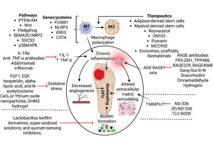
Immunomodulation strategies for nonhealing diabetic foot ulcers (DFUs). Created in BioRender. Rai, V. (2026) https://BioRender.com/cwvna0i.
A study by Yang et al. [57], using diabetic rats and human dermal fibroblasts, reported the role of Forkhead box protein M1 (FOXM1) in accelerating wound healing in diabetic mice involving M2 macrophage polarization through Hedgehog signaling. This suggests that macrophage polarization is an attractive target for promoting DFU healing. Another study using a mouse model reported the effects of MCC950 in promoting diabetic wound healing by inhibiting NLRP3 inflammasome activation and modulating macrophage polarization in a myeloid-derived suppressor cell (MDSC)-dependent manner [54] (Figure 2). Accelerated wound healing in diabetic mice was reported with resveratrol targeting macrophage polarization [55]. Qi et al. [58] reported that AuPt@melanin-incorporated (GHM3) hydrogel decreases local glucose and ROS levels and promotes wound healing in diabetic rats’ wounds by promoting M2 macrophage polarization. The role of other drugs and biomaterials, including melatonin-stimulated exosomes, insulin, CSO, pUBM, quercetin, docosahexaenoic acid, and biomaterials containing sulfated chitosan (SCS)-doped collagen type I (Col I/SCS), deep eutectic solvent (DESs), hyaluronic acid (HA) and a pH-controllable hydrogen sulfide donor named JK1 (HA-JK1), konjac glucomannan-modified SiO2 nanoparticles (KSiNPs), Lactococcus lactis thermo-sensitive hydrogel, adipose-derived stem cells (ADSCs), MSCs, human (h)MSCs, bone marrow-derived MSCs (BM-MSCs), and ADSCs in promoting wound healing in rats, mice, and humans by targeting macrophage polarization has been discussed [23]. A recent study using obese diabetic mice (B6.Cg-Lepob/J, ob/ob) reported the role of the M2 macrophage [Tol (tolerization)/Pol (polarized) M2 monocytic cells] secretome in promoting wound healing [59]. These studies in animal models support the notion of targeting macrophage polarization towards M2 macrophages to promote healing in DFUs; however, they warrant further research in large animal models and clinical trials before translating to clinics.
Exosomes are small (40–150 nm) extracellular vesicles secreted by cells that function as messengers, delivering cargo like proteins, mRNA, and growth factors to regulate cellular behavior, reduce inflammation, and facilitate tissue regeneration. They are essential for intercellular communication and are increasingly used in skincare to boost collagen and repair skin, as well as in studies for treating chronic disease [60]. Many studies have reported the role of exosomes in promoting M2 macrophage polarization and wound healing in diabetic wounds. For example, double-layer microneedle-based wound dressing systems (MEs@PMN) were shown to promote wound healing in diabetic Sprague Dawley rats by promoting M2 macrophage polarization and angiogenesis, and attenuating inflammation [61]. Another study using db/db mice reported enhanced wound healing potential of epidermal stem cell-derived exosomes via miR-203a-3p/SOCS3-mediated induction of M2 macrophage polarization [62]. Another study using C57BL/6 mice reported that exosomes from ADSCs promote wound healing by promoting M2 macrophage polarization, reducing inflammatory response, and increasing collagen production involving the circ-Rps5/miR-124-3p axis [63]. Liu et al. [64] reported the role of melatonin-pretreated MSC-derived exosomes in promoting wound healing in db/db mice by increasing M2 macrophage polarization involving activated phosphatase and tensin homolog (PTEN)/AKT signaling pathway. These results support the notion that exosomes promoting M2 macrophage polarization may have therapeutic significance in promoting wound healing in DFU. Though exosome-mediated promotion of wound healing has shown encouraging results in preclinical studies (rodent models), it has not yet been translated into clinical practice due to significant bottlenecks in standardization, manufacturing, and in-vivo efficacy. Although MSC-derived exosomes effectively modulate inflammation, angiogenesis, and tissue remodeling, their transition from bench to bedside faces several critical challenges, as discussed in the limitations section below.
A recent study [52] using bioinformatics analysis reported that ADSCs regulate the gene expression of EREG and CSTA to promote M2 macrophage polarization and DFU wound healing. Another study reported that a hydrogel of zwitterionic poly(sulfobetaine methacrylate) (SB) incorporated with keratin-exfoliated MoS2 and bee-wax nanoparticles to deliver phenytoin promotes wound healing by promoting M2 macrophage polarization and attenuating inflammation [65]. A phase 3 large-scale, international Multi-Regional Clinical Trial (MRCT) in Taiwan (China) investigating the efficacy and safety of ON101, a topical new drug regulating M1/M2-macrophage polarization, reported excellent therapeutic efficacy in promoting healing in Wagner grade 3 and 4 DFUs in the real-world scenario. The study involved 133 patients with ulcer severity varying between Wagner grades 1 to 4, and the average ulcer complete healing rate was 71%, with no adverse events [66]. Previously, ON101 was investigated in China with 236 DFU patients who applied it topically and showed improved healing in Wagner Grade 1 and 2 DFUs after a 12-week follow-up [53]. It should be noted that these results are from a certain geographical area or a small set of population; thus, for translating these strategies to clinics for the general population, there is a need for large-scale randomized controlled trials.
Targeting inflammatory cytokines, particularly inhibiting TNF-α, IL-1, and IL-6, is a key strategy for treating DFUs by reversing chronic inflammation and fostering a regenerative environment. This is because chronic inflammation is the cause of keeping the wound in the inflammatory phase of wound healing. One of the strategies is polarizing macrophages towards the M2 phenotype, as discussed above. Promoting IL-10, IL-4, and IL-13 is another strategy crucial to shift from chronic inflammation to healing [67]. In this section, targeting cytokines will be discussed.
TNF-α plays an important role in maintaining chronic inflammation, as shown in Figure 1. Targeting it may reduce inflammation, and the wound may enter the proliferation phase, promoting wound healing in DFUs. The insight to target TNF-α came from a study by Goren et al. [68], revealing restoration of impaired wound healing in ob/ob mice with reduced number of activated viable macrophages in the wound after short-term systemic administration of TNF-α neutralizing monoclonal antibodies (V1q) or monocyte/macrophage-expressed EGF-like module-containing mucin-like hormone receptor-like (Emr)-1 (F4/80) antibody (Figure 2). The results of this study indicated that targeting “activated” TNF-α-expressing macrophages may be a novel therapeutic target to promote wound healing in diabetes. A study comparing various TNF-α inhibitors administered subcutaneously reported that adalimumab and infliximab effectively promote wound healing and show similar levels of efficacy throughout the healing process in db/db mice, whereas golimumab, etanercept, and certolizumab pegol showed no significant effects on wound healing and were less effective (Figure 2). This functional difference between various inhibitors may be due to differences in functional Fc (fragment crystallizable) domains. The accelerated cutaneous wound healing in mice with adalimumab was mediated via improved epidermal closure and granulation tissue formation [69]. Both of these studies used anti-TNF-α therapy to promote wound healing (without mentioning any side effects), one systemically for the short term and the other subcutaneously. Both systemic and subcutaneous (short-term and long-term) may have different effects as well as side effects. Systemic anti-TNF therapies (e.g., adalimumab, infliximab, etanercept) are highly effective for chronic inflammatory diseases, but their safety profile changes over time. While short-term use often triggers localized immune/hypersensitivity responses, long-term use is associated with systemic cumulative risks like reduced immunity and drug tolerance [70, 71]. It should also be noted that systemic administration causes more widespread, serious adverse effects, while local delivery isolates risks to the treated area. Further, it should also be noted that systemic anti-TNF therapy disrupts the body’s immune defenses, increasing the risk of both systemic and local infections. However, the presentation, mechanism, and risk profiles differ significantly [72, 73]. These aspects should be considered while designing therapeutics to promote wound healing in DFUs.
IL-1β is another cytokine whose high levels inhibit fibroblast proliferation and migration, delaying healing in DFUs [74]. IL-1β impaired diabetic wound healing by regulating the expression of MMP-2, MMP-9, and TIMPs involving the p38 MAPK pathway [75]. Thus, IL-1β may be an attractive target to promote wound healing. This notion is supported by the fact that an IL-1β neutralizing antibody blocking the IL-1β pathway resulted in improved wound healing in diabetic mice. Improved healing was associated with a switch of proinflammatory to a healing-associated macrophage phenotype and increased levels of wound growth factors [76]. Another study evaluated the effects of locally administered IL-1 receptor antagonist (IL-1Ra; Anakinra) on wound healing in a diabetic mouse model. The study reported that IL-1Ra therapy decreases time to closure in splinted diabetic wounds associated with significantly decreased inflammation (decreased expression of neutrophils and macrophages) [77]. The role of IL-1Ra on wound healing was evaluated by targeting the IL-1-IL-1 receptor (IL-1-IL-1R1) axis, which delays wound healing. The results showed enhanced wound healing in diabetic mice with matrix-binding IL-1Ra associated with a decreased number of pro-inflammatory cells (macrophages and neutrophils), cytokines (IL-1β, IL-6, and CXCL1), senescent fibroblasts, decreased MMP-2 and MMP-9, along with higher levels of TIMP-1, anti-inflammatory cytokines (TGF-β, IL-4, and IL-10) and growth factors (FGF-2, PDGF-BB, and VEGF) [78]. These results indicate that targeting IL-1β may be a potential therapeutic target to promote wound healing in diabetes.
Not only inhibiting cytokines to promote wound healing in DFUs, but also increasing the expression of cytokines like the IL-22 family may promote wound healing. This notion is supported by the fact that mice deficient in IL-22R showed delayed wound healing. IL-22R is the common receptor chain for IL-20, IL-22, and IL-24. Kolumam et al. [79] reported that IL-20, IL-22, and IL-24 promote wound healing in type II diabetic db/db mice. Improved wound healing was mediated by increased expression of genes involved in reepithelialization, tissue remodeling, and innate host defense mechanisms [79]. The studies in animal models support the notion that targeting cytokines may promote wound healing in DFUs, but the evidence is lacking in humans, and there is a need for large-scale randomized clinical trials.
Excessive and uncontrolled MMPs, particularly MMP-9, inhibit DFU healing by degrading ECM and prolonging inflammation. Thus, targeting MMPs should be considered to attenuate inflammation and promote ECM remodeling. Gao et al. [80] reported accelerated diabetic wound healing in db/db mice using compound ND-336, a highly selective inhibitor of gelatinases (MMP-2 and MMP-9) and MMP-14 (Figure 2). This effect was mediated by attenuating inflammation and by enhancing angiogenesis and re-epithelialization. The study further showed that combining ND-336 with the topical MMP-8 recombinant protein has more pronounced effects. Another study showed improved wound healing in diabetic mice with the MMP-9 inhibitor (R)-ND-336 alone or in combination with linezolid. (R)-ND-336 is a highly selective, small-molecule inhibitor of MMP-9, which reduces inflammation and enhances angiogenesis, contributing to enhanced wound healing [81] (Figure 2).
Technology Lipido-Colloid Nano-OligoSaccharide Factor (TLC-NOSF) is another strategy for targeting MMPs. A systematic review of 16 randomized clinical trials (13 with collagens and 3 with TLC-NOSF dressing) reported the beneficial effects of inhibiting MMPs in promoting wound healing, reduction in wound size, and improved healing rates; however, there were substantial differences in evidence for different types of wounds, including DFUs, venous leg ulcers, pressure ulcers, or wounds of mixed origin. TLC-NOSF gel directly binds to and specifically targets MMP-2 and MMP-9, and TLC-NOSF dressing significantly promotes healing in hard-to-heal neuroischemic DFUs and venous leg ulcers [82] (Figure 2). Thus, there is a need for more research to generate evidence and potentiate the notion of targeting MMPs to promote wound healing in DFUs.
Accumulated AGEs bind to RAGE and trigger chronic inflammation, oxidative stress, and impaired cellular regeneration, preventing normal wound healing in DFUs. Inhibiting this pathway is a promising therapeutic strategy for chronic DFUs. Lin et al. [39] reviewed the role of small-molecule inhibitors of RAGE (FPS-ZM1, TPP488, RAGE229), RAGE antibodies, RAGE gene therapy, and RAGE scavengers in promoting wound healing in diabetic ulcers in in-vitro, mouse model, and human patients [83]. In this section, we have summarized other recent studies (Table 3, Figure 2) supporting the notion of targeting the AGE-RAGE axis to promote wound healing in diabetic ulcers.
Therapeutic strategies targeting the AGE-RAGE axis to promote wound healing in diabetes.
| Study | Model | Strategy | Mechanisms/Outcomes |
|---|---|---|---|
| Anti-RAGE antibody [84] | Diabetic male C57BL/6 mice | Topical application of the RAGE antibody on the wound | ↑ Neutrophil phagocytosis by macrophages.↑ Phenotypic switch to M2 macrophages.Enhanced wound healing. |
| RAGE406R [85]* | Diabetic mice | Prevents the formation of the RAGE-DIAPH1 complex | Accelerate wound healing.Decrease systemic inflammation. |
| Cinnamaldehyde [86] | C57BL/J6 diabetic mice | Daily intraperitoneal injections of cinnamaldehyde | Improved wound healing.Accelerated wound closure.↓ Inflammatory infiltration and oxidative stress.↑ CD31 expression (angiogenesis).M1 to M2 polarization. |
| Resina Draconis hydrogel [87] | RAW264.7 cellMale C57BL/6J diabetic mice | Hydrogels were applied to the wounds | Accelerated wound healing.↓ Oxidative stress.↑ M2 macrophage polarization. |
| RAGE229 [88] | Murine and human SMCsBTBR ob/ob mice | Topical injection of RAGE229 two times daily | ↓ Expression of TNF-α, IL-6, and CCL2/JE-MCP-1.Accelerate wound healing. |
| Palladium hydride (PATP) hydrogel [89] | Male C57BL/6 diabetic mice | Co-blocks the HMGB1-RAGE axis “head-to-tail” (upstream and downstream both)PATP hydrogels were applied to the wounds | ↓ TNF-α, iNOS, and IL-1α levels in the wound.↓ ROS, RAGE, and HMGB1 levels.Promotes wound healing with normal skin architecture.Increased neovascularization. |
| Dang-Gui-Si-Ni [90] | Male Sprague-Dawley diabetic rats | Oral gavage to rats after wounding | Accelerated wound healing.Decreased inflammation (↓ IL-1β, IL-6, TNF-α, AGEs, and RAGE levels).Modulated (↑) TGF-β1 and Smad2/3 protein expression. |
| Dracorhodin [91] | Sprague-Dawley diabetic rats | Wounds were treated with dracorhodin | Accelerated wound healing in a dose-dependent manner.↑ Collagen synthesis, angiogenesis, and growth factor levels.↓ Inflammation and ROS levels. |
| Antimicrobial hydrogel with RAGE and MMP-9 inhibitors [92] | In vitro cell (RAW 264.7) and in vivo diabetic Wistar rat wound model | Immuno-gel was applied to wounds | Significantly decreased MMP-9 and NF-κB expression.Enhanced M2 macrophages and pro-healing cytokines. |
| Rosiglitazone and S-nitroso glutathione (nanoparticles/hydrogel composite) [93] | Diabetic SD rats | RAGE inhibitor/exogenous nitric oxide dressing to the wound every other day | Significantly improved wound healing.↑ Wound closure rate, collagen fiber production, and angiogenesis.↓ Inflammation (↓ IL-1β, TNF-α, and IL-6). |
| GPP@ZnBG hydrogels [94] | Diabetic mice and human subjects | Hydrogel was applied to the wound in mice and to DFUs in humans | ↑ Angiogenesis, collagen formation, and tissue repair.↓ Inflammation.Promote wound repair.Have antibacterial effects. |
* Article preview only is available. DIAPH1: diaphanous-related formin 1; TNF-α: tumor necrosis factor alpha; IL: interleukin; NF-κB: nuclear factor kappa beta; ROS: reactive oxygen species; HMGB1: high-mobility group box protein 1; AGEs: advanced glycation end products; RAGE: receptor for advanced glycation end products; iNOS: inducible nitric oxide synthase; CCL2: C-C motif chemokine ligand 2; MCP-1: monocyte chemoattractant protein-1; TGF-β: transforming growth factor beta.
Excessive oxidative stress caused by hyperglycemia-induced ROS and reduced antioxidant capacity is a primary driver of nonhealing DFUs. This imbalance damages cells, promotes chronic inflammation, impairs angiogenesis, and delays collagen deposition, leading to persistent, stubborn, and often infected wounds. Thus, targeting oxidative stress may be a potential immunomodulatory strategy to promote healing. Effective reduction of oxidative stress and enhancement of wound healing have been reported with antioxidants like FGF-1, EGF, hesperidin, alpha-lipoic acid, and N-acetylcysteine. Further, nanotechnology-loaded antioxidants (CeO2 nanoparticles, Yttrium oxide nanoparticles) offer a promising approach [95] (Figure 2). AuPt@melanin-incorporated (GHM3) hydrogel facilitates hyperthermia-enhanced local glucose depletion and ROS scavenging and promotes wound healing in diabetic rats. The enhanced healing was associated with decreased TNF-α, downregulation of the ratio of M1/M2 macrophages, increased angiogenesis, collagen deposition, and cell proliferation and differentiation. These effects were more pronounced when GHM3 was combined with NIR laser therapy [58].
A study evaluated the effects of hyperbaric oxygen therapy (HBOT) on inflammation in 15 DFU patients with Wagner stages 2–4 ulcers by investigating oxidative stress regulators, inflammatory cytokines, and NLRP3 inflammasome gene expression (NCT06502808). The study reported increased gene expression of SOD1 and GPX2 genes (oxidative stress regulators) and IL-1β, IL-12, IL-4, and NLRP3, while decreased expression of TNF-α. This change in gene expression was associated with healed wounds and suggests the effects of HBOT in promoting wound healing in DFUs by targeting oxidative stress [96].
Immunomodulation of biofilms in DFUs targets the chronic inflammatory microenvironment caused by bacterial communities, aiming to shift macrophages from M1 to M2 phenotype. Emerging therapies, including Lactobacillus biofilm derivatives, super-oxidized solutions, and quorum-sensing inhibitors, reduce biofilm biomass and modulate immune response, breaking the cycle of persistent inflammation and enhancing wound healing (Figure 2). Lactobacillus biofilm derivatives are bacteria-free derivatives that modulate immune function and systemic metabolic reprogramming, suppress the JAK-STAT1 signaling pathway, alleviate the local inflammatory microenvironment, and promote neovascularization and tissue repair in diabetic wounds [97]. Super-oxidized solutions are non-antibiotic agents that improve biofilm integrity, decrease excessive inflammation, and support tissue regeneration [98]. Quorum-sensing (QS) inhibitors are molecules that can reduce biofilm formation by up to 50% by targeting QS signaling molecules or their receptors, or downstream regulatory factors [98, 99]. Mashamba et al. [100] reported the highest biofilm inhibition (73%) against E. coli with Warburgia salutaris aqueous extract and antagonism of N-acyl homoserine lactone signaling with Euclea natalensis, Aloe ferox, and Warburgia salutaris compounds. These results indicated the antipathogenic and antibiofilm phytomedicine development role of these compounds in nonhealing DFUs. The goal of immunomodulation of biofilm is to reprogram macrophages to move away from an immune-suppressive phenotype that cannot clear the infection, reducing excessive inflammatory cytokines (e.g., IL-1β, TNF-α).
A double-blind, randomized controlled trial with 172 patients from Indonesia reported the advantages of targeting biofilms in promoting wound healing in DFUs. The intervention group received standard of care followed by wound cleansing based on the results of wound blotting and antimicrobial dressing, while the control group received standard of care and regular dressing. The intervention group showed significant improvement in DFU healing after 2 weeks of intervention. These findings suggest the role of targeting biofilms to promote DFU healing [101].
Targeting chronic inflammation in DFU has clinical relevance because the aim of the clinicians (or podiatricians) is to modulate specific cytokine and macrophage pathways to suppress excessive oxidative damage, prevent osteomyelitis, and promote tissue regeneration—ultimately reducing the high risk of limb amputation [67, 102]. As discussed above, the aim of the clinicians is to target macrophage polarization towards M2 macrophages, inflammatory cytokines, and unresolved activated neutrophils. For example, G-CSF (granulocyte-colony stimulating factor) is used to improve neutrophil antimicrobial function [103]. Hyperglycemia activates inflammatory cascades (like the AGE-RAGE axis) that upregulate IL-6 and TNF-α. Targeted anti-inflammatory drugs like treprostinil regulate prostaglandin I2 pathways to interrupt this cycle and have shown clinical efficacy [104]. Furthermore, the antihyperglycemic agents such as metformin, glucagon-like peptide 1 receptor agonists, and dipeptidyl peptidase 4 enzyme inhibitors not only help in glycemic control but also regulate overall metabolic imbalance, angiogenesis, inflammation, and tissue regeneration in DFU [105].
Targeting biofilm in DFUs is crucial because biofilms are a primary driver of wound chronicity, treatment failure, and lower-limb amputations. They shield microbes from antibiotics and host immunity, making eradication extremely difficult without mechanism-based therapies [106]. Super-oxidized solutions could be more effective for sustainable wound care strategies by improving infection control and reducing chronic wound burden [98]. Because standard topical antibiotics have limited efficacy against established biofilms, comprehensive management requires a multifaceted approach to disrupt the EPS matrix and eradicate the pathogens. Sharp debridement is considered the “gold standard” to mechanically remove necrotic tissue and surface biofilm, though biofilms can rapidly reform within days without further intervention. Antimicrobial dressings utilizing cadexomer iodine, silver, and antimicrobial peptides have shown clinical promise in managing biofilm and exudate while reducing inflammation [107]. To overcome the limitations of surface-level debridement, researchers are validating novel targeted therapies. These include matrix-degrading enzymes, quorum sensing inhibitors, bacteriophage therapy (which uses viruses that target specific bacteria), cold plasma, and electroceutical dressings [108, 109].
Another important aspect in treating DFUs is stewardship principles to guide the responsible use of resources, which means the treatment emphasizes targeted diagnostics and conservative management to prevent overtreatment, reduce antimicrobial resistance, and spare healthy bone. These principles require bone biopsies for culture, focused foot-sparing surgery, and individualized, strictly time-limited antibiotic courses [110, 111].
The clinical evidence of improved healing with surgical debridement is supported by the notion of complete healing of DFU in a 72-year-old woman with one session of surgical debridement and ten sessions of maggot therapy (one session every two days) using sterile Lucilia sericata [112]. A retrospective review of 10 cases using a reconstituted bilayer matrix for DFUs refractory to healing after topical wound care and offloading for longer than 4 weeks reported 90% of wounds closed at 12 weeks, with a mean wound area reduction of 85% at 6 weeks and 94% at 12 weeks. No adverse events, pain, or discomfort were reported [113]. Another case study reported wound closure in a 46-year-old male with DFU treated with surgical treatment followed by Med-honey dressing and human amniotic membrane [114]. A meta-analysis with 22 randomized control trials (RCTs) (n = 1,148) reported that biological and enzymatic debridement reduce the wound area (mean decrease 29.6% and 21.8%; respectively). The report concluded the efficacy of biological debridement over surgical and standard wound care and of enzymatic debridement over autolytic debridement [115]. However, another meta-analysis including 19 RCTs (n = 900) reported the superiority of enzymatic debridement over standard care and other debridement methods [116]. Clinical trial NCT04723134 is evaluating the safety and efficacy of a folic acid wound treatment (FAWT) in promoting wound healing in DFUs.
The studies discussed above suggest that immunomodulation of diabetic wound microenvironment by targeting inflammation, macrophage polarization, oxidative stress, and the AGE-RAGE axis has a promising future, though large scale clinical trials are warranted. The role of various other natural therapeutics discussed by Liu and Yu [117] was tested in in-vitro and in-vivo models, showing enhanced wound healing involving decreased inflammation, oxidative stress, M1 macrophages, and increased collagen deposition, M2 macrophages, cell proliferation, and angiogenesis. The studies discussed by Liu and Yu [117] support the notion that immunomodulation may promote wound healing in DFUs. Further, studies discussed in this section were conducted in animal models (mice and rats), with only a few studies in humans. Thus, there is a need for clinical trials to test the results of these studies. But the question arises, why are these results limited to preclinical studies and have not been translated to clinics? The reason behind this is limitations in translation, as discussed below.
Preclinical studies often fail to translate to human clinics due to fundamental biological differences, inappropriate disease models, and poor study design, leading to a nearly 95% failure rate in drug development. Major limitations include inter-species differences (genetics, metabolism), lack of human disease diversity (patient heterogeneity), and flawed preclinical methodologies. Animal models, particularly in complex diseases (like multifactorial DFUs), frequently misrepresent the human disease process and often fail to replicate human pathophysiology (physiology, drug metabolism, and immune responses) or the full complexity of the disease. Lack of standardization, poor statistical design, small sample sizes, and publication bias (publishing only positive results) often lead to poor reproducibility in animal studies. Laboratory animals are often genetically identical and kept in uniform environments, whereas human populations are diverse with varying genetics, lifestyles, and comorbidities, affecting drug response. Physiological, immune, and genetic differences between animals and humans mean drugs can behave differently, causing unexpected toxicity or lack of efficacy. Lack of variation in laboratory conditions (using both sexes and multiple strains) may be a reason for the unsuccessful translation of preclinical studies to clinics. However, preclinical results are not independently validated before moving to clinical trials [118, 119].
One of the options is to replace animals with modern technologies like using human induced pluripotent stem cells (iPSCs), tissue engineering, organoids, 3D modeling (culturing), microfabrication approaches, microfluid technology, organ on a chip technique, and in-silico bioinformatics; however, these technologies have a long way to go. These techniques have limitations in not displaying the harmful effects of the drug being tested, such as in animals, and providing a more accurate prediction of side effects [120]. This suggests that a combined effort of all techniques with a data-driven approach may be beneficial in translating preclinical results to clinics. Another strategy will be to improve and apply rigorous statistical design. As stated above, weak statistical power led to poor clinical translation, enforcing a priori sample size calculations, performing multi-site confirmation trials across different laboratories, and pre-register study protocols to eliminate bias and ensure highly reproducible results. Furthermore, designing preclinical studies with endpoints that mirror human clinical protocols may also help in advancing the results of preclinical studies to clinics [121, 122].
Coming to the small molecules and natural compounds, poor solubility, poor bioavailability, chemical instability, complex structure, large molecular size, issues with crossing the cell membrane (penetration), cytotoxicity at higher doses, purification status, manufacturing cost, release profile, and characterization of the drug molecule are common limitations in translating the preclinical studies to clinics [117]. The limitations with small-molecule drugs in trials include low selectivity (off-target effects), limited ability to target complex protein-protein interactions, high toxicity risks, and poor pharmacokinetic profiles, such as low oral bioavailability or short half-lives. These challenges lead to high attrition rates during clinical development, often requiring frequent dosing [123]. Further, the small molecule under consideration for a target may act differently in animal models and humans. An estimated 80% of the human proteome is traditionally considered “undruggable” by small molecules [124]. Another issue may be the dose of a drug effective in humans based on animal model research. It is notoriously difficult to predict the exact dose required to safely maintain a therapeutic concentration at the target site in humans based on animal data, leading to inadequate clinical exposure. Often, the dose required to achieve therapeutic efficacy in humans is higher than the maximum tolerated dose, leading to unmanageable side effects [125]. To mitigate the small molecule limitations from preclinical models to the clinic, integrating the Developability Classification System principles and Physiologically Based Pharmacokinetic (PBPK) Modeling early in the pipeline may be helpful. This can bridge the gap between animal models and human trials [126].
The primary limitations of using phytochemicals (natural compounds) in preclinical and clinical trials include poor bioavailability, lack of standardized preparations, low efficacy in humans compared to animal models, and potential drug interactions or toxicities at high doses. Furthermore, many preclinical studies fail to translate, and identifying optimal dosages for human trials remains challenging [127]. When administered, these compounds are rapidly metabolized, poorly absorbed, or broken down by the digestive system, meaning they rarely reach target tissues at the high concentrations proven effective in cell cultures. Additionally, many potent phytochemicals are insoluble in water, chemically unstable, and subject to extensive first-pass metabolism in the liver, meaning only a tiny fraction of an oral dose ever enters the bloodstream [128, 129]. As discussed above, complex clearance mechanisms and biological barriers of the human body may be a reason for failure of phytochemicals. These limitations may be mitigated by utilizing advanced drug delivery systems, structural modifications, and rigorous, standardized trial designs to bridge the translational gap [127].
Bioabsorbable scaffolds and hydrogels are emerging strategies for designing novel therapeutics that promote DFU healing. Hydrogels in preclinical and clinical trials face limitations regarding poor mechanical strength, degradation byproducts, and limited long-term stability. Key challenges include controlling drug burst release, achieving long-term bio-integration, potential immunogenicity, cost-effectiveness, and scaling up for regulatory approval. Limited reporting of key design parameters (e.g., crosslinking ratios, viscosity) hinders the ability to create meta-analyses of clinical outcomes. These challenges often make translating promising preclinical results to clinical practice difficult [130]. The issues of scaling limits, mechanical mismatches, poor vascularization, and component manufacturing complexity associated with scaffolds may be mitigated by utilizing hybrid materials, establishing dynamic biomimetic testing platforms, adopting predictive animal models, and ensuring scalable manufacturing compliance [131].
Tissue engineering faces significant limitations in preclinical and clinical trials, primarily driven by the difficulty of replicating complex human tissue architecture (mechanical mismatch), inadequate revascularization of implants, immune rejection, and low cell survival rates in vivo. Key hurdles include sourcing sufficient autologous cells, high production costs, strict regulatory requirements, and the poor translation of small animal study findings (poor predictive model) to human patients [132]. The issues of tissue engineering and stem cells may be mitigated by overcoming cell sourcing and scalability using Good Manufacturing Practice (GMP), automated bioreactors, and induced pluripotent stem cells (iPSCs). Enhancing engraftment and survival of cells by improving bioabsorbable scaffolds and using chemoattractant scaffolds that recruit the patient's own native stem cells directly to the injury site [133–135].
As discussed above, nanoparticles are often used with engineered tissues and hydrogels to deliver drugs. Significant limitations associated with nanoparticles in preclinical and clinical trials include low translational success due to poor prediction of human biological processes, poor efficacy in humans compared to animal models, rapid immune clearance by the mononuclear phagocyte system, high excretion rate, protein corona formation, and unexpected immunotoxicity. Other challenges involve complex, inconsistent ADME (absorption, distribution, metabolism, excretion) profiles and difficulties in scaling production while maintaining stability [117, 136, 137]. These limitations and the translational gap may be mitigated by addressing critical discrepancies in biological complexity, manufacturing scalability, and patient heterogeneity. Bridging this gap requires adopting robust characterization protocols, physiologically relevant disease models, and standardized regulatory strategies early in development [138–140].
Using exosomes is another strategy for therapeutics promoting wound healing in DFUs. The primary limitations of using exosomes in preclinical and clinical trials include a lack of standardized, scalable isolation and purification methods, low manufacturing yields, and poor long-term stability. Furthermore, challenges in maintaining consistent cargo loading, determining precise dosing, and avoiding potential immune reactions or undesired off-target effects impede clinical translation [141–145]. Exosomes are unstable at room temperature and prone to degradation, leading to over 70% loss of activity, requiring specialized, costly storage like cryopreservation. Due to complex biological origins, isolating specific exosomes and confirming their homogeneity is difficult, as they are often contaminated with other extracellular vesicles (EVs). Poor loading efficiency for drug delivery and difficulties in achieving specific, targeted delivery to target tissues in vivo limit therapeutic efficacy. There are few precedents for exosome therapy regulatory approval, raising concerns about potential immunogenicity, unknown donor history, and long-term side effects. Further investigation is required to optimize manufacturing techniques to ensure reproducible and effective clinical applications [141–145]. Mitigating the gap between exosome preclinical studies and clinical trials requires overcoming strict manufacturing hurdles, establishing standardized potency assays, addressing pharmacokinetic limitations, and navigating complex regulatory frameworks. For exosome production, preclinical research often relies on manual, low-yield methods like ultracentrifugation, which are inadequate for commercial-scale clinical trials. Transitioning from static flasks to hollow-fiber bioreactors to significantly increase exosome yield while strictly maintaining animal-free, Good Manufacturing Practice (GMP) conditions may improve the outcomes. Furthermore, replacing traditional ultracentrifugation with scalable techniques like tangential flow filtration (TFF) and size exclusion chromatography (SEC) to remove contaminants reliably may also help in improving outcomes [142, 144, 146].
Overcoming preclinical to clinical trial limitations requires improving the predictive value of animal models, increasing methodological rigor, and implementing adaptive clinical designs to address the failure rate due to efficacy or toxicity gaps. Key strategies include utilizing complex models, independent replication, early human data, and strict adherence to Good Laboratory Practice guidelines [120, 125, 147, 148]. Improved preclinical models may be a strategy for improved outcomes. The strategies involve shifting beyond traditional mouse models to more predictive tools, such as human organ-on-a-chip technologies, 3D tissue models, and computer simulations, to better represent human biology. Rigorous study design may be another strategy to ensure high internal and external validity by using randomized, blinded studies with clear inclusion/exclusion criteria, like clinical trials. Independent validation to reproduce key preclinical findings in independent labs and across different species to enhance robustness before advancing to human trials. Implementing adaptive trial designs that allow for modifications based on interim data can reduce the time and expense of clinical trials. Gathering human data earlier by starting phase 1 with or in patient populations, rather than relying solely on healthy volunteers, may improve outcomes. Furthermore, focusing on optimizing drug-like properties and verifying therapeutic targets using genomic/proteomic data for better candidate selection will help improve outcomes of pre-clinical and clinical trials [120, 125, 147, 148].
In relation to the healing in DFUs, along with the above future directions, the focus of future research should be on the development of advanced delivery systems, incorporation of antimicrobials and/or debriding enzymes alongside natural active compounds, integration of novel therapeutic agents with standard care, identification of more specific biomarkers, or a panel of biomarkers for diagnosis as well as treatment outcome, personalized medicine based on transcriptomic, proteomic, or epigenomic analysis, and elucidation of evidence-based medicine through large-scale clinical trials after the preclinical studies [117].
In conclusion, immunomodulation strategies have shown promising results in preclinical trials, but most of them have not been translated to clinics. Enhancing preclinical results for clinical translation requires improving research rigor, utilizing advanced human-relevant models, and ensuring transparent, standardized reporting. Research focus should be on fostering seamless communication between teams, validating biomarkers early, personalized medicine, and including multi-omics while designing a therapeutic strategy in a pre-clinical trial with the aim of translating it to clinics. Focus of research should be on designing strategies targeting chronic inflammation (macrophages polarization), angiogenesis, and ECM remodeling to promote wound healing in DFUs. Key strategies include advanced biomaterial delivery and targeted biological and metabolic interventions. Applying specialized 3D-crosslinked hydrogels loaded with active agents acting as localized anti-inflammatory reservoirs in the wound bed and using ROS-scavenging nanomaterials to clear harmful free radicals and protect healthy regenerating cells at the wound edge may be an attractive strategy.
ADSCs: adipose-derived stem cells
AGEs: advanced glycation end products
AKT: protein kinase B
CXCL: C-X-C motif ligand
DFU: diabetic foot ulcer
ECM: extracellular matrix
EGF: epidermal growth factor
EPCs: endothelial progenitor cells
EPSs: extracellular polymeric substance
FGF: fibroblast growth factor
ICAM-1: intercellular adhesion molecule 1
IFN: interferon
IL: interleukin
JAK: Janus kinase
MCP-1: monocyte chemoattractant protein-1
MDSC: myeloid-derived suppressor cell
MMPs: matrix metalloproteinases
MSCs: mesenchymal stem cells
NETs: neutrophil extracellular traps
NF-κB: nuclear factor kappa beta
NLRP3: NOD-, LRR- and pyrin domain-containing protein 3
PAD: peripheral arterial disease
PDGF: platelet-derived growth factor
PI3K: phosphatidylinositol 3‐kinase
RAGE: receptor for advanced glycation end products
RCTs: randomized control trials
ROS: reactive oxygen species
SCS: sulfated chitosan
STAT: signal transducer and activator of transcription
TGF-β: transforming growth factor beta
TIMP: tissue inhibitors of matrix metalloproteinase
TNF-α: tumor necrosis factor alpha
TSP: thrombospondin
VEGF: vascular endothelial growth factor
VR: Conceptualization, Visualization, Writing—original draft, Writing—review & editing. The author read and approved the submitted version.
The author declares that there are no conflicts of interest.
Not applicable.
Not applicable.
Not applicable.
Not applicable.
The authors received no specific funding for this study.
© The Author(s) 2026.
Open Exploration maintains a neutral stance on jurisdictional claims in published institutional affiliations and maps. All opinions expressed in this article are the personal views of the author(s) and do not represent the stance of the editorial team or the publisher.
Copyright: © The Author(s) 2026. This is an Open Access article licensed under a Creative Commons Attribution 4.0 International License (https://creativecommons.org/licenses/by/4.0/), which permits unrestricted use, sharing, adaptation, distribution and reproduction in any medium or format, for any purpose, even commercially, as long as you give appropriate credit to the original author(s) and the source, provide a link to the Creative Commons license, and indicate if changes were made.



View: 483
Download: 31
Times Cited: 0


