 Review


Affiliation:
Faculty of Engineering, Department of Biomedical Engineering, Karabuk University, Karabuk 78050, Turkey
Email: abedinielnaz@gmail.com
ORCID: https://orcid.org/0000-0001-6238-0860


 ,
,
Affiliation:
Faculty of Engineering, Department of Biomedical Engineering, Karabuk University, Karabuk 78050, Turkey
Explor BioMat-X. 2026;3:101371 DOI: https://doi.org/10.37349/ebmx.2026.101371
Received: March 23, 2026 Accepted: July 15, 2026 Published: August 24, 2026
Academic Editor: Kuladip Jana, Division of Molecular Medicine at Bose Institute, India
Biodegradable hydrogels are injected in situ to create scaffolds in complex tissue defects with a minimally invasive approach. The current narrative review critically discusses their design principles such as polymer type (natural, synthetic and hybrid systems), crosslinking processes (physical, chemical, and self-crosslinking strategies), and optimization of their rheological properties for clinical injectability. Various advanced biofunctionalization strategies such as cell encapsulation, spatiotemporal delivery of growth factors, extracellular matrix mimicry via fiber-reinforced composites, and active immunomodulation are assessed for their application in tissue-specific regeneration in cartilage, bone, cardiac, neural, skin, and dental applications. While there has been significant progress in preclinical work, there are significant translational challenges that remain: mechanical mismatch with load-bearing native tissues, natural polymer batch-to-batch variability, unpredictable degradation rates, and a complex regulatory pathway for combination products. We explore under-explored areas such as 4D bioprinting for dynamic shape morphing, the design of materials through artificial intelligence, and closed-loop theranostic platforms that combine real-time biosensing with on-demand therapeutic release. This review suggests that the interdisciplinary convergence of materials science, bioengineering, and regulatory science is necessary to tackle these challenges and make injectable hydrogels a standard-of-care regenerative therapeutic.
The surgical intervention and tissue repair paradigm has moved gradually to less invasive surgeries that do not require as much invasiveness as the conventional surgeries used to be performed. The traditional methods of surgery can be characterized by major collateral tissue trauma, lengthy hospital stays, high chances of nosocomial infection, and prolonged postoperative rehabilitation. Minimally invasive procedures, on the contrary, involve small cuts or catheter-based deliveries that severely reduce patient morbidity and healthcare expenditures [1]. In this background, the medical need for in situ tissue engineering has increased. In situ tissue engineering builds on the biological resources and regenerative capacity of the body and uses the biomaterials implanted, in direct relation to the location of the defect, to repair or replace damaged tissues by using the biological resources and regenerative capability of the body at the location of the defect [2]. The method circumvents the complicated, time-intensive, and expensive in vitro tissue culture steps of conventional tissue engineering that provide the off-the-shelf therapeutic modality, which is highly adaptable to the immediate anatomical requirements of the patient [3].
The most basic component of this minimally invasive revolution is injectable biodegradable hydrogels. These are three-dimensional (3D) polymeric networks that are highly hydrated and can be introduced in the form of low-viscosity precursor solutions, which can undergo sol-gel transitions at the site and ideally conform to the discontinuous geometries of tissue defects and are subsequently solidified into robust scaffolds. This situation has seen the state of the field with astonishing breakthroughs in chemical and physical tunability of these materials. Scientists have been able to fabricate hydrogels that not only act as mechanical scaffolds but also contribute to the regenerative response actively due to the local delivery of bioactive signals and the regulation of the local immune response [1, 2]. Moreover, dynamic chemistries have resulted in the creation of stimuli-responsive and self-healing hydrogel-based systems that are smart and dynamically regulated to the physiological microenvironment [3].
The overall objective of this review is to help systematize the basic design, advanced bio-functionalization, and variety of clinical uses of injectable biodegradable hydrogels in in situ tissue engineering. The area covers an in-depth analysis of the polymer choice, crosslinking processes, rheological characteristics, and rheological degradation. It also examines the methods of functionalization of these matrices to provide cells and growth factors, recapitulate the native extracellular matrix (ECM), and regulate immune responses. This review article will provide a comprehensive perspective of the state of injectable hydrogel technology within the regenerative medical context and future opportunities through an analysis of tissue-specific applications and state-of-the-art capabilities, as well as a critical discussion of clinical translation and regulatory issues.
This is a narrative review that aims to collate and summarise the latest developments in the field of in situ tissue engineering through the use of injectable biodegradable hydrogels. The comprehensive literature search was performed using the PubMed, Web of Science, and Scopus databases from 2015 to 2025. The search terms used were: injectable hydrogel, biodegradable, in situ tissue engineering, crosslinking, biofunctionalization, and regenerative medicine. The inclusion criteria are: (i) peer-reviewed original research articles and review papers; (ii) research on biodegradable injectable hydrogels; and (iii) reports on design principles, biofunctionalization and/or clinical applications. The following were excluded: (i) non-biodegradable hydrogel systems; (ii) non-injectable fabrication methods for scaffolds (preformed solid scaffolds); (iii) studies that were not available as full texts in English. As a narrative review format, there was no formal systematic review protocol (such as PRISMA) used; instead, thematic synthesis was used to structure the findings according to the type of polymer, crosslinking mechanism, and application in different tissues with a focus on potential gaps in the research and future directions.
The initial phase of constructing an injectable hydrogel consists of the careful choice of the base polymer that determines the intrinsic biocompatibility level, the mechanical strength, as well as the ability of the material to provide biological signals. The polymers applied in this field can be generally classified as natural, synthetic, and hybrid systems [1, 2]. Natural polymers are biologically derived, including proteins (collagen and gelatin), polysaccharides [hyaluronic acid (HA), chitosan, and alginate]. The most common protein in the mammalian ECM, called collagen, has its own cell-adhesion motifs (e.g., RGD sequences) and is highly biocompatible, which makes it an excellent choice to facilitate cellular attachment and proliferation [3]. HA is a non-sulfated glycosaminoglycan, which is essential in cell signaling, wound healing, and tissue hydration through interactions with receptors on the cell surface, including CD44 [4]. Chitosan, which is a product of the deacetylation of chitin, has distinct cationic properties that enable it to engage in electrostatic interactions with negatively charged cell membranes and bioactive compounds in addition to having natural antimicrobial effects [5]. Alginate is a brown algae-derived polyanionic copolymer that is renowned for its fast, gentle ionic crosslinking properties in the presence of divalent cations such as calcium, and, therefore, it is quite consistent with sensitive cell encapsulation [6]. A denatured form of collagen, gelatin still contains most of the bioactive sequences of its origin, but is more soluble and less immunogenic, making it very versatile for chemical modifications, including the production of gelatin methacryloyl (GelMA) [7].
Synthetic polymers, on the other hand, are designed to offer highly specific control over mechanical properties, degradation kinetics, and batch-to-batch reproducibility to natural polymer variability and immunogenicity [1, 2]. Poly(ethylene glycol) (PEG) is the gold standard in terms of synthetic hydrogel precursors because it has great hydrophilicity, end-group chemistry that is tunable, and stealth characteristics that prevent non-specific adsorption followed by immune recognition of the conjugated protein [4]. Polymers that are commonly used as macromers, or as hydrophobic blocks in amphiphilic copolymers, to confer biodegradability through hydrolysis of ester bonds and to tune the mechanical rigidity of the end product in the form of hydrogel are aliphatic polyesters, including poly(lactic-co-glycolic acid) (PLGA) and poly(ε-caprolactone) (PCL) [5]. The main weakness of entirely synthetic systems is that they are inert, that is, not naturally bioactive; they do not support cell adhesion or cell proliferation directly without biochemical functionalization [6]. The design of injectable biodegradable hydrogels involves a systematic approach encompassing polymer selection, crosslinking strategy, rheological optimization, and controlled biodegradation. Figure 1 schematically illustrates these interconnected design principles, including (Figure 1A) the classification of natural and synthetic polymers, (Figure 1B) the diverse crosslinking mechanisms available for in situ gelation, (Figure 1C) the critical rheological properties governing injectability and structural recovery, and (Figure 1D) the biodegradation timeline aligned with tissue regeneration kinetics.
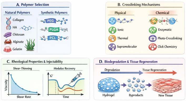
Schematic illustration of design principles for injectable biodegradable hydrogels. (A) Polymer selection and classification showing natural (collagen, HA, chitosan, alginate, gelatin) and synthetic (PEG, PLGA, PCL) components; (B) crosslinking mechanisms including physical (ionic, thermal, supramolecular) and chemical (enzymatic, photo-crosslinking, click chemistry) strategies; (C) rheological properties and injectability demonstrating shear-thinning behavior and modulus recovery post-injection; (D) biodegradation kinetics and timeline matching with tissue regeneration. HA: hyaluronic acid; PEG: poly(ethylene glycol); PLGA: poly(lactic-co-glycolic acid); PCL: poly(ε-caprolactone); G': storage modulus. Created with Canva.
In order to exploit the synergistic benefits of the two classes, hybrid systems that incorporate natural and synthetic components have been greatly developed. These composite hydrogels are expected to combine the strong and tunable mechanical behavior and reproducibility of synthetic polymers with comprehensive biological signaling and cell-instructive behavior of natural polymers [7]. For example, PEGylation of collagen or HA produces a matrix that is resistant to degradation by enzymes but contains important ECM signals to encapsulated cells. These hybrid strategies are the future of material design, which allows for the development of highly tailored microenvironmental settings depending on the particular use in tissue engineering [3]. A comparative overview of natural and synthetic polymers commonly employed in injectable hydrogel formulations is presented in Table 1, highlighting their respective advantages, limitations, and key tissue engineering applications.
Comparison of natural and synthetic polymers for injectable hydrogels.
| Polymer type | Examples | Advantages | Limitations | Key applications |
|---|---|---|---|---|
| Natural polymers ※ | Collagen, HA, chitosan, alginate, gelatin | Biocompatibility, bioactive signaling, cell adhesion | Batch variability, immunogenicity risk, and limited mechanical strength | Cartilage, bone, wound healing |
| Synthetic polymers | PEG, PLGA, PCL | Tunable properties, reproducibility, and low immunogenicity | Lack of bioactivity, slow degradation in some cases | Bone, neural, cardiac |
| Hybrid/Composite | PEG-collagen, GelMA | Combines bioactivity with mechanical tunability | Complex synthesis | Soft tissue, cartilage, skin |
※ Collagen and hyaluronic acid are the most commonly studied natural polymers to use as injectable hydrogels because of their natural ECM components and biological signaling properties.
The crosslinking mechanism of a polymer solution determines the conversion of this solution into a soluble system in a solid hydrogel network in situ, which is the main determinant of the kinetics of gelation, mechanical integrity, and biocompatibility of the system. The crosslinking techniques are broadly divided into physical and chemical techniques [8, 9]. In non-covalent interactions, physical crosslinking has the benefit of mild conditions of gelation, and non-toxic chemical crosslinkers and initiators are not required. Ionic crosslinking can be traditionally well illustrated by the response of the carboxylate groups of alginates and divalent cations (i.e., Ca2+) to form a gel in a few seconds to a few minutes [10]. To achieve thermal gelation, polymers with lower critical solution temperature (LCST) are used, including poly(N-isopropylacrylamide) (PNIPAM) or Pluronic F127, which remain in a liquid state at room temperature and undergo sol-gel transition at physiological temperature (37°C) [11]. Supramolecular crosslinking takes advantage of highly specific reversible interactions, including host-guest chemistry (e.g., cyclodextrin and adamantane) or hydrogen bonding in peptide assemblies. Physically crosslinked hydrogels are highly biocompatible and also have strong shear-thinning properties; however, as a rule, they have lower mechanical properties and can dissolve readily in vivo because their bonds are reversible [12].
Chemical crosslinking, in its turn, is a mechanism of building robust covalent bonds between polymer strands, obtaining a hydrogel based on high mechanical strength and tunable degradation curves [6]. Enzyme crosslinking involves the use of biological catalysts, e.g., transglutaminase or horseradish peroxidase (HRP), in the presence of hydrogen peroxide (H2O2) to catalyze bond formation under physiological conditions with the potential to offer high cytocompatibility and moderate to high mechanical stiffness [8]. Photo-crosslinking has been extensively used because of its ability to control the spatial and temporal gelation. GelMA systems or PEG diacrylate (PEGDA) polymerize (seconds to minutes) when subjected to ultraviolet (UV) or visible light in the presence of a photoinitiator [13]. Nevertheless, due to the possible cytotoxicity of free radicals and UV light, it is important to optimize it. Click chemistry has become an effective, bioorthogonal crosslinking approach in chemistry. Other reactions like thiol-Michael addition, Diels-Alder, and azide-alkyne cycloadditions are processed quickly and under mild conditions without the production of harmful byproducts, producing a hydrogel of high structural fidelity and mechanical stability [10, 11]. The decision between physical and chemical crosslinking or a two-crosslinking strategy, which uses both, is solely in accordance with the mechanical demands of the target tissue and the sensitivity of the biological cargo that is encapsulated in it [12]. The principal crosslinking mechanisms for injectable hydrogels, including their underlying chemistry, gelation times, mechanical properties, and self-crosslinking capabilities, are summarized comparatively in Table 2.
Comparison of crosslinking mechanisms for injectable hydrogels.
| Mechanism type | Chemistry | Gelation time | Mechanical properties (G') | Self-crosslinking capability |
|---|---|---|---|---|
| Physical-Ionic | Alginate + Ca2+, chitosan + tripolyphosphate | Seconds–minutes | Low–moderate (~10–1,000 Pa) | Limited (requires ion source) |
| Physical-Thermal | Pluronic F127, PNIPAM | Minutes | Moderate (~1–10 kPa) | Yes (temperature-dependent) |
| Physical-Supramolecular | Cyclodextrin-polymer, peptide assemblies | Seconds–minutes | Low–moderate | Yes (host-guest, H-bonding) |
| Chemical-Enzymatic | Transglutaminase, HRP/H2O2 | Minutes | Moderate–high (~1–20 kPa) | No (requires enzyme) |
| Chemical-Photo-crosslinking* | GelMA/PEGDA + UV/Vis | Seconds–minutes | Tunable (~0.1–100 kPa) | No (requires photoinitiator + light) |
| Chemical-Click chemistry | Thiol-ene, Diels-Alder, azide-alkyne | Minutes–hours | High (~5–50 kPa) | Limited (requires catalyst) |
* Photo-crosslinked GelMA-based hydrogels are already in a preclinical phase of testing as a cartilage repair and ocular surface reconstruction material.
Self-crosslinking is a new type of gelation method that allows polymers to crosslink without external crosslinkers, initiators, or energy input. This method has marked benefits for clinical translation, removing cytotoxic reagents and making the injection protocol a one component system. The spontaneous formation of imine-based bonds between amine-containing molecules (such as HA, oxidized alginate) and aldehyde-containing molecules (such as gelatin, chitosan) is the basis of Schiff base crosslinking [9]. Likewise, the boronic acid functionalized polymers can be self-crosslinked to form a pH-responsive gel by using cis-diol rich molecules such as the catechol functionalized polymers or natural polysaccharides, which are present in the surrounding environment in this case [12]. Another route of self-crosslinking involves the formation of disulfide bonds between the polymer chains comprising thiol groups, which occurs under ambient oxidation or under endogenous generation of the reactive oxygen species that catalyse the network formation, without the need of any further initiator [12]. The gelation time of such systems is usually on the order of seconds to minutes and can be controlled by adjusting the polymer concentration, degree of functionalization, and the microenvironmental conditions (e.g., ionic strength and pH).
The time from the mixing or injection of the precursors to the formation of a self-supporting gel network (gelation time) is an important design parameter that has a direct influence on clinical handling, cell viability, and implant localization. Gelation time less than 10 s could cause premature crystallization in the syringe/catheter, leading to needle occlusion and poor filling of the defect. On the other hand, too long a gelation time (> 30 min) allows for leakage of the material at the injection site, dilution by physiological fluids, and breakage of the structure [10, 14].
For most in situ tissue engineering applications, the ideal gelation time range is between 30 seconds and 10 minutes: this provides adequate time for the surgeon to manipulate the implant and to ensure that the network has formed before the implant is in place [10]. There are several approaches to tune gelation time: (i) polymer concentration and molecular weight, (ii) degree of functionalization, (iii) temperature, (iv) concentration of the catalyst and (v) pH and ionic strength, especially important when the gelation is induced by ionic and Schiff base crosslinking, where physiological conditions (pH 7.4, ionic strength ~150 mM) are often the optimum gelation environment [8, 10, 11].
Figure 1 provides a comprehensive schematic overview of the design principles for injectable biodegradable hydrogels, from polymer selection and crosslinking strategies to rheological behavior and biodegradation kinetics, which are discussed in detail in the following sections. The rheological nature of an injectable gel is the key determinant of its clinical utility, as it determines the behavior of the gel during extrusion with a syringe and its further behavior in vivo. A necessary condition for injectability is shear-thinning, or non-Newtonian reduction of viscosity due to applied shear stress [15]. In this property, the highly viscous hydrogel precursor to easily pass through fine-gauge needles when it is injected. The extent of shear-thinning can be commonly determined as a power-law fluid model with an index of shear-thinning behavior (n) below 1, showing shear-thinning; a smaller index (n) of shear-thinning is usually easier to administer and reduces the extensional forces, which can damage the encapsulated cells [16].
Once injected, the hydrogel should quickly regain its structural integrity to avoid leakage at the site of the defect and to give instant mechanical support. This would require optimization of the storage modulus (G'), which is the elastic, solid-like character of the gel, and the loss modulus (G''), which is the viscous, fluid-like character. To achieve effective tissue engineering, the G of the hydrogel should be carefully adjusted to the mechanical stiffness of the desired tissue [12]. An example of this is the use of hydrogels to repair the soft neural tissue [low G' (0.1–1 kPa)] to avoid the mechanical crushing of the fragile neurons, and to repair the load-bearing articular cartilage [high G' (100 kPa–1 MPa)] to resist physiological joint forces [17]. Moreover, the yield stress, i.e., the lowest stress needed to start flow, should be properly balanced. A high enough yield stress will also guarantee that the hydrogel is sturdy and in shape during rest due to gravity and gentle physiological actions, and yet low enough to be injected manually by the clinician [18]. The kinetics of recovery after injection are also crucial; self-healing hydrogels with fast recovery of modulus (within seconds to minutes) after the withdrawal of shear stress are very desirable, as they guarantee the localization and functionalization of the implant in the dynamic in vivo environment immediately [15, 16].
Injectability—the ability of an injectable hydrogel to flow through a clinically relevant needle gauge under manual or syringe pump-driven force without altering the integrity of the material or the viability of the encapsulated biological cargo—is the most fundamental determinant of the clinical utility of an injectable hydrogel. The ability to be extruded is not an absolute characteristic but rather a multifaceted parameter that includes the rheological behavior, the needle geometry, the extrusion force and the survivability of the cells. Extrusion force is usually determined by measuring the force needed to push the material into a syringe using a texture analyzer or rheometer with a syringe fixture, and is typically well below 10–20 N for manual injection through 18–23 G needles [15, 16].
The power-law index (n < 1) is a good marker of shear-thinning properties, but the injectability requirements also include a high yield stress to prevent gravitational collapse after injection and a fast modulus recovery to ensure that the material is localized in the defect site [15]. The shear rate imposed on the cells during passage through the needle lumen can reach values of more than 103 s–1, causing extensional stresses that can be harmful to the cell membranes, thereby further limiting the injectability window. Therefore, a complete injectability profile must require measurements of: (i) steady-shear viscosity at physiologically relevant shear rates (0.01–1,000 s–1); (ii) yield stress via oscillatory amplitude sweeps; (iii) thixotropic recovery time after cessation of shear; and (iv) cell viability following injection under clinically realistic shear conditions (extrusion) to ensure cell viability and biocompatibility after the injection [15, 18].
To enable a hydrogel to be used as a temporary scaffold in tissue engineering effectively, the biodegradation time of a hydrogel should be regulated to align with the pace at which new tissues are being formed. In case of an uncontrolled degradation rate of the hydrogel, it will not be able to offer sufficient mechanical stability and spatial orientation to the growing cells, which will cause defective collapse. On the other hand, too slow degradation may inhibit the deposition of ECM and the incorporation of tissue and may provoke a chronic foreign body response [1]. Biodegradation normally takes place through two major routes, which include hydrolytic and enzymatic degradation. Synthetic polyesters such as PLGA, PCL, etc., have hydrolytic degradation, wherein the ester bonds within the polymer backbone are broken by the presence of water molecules. The copolymer ratio (i.e., the ratio of hydrophilic lactic acid to the hydrophobic glycolic acid in the PLGA), the molecular weight, and the crosslinking density can all be adjusted to fine-tune the rate of hydrolysis [19].
Enzymatic degradation has been mainly applied in natural polymer systems, whereby a particular enzyme in the microenvironment of the body cuts through the polymer chains. As an illustration, HA is broken down by endogenous hyaluronidases, whereas collagen is broken down by collagenases [6]. In order to confer enzyme degradability to synthetic or highly crosslinked networks, scientists often functionalize the hydrogel backbone with matrix metalloproteinase (MMP)-sensitive peptide sequences. This will enable the hydrogel to be locally and specifically degraded by the enzymes released by migrating and proliferating cells to facilitate cell-driven remodeling that perfectly coordinates tissue regeneration [7]. More so, the biocompatibility of the degradation products is a crucial factor. One of the products of hydrolytic degradation of PLGA is lactic and glycolic acid, which may cause a local decrease in pH and, therefore, inflammation and cellular toxicity. To overcome this, it is common to use buffering methods, including the addition of basic inorganic nanoparticles (e.g., hydroxyapatite), to counteract the acidic byproducts and keep the physiological microenvironment under constant degradation [20]. Advanced biofunctionalization strategies transform injectable hydrogels from passive scaffolds into active therapeutic platforms. Figure 2 illustrates four major approaches: (Figure 2A) cell encapsulation with viability maintenance during injection, (Figure 2B) growth factor delivery via physical entrapment, chemical conjugation, and affinity-based controlled release, (Figure 2C) ECM mimicry through adhesive peptides, MMP-sensitive sequences, and decellularized ECM (dECM)-derived hydrogels, and (Figure 2D) immunomodulation via macrophage polarization modulation.
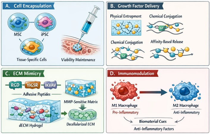
Biofunctionalization strategies for enhanced therapeutic efficacy. (A) Cell encapsulation approaches, including MSC, iPSC, and tissue-specific cell types with viability maintenance during injection; (B) growth factor delivery systems using physical entrapment, chemical conjugation, and affinity-based controlled release; (C) ECM mimicry through RGD, YIGSR, and IKVAV adhesive peptides, MMP-sensitive sequences, and dECM-derived hydrogels; (D) immunomodulation mechanisms depicting macrophage M1-to-M2 polarization modulation by material properties and anti-inflammatory factor incorporation. MSC: mesenchymal stem cell; iPSC: induced pluripotent stem cell; ECM: extracellular matrix; MMP: matrix metalloproteinase; dECM: decellularized extracellular matrix; RGD: Arg-Gly-Asp. Created with Canva.
There is no greater therapeutic benefit of injectable hydrogels than encapsulation and delivery of living cells directly to the site of injury. The most commonly used cell type, as it is the most versatile, is mesenchymal stem cells (MSCs) because of their strong multipotency, powerful paracrine signaling, and natural immunomodulatory activity. MSCs, when encapsulated in matrices like PEG, GelMA, or alginate, release a plethora of trophic factors that induce endogenous repair, angiogenesis, and inhibit harmful inflammation [1, 3]. iPSCs are a groundbreaking candidate for patient-specific therapies, whereby they represent a limitless supply of autologous cells that can be developed into all of the somatic lineages. Nevertheless, their differentiation can only be encapsulated in a highly informative hydrogel microenvironment to strictly regulate the differentiation paths and avoid teratoma formation [5].
Tissue-specific cells are commonly encapsulated to create specific repair: chondrocytes in the case of articular cartilage healing, osteoblasts in the case of bone healing, fibroblasts and keratinocytes in the case of skin repair, and cardiomyocytes in the case of myocardial infarction (MI) healing [15]. Another important issue with cell encapsulation is ensuring that the cells remain highly viable throughout the injection procedure. Shear and extensional stresses that occur during extrusion in a needle may cause drastic mechanical harm to the cell membrane, with the result being either apoptosis or necrosis. To address this, hydrogels are developed with a significant shear-thinning nature to lubricate the cells when they flow. Cytoprotective additives, like the ROCK inhibitor Y-27632, are also usually added to the precursor solution to inhibit anoikis (apoptosis caused by cell-matrix interaction loss) and improve post-injection survival. Cell seeding density is also important to achieve sufficient cell-cell communication and avoid nutrient and oxygen deficit inside the core of the avascular hydrogel [17, 20, 21].
In addition to offering a physical scaffold, injectable hydrogels can be utilized as advanced depots for localized and controlled delivery of strong growth factors, which are crucial in assembling intricate cellular processes like migration, proliferation, and differentiation. Vascular endothelial growth factor (VEGF) to induce angiogenesis, bone morphogenetic protein-2 (BMP-2) to induce osteogenesis, fibroblast growth factor (FGF) to induce cellular proliferation and wound healing, and transforming growth factor-beta (TGF-β) to induce chondrogenic differentiation are some of the important factors used as growth factors in the tissue engineering process [2, 10]. The delivery mechanism should be well-designed in order to avoid rapid diffusion and enzyme degradation of these short-lived proteins in the body.
The three most common types of controlled release mechanisms are physical entrapment, chemical conjugation, and affinity-based systems. Mechanical retention in the mesh of the hydrogel can tend to lead to a burst release profile that could be useful in immediate signaling, but does not achieve long-term levels of therapeutics. Chemical conjugation entails the conjugation of the growth factor to the polymer backbone, which ensures that the growth factor is released with sustained high release rates that depend on the rate at which the hydrogel breaks down [17, 22]. Affinity-based systems are a copy of the native ECM, which is based on particular binding interactions; in one example, the reversible, high-affinity sequestration of VEGF and FGF was achieved by including heparin or heparin-binding peptides in the hydrogel, enabling the system to protect these proteins and deliver them slowly upon demand by the cell [23]. The further development of regenerative strategies depends on dual and multiple factor delivery to replicate the signaling cascades of natural tissue development that are complex and spatiotemporal. For example, a sequential release of VEGF and the platelet-derived growth factor (PDGF) is applied so as to initially sprout new blood vessels and then to mature and stabilize them. On the same note, the delivery of BMP-2 and TGF-β using the method of co-delivery in spatially graded hydrogels is utilized to regenerate complex osteochondral interfaces [24].
The biochemical and structural complexity of the native ECM is a key requirement for an injectable hydrogel to serve as an artificial provisional matrix. Natural polymers can be extensively modified or completely synthetic, and have no particular sites for integrin to promote cell adhesion, which is critical for cell survival, spreading, and mechanotransduction. This is overcome by the functionalization of the hydrogel network with short synthetic adhesive peptides that mimic natural ECM proteins [3, 11]. The most frequent ones are the RGD (Arg-Gly-Asp) motifs, which are present in fibronectin, which binds many integrin receptors and are present in all cell adhesion. As the peptides can be more specific, tissue-specific peptides, such as YIGSR (from laminin) to promote endothelial and neural cell adhesion, and IKVAV (from laminin) to promote neural axon growth and guidance specifically, can be used [6].
Injectable fiber materials for ECM structural mimicry. The main drawback of traditional injectable hydrogels is their low mechanical strength and fracture toughness when compared with the fibrillar structure of native ECM with interwoven collagen fibers that offer anisotropic mechanical resilience. Micro- or nanoscale fibrous elements can be integrated into fiber-reinforced injectable hydrogels to compensate for this lack of structural hierarchy in native connective tissue. The short fibers (e.g., PCL, PLA, or collagen) can be added to the hydrogel precursors to create interpenetrating networks, substantially increasing tensile modulus, toughness, and resistance to crack propagation [25]. Other reinforcements are available, such as the high-aspect-ratio rod-like reinforcements, cellulose nanocrystals (CNCs), and nanofibrillated cellulose, which align during shear flow through the injection needle, forming anisotropic structures upon gelation that mimic the aligned collagen fibrils in tendons, ligaments, and cardiac tissue [25]. In recent advances, dECM-derived fibers have been investigated as both biomimetic mechanical reinforcement and biochemical cues, which provide tissue-specific cues [21]. Such fiber-reinforced systems are especially attractive to applications that require load-bearing, like cartilage, bone, and tendon regeneration, where the native ECM has a strong fibrous architecture and mechanical properties are anisotropic.
Adhesive cues require the hydrogel to be sensitive to cellular remodelling, in addition to dynamic ECM mimicry. Another way of facilitating migration of cells is to incorporate degradation-sensitive peptide sequences that are susceptible to degradation by MMPs (such as GGGPQGIWGQGGG) into the crosslinking junctions of the synthetic hydrogels, leaving room within the matrix for the proliferation and deposition of newly secreted ECM molecules [7]. This is the grand idea of biomimicry, and the use of dECM based hydrogel has gained huge popularity. The cellular component of tissues can be completely removed, leaving behind the complex milieu of the structural proteins, and the components are solubilized and delivered as an injectable and temperature-responsive hydrogel dECM. Their excellent biochemical cues and unmatched tissue-specific biochemical cues have shown remarkable potential in a variety of complex biomedical applications, such as the repair of cardiac and skeletal muscle, where the complex native architecture is hard to recapitulate synthetically [20, 21].
The multifaceted biofunctionalization strategies employed to enhance the therapeutic efficacy of injectable hydrogels are schematically depicted in Figure 2, encompassing cell encapsulation, controlled growth factor delivery, ECM mimicry, and active immunomodulation. Any biomaterial implantation will inevitably result in an immune response to it, which is initiated by the innate immune system. Traditionally, biomaterial design targeted immune evasion, but current designs are geared towards active immunomodulation to enlist the immune system to aid in positive tissue regeneration. The core of this process is the polarization of the macrophages. Macrophages are highly plastic: they can exist on a continuum between an M1 phenotype (pro-inflammatory secreting TNF-α and interleukin-6 (IL-6), clearing of debris and pathogen) and an M2 phenotype (anti-inflammatory secreting IL-10 and TGF-β, eliminating inflammation and facilitating tissue repair) [1, 2]. The chronic inflammation and fibrous encapsulation of the hydrogel are the effects of long-term M1 activation, but the transition to M2 is the key to successful integration and regeneration [10].
Hydrogels that are injectable can be designed to actively induce this M1-to-M2 transition. One method is the direct delivery and targeted release of anti-inflammatory molecules, including IL-4, IL-10, or corticosteroids like dexamethasone that highly bias the macrophages towards the pro-regenerative M2 state [26]. Moreover, the innate physical and chemical characteristics of the material per se have a significant effect on the behavior of immune cells. It has also been shown in the literature that soft hydrogels (low G) are associated with the promotion of the M2 phenotype, and stiff matrices with an increase in M1 activation. Other critical issues are surface chemistry, hydrophilicity, and the nature of degradation products. Also, macrophage morphology can be physically perturbed by introducing fibrous spaces or particular topographical geometries on the micro- and nanoscale that promote elongation associated with M2 polarization and establish an immunopermissive microenvironment that facilitates tissue repair [27].
Despite significant advances, each tissue-specific application of injectable hydrogels faces distinct critical limitations and translational challenges. Table 3 provides a comprehensive overview of these challenges across cartilage, bone, cardiac, neural, skin, and dental applications, along with key supporting references. Articular cartilage is a non-vascular, aneural tissue with a disastrously poor self-healing potential. Cartilage tissue engineering injectable hydrogels should have rigorous load-bearing conditions because the knee joint is subjected to compressive moduli of between 0.5 and 2 MPa during normal ambulation. The hydrogel should have strong viscoelastic characteristics in order to absorb the mechanical energy and shield the encapsulated chondrocytes [28]. The most notable polymer system in this area is HA and GelMA. HA is a significant portion of native cartilage ECM, which is important in lubrication and interacts with the CD44 receptors in order to sustain chondrocyte phenotype. Photo-crosslinked GelMA can be tuned to provide very soft stiffness, which is comparable to native cartilage mechanics. The expression of TGF-β3 within these matrices is commonly used to induce the firm chondrogenic differentiation of encapsulated MSCs [29]. Moreover, the repetitive loading cycle (repetitive) of the joints requires the application of self-healing hydrogels. Systems based on dynamically binding covalent bonds (e.g., imine, boronate ester bonds) are capable of automatically repairing micro-fractures caused by mechanical load, and hence the structural integrity of the implant during prolonged physiological loading [30, 31].
Skeletal tissue engineering must have scaffolds, which are mechanically stable but also osteoconductive and osteoinductive. Bone repair injectable hydrogel is normally developed as a composite material to address these needs. Inorganic phases are added to the polymeric network in order to obtain osteoconductivity, the most common of them being hydroxyapatite (HA) nanoparticles or bioactive glass. These minerals give a biomimetic scaffold that allows the adhesion, proliferation, and deposition of mineralized matrix by osteoblasts [31, 32]. Osteoinductivity can be provided by the provision of strong morphogens, mainly BMP-2, or by the joint action of bioactive ions like magnesium and zinc, which have been demonstrated to promote osteogenic pathways and bone consolidation. The key limitation of treating large bone defects is the inability to vascularize the bone quickly, resulting in core necrosis of the implant. To overcome this, more sophisticated injectable models use vascularization schemes, such as controlled release of VEGF, prevascularization micro-constructs, or co-delivery of angiogenic and osteogenic signals in space and time to maintain the simultaneous formation of a supporting vascular network and new bone formation [22, 33].
MI causes the irreversible death of billions of cardiomyocytes, which causes unfavorable remodelling of the ventricles, thinning of the walls, and subsequent heart failure. Injectable hydrogels provide a minimally invasive approach to the delivery of therapeutics to infarcted myocardium by endocardial or epicardial catheter-based injections. The acellular hydrogels are used to provide mechanical support to the weaker ventricular wall, in order to reduce the wall stress, pathological dilation, and aid homing of cells endogenously [2, 10]. In order to overcome the electrical uncoupling of fibrotic scar tissue, conductive hydrogel systems have been developed. These scaffolds can conduct electrical signals across the scar to stimulate the synchronous beating of endogenous cardiomyocytes, eliminating the probability of arrhythmias by incorporating conductive nanomaterials, such as carbon nanotubes, graphene oxide, or gold nanowires into the hydrogel matrix [17]. A significant problem in the context of exogenous cell delivery (e.g., iPSC-derived cardiomyocytes) is that of low cell retention and survival in the post-MI environment, which is hostile, ischemic, and inflammatory. The kinetics of fast gelation are important in reducing cell washout caused by the beating heart. Moreover, the hydrogels are functionalized to release anti-apoptotic factors such as insulin-like growth factor-1 (IGF-1) or are made with oxygen-releasing microparticles to maintain cell viability in the hypoxic core until host vascularization takes place [18, 22].
The central nervous system (CNS) poses special threats to regeneration because it is extremely soft, highly circuitous, and has inhibitory molecules that develop glial scars after injury. Neural repair injectable hydrogels should be ultra-soft (G' < 1 kPa) to be consistent with the mechanics of the brain and spinal cord tissue and avoid mechanical trauma. RADA16, which is a type of self-assembling peptide nanofiber (SA), is very effective in this regard. They create nanoscale fibrous structures that closely resemble the native neural ECM, which offer a permissive zone for neurite outgrowth. Targeted axon elongation is further increased by the functionalization of these peptides with IKVAV sequences [10, 34]. It is necessary to deliver neurotrophic factors, such as nerve growth factor (NGF), brain-derived neurotrophic factor (BDNF), and neurotrophin-3 (NT-3), to the lesion site to promote neuronal survival and axonal regeneration across the lesion site [35]. Moreover, neural regeneration has been proven to be stimulated by electrical stimulation. As a result, electrically conductive hydrogels (10–3–10–1 S/m) are being produced by using electrically conductive polymers such as polypyrrole or PEDOT or carbon-based nanomaterials such as graphene oxide. There is also a current investigation into piezoelectric biomaterials, which can generate transient electrical charges in response to sub-minimal mechanical deformations, to provide wireless, localized electrical stimulation to regenerating neurons [27].
Multifunctional interventions are necessary in skin regeneration, especially in non-healing wounds, e.g., diabetic foot ulcers and pressure ulcers, which are chronic wounds. Chronic wounds are typified by the presence of persistent inflammation, impaired angiogenesis, and excess exudate. Injectable hydrogels are the most appropriate advanced wound dressing since they keep the wound bed moist, absorb excessive exudates, and fit well into deep and irregular wound beds [26]. Antimicrobial functionalization is the most important since chronic wounds are highly prone to infection and biofilm development. It is done by either embedding silver nanoparticles, incorporating antimicrobial polymers that are inherently antimicrobial, such as chitosan, or by loading the broad-spectrum antimicrobial peptides [21]. Bilayer dermal-epidermal systems are designed for the case of full-thickness skin defects. The bottom layer, which can be made of collagen or HA, is intended to encourage infiltration and vascularization of dermal fibroblasts, whereas the top layer is designed to encourage seeding of the skin with keratinocytes and epidermal stratification, resulting in the restoration of the complex, stratified architecture of native skin [36, 37].
Craniofacial and dental tissues have unique regenerative challenges because they are complex composite tissues, composed of mineralized enamel and dentin, vascularized pulp and periodontal ligaments that hold teeth in place within the alveolar bone. The ability of injectable hydrogels to fill irregular root canal structures, extraction sockets, and periodontal defects that cannot be preformed with scaffolds has led to their widespread usage in dental tissue engineering [9, 21].
Pulp regeneration: To regenerate dental pulp in RCs, materials must promote angiogenesis, innervation, and odontoblast differentiation and withstand the narrow, tapered RCs. Injectable alginate, chitosan, and collagen-based hydrogels containing dental pulp stem cells (DPSCs) or stem cells from human exfoliated deciduous teeth (SHED) have been shown to have the ability to generate vascularized pulp-like tissue and lay down reparative dentin [21]. The co-delivery of pro-angiogenic factors such as VEGF and pro-odontogenic factors such as BMP-2 within these hydrogels supports the angiogenesis and dentin formation that are both necessary for pulp regeneration [38].
The aim of Periodontal Ligament and Alveolar Bone Regeneration is to restore the attachment lost due to periodontal disease. To regenerate the periodontal ligament–cementum–bone interface, stem cells of the periodontal ligament (PDLSCs) and growth factors (e.g., FGF-2, PDGF-BB) can be delivered through the injection of such hydrogels. The composite hydrogels based on bioactive glass or hydroxyapatite nanoparticles embedded in a gelatin or chitosan matrix are osteoconductive and mechanically stable, and the crosslinks can be degraded by enzymes thus allowing for the progression of the remodeling in a controlled manner as new periodontal attachment forms [31, 32]. These injectable systems offer a great benefit in minimally invasive periodontal regeneration procedures in which the trauma of surgery is reduced, and the better compliance of the patient is achieved when compared to conventional guided tissue regeneration membranes.
Enamel and dentin remineralization: The injectable hydrogels containing calcium phosphate precursors or bioactive ions (Mg2+, Sr2+, F–) can induce biomimetic remineralization of demineralized dentin and halt caries progression without the need to involve enamel. The fixation of the mechanical integrity in carious lesions by bottom-up mineralization via peptide amphiphile nanofiber hydrogels that template hydroxyapatite nucleation is a promising approach.
Key limitations: Although the preclinical results have been encouraging, there are a number of unique challenges that the oral environment poses for translation of a product to the market, including the presence of the oral microbiome (a challenge to avoid bacterial contamination and biofilm formation), the need for materials to resist masticatory forces (5–15 MPa compressive stress), and the regulatory complexities of combination products in a confined anatomical space. In addition, the long-term stability of the hydrogel-based dental restoration under cyclic wet-dry and thermal loads and changes in the oral cavity has not been sufficiently characterized [31].
The active and dynamic environment of the in vivo system frequently exposes implanted hydrogels to unremitting load, which results in micro-cracks and structural failure. Self-healing hydrogels solve this shortcoming by self-healing structural damage, thus increasing their service life. This is made possible by integrating dynamic bonds that are reversible. Dynamic covalent chemistry methods make use of bonds that can be broken and reformed under physiological conditions, including imine bonds (formation of Schiff bases between amines and aldehydes), boronate ester bonds, and disulfide exchange reactions. These are bonds that offer a balance of strong mechanical strength and high speed of reversibility [12, 30]. On the other hand, supramolecular self-assembly is based on non-covalent interactions, such as host-guest interactions (e.g., adamantane in the hydrophobic cavity of 8-cyclodextrin) and multiple arrays of hydrogen bonds and metal-ligand coordination. Such systems allow self-recovery without the interference of external stimuli [35]. The effectiveness of these materials is strictly measured by the rheology strain amplitude sweeps and step-strain recovery tests. The efficiency of optimal injectable systems has been in the recovery of the modulus with a modulus above 90 percent in minutes after the destructive shear strains are applied, and is therefore able to withstand the injection and subsequent physiological loading [36].
Smart hydrogels are programmable biomaterials which exhibit macroscopic mechanical, chemical or swelling changes when stimulated by particular environmental signals, providing on-demand controlled therapeutic delivery and adaptive tissue integration. These stimuli can be divided into physical, chemical, and biological classes of stimuli, each with varying advantages for specific regenerative interventions.
Physical stimuli: Temperature-responsive hydrogels are based on polymers with a LCST that change from sol to gel in response to the physiological temperature of 37°C when the temperature crosses this threshold. Polymers with a LCST that transition from sol to gel with increasing temperature, such as PNIPAM (LCST ≈ 32°C) and Pluronic F127, are used in these hydrogels as they remain liquid at room temperature of 20–25°C, but undergo a rapid sol-gel transition when the temperature exceeds 37°C [11]. This intrinsic thermosensitivity allows for easy mixing of therapeutic cargo at the benchtop before it is injected and removes the need for external crosslinkers. Photochromic materials, including azobenzene (which can isomerize under UV/visible light, changing the permeability of the gel) and spiropyran (which can open the ring upon UV light, causing dissolution of the gel), were used in light-responsive hydrogels for spatiotemporally controlled drug delivery and implant removal on demand [10]. Magnetic-responsive systems are based on the insertion of superparamagnetic iron oxide nanoparticles (SPIONs, Fe3O4, 10–50 nm) that, upon exposure to an external alternating magnetic field (AMF, 100–500 kHz), produce local hyperthermia or mechanically deform, and cause pulsatile drug release [39].
Chemical stimuli: Pathological tissues have different pH microenvironments, which are exploited by pH-responsive hydrogels. Extracellular acidosis (pH 6.5–6.8) is found in tumor tissue, in inflamed wounds, and in ischemic tissues (pH 5.5–6.5). Ionizable groups (such as carboxylic acids, pKa ~4–5, or amines, pKa ~9–10) induce charge-state transitions within these pH ranges that lead to a swelling-mediated burst release of therapeutics encapsulated within a hydrogel [8, 10, 40]. For instance, chitosan-based hydrogels become protonated in an acidic environment, resulting in electrostatic repulsion and enlargement of the space mesh, which promotes the diffusion of drugs. Ion-responsive systems such as alginate hydrogels gel in the presence of a physiological concentration of divalent cations (Ca2+ 5–10 mM) and crosslink in situ when exposed to the interstitial fluid of tissue [10]. Redox-responsive hydrogels take advantage of the 100- to 1,000-fold difference in the concentrations of glutathione (GSH) in intracellular (2–10 mM) and extracellular (2–20 µM) compartments. In the oxidizing extracellular space, the disulfide crosslinks remain intact and are cleaved in the reducing intracellular environment, resulting in triggered release of the intracellular payload [41].
Biological stimuli: Enzyme-responsive hydrogels contain peptide linkers that are designed to be selectively cleaved by the enzyme that is overexpressed in diseased or remodeling tissues. MMP-sensitive sequences (MMP-2/MMP-9-sensitive) are degraded by MMP-2 and MMP-9, which are selectively expressed in tumor microenvironments, infarcted myocardium, and in healing wounds, allowing site-specific drug release in conjunction with cellular remodeling activity [7]. Likewise, elastase-sensitive or hyaluronidase-sensitive linker groups allow for degradation in tissues where the enzyme levels are high.
Researchers are progressively designing complex biohybrid and composite spaces to solve the mechanical drawbacks and release kinetics of monolithic hydrogels, which are inherently single-phase. Microsphere reinforcement is a type of reinforcement that incorporates rigid polymeric microspheres (e.g., PLGA or PCL) into the soft hydrogel base. Not only does this substantially increase the compressive modulus of the composite, but it is also capable of offering sustained, dual-phase drug release profiles whereby the hydrogel would be used to provide an initial burst and microspheres provide long-term delivery [18, 33]. Nanofiber reinforcement makes use of electrospun nanofibers (PCL or PLA) or CNCs in order to form interpenetrating networks. These nanoscale reinforcements can be likened to collagen fibrils that are found in native ECM, and the tensile properties and toughness of the hydrogel are dramatically enhanced [25]. On the biological scale, organoid encapsulation approaches are driving tissue engineering for the repair of complicated organs. By using protective and instructive hydrogel carriers to entrap pre-formed mini-organ constructs (e.g., intestinal or liver organoids), researchers can package highly functional tissue units that can be rapidly integrated. In addition, multi-compartment materials like core-shell microspheres or spatially graded hydrogels can be used to spatially separate incompatible bioactives or generate biomimetic in situ gradients (e.g., a cartilage to bone gradient) within a single injectable construct [20].
The intersection of diagnostics/therapeutics has led to theranostic hydrogels that not only provide therapies, but also real-time disease progression and material degradation. Hydrogels that are imaging compatible are designed by embedding contrast agents within the polymer structure. The SPIONs are incorporated into magnetic resonance imaging (MRI)-visible hydrogels, whereas quantum dots or FITC-labeled polymers are used to enable the process of fluorescent tracking. Non-invasive visualization of the precise location, degradation rate, and integration of the hydrogel after injection is enabled by ultrasound-trackable formulations using echogenic microbubbles [42]. Going a step higher, biosensor integration entails the integration of miniaturized electrochemical glucose sensors, pH sensors, or oxygen sensors into the hydrogel matrix. This allows the local metabolic microenvironment to be monitored continuously and in real-time, giving invaluable information on tissue healing or disease recurrence [27]. The final product of this technology is the diagnostic-therapeutic system of a closed loop. On these sophisticated platforms, the built-in sensor identifies a given disease biomarker and automatically activates an on-demand drug release effect. One of the prototype solutions can be a glucose-reactive hydrogel that constantly checks the level of glucose in the blood and automatically transmits accurate doses of insulin only when hyperglycemia is observed, which also represents a groundbreaking way of managing diabetes [20].
Critical limitations and challenges in tissue-specific applications of injectable hydrogels.
| Tissue application | Critical weaknesses and limitations | Key references |
|---|---|---|
| Cartilage | Poor long-term mechanical durability under cyclic loading; inadequate recapitulation of zonal cartilage architecture (superficial, middle, deep zones); risk of fibrocartilage formation rather than hyaline cartilage; limited vascularization impairs nutrient delivery to encapsulated cells | [28–30] |
| Bone | Insufficient vascularization of large defects leading to core necrosis; mismatch between hydrogel degradation rate and bone remodeling timeline;potential immune reactions and fibrous encapsulation at the defect site; difficulty in achieving load-bearing mechanical strength comparable to native cortical bone | [31–33, 43] |
| Cardiac | Low cell retention and survival in hostile post-MI environment; risk of arrhythmia induction by conductive fillers; mechanical mismatch with beating myocardium; challenge of achieving electromechanical integration with host tissue | [2, 10, 17, 22] |
| Neural | Risk of glial scar formation and foreign body response; difficulty in guiding long-distance axonal regeneration across lesion site; potential neurotoxicity of conductive nanomaterials; mechanical mismatch with extremely soft neural tissue (E ~0.1–1 kPa) | [2, 10, 27, 35] |
| Skin | Susceptibility to bacterial colonization and biofilm formation in chronic wounds; excessive exudate absorption leading to hydrogel swelling and mechanical weakening; uneven vascularization in full-thickness defects; risk of hypertrophic scarring | [21, 26, 36, 40] |
| Dental | Hostile oral microbiome and contamination risk; inadequate mechanical strength under masticatory loads; long-term degradation under cyclic wet-dry and thermal oral environment; limited clinical evidence beyond Phase I trials | [9, 21, 31] |
Though tremendous success has been accomplished in the laboratory, many manufacturing, regulatory, and economic challenges have yet to be addressed for the translation of injectable biodegradable hydrogels to the medical clinic.
Manufacturing and scale-up challenges. A very important problem is the scale-up of manufacturing, particularly for systems containing natural polymers, which have natural batch-to-batch variation in molecular weight and degree of substitution, and contain traces of endotoxins. This difference in properties adversely affects the reproducibility of mechanical properties, gelation time, and biological activity, which are all within narrow limits for regulatory approval [1, 2]. It is important to have a set of GMP-compliant and absolutely reproducible synthesis protocols. Furthermore, the cost and operation of obtaining shelf-stable formulations that preserve polymers and bioactive cargo after reconstitution at the bedside involve complex lyophilization procedures, which further increases complexity [1, 2].
Sterilization-induced material degradation. Another significant impediment is the sterilization effects on material properties. Conventional procedures may be very harsh; gamma irradiation can result in chain cleavage and degradation of the molecular weight of polymers like HA, while ethylene oxide (EtO) sterilization can produce toxic molecules that can affect the cytocompatibility. For this reason, manufacturers often have to resort to very costly aseptic production processes, or to complex terminal sterilization methods without compromising the sensitive chemistry of the hydrogel [44]. This material incompatibility of the sterilization material is a significant translational challenge because there is no universally accepted sterilization protocol which gives preservation of the structural integrity and biological activity of various hydrogel chemistries.
Regulatory complexity and jurisdictional ambiguity. The advanced materials are known for their confusing regulatory process, set up by the FDA (Food and Drug Administration) and EMA (European Medicines Agency). Injectable hydrogels can also be used as combination products (device, biologic, and/or drug) with regulatory confusion about jurisdiction and delays in approvals [10]. In the case of substantial equivalence to an existing device, acellular hydrogels would be less complicated and require the pathway known as the 510(k) pathway; new materials typically would go through the more involved and costly Premarket Approval (PMA) pathway. Hydrogels with live cells in the European Union are designated as Advanced Therapy Medicinal Products (ATMPs), which are exposed to the greatest regulatory oversight in terms of safety, efficacy, and mechanism of action [10]. However, the absence of standardized international characterization, testing, and clinical drug evaluation of injectable hydrogels further hinders multinational clinical trials and market access.
Economic and reimbursement barriers. Clinics are not only affordable, but an important part of adoption. GMP-grade biologics are costly, the autologous cell expansion process is complex and involves scarce resources, and the manufacturing processes are complex. For popularizing such therapies, the developer should be able to demonstrate that the initial costs will be recovered in the long-term savings, measured by reduced hospital visits, reduced re-operation rates, and the huge improvement in patient quality of life that such treatments will provide the developer with good reimbursement routes with health care payers [17]. Long-term (5–10 years) clinical outcomes are available for a few hydrogel systems; however, this results in a limited evidence base for favorable reimbursement by insurance providers and national health services for most hydrogel systems. The current clinical trial landscape for injectable hydrogels encompasses several tissue-specific applications at various stages of development. Table 4 summarizes representative clinical applications, including the hydrogel system, cell or growth factor delivery, clinical stage, and key outcomes reported to date.
Summary of clinical applications of injectable hydrogels.
| Issue type | Hydrogel system | Cell/GF delivery | Clinical stage | Key outcomes |
|---|---|---|---|---|
| Cartilage | Hyaluronic acid (HA) hydrogel | TGF-β3, chondrocytes | Phase II/III clinical trials | Improved cartilage regeneration, reduced pain scores |
| Bone | Calcium phosphate/collagen composite | BMP-2 | Phase II clinical trials | Accelerated bone union, osteoconductive scaffold integration |
| Cardiac | Alginate hydrogel (IK-5001) | None (acellular) | Phase II (AUGMENT-HF) | Reduced infarct expansion, improved LV geometry |
| Wound healing | Collagen/HA wound filler | PDGF-BB (becaplermin) | FDA-approved | Accelerated chronic wound closure in diabetic ulcers |
| Neural | Self-assembling peptide (RADA16) | NGF | Phase I | Safety established; early signs of functional improvement |
Clinical trial landscape. However, the current clinical trials pipeline remains, and several hydrogel systems are being developed for cartilage, bone, cardiac, and wound healing applications, as outlined in Table 4. Most of the trials, however, are still in the early stages (Phase I/II), and a few Phase III randomized controlled trials have shown a superiority over the standard of care. In addition to the wide range of patients, defect sizes and co-morbidities, the diseases included in each study vary in the breadth or narrowness of their scope, which makes it difficult to perform a meta-analysis and evidence synthesis [17].
Injectable biodegradable hydrogels are a breakthrough technology in minimally invasive tissue engineering. These materials provide highly tailorable in situ-forming scaffolds that can adhere to irregular defect structures with a biomimetic microenvironment that supports cellular regeneration by integrating the complex polymer chemistry and dynamic biological systems in a seamless manner. This review has outlined in a systematic way the critical design principles, and it goes up to the wise choice of natural, synthetic, and hybrid polymers and the strain-controlled crosslinking processes, rheological properties, and degradation kinetics. Moreover, by combining advanced biofunctionalization approaches, such as directed cell encapsulation, spatiotemporally regulated growth factor delivery, mimicry of the ECM, and active immunomodulation, these hydrogels have been upgraded to not only passive fillers but also active regulators of tissue repair in a wide range of applications in cartilage, bone, cardiac, neural, and skin regeneration.
Nevertheless, even though these technologies have experienced deep-rooted preclinical success, there are a number of contemporary constraints that hinder the smooth transfer of the technology. The most common is a mechanical incompatibility between hard and soft tissues, where, unfortunately, mechanical incompatibility persists between soft hydrogels and hard and load-bearing native tissues, which tends to undermine stability and mechanical integrity in vivo. In addition, the issue of possible immunogenicity of degradation products, the lot-to-lot variability of natural polymers, and the unimaginable regulatory complexity of the combination products and cell-bearing matrices remains a major impediment to commercialization.
In the future, the research directions should be realized on how to overcome these barriers by converging injectable hydrogels with other advanced technologies. The fusion of 4D bioprinting and injectable hydrogel technology is a revolutionary territory. Compared with conventional 3D bioprinting, which produces rigid scaffolds with predefined geometries, 4D bioprinting also adds the fourth dimension, timing; that is, shape-morphing hydrogels which can change their shape in response to physiological stimuli after implantation [12]. In this paradigm, hydrogels are designed to possess anisotropic swelling capabilities or contain shape-memory polymers with programmed in vivo changes in shape when exposed to temperature, pH or enzymatic changes. A flat injectable hydrogel sheet could, for example, self-fold to form a tubular conduit after injection into a nerve guidance channel, or a bone defect filler could expand to conform to the shape of an irregular fracture site when it is heated to body temperature. This allows for the elimination of pre-surgical customization of scaffolds and the ability to deliver complex and patient-specific geometries using narrow gauge needles. With strategic patterning of crosslinking density, fiber orientation or stimuli-responsive polymer domains in the hydrogel precursor, the programmable shape transformation is unprecedented and capable of adapting to the dynamic tissue remodeling environment.
3D: three-dimensional
BMP-2: bone morphogenetic protein-2
CNCs: cellulose nanocrystals
dECM: decellularized extracellular matrix
ECM: extracellular matrix
FGF: fibroblast growth factor
G': storage modulus
GelMA: gelatin methacryloyl
HA: hyaluronic acid
IL-6: interleukin-6
LCST: lower critical solution temperature
MI: myocardial infarction
MMP: matrix metalloproteinase
MSCs: mesenchymal stem cells
PCL: poly(ε-caprolactone)
PDGF: platelet-derived growth factor
PEG: poly(ethylene glycol)
PLGA: poly(lactic-co-glycolic acid)
PNIPAM: poly(N-isopropylacrylamide)
SPIONs: superparamagnetic iron oxide nanoparticles
TGF-β: transforming growth factor-beta
UV: ultraviolet
VEGF: vascular endothelial growth factor
EA: Conceptualization, Investigation, Writing—original draft, Visualization, Writing—review & editing, Project supervision. DA: Supervision, Writing—review & editing. Both authors read and approved the submitted version.
The authors declare that there are no conflicts of interest.
Not applicable.
Not applicable.
Not applicable.
Not applicable.
Not applicable.
© The Author(s) 2026.
Open Exploration maintains a neutral stance on jurisdictional claims in published institutional affiliations and maps. All opinions expressed in this article are the personal views of the author(s) and do not represent the stance of the editorial team or the publisher.
Copyright: © The Author(s) 2026. This is an Open Access article licensed under a Creative Commons Attribution 4.0 International License (https://creativecommons.org/licenses/by/4.0/), which permits unrestricted use, sharing, adaptation, distribution and reproduction in any medium or format, for any purpose, even commercially, as long as you give appropriate credit to the original author(s) and the source, provide a link to the Creative Commons license, and indicate if changes were made.



View: 691
Download: 16
Times Cited: 0


