Review

Affiliation:
1Department of Manufacturing Pharmacy, Faculty of Pharmacy, Mahidol University, Bangkok 10400, Thailand
Email: mudassir.far@student.mahidol.ac.th
ORCID: https://orcid.org/0000-0002-0586-3476
Affiliation:
2Department of Pharmaceutics, Faculty of Pharmacy, Bahauddin Zakariya University, Multan 66000, Pakistan
Explor BioMat-X. 2026;3:101368 DOI: https://doi.org/10.37349/ebmx.2026.101368
Received: April 15, 2026 Accepted: June 11, 2026 Published: June 24, 2026
Academic Editor: Vasif Hasirci, Middle East Technical University, Turkey
Hydrogels are increasingly explored as functional wound-dressing materials because they combine high water content, biocompatibility, structural tunability, and the ability to localize therapeutic delivery at injured tissue. As biomaterial platforms, hydrogel dressings can maintain a moist microenvironment, absorb exudate, protect the wound bed, and carry bioactive agents that modulate infection, inflammation, oxidative stress, and tissue regeneration. This review examines hydrogel wound dressings from a materials centered perspective. First, skin structure, wound-healing physiology, and major barriers to repair are outlined to define the biological requirements for effective dressings. Next, the chemical composition of natural, synthetic, and composite hydrogels, their crosslinking strategies, swelling behavior, and drug-loading and release mechanisms are discussed in relation to wound healing performance. Recent progress in infection responsive, stimuli responsive, growth factor delivering, antimicrobial peptide loaded, and self-healing hydrogel systems is then summarized. The present review highlights how composition, network architecture, and responsiveness govern biomedical function and localized drug delivery in wound care. These insights provide a materials centered framework that connects hydrogel composition, network architecture, responsiveness, and localized delivery behavior with wound healing performance, thereby supporting the rational design of next generation hydrogel biomaterials for difficult to heal wounds.
Hydrogel wound dressings are best understood as functional biomaterials for localized therapy at injured skin rather than as classical transdermal systems designed for systemic delivery across intact tissue. In wound care, biomaterials maintain a hydrated protective interface while delivering bioactive agents directly to the wound bed to control infection, attenuate inflammation, manage oxidative stress, and promote regeneration. These materials centered view positions hydrogel dressings at the intersection of wound-healing biology, soft biomaterials design, and localized drug delivery [1]. Unlike previous reviews that mainly summarize hydrogel dressings according to polymer type, wound healing stage, or therapeutic cargo, this review emphasizes the relationship between hydrogel composition, network architecture, responsiveness, and localized drug delivery performance in wound care [2]. The unique contribution of this article is to integrate materials design principles with wound healing requirements, showing how crosslinking chemistry, swelling behavior, drug loading strategy, and stimuli responsive mechanisms collectively determine biological performance. By connecting hydrogel structure to practical wound dressing functions, this review provides a material centered framework for designing next generation hydrogel systems for infected, diabetic, and difficult to heal wounds. To maintain focus, this review emphasizes hydrogel systems designed for wound bed application and localized therapeutic delivery rather than general hydrogel based drug delivery platforms for unrelated administration routes.
The skin is the body’s largest organ and acts as a dynamic barrier against physical, chemical, and microbial insults while limiting trans epidermal water loss [3]. Structurally, it consists of the epidermis, dermis, and hypodermis, each of which contributes to barrier integrity, immune defense, and tissue homeostasis. Because the skin is both a barrier and a therapeutic target, its architecture is relevant not only to conventional transdermal delivery but also to wound-healing biomaterials. In hydrogel dressings, transport is directed primarily toward the injured tissue and wound microenvironment rather than across intact skin into the bloodstream. Nevertheless, skin structure, hydration, and local diffusion pathways remain important design considerations because they influence drug retention, tissue contact, and local therapeutic performance at the wound interface [4].
A wound can be broadly defined as any injury or disruption to the integrity of biological tissues, including the skin, mucous membranes, and internal organs. Damage occurs when the protective epithelial covering is broken, thereby impairing the structure and function of the underlying tissues. Prompt restoration of the injured barrier is essential for maintaining homeostasis because untreated wounds may lead to infection and other complications. Wounds range from minor tissue disruptions, such as incisions, to extensive tissue damage, such as burns. Accurate wound assessment is therefore essential for selecting appropriate treatment strategies [5]. Wounds can be classified according to their nature and healing pattern as either acute or chronic. Acute wounds usually heal within 8–12 weeks and typically result from mechanical injuries such as abrasions, avulsions, crush injuries, cuts, fishhook injuries, incisions, and lacerations [6]. By contrast, chronic wounds heal slowly, often lack a predictable healing timeline, and frequently recur. Delayed healing is associated with conditions such as diabetes, heavily exuding wounds, persistent infection, and antibiotic resistance [7].
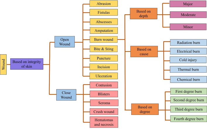
Despite the high prevalence, complexity, and diverse causes of wounds, establishing a universally accepted classification system remains challenging. As summarized in Figure 1, wound classification remains important for diagnosis, management, and treatment because it helps anticipate infection risk and guides therapeutic selection [8]. Burn wounds represent a clinically important category of skin injury because they involve thermal, chemical, electrical, or radiation related tissue damage and may vary substantially in depth and severity. From a dressing-design perspective, burn wounds often require protection from infection, maintenance of a moist environment, absorption of exudate, pain reduction, and support for re-epithelialization [9]. Hydrogel dressings are therefore relevant to burn management because their high-water content can provide cooling, hydration, and a protective interface while enabling localized delivery of antimicrobial, anti-inflammatory, or regenerative agents. However, the selection of a hydrogel system should consider burn depth, exudate level, infection risk, and the need for mechanical stability.
Healthy skin maintains a delicate balance between the epidermal and dermal layers. When this protective barrier is disrupted, the wound healing cascade is initiated and proceeds through hemostasis, inflammation, proliferation, and remodeling. These stages involve cell migration, angiogenesis, re-epithelization, collagen deposition, and extracellular matrix remodeling. Numerous growth factors in these events, as summarized in Table 1 [10]. Hemostasis begins with vasoconstriction, clot formation, and the release of mediators that promote subsequent healing events [11]. The inflammatory phase includes an early neutrophil dominated response followed by later monocyte and macrophage activity. During the proliferative phase, fibroblasts migrate to the wound bed, deposit extracellular matrix, and support new tissue formation. The final remodeling phase, illustrated in Figure 2, may last for weeks or longer and involves collagen synthesis, reorganization, and degradation under cytokine regulation [12]. Together, these tightly regulated events restore tissue integrity and improve the mechanical strength of healed skin. These biological events define the key design requirements for hydrogel wound dressings, including moisture regulation, exudate management, antimicrobial protection, inflammation control, oxygen permeability, mechanical flexibility, and support for re-epithelialization and tissue remodeling.
Various growth factors involved in wound healing.
| Sources | Growth factors | Properties |
|---|---|---|
| Macrophages, lymphocytes, keratinocytes, fibroblasts | GM-CSF | Promotes epidermal cell proliferation |
| Macrophages, lymphocytes | IL-1 | Promotes fibroblast proliferation and neutrophil chemotaxis |
| Platelets, macrophages, epithelial cells | TGF-alpha | Promotes cell proliferation and granulation tissue formation |
| Fibroblasts, neutrophils, keratinocytes, platelets | TGF-beta | Supports angiogenesis and collagen metabolism |
| Fibroblasts and plasma | IGF-1 | Mediates fibroblast proliferation and supports collagen and sulfated proteoglycan synthesis |
| Platelets, endothelial cells, fibroblasts | PDGF | Stimulates collagen metabolism and the proliferation of fibroblasts and neutrophils |
| Lymphocytes, fibroblasts, and monocytes | G-CSF | Promotes the production and function of monocytes and neutrophils |
| Mast cells, T lymphocytes, macrophages | TNF | Promotes fibroblast proliferation |
| Macrophages, keratinocytes, platelets | EGF | Promotes keratinocyte proliferation, differentiation, and migration at the wound site |
| Endothelial cells, smooth muscle cells, macrophages, fibroblasts | FGF | Promotes angiogenesis, fibroblast and epithelial cell proliferation, and wound contraction |
| Endothelial cells, keratinocytes, fibroblasts, tumor cells | HGF | Promotes neovascularization, granulation tissue formation, and re-epithelialization |
| Fibroblasts | KGF | Promotes keratinocyte proliferation and migration |
| Platelets, neutrophils | VEGF | Promotes collateral vessel formation and angiogenesis |
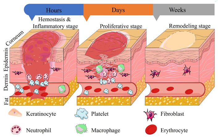
Stages of wound healing. Adapted with permission from https://doi.org/10.3390/life11070665. © 2021 by the authors. Licensed under a CC-BY 4.0.
Hydrogels are three dimensional crosslinked polymeric networks that absorb and retain large amounts of water while maintaining structural integrity. In wound dressings, their high water content, soft tissue like properties, and tunable porosity support moisture balance, exudate absorption, oxygen exchange, and localized delivery of therapeutic agents. Major hydrogel based wound dressing systems and their localized delivery functions are summarized in Table 2. The swelling and deswelling behavior can be regulated by polymer composition, crosslinking density, pH, ionic strength, temperature, and wound specific biochemical cues (Figure 3). Therefore, hydrogel design should be considered in relation to wound bed requirements, including hydration, infection control, inflammation modulation, mechanical conformity, and controlled release. To clarify the differences among responsive hydrogel systems, Table 3 compares their main triggers, release behaviors, wound care relevance, and limitations.
Hydrogel based wound dressing systems for localized drug delivery.
| Hydrogel system | Therapeutic function | Role | Mechanism | Application |
|---|---|---|---|---|
| Chitosan based hydrogel [13] | Antibacterial/anti-inflammatory agents | Infection control and inflammation reduction | Diffusion, degradation, or pH-responsive release | Infected and diabetic wounds |
| Alginate based hydrogel [14] | Antimicrobials, growth factors, ions | Exudate absorption and moist healing | Swelling controlled release | Exuding wounds |
| Hyaluronic acid hydrogel [15] | Growth factors, antioxidants, peptides | Cell migration and tissue regeneration | Enzyme- or degradation-mediated release | Chronic wounds |
| Gelatin/collagen hydrogel [16] | Growth factors, cells, hemostatic agents | ECM support and granulation tissue formation | Entrapment and sustained diffusion | Regenerative wound dressings |
| Composite hydrogel [16] | Antimicrobials, antioxidants, nanozymes | Multifunctional wound repair | Combined swelling, degradation, and stimuli response | Infected diabetic wounds |
| Self-healing hydrogel [17] | Antibacterial nanoparticles or peptides | Adhesion, flexibility, and repeated wound movement | Dynamic covalent/reversible bonding | Irregular or mobile wound sites |
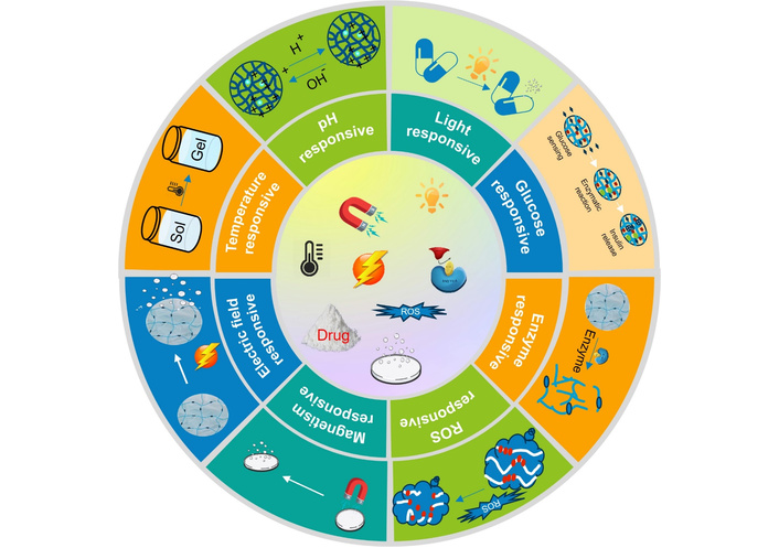
Classification of smart, responsive hydrogels for wound healing. ROS: reactive oxygen species; sol: solution.
Comparison of major responsive hydrogel types for wound dressing and localized drug delivery.
| Hydrogel type | Trigger | Typical release behavior | Wound-care relevance | Limitation |
|---|---|---|---|---|
| pH responsive hydrogels | Acidic/basic wound pH | pH triggered swelling, degradation, or bond cleavage | Infection and chronic wound microenvironments | Variable wound pH may affect predictability |
| Temperature responsive hydrogels | Local temperature change | Sol–gel transition or thermally controlled diffusion | Injectable or in situ forming dressings | Limited response range near physiological temperature |
| Glucose responsive hydrogels | Elevated glucose/glucose oxidase reaction | Glucose-dependent release and antibacterial activity | Diabetic wound healing | Requires careful control of oxidative by-products |
| ROS responsive hydrogels | Excess reactive oxygen species | Oxidative degradation and drug release | Inflamed and infected wounds | Over-response may weaken the network |
| Enzyme-responsive hydrogels | Bacterial enzymes or matrix metalloproteinases | Enzyme mediated degradation or cargo release | Infection responsive and chronic wound therapy | Enzyme levels vary between wounds |
| Multi stimuli responsive hydrogels | Combined pH, glucose, ROS, enzymes, or temperature | More selective and adaptive release | Complex chronic wound microenvironments | More complex formulation and validation |
Multifunctional hydrogel biomaterials can integrate several wound healing functions within a single network. For example, injectable chitosan phenylboronic acid hydrogels incorporating catechol and epigallocatechin gallate groups can scavenge reactive oxygen species and reduce oxidative stress. Self-healing hyaluronic acid hydrogels releasing salvianolic acid B can attenuate inflammation and accelerate diabetic wound repair in vivo. Antimicrobial effects can arise intrinsically, as in carboxymethyl chitosan hydrogels that inhibit Escherichia coli and Staphylococcus aureus, or through photothermal strategies using polydopamine nanoparticles. Rapid hemostasis may be promoted by bioadhesive catechol modified chitosan systems, whereas magnetic curcumin releasing nanocomposite hydrogels can stimulate collagen maturation and dermal reconstruction during the proliferative phase [18]. Li et al. reported a multifunctional fluorescent hydrogel containing a multi enzyme like nanocomposite for diabetic wound healing; the fabrication concept and cascade mechanism [19].
Hydrogels can be prepared from natural biopolymers such as polysaccharides (alginate, chitosan, cellulose, and amylopectin) and proteins (collagen and gelatin), as well as from synthetic polymers such as poly(ethylene glycol) (PEG), poly(vinyl alcohol) (PVA), and poly(acrylic acid) [20]. Composite hydrogels that combine natural and synthetic polymers may offer the advantages of both classes, as summarized in Table 4. Selecting an appropriate polymer system remains challenging because the structural diversity of available materials must be balanced against the chemical, mechanical, biological, and interfacial properties required for a given wound application. Thus, the selection of natural, synthetic, or composite hydrogel matrices should be guided by the wound type, exudate level, infection risk, required residence time, mechanical demands, and intended therapeutic release profile.
Natural and synthetic polymers used in hydrogel formulations and their properties.
| Polymers | Properties | Uses | Sources | Reference |
|---|---|---|---|---|
| Natural polymers | ||||
| Carrageenan | Highly stable, biodegradable, biocompatible, bioadhesive, antibacterial, and characterized by high tensile strength | Stimulates angiogenesis and re-epithelialization and supports wound healing | SeaweedsAlgaeRed marine algae | [21] |
| Guar gum | Non-ionic, non-toxic, hydrophilic, biocompatible, and biodegradable | Promotes epithelialization, differentiation, and regeneration | Cyamopsis tetragonolobus | [22] |
| Chitosan | Biocompatible, biodegradable, antibacterial, and anti-inflammatory | Promotes keratinocyte and fibroblast activity and supports wound healing and tissue engineering | Fungal cell wallCrustacean exoskeleton | [23] |
| Sodium alginate | Biocompatible and biodegradable; forms a hydrophilic, porous gel with high swelling capacity | Absorbs wound fluid and facilitates the healing process | Algae | [24] |
| Hyaluronic acid | Stimulates cell motility and is biocompatible, non-immunogenic, biodegradable, and strongly hydrophilic | Accelerates wound closure and promotes wound healing, as reflected by increased neo-epidermal thickness | Animal tissuesBacterial fermentation | [25] |
| Collagen | Natural polymer that is biocompatible, hemostatic, non-toxic, and able to support tissue development and fibroblast activity | Promotes epithelialization and wound healing | BovineMarine | [26] |
| Dextran | Biocompatible, biodegradable bacterial hydrophilic polysaccharide | Promotes angiogenesis, skin regeneration, and tissue repair | LactobacilliLeuconostoc species | [27] |
| β-Glucan | Antibacterial, antiviral, anti-inflammatory, biocompatible, and capable of enhancing immune responses | Can reinitiate the healing process and thereby reduce wound size | Cell wall of bacteriaLichensOats and barley | [28] |
| Synthetic polymers | ||||
| Polyethylene glycol | Hydrophilic polymer, biodegradable, and non-toxic | Shows potential in wound healing applications | Condensed ethylene oxide | [29] |
| Polyvinyl alcohol | High elasticity, water retention capacity and tensile strength | Accelerates chronic diabetic wound healing | Polyvinyl acetate | [30] |
| Polyvinylpyrrolidone | Highly stable, non-toxic, and biocompatible | Promotes burn wound healing | N-Vinyl-2-pyrrolidone monomer | [31] |
| PNIPAAM | Thermally stable, hydrophilic polymer with high mechanical strength and good biocompatibility | May reduce scar formation | Free radical polymerization of N-isopropylacrylamide | [32] |
Composite hydrogels are particularly important in wound dressing design because they combine the biological advantages of natural polymers with the mechanical strength, tunability, and reproducibility of synthetic polymers. Natural components such as chitosan, alginate, collagen, gelatin, and hyaluronic acid can improve biocompatibility, biodegradability, cell adhesion, hemostasis, and intrinsic wound healing activity. Synthetic polymers such as PEG, PVA, polyacrylic acid, and PNIPAAM can improve mechanical stability, elasticity, swelling control, and controlled release behavior. In composite systems, the balance between natural and synthetic components allows modulation of porosity, degradation rate, adhesive strength, exudate absorption, and drug-release kinetics. Such hybrid networks are therefore useful for infected, exuding, diabetic, and irregular wounds where a single polymer hydrogel may not provide sufficient mechanical durability and biological activity. Natural polymer based hydrogels are often favored because of their biodegradability, biocompatibility, relatively low production cost, and environmental friendliness. Biodegradable systems can also provide improved flexibility and better control over drug stability and diffusion. In some cases, they enable site-specific delivery when the matrix is degraded by biological triggers such as microbial enzymes [33]. These systems may respond to enzymes, temperature, pH, and other cues while also exhibiting bioadhesive behavior, thereby supporting self-regulated delivery. Techniques such as grafting, physical blending, and block copolymerization can be used to improve mechanical performance and tune biodegradation rates. Naturally derived polymers typically show excellent biocompatibility, low cytotoxicity, minimal immunogenicity, and the ability to promote cell adhesion, tissue proliferation, and regeneration. In particular, polysaccharide-based hydrogels have emerged as attractive wound biomaterials, as illustrated in Figure 4 [34, 35].
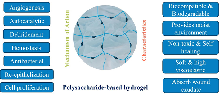
Properties of polysaccharide-based hydrogels and their mechanisms in wound dressing applications.
Hydrogel synthesis methods can generally be classified into physical crosslinking and chemical crosslinking. Physical crosslinking involves reversible interactions such as ionic interactions, hydrogen bonding, hydrophobic association, and complex coacervation. Chemical crosslinking involves covalent bond formation between polymer chains through chemical crosslinkers, enzymatic reactions, photo-polymerization, or irradiation induced free radical reactions. Therefore, irradiation crosslinking is best considered a chemical crosslinking method because high energy radiation generates reactive radicals that form covalent network structures. The choice of crosslinking method should balance mechanical stability, biocompatibility, sterilization compatibility, degradation behavior, and the ability to preserve the activity of incorporated therapeutic agents for wound dressing applications.
Physical crosslinking includes ionic crosslinking, complex coacervation, and hydrogen-bond-based network formation. Ionic crosslinking relies on electrostatic interactions between oppositely charged polymers to form hydrogels. Pessoa et al. developed ionically crosslinked alginate-chitosan hydrogel beads for oral insulin delivery [36], while carrageenan-based hydrogels have been fabricated using KCl and CaCl2 as crosslinkers [37]. The main advantages of ionic crosslinking are mild reaction conditions, avoidance of potentially toxic chemical crosslinkers, cost-effectiveness, and ease of preparation. However, ionically crosslinked networks often exhibit relatively weak mechanical properties and may be unstable under conditions that disrupt ionic interactions [38].
Complex coacervation produces hydrogels through electrostatic complexation between oppositely charged polyelectrolytes. Zhao et al. [39] developed multilayer hydrogel beads composed of chitosan, alginate, and octenyl succinic anhydride starch encapsulating Perilla frutescens L. essential oil using this approach. The method offers facile preparation, good cytocompatibility, and high loading capacity for bioactive agents, although the resulting coacervate systems can be highly sensitive to pH, polymer concentration, temperature, and ionic strength. Hydrogen bonding can also create physically crosslinked networks between polymers capable of forming reversible interactions. For instance, a dual-crosslinked injectable carboxymethylated starch hydrogel incorporating Ca2+ and Mg2+ ions promoted in situ bone formation by supporting cell migration and new bone regeneration after implantation [40]. Although hydrogen bonded hydrogels can be prepared using simple and nontoxic chemistry and may display tissue like, cell responsive behavior, their stability can be compromised under physiological conditions such as changing pH or solvent environments.
Chemical crosslinking forms covalent bonds between polymer chains, commonly through bifunctional crosslinkers such as glutaraldehyde. Gelatin hydrogels, which are widely used in biomedical applications, often require covalent crosslinking to improve thermal stability. Glutaraldehyde has been used to provide functional groups for covalent immobilization in chitosan-based bead systems [41]. For wound dressing applications, crosslinker selection should also consider network stability, residual reagent, and biocompatibility. Irradiation crosslinking uses high-energy gamma or UV irradiation to generate free radicals on polymer chains, thereby forming crosslinked networks. Wahba reported that incorporating polyethylene glycol diacrylate improved the material properties of PEGDA/gelatin hybrid hydrogels, with the final characteristics being tunable through changes in irradiation dose and PEGDA/gelatin composition [42]. This approach enables precise control over hydrogel properties and facilitates cell encapsulation. Although the required equipment can be complex and expensive, irradiation techniques offer a clean and initiator free route for hydrogel fabrication. Prolonged irradiation may, however, induce polymer degradation.
The effective delivery of therapeutic agents from a hydrogel matrix depends strongly on the chemical and physical characteristics of both the cargo and the network. Three main drug-loading strategies are commonly used: diffusion, entrapment, and tethering. In the diffusion method, the hydrogel is immersed in a solution containing the therapeutic agent, and uptake depends on the size and chemical nature of the molecule as well as the mesh structure of the hydrogel network [35]. Entrapment occurs during gelation and is particularly suitable for large biomolecules, such as proteins and peptides, that cannot readily diffuse through small pores. Because both diffusion and entrapment allow relatively free movement of the drug within the network, they are often associated with an initial burst release. To reduce this effect, therapeutic agents may be linked covalently or physically to the hydrogel network in a strategy known as tethering. Tethering helps limit premature exposure and enables more controlled and sustained release [43]. Incorporating therapeutic agents such as antimicrobials, anti-inflammatory compounds, growth factors, enzymes, and stem cells can further enhance hydrogel efficacy. For example, a multifunctional chitosan/gallic acid glucomannan hydrogel promoted healing of antibiotic-resistant, bacteria infected wounds in diabetic models by scavenging reactive oxygen species and modulating inflammation [44]. Hydrogel systems can also help prevent microbial invasion and proliferation at the wound site [45].
Swelling capacity and moisture retention are key hydrogel properties that determine wound hydration and exudate absorption. Because swelling behavior is governed by the chemical and physical characteristics of the network, it is also an important determinant of drug-delivery performance. Hydrogels containing hydrophilic polymers generally exhibit high water uptake, whereas increasing the degree of crosslinking typically reduces swelling. Owing to their high-water content, often exceeding 90%, these materials closely resemble soft natural tissues and can be crosslinked through covalent bonds, van der Waals interactions, physical entanglements, or hydrogen bonding. An optimal degree of crosslinking enables sufficient swelling and fluid uptake while preventing premature degradation. For example, a hyaluronic acid/gelatin hydrogel with an 11-fold swelling capacity enabled sustained thrombomodulin release and promoted diabetic wound healing, whereas a pectin/Bletilla striata hydrogel with high moisture retention accelerated wound-fluid absorption [9].
Drug release from hydrogels commonly occurs by passive diffusion, although diffusion-controlled, chemically controlled, and swelling controlled mechanisms may each become rate-limiting depending on the system. Hydrogel mesh size is a key determinant of drug diffusion because it is influenced by the chemical structure, degree of crosslinking, and responsiveness of the network to external stimuli. Important physical properties such as mechanical strength, diffusivity, and degradability are all linked to mesh size, which typically ranges from 5 to 100 nm in swollen biomedical hydrogels [9]. Drug release from hydrogel wound dressings is controlled by several interrelated mechanisms. In diffusion controlled systems, the therapeutic agent migrates through water filled pores of the hydrogel network, and the release rate depends on mesh size, polymer concentration, crosslinking density, drug molecular weight, and drug polymer interactions. In swelling controlled systems, water uptake expands the polymer network and increases chain mobility, thereby allowing drug molecules to diffuse more readily from the matrix. In degradation controlled systems, hydrolytic, enzymatic, or oxidative breakdown of the network gradually releases entrapped or conjugated agents. Chemically controlled release may also occur when drugs are linked to the hydrogel through cleavable covalent bonds or reversible physical interactions. In stimuli responsive systems, wound-specific triggers such as acidic pH, elevated glucose, bacterial enzymes, matrix metalloproteinases, reactive oxygen species, or temperature changes can accelerate network swelling, bond cleavage, or matrix degradation. Therefore, release performance is influenced not only by the hydrogel composition and crosslinking strategy but also by wound pH, exudate level, enzyme activity, infection status, and the physicochemical properties of the loaded drug.
The traditional wound dressings mainly provide physical coverage and absorb exudate, advanced hydrogel dressings can actively participate in healing because of their moisture management, gas exchange capacity, and ability to incorporate therapeutic biomolecules.
Prolonged infection with resistant organisms such as Pseudomonas aeruginosa, Staphylococcus aureus, and Enterobacter species remains a major barrier to chronic wound healing. Infection-responsive carriers therefore offer an attractive strategy for on-demand delivery directly at the wound site. For example, a hyaluronic acid-based antibacterial hydrogel was designed to release the antimicrobial peptide epsilon-poly-L-lysine in response to hyaluronidase produced by pathogenic bacteria [46]. In another study, a glucose responsive hydrogel treated methicillin-resistant Staphylococcus aureus-infected diabetic wounds in rats by combining glucose oxidase activity, pH-triggered release of copper nanoclusters, and enhanced conductivity to improve antibacterial action and tissue repair [47].
A variety of stimuli responsive hydrogels have been explored for diabetic wound treatment, including pH responsive hydrogels containing acidic or basic groups that swell or deswell in response to pH changes; temperature responsive hydrogels such as poly(N-isopropylacrylamide), which undergo phase transitions around their critical solution temperature; glucose-responsive hydrogels based on glucose oxidase, phenylboronic acid, or concanavalin A; reactive oxygen species responsive hydrogels with bonds that degrade in oxidative environments to release drugs; matrix metalloproteinase-responsive hydrogels that are degraded by enzymes overexpressed in wounds; and dual or multi stimuli responsive systems that combine pH, glucose, reactive oxygen species, or thermal sensitivity to better match the complex wound environment. Collectively, these systems demonstrate the strong potential of smart hydrogels that sense pathological microenvironments and regulate therapeutic delivery to accelerate healing [48]. Hu et al. reported a photo-enzyme-polymerized hydrogel with photo-switchable redox behavior based on flavin adenine dinucleotide-centered dihydrolipoamide dehydrogenase [49].
Hydrogel-based carriers that enable sustained and localized delivery of regenerative growth factors such as EGF, FGF, and VEGF represent an expanding area of research aimed at addressing the growth factor deficiency associated with chronic wounds. For example, PDGF loaded hydrogel systems developed for neuropathic diabetic foot ulcers have been reported to promote substantial granulation tissue formation [50].
Hydrogels loaded with naturally derived antimicrobial peptides, such as cathelicidins and defensins, can disrupt critical bacterial processes while also promoting innate immune responses, making them promising for the prevention and treatment of wound infection [51]. For example, a patented chitosan hydrogel containing temporin antimicrobial peptides exhibited broad-spectrum activity against commonly isolated multidrug-resistant wound pathogens [52].
Liu et al. developed an ultra-stretchable, tissue-adhesive, shape-adaptive, self-healing, and on-demand removable PBOF hydrogel dressing for infected wounds in highly mobile skin regions [53]. The hydrogel was prepared through multiple reversible interactions among polyvinyl alcohol, borax, oligomeric procyanidin, and ferric ions. The incorporation of ferric ion/polyphenol chelates endowed the hydrogel with photothermal antibacterial activity, while the dynamic network provided tissue adhesion, stretchability, shape adaptability, and self-healing behavior. Under near-infrared irradiation at 808 nm, the PBOF hydrogel showed strong antibacterial activity against both Staphylococcus aureus and Escherichia coli, representing Gram-positive and Gram-negative bacteria, respectively [53].
Hydrogel wound dressings are evolving from passive coverings into functional biomaterial platforms that integrate hydration control, structural compliance, and localized drug delivery. Across natural, synthetic, and composite systems, composition, network architecture, and responsiveness strongly influence swelling, cargo retention, release behavior, and biological performance. Recent advances in infection-responsive, stimuli responsive, growth factor delivering, antimicrobial peptide loaded, and self-healing hydrogels highlight the promise of materials informed design for difficult to heal wounds.
Future progress will depend on translating these materials from proof-of-concept platforms into clinically practical systems with improved manufacturability, cost efficiency, and reproducible therapeutic performance. Greater integration of advanced fabrication, bioresponsive design, and multifunctionality should support next generation hydrogel biomaterials that better address the complexity of chronic wound healing.
During the preparation of this work, the authors used the Grammarly tool for language editing, grammar improvement, and formatting assistance. After utilizing the tool/service, the authors reviewed and edited the content as necessary and take full responsibility for the final content of the publication.
MF: Conceptualization, Supervision, Methodology, Investigation, Data curation, Visualization, Writing—original draft, Writing—review & editing. SS: Conceptualization, Formal analysis, Validation, Writing—review & editing. Both authors read and approved the final manuscript.
The authors declare that they have no conflicts of interest.
Not applicable.
Not applicable.
Not applicable.
Not applicable.
Not applicable.
© The Author(s) 2026.
Open Exploration maintains a neutral stance on jurisdictional claims in published institutional affiliations and maps. All opinions expressed in this article are the personal views of the author(s) and do not represent the stance of the editorial team or the publisher.
Copyright: © The Author(s) 2026. This is an Open Access article licensed under a Creative Commons Attribution 4.0 International License (https://creativecommons.org/licenses/by/4.0/), which permits unrestricted use, sharing, adaptation, distribution and reproduction in any medium or format, for any purpose, even commercially, as long as you give appropriate credit to the original author(s) and the source, provide a link to the Creative Commons license, and indicate if changes were made.

View: 809
Download: 15
Times Cited: 0